Báo cáo y học: " Susceptibility to ozone-induced airway inflammation is associated with decreased levels of surfactant protein D" pdf
Bạn đang xem bản rút gọn của tài liệu. Xem và tải ngay bản đầy đủ của tài liệu tại đây (405.21 KB, 9 trang )
BioMed Central
Page 1 of 9
(page number not for citation purposes)
Respiratory Research
Open Access
Research
Susceptibility to ozone-induced airway inflammation is associated
with decreased levels of surfactant protein D
S Kierstein
1
, FR Poulain
2
, Y Cao
1
, M Grous
3
, R Mathias
2
, G Kierstein
1
,
MF Beers
1
, M Salmon
3
, RA Panettieri Jr
1
and A Haczku*
1
Address:
1
University of Pennsylvania, Philadelphia, PA, USA,
2
University of California, Davis, CA, USA and
3
GSK, King of Prussia, PA, USA
Email: S Kierstein - ; FR Poulain - ; Y Cao - ;
M Grous - ; R Mathias - ; G Kierstein - ;
MF Beers - ; M Salmon - ; RA Panettieri - ;
A Haczku* -
* Corresponding author
Abstract
Background: Ozone (O
3
), a common air pollutant, induces exacerbation of asthma and chronic
obstructive pulmonary disease. Pulmonary surfactant protein (SP)-D modulates immune and
inflammatory responses in the lung. We have shown previously that SP-D plays a protective role
in a mouse model of allergic airway inflammation. Here we studied the role and regulation of SP-
D in O
3
-induced inflammatory changes in the lung.
Methods: To evaluate the effects of O
3
exposure in mouse strains with genetically different
expression levels of SP-D we exposed Balb/c, C57BL/6 and SP-D knockout mice to O
3
or air. BAL
cellular and cytokine content and SP-D levels were evaluated and compared between the different
strains. The kinetics of SP-D production and inflammatory parameters were studied at 0, 2, 6, 12,
24, 48, and 72 hrs after O
3
exposure. The effect of IL-6, an O
3
-inducible cytokine, on the expression
of SP-D was investigated in vitro using a primary alveolar type II cell culture.
Results: Ozone-exposed Balb/c mice demonstrated significantly enhanced acute inflammatory
changes including recruitment of inflammatory cells and release of KC and IL-12p70 when
compared with age- and sex-matched C57BL/6 mice. On the other hand, C57BL/6 mice had
significantly higher levels of SP-D and released more IL-10 and IL-6. Increase in SP-D production
coincided with the resolution of inflammatory changes. Mice deficient in SP-D had significantly
higher numbers of inflammatory cells when compared to controls supporting the notion that SP-D
has an anti-inflammatory function in our model of O
3
exposure. IL-6, which was highly up-regulated
in O
3
exposed mice, was capable of inducing the expression of SP-D in vitro in a dose dependent
manner.
Conclusion: Our data suggest that IL-6 contributes to the up-regulation of SP-D after acute O
3
exposure and elevation of SP-D in the lung is associated with the resolution of inflammation.
Absence or low levels of SP-D predispose to enhanced inflammatory changes following acute
oxidative stress.
Published: 01 June 2006
Respiratory Research 2006, 7:85 doi:10.1186/1465-9921-7-85
Received: 22 February 2006
Accepted: 01 June 2006
This article is available from: />© 2006 Kierstein et al; licensee BioMed Central Ltd.
This is an Open Access article distributed under the terms of the Creative Commons Attribution License ( />),
which permits unrestricted use, distribution, and reproduction in any medium, provided the original work is properly cited.
Respiratory Research 2006, 7:85 />Page 2 of 9
(page number not for citation purposes)
Background
Ozone (O
3
), an ubiquitous, oxidizing, and highly toxic air
pollutant is generated photo-chemically from nitric
oxides and hydrocarbons. O
3
is associated with an imme-
diate impairment of lung function and contributes to
increased morbidity in patients with asthma and chronic
obstructive pulmonary disease (COPD) [1,2]. Even in
healthy subjects, short-term exposure to O
3
increases lev-
els of the vascular adhesion molecules P-selectin and
ICAM-1 in airway lavages and bronchial tissue and
induces influx of neutrophils and mast cells [3]. In mouse,
it has been shown that the quality and time course of the
cellular response vary considerably between different
inbred strains. Some strains like 129/J and DBA/2J
respond with an early peak of polymorphonuclear cells
six hours after exposure, whereas C57BL/6J mice reach the
peak of inflammation 24 hrs after exposure. Additionally,
A/J and C3H/HeJ mice respond with only minimal cellu-
lar influx [4]. The O
3
-induced acute pathological changes
are characterized by an influx of neutrophils and airway
hyperresponsiveness (AHR). Long-term or chronic expo-
sure to O
3
, however, attenuates inflammatory responses,
a phenomenon referred to as adaptation [5]. The early
adaptive response (within 18 hrs after O
3
exposure) is
largely IL-6 dependent but the late adaptive response (sev-
eral days after exposure) involves mobilization of pulmo-
nary antioxidants and leads to hypertrophy and
metaplasia of epithelial cells in the upper as well as in the
lower respiratory tract [5-8]. The mechanisms influencing
the severity of the O3-induced pulmonary reaction and
the molecules involved in the modulation of this
response are yet to be fully determined.
Surfactant protein-D (SP-D), a pattern-recognition mole-
cule of the pulmonary innate immune system, enhances
the phagocytosis and clearance of various inhaled patho-
gens, allergens, and apoptotic cells in the lung and serves
as a potent immuno-modulator [9-11]. SP-D possesses
anti- as well as pro-inflammatory functions depending on
binding specificities and orientation against cell surface
receptors [12]. SP-D also inhibits T-cell activation and
allergic inflammatory events and it may function as a local
regulator of a T-helper type 2 (Th2) inflammation [13-
15]. The expression of SP-D is regulated developmentally
but SP-D levels increase from baseline constitutive expres-
sion under a variety of lung inflammatory conditions
[16,17]. We have previously shown that SP-D production
induced during allergic inflammation is mediated by the
Th2 cytokine IL-4 [13,18]. However, little is known
regarding the role and regulation of SP-D in non-antigen-
related inflammatory changes of the lung. Recently, Casey
and colleagues proposed an anti-inflammatory role of SP-
D in a mouse model of bleomycin-induced lung injury
[19]. Since different mouse strains not only vary in their
airway responses to O
3
but also express different levels of
SP-D, we hypothesized that there is a causal relationship
between these two characteristics. To test our hypothesis
and to better characterize the role SP-D may play in O
3
-
induced inflammation, we used mice with reportedly dif-
ferent SP-D levels [13] and mice lacking SP-D [20] We
found that mice expressing high levels of SP-D had signif-
icantly less severe inflammatory responses as compared to
mice with low or no SP-D. Additionally, the O
3
-inducible
cytokine IL-6 selectively induced the expression of SP-D in
vitro.
Methods
Animals
All experimental animals used in this study were housed
under pathogen-free conditions. Experiments were per-
formed between 8 and 12 weeks of age. Animals received
water and food ad libitum. The protocols were approved by
the Institutional Animal Care and Use Committee of the
University of Pennsylvania and GlaxoSmithKline.
Modes of O
3
exposure
To evaluate the effects of O
3
exposure in mouse strains
with different SP-D levels, we used Balb/c and C57BL/6
mice and exposed them to 3.0 ppm O
3
for a 2 hrs period.
BAL SP-D levels, cellular and cytokine content were evalu-
ated 6 hrs later. To define the kinetics of the O
3
-induced
inflammation and SP-D production in more detail,
C57BL/6 mice (Jackson Laboratory, Bar Harbor, ME) were
exposed to 3.0 ppm O
3
or air for a 2 hrs period and stud-
ied 2, 6, 12, 24, 48, and 72 hrs later. Finally, to study the
effects of a complete lack of SP-D, SP-D knockout mice
[20] were exposed to either 3.0 ppm O
3
for 2 hrs or to 0.5
ppm O
3
for 24 hrs. BAL was performed 12 hrs (2 hrs expo-
sure) and 24 and 48 hrs (24 hrs exposure) later. In all
experiments age- and strain-matched controls were
exposed to room air concurrently. The levels and exposure
times were based on a previous pilot study (unpublished)
and were chosen to accommodate all three different
mouse strains and to allow us to study and compare the
temporal inflammatory changes. After exposure, groups
of mice (n = 6) were euthanized and BAL was performed.
Bronchoalveolar lavage (BAL)
Differential cell count
BAL was performed as previously described [13]. Briefly,
mice were euthanized with an i.p. injection of a mixture
of ketamine and xylazine (100 mg/kg and 20 mg/kg
respectively). A tracheotomy was performed and the tra-
chea was canulated with a 20 gauge blunt end needle. Lav-
age was carried out once with 0.7 ml and twice with 1 ml
sterile PBS. The recovered BAL from three lavages was
pooled. BAL was centrifuged at 4°C for 10 min. at 400 g
and the pellet was resuspended in 1 ml of PBS. Total cell
counts were determined from an aliquot of the cell sus-
pension. Differential cell counts were done on cytocentri-
Respiratory Research 2006, 7:85 />Page 3 of 9
(page number not for citation purposes)
fuge preparations (Cytospin 3; Thermo Shandon,
Pittsburgh, PA) stained with Kwik™Diff (Thermo Shadon,
Pittsburgh, PA), and 200 – 500 cells were counted from
each individual.
Cytokine assays, SP-D Western blots and ELISA
Cytokine and chemokine levels in the cell-free BAL were
determined as part of a Luminex
®
100™ assay System
(Luminex Corporation, Austin, TX) and Endogen
®
Search-
Light™ Mouse Cytokine and Chemokine arrays (Pierce
Biotechnology Inc., IL), respectively, and was performed
according to the manufacturer's instructions.
Total protein from cell free supernatant of the BAL fluids
was assessed using the Bradford Assay (BioRad, CA).
Western blots for SP-D levels in cell-free BAL fluid were
performed as previously described [18]. Briefly, 4 µg of
total protein were loaded and run on an SDS-PAGE and
transferred onto nitrocellulose membranes. Membranes
were incubated with a rabbit polyclonal anti-SP-D anti-
body (Chemicon Int., Temecula, CA), followed by incu-
bation with horseradish peroxidase conjugated goat anti-
rabbit IgG (Bio-Rad, CA). Specific binding was visualized
by enhanced chemiluminescence with ECL Kit (Amer-
sham, IL). The intensity of the signals was quantified with
GelPro Analyzer 4.0 (Media Cybernetics Inc., NJ) soft-
ware. The band density values obtained from individuals
were expressed as percentage of the band intensities of
treated animal to non-treated, naïve samples. To be able
to compare different mouse strains the mean baseline lev-
els in each strain were assigned the value 100 % (± SEM).
SP-D protein recovered from BAL was quantified by ELISA
using an in-house rabbit polyclonal anti-SP-D antibody
[18]. Aliquots of the BAL samples neat or diluted with
blocking buffer (1 % BSA, 2 % normal goat serum, 0.5 %
Tween-20 in Dulbecco's Phosphate-buffered saline) were
applied to 96-well Nunc-Immuno Max iSorp plates (Nal-
gene Nunc International, Denmark). Each assay plate
included a standard of purified SP-D peptide (0.31 to 40
ng/ml) [18]. Polyclonal anti-SP-D antiserum was applied
as a primary antibody (1:10,000) and horseradish peroxi-
dase conjugated goat anti-rabbit IgG (1:1,000) was used
as the secondary antibody. Colorimetric detection was
performed using ABC reagent (Vectastain ABC kit, Vector
Laboratories, Burlingame, CA) according to the manufac-
turer's instructions. Color intensity was measured at 405
nm using an automated microplate reader (Bio-Rad, Her-
cules, CA) and analyzed with Bio-Rad Microplate man-
ager software. Overlapping serial dilution curves of the
SP-D peptides and the purified SP-D protein showed a
semi-logarithmic relationship between OD and concen-
tration. ELISA for SP-A was performed as published previ-
ously [13].
Alveolar type II cell culture
Lung alveolar type II cells were isolated from neonatal
Sprague-Dawley rats (Charles River Laboratories, Wilm-
ington, MA) as previously described [3,18]. Our method
yields approximately 60% of type II cells (positive for the
lamellar body protein ABCA3). Major contaminating cell
types are macrophages and fibroblasts. The viability of
type II cells in our culture system is about 85–95 %. Cells
were cultured in serum-free Weymouth's MB 752/1
medium (Invitrogen, Carlsbad, CA) containing DCI [Dex-
amethasone (10 nM), 8-Br-cAMP (100 µM) and Isobutyl-
methylxantine (100 µM) all from Sigma, St. Luis, MO)] in
the presence or absence of IL-6 (BD Pharmingen, San
Diego, CA) for 4 days. Western blots for intra-cellular SP-
D were performed as described above.
Data analysis
Statistical analysis was performed with Prism4 software
(GraphPad Inc., San Diego, CA). Multiple comparisons
were performed by one-way-ANOVA followed by Barlett's
test or Post test for linear trend. Student t-test was used for
two-group comparisons. Data are expressed as mean ±
SEM, p < 0.05 was considered statistically significant.
Results
A relative SP-D deficiency in Balb/c mice was associated
with exaggerated inflammatory changes 6 hrs after O
3
exposure
We have previously reported that SP-D levels in Balb/c
and C57BL/6 mice differ under normal conditions as well
as upon allergen sensitization and challenge [13]. Since
SP-D is a potent immuno-regulator we were interested in
evaluating whether these mouse strains would show
quantitative differences in their inflammatory response to
O
3
. In these experiments BAL SP-D levels in the different
moue strains were normalized to 100%, i.e. their mean
baseline level. We previously published results of a direct
comparison between naïve Balb/c and naïve C57BL/6
mice in which Western blot analysis demonstrated that
C57BL/6 mice had approximately twice as much SP-D as
Balb/c mice [13]. As shown in Fig. 1A, O
3
-exposure caused
a significant drop in SP-D levels in Balb/c (but not in
C57BL/6) in comparison with air exposed controls (p =
0.0249). Six hours after O
3
exposure, the amount of SP-D
recovered from the BAL (and normalized to the baseline
)
was significantly lower in Balb/c mice compared with
C57BL/6 mice (p = 0.0027).
Balb/c mice also had significantly more inflammatory
cells (mainly neutrophils, approx. 50 % of total cell
counts) compared to C57BL/6 mice (p = 0.0316; Fig. 1B).
Moreover, Balb/c mice had significantly higher total pro-
tein content in their BAL as compared to C57BL/6, indi-
cating more severe lung injury (p = 0.028; Fig. 1C). The
levels of the pro-inflammatory cytokine IL-12p70 and the
Respiratory Research 2006, 7:85 />Page 4 of 9
(page number not for citation purposes)
neutrohpil chemo-attractant KC were significantly higher
in Balb/c as compared to C57BL/6 mice (p = 0.0134 and
p = 0.0001, respectively; Fig. 1D–E) after O
3
challenge. In
contrast, C57BL/6 mice released more IL-10 and IL-6 (p <
0.0001 and p < 0.0001, respectively; Fig. 1F–G). Absolute
cytokine levels are indicated in the figure legend.
Kinetics of SP-D during O
3
-induced inflammatory changes
To study the kinetics of SP-D changes in the context of O
3
-
induced inflammation we used C57BL/6 mice (the "SP-D
high" strain) and followed the onset and resolution of the
inflammation at 0, 2, 6, 12, 24, 48, and 72 hrs after O
3
exposure. ELISA for SP-D and SP-A recovered from the
BAL fluid of O
3
-exposed mice showed significant eleva-
tion of SP-D levels with approximately 50 % increase from
baseline 12 hrs post-exposure. SP-D continued to increase
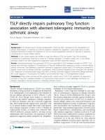
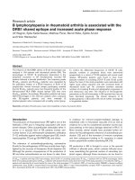
A decrease in BAL SP-D levels was associated with significantly increased inflammation in Balb/c mice 6 hrs after O3 exposure.Figure 1
A decrease in BAL SP-D levels was associated with significantly increased inflammation in Balb/c mice 6 hrs
after O3 exposure. Groups of Balb/c (grey bars) and C57BL/6 (black bars) mice were exposed to O
3
or room air for 2 hrs
and BAL was performed 6 hrs later. (A) Ozone exposed Balb/c mice had significantly reduced SP-D levels as compared to air
exposed Balb/c or O
3
exposed C57BL/6 mice. SP-D was detected by Western blot analysis of the cell-free supernatant of the
BAL (top panel) and was performed using our in-house rabbit polyclonal anti-SP-D antibody. Two representative samples out
of a total of six are shown in each group. SP-D expression was quantified by densitometric analysis. Results are expressed as %
of naïve control levels. * p = 0.0249 vs. room air; p = 0.0027 vs. C57BL/6. (B) Balb/c mice had significantly higher numbers of
inflammatory cells in their BAL as compared to C57BL/6 mice. Cells were counted using a Coulter counter and results are
expressed as cell number/ml (*p = 0.0316). (C): The BAL total protein content was significantly higher in Balb/c mice com-
pared to C57BL/6 mice. Total protein was measured by Bradford assay in the cell-free supernatant *p = 0.028. Absolute pro-
tein contents were 919,6 (± 51,4) and 3262,3 (± 281.0) in air and O
3
exposed Balb/c mice, respectively, and 772 (± 26.4) and
2110 (± 36.4) in air and O
3
exposed C57BL/6 mice, respectively.(D-G): Cytokine expression was studied by Endogen
®
Search-
Light™ and Luminex
®
100™ technologies. O
3
induced the release of IL-12p70 (34 pg/ml ± 4 in Balb/c, 18.7 pg/ml ± 1 in C57BL/
6), IL-6 (2,393 pg/ml ± 119 in Balb/c, 412 pg/ml ± 68.7 in C57BL/6), IL-10 (110 pg/ml ± 16 in Balb/c, 14.2 pg/ml ± 1.6 in C57BL/
6) and KC (1,896 pg/ml ± 224 in Balb/c, 136.8 pg/ml ± 27.7 in C57BL/6). Cytokine and chemokine levels are expressed as %
increase from control levels. The O
3
- induced pro-inflammatory cytokine IL-12p70 and KC levels were significantly higher (*p
= 0.0134 and *p = 0.0001, respectively) whereas the immunosuppressive IL-10 and the immunoregulatory IL-6 levels were sig-
nificantly lower (*p < 0.0001 and *p < 0.0001, respectively) in Balb/c mice than in C57BL/6 mice. (A-G): Mean ± SEM of n = 6
in each groups.
D
Balb/c C57BL/6
IL-12p70
% increase after O
3
100
0
200
F
% increase after O
3
*
Balb/c C57BL/6
IL-10
200
0
400
E
% increase after O
3
*
Balb/c C57BL/6
KC
500
0
1000
% increase after O
3
3000
0
6000
Balb/c C57BL/6
IL-6
*
G
A
SP-D levels (% of control)
*
Balb/c C57BL/6
Air
Balb/c C57BL/6
Ozone
50
100
150
0
*
B
Balb/c C57BL/6
Air
Balb/c C57BL/6
Ozone
Total cells (x1000)
100
300
0
*
400
200
Total BAL protein (%)
100
200
300
0
*
Balb/c C57BL/6
Air
Balb/c C57BL/6
Ozone
C
400
Balb/c C57BL/6
Air
Balb/c C57BL/6
Ozone
Respiratory Research 2006, 7:85 />Page 5 of 9
(page number not for citation purposes)
until the last time point of the experiment (72 hrs) when
SP-D levels were about 150 % above control levels (p =
0.0022; Fig. 2A). SP-A levels on the other hand did not
change significantly. The SP-D ELISA results were verified
using Western blot analysis. The two different methods
showed a significant positive Spearman correlation r =
0.86 (p = 0.0238). Inflammatory cells were detected 2 hrs
after O
3
-exposure, and the numbers were significantly
increased compared to naïve animals and peaked around
12 hrs post-exposure (p < 0.0001; Fig. 2B). A slight
increase in the numbers of lymphocytes (up to 3 % of
total cell counts) was also observed with a time course
comparable with that of neutrophilic cells (p = 0.0003;
not shown). Eosinophil counts showed a transient peak at
6 hrs, but their number remained less than 1 % of total
cell counts at all time points (not significant; not shown).
Airway neutrophilia resolved markedly within 72 hrs after
O
3
exposure indicating an inverse relationship between
the rise of SP-D levels and the decrease of inflammatory
cells, including neutrophils and lymphocytes (Fig. 2A and
2B). There was however no statistical correlation between
these parameters. The neutrophil influx was preceded by a
significant increase in KC levels but there was no signifi-
cant correlation between this chemokine and neutrophil
recruitment. Release of KC into the airways started 2 hrs
after O
3
challenge and reached a peak at 6 hrs (p < 0.0001;
not shown). KC levels were back to baseline by 24 hrs.
Interestingly, IL-6 was highly induced, with a peak at the
6 hrs time point and a return to baseline levels 48 hrs
post-exposure (p < 0.0001; Fig. 2C). Release of the anti-
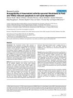
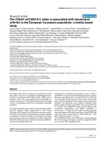
Kinetics of O
3
-induced SP-D and inflammatory changes in C57BL/6 miceFigure 2
Kinetics of O
3
-induced SP-D and inflammatory changes in C57BL/6 mice. Groups of C57BL/6 mice were exposed to
3 ppm O
3
or room air and studied 0, 2, 6, 12, 24, 48, and 72 hrs later. (A): SP-D and SP-A recovered from the BAL were quan-
tified by an in-house ELISA. Results are expressed as % change from naïve controls. SP-D levels gradually increased until the 72
hrs time point (p = 0.0022, ANOVA and post test for linear trend) whereas SP-A levels did not change significantly. (B): The
number of neutrophilic granulocytes was assessed by counting total number of BAL cells in the BAL fluid and performing differ-
ential cell counting on Kwick™Diff cytospin preparations. Results are expressed as absolute cell number/ml of BAL fluid. Neu-
trophilic inflammation peaked around 12 hrs post exposure (p < 0.0001) and largely resolved by 72 hrs after O
3
exposure. (C-
D) IL-6 and IL-10 levels were assessed as part of a Luminex
®
100™ assay and showed significant increases 2–6 hrs after O
3
exposure (p < 0.0001) and 6–24 hrs after O
3
exposure (p = 0.0007), respectively. (A-D): Mean ± SEM of n = 6 in each groups.
pg/ml
250
0
500
C
D
B
Cell count (x1000)
70
0
140
Neutrophils
02612244872
Time (h)
A
BAL SP-D (% of control)
200
300
100
0
02612244872
Time (h)
SP-D SP-A
02612244872
Time (h)
IL-6
02612244872
Time (h)
pg/ml
10
20
0
IL-10
Respiratory Research 2006, 7:85 />Page 6 of 9
(page number not for citation purposes)
inflammatory cytokine IL-10 was delayed by several
hours. IL-10 levels were slightly but significantly increased
with highest values between 6–24 hrs after O
3
challenge
(p = 0.0007; Fig. 2D).
SP-D deficient mice have increased cellular inflammation
following O
3
exposure
To further evaluate the anti-inflammatory role of SP-D in
the O
3
-induced immune response we used SP-D deficient
mice and compared them with age- and sex-matched
C57BL/6 wild-type controls. SP-D deficient mice showed
a baseline inflammation that was further increased after
O
3
exposure. The O
3
-induced cellular response was signif-
icantly higher in SP-D -/- mice compared to wt C57BL/6
mice 12hrs after acute O
3
exposure (p = 0.0106; Fig. 3A).
In addition, when mice were exposed to O
3
for 24 hrs
(0.5ppm O
3
), a sub-acute exposure, this finding was con-
firmed, since SP-D -/- mice had increased numbers of
inflammatory cells both 24 and 48 hrs after cessation of
O
3
exposure (Fig. 3B, p = 0.0082). Unlike after acute O
3
exposure (Fig. 2B), neither the wild type nor the SP-D-/-
mice showed signs of resolution of cellular infiltration at
the 48 hrs time point after sub-acute exposure. On the
contrary, inflammatory cell numbers were further
increased (Fig. 3B).
IL-6 selectively induces the production of SP-D in vitro
We have shown previously that SP-D induction in allergic
inflammation is dependent on IL-4 and IL-13 [15,18].
Although, none of these Th2 type cytokines was induced
in the present model of O
3
challenge, the production of
SP-D was highly up-regulated. Therefore we tested the
possibility that one of the O
3
-inducible cytokines is capa-
ble of promoting SP-D expression. Since IL-6 showed the
most pronounced changes following O
3
challenge, and
since it is a pluripotent immuno-regulatory cytokine, we
chose to investigate its effects on SP-D gene expression in
vitro. As shown in Fig. 4A, in vitro stimulation of primary
rat alveolar type II cells revealed that IL-6 is indeed capa-
ble of directly up-regulating SP-D production. The effect
of IL-6 was dose dependent (Fig. 4B) and selective for SP-
D, because SP-A production was not changed (Fig. 4A).
Discussion
Our results confirm the findings of other investigators
showing that acute O
3
exposure induces a rapid onset and
resolution of airway inflammatory changes characterized
by a KC-driven neutrophilic inflammation and moder-
ately increased numbers of lymphocytes, eosinophils and
macrophages [4,6]. Altered levels of surfactant protein D
have been reported in association with a number of differ-
ent pathological conditions of the lung [13,15,18,21-24].
Here we show that O
3
exposure induces a delayed expres-
sion of SP-D. Our data also show that the susceptibility to
O
3
-induced inflammatory changes varies between differ-
ent mouse strains and appears to be associated with differ-
ent levels of SP-D. C57BL/6 mice that express high levels
of SP-D also produce high levels of the anti-inflammatory
cytokine IL-10 and high levels of IL-6. In contrast, Balb/c
mice release significantly more KC and IL-12p70. Elevated
SP-D deficient mice have increased airway inflammation following O
3
exposureFigure 3
SP-D deficient mice have increased airway inflammation following O
3
exposure. (A) SP-D -/- mice and age-matched
C57BL/6 controls were exposed to 3 ppm O
3
for 2 hrs or (B) to 0.5 ppm for 24 hrs. Influx of neutrophilic granulocytes was
assessed on cytospin preparations stained with Kwick™Diff. In both models cellular inflammation in SP-D -/- mice was signifi-
cantly higher compared to wt mice (A) Student t-test *p = 0.0106 (B) ANOVA and Barlett's test *p = 0.0082.
Air
SP-D -/-
wt
Ozone
SP-D -/-
wt
*
*
A
Neutrophils (x1000)
400
0
800
B
SP-D -/-
wt
air 24 48 air 24 48
300
600
0
*
Neutrophils (x1000)
Respiratory Research 2006, 7:85 />Page 7 of 9
(page number not for citation purposes)
levels of SP-D are associated with the resolution of the O
3
-
induced inflammation and low levels or lack of SP-D pre-
dispose to a severe inflammatory response.
The drop in SP-D levels seen in Balb/c mice 6 hrs after O
3
exposure could be due to a direct damage and/or apopto-
sis of SP-D producing epithelial cells [25]. It is likely that
this acute phenomenon affects stored SP-D only, because
de-novo mRNA expression occurs only about 6 hrs after
allergen challenge or pulmonary infection and increased
levels of SP-D protein were only detected at about 12 hrs
(Fig. 2A) [13,18,26]. Additionally, the size of the extracel-
lular SP-D pool might be important in the protection
from O
3
-induced epithelial injury. Although the authors
did not specifically investigate the role of SP-D, Li and col-
leagues demonstrated that endotoxin pre-treatment,
which is expected to induce SP-D production, protects
against O
3
-induced cell death and pulmonary inflamma-
tion [27,28]. This could explain why C57BL/6 mice with
their higher levels of SP-D were more protected from the
acute effects of O
3
. Although it has been shown previously
that different mouse strains vary in their acute O
3
-induced
pulmonary response [4], no host factors responsible for
the individual susceptibility have been identified. Savov
and co-workers identified several chromosomal regions
that appeared to be associated with the physiologic and
biologic phenotypes [4]. Their in silico genome scan indi-
cates that a locus between 30 and 40 megabases (Mb) on
mouse chromosome (MMU) 14 contains one or more rel-
evant genes. It is noteworthy, that the gene coding for SP-
D is located on MMU 14 in the area of 37.2 Mb. Addition-
ally, two recent reports identified sequence polymor-
phisms in the human SP-D gene that lead to differences in
constitutive serum levels and influence the multimeric
assembly and function of SP-D protein [29,30].
Neutrophils play a vital role in the pulmonary host
defense. However, due to their release of large amounts of
histo-toxic and pro-inflammatory agents, these cells can
cause significant tissue damage. Hence, a stringent control
of neutrophil priming, recruitment, activation, apoptosis
and clearance is crucial to confine tissue damage [31].
Mice genetically deficient in SP-D have chronic inflamma-
tion and hyper-activated macrophages further strengthen-
ing the important role of SP-D as a local regulator of the
innate immune response [20,32,33]. In a recent publica-
tion White and colleagues demonstrated that SP-D may
either inhibit or enhance neutrophil respiratory burst
responses to influenza A virus. Their data also suggest that
the effects of SP-D are modulated by the presence of other
respiratory innate immune proteins such as SP-A, and on
the multimerization state of SP-D [34]. Other studies
show that administration of recombinant SP-D enhances
the up-take of apoptotic cells and reduces the production
of pro-inflammatory cytokines [35,36]. In line with those
results, our study shows that levels of KC, the main
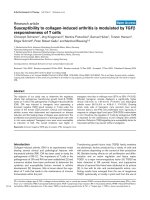
In vitro production of SP-D by alveolar type II cells after IL-6 stimulationFigure 4
In vitro production of SP-D by alveolar type II cells after IL-6 stimulation. (A) Primary rat alveolar type II cells were
cultured in the presence or absence of DCI and IL-6 for up to 4 days and intra-cellular SP-D and SP-A levels were assayed by
Western blot analysis. Day 0 SP-D and SP-A signals were obtained from freshly isolated alveolar type II cells. To maintain the
SP-D producing phenotype, cells have to be cultured in the presence of DCI (10 nM dexamethason, 100 nM IBMX, 10 nM
cAMP) that in turn stimulates SP-D production. IL-6 selectively stimulated the production of SP-D but did not induce up-regu-
lation of SP-A. (B) Increase of SP-D after IL-6 stimulation at day 4 was slightly dose dependent. All levels were compared to
non-stimulated alveolar type II cells cultured in DCI containing medium (DCI control).
SP-D
SP-A
A
DCI
IL-6
Day
+
-
+
+
4441
+
-
% increase from DCI control
B
100
200
0
300
12.5 25 50 100 200
IL-6 (ng/ml)
0
Respiratory Research 2006, 7:85 />Page 8 of 9
(page number not for citation purposes)
chemo-attractant for neutrophils, significantly dropped in
concert with a significant elevation of SP-D 12 hrs after O
3
exposure in C57BL/6 mice. However, whether or not SP-
D has a direct regulatory effect on KC remains to be deter-
mined and is the current focus of our studies.
Ozone exposure does not induce the release of classical
Th2 type cytokines such IL-4 or IL-13 which have been
shown to stimulate the expression of SP-D [18]. However,
in our study we show that O
3
induced a significant rise in
BAL SP-D suggesting that there are other mechanisms to
promote SP-D expression during a non-allergen induced
inflammation. Indeed, our in vitro studies using rat alveo-
lar type II cells show that IL-6 is capable of inducing SP-D
production. In a different model of airway inflammation,
elicited by allergic sensitization and challenge, we have
previously shown that a rapid release of pro-inflamma-
tory cytokines is followed by a relatively slow, gradual ele-
vation of SP-D protein levels in the airways, with a peak
48 hours after allergen challenge [15]. In accordance to
that, SP-D protein levels were still increased at 48 and 72
hrs when IL-6 levels were already back to normal. IL-6 can
transduce its signal either via C/EBPβ or via Stat3 activa-
tion. The SP-D promoter region harbours binding sites for
both of these transcription factors [37]. Whether IL-6-
dependent SP-D gene expression is promoted by C/EBPβ
or Stat3 or synergistically by both of them remains to be
clarified. Interestingly, IL-6 deficient mice have signifi-
cantly less BAL protein, neutrophils and soluble TNF
receptors after exposure to sub-acute levels of O
3
[38]. On
the other hand mice over-expressing IL-6 are protected
from lung injury caused by chronic hypoxia [39]. These
findings point out the pluripotent functions of IL-6 as an
anti- as well as a pro-inflammatory cytokine. The reports
by Johnston et al. and Ward et al. also provide indirect
support of our findings that IL-6 is important in launching
a protective pulmonary response. Our results showed that
the neutrophil chemoattractant KC and the immunosup-
pressive cytokine, IL-10 were also elevated in the BAL fluid
after O
3
exposure. While the possibility was raised that
these mediators could contribute to the up-regulation of
SP-D, we found no evidence that lung epithelial cells
would express receptor or show any functional response
to exogenous IL-10 or KC [40].
Conclusion
Strain dependent differences in SP-D production are asso-
ciated with differences in the severity of the inflammation.
The importance of SP-D in the protection against the ini-
tial O
3
-induced injury and during the resolution of the
inflammation is confirmed in SP-D knockout mice, as the
absence of SP-D resulted in enhancement of the inflam-
mation in these animals. We propose that IL-6 may con-
tribute to the up-regulation of SP-D expression, which in
turn inhibits pro-inflammatory changes and promotes
resolution of the inflammation following O
3
exposure.
Competing interests
The author(s) declare that they have no competing inter-
ests.
Authors' contributions
SK participated in the animal experiments, BAL cell
counts, Western blots, data analysis and prepared the
manuscript.
FRP supervised the animal experiments using SP-D defi-
cient mice and advised on data analysis.
YC participated in most of the animal experiments and
developed the ELISA for SP-D.
MG performed the time course study in C57BL/6 mice.
RM performed the animals experiments using SP-D -/-
mice.
GK developed a template for statistical data analysis and
participated in the preparation of the manuscript.
MFB gave helpful advice for data analysis and preparation
of the manuscript.
MS participated in the design of the experiment, took part
in the time course study and gave helpful advice for the
preparation of the manuscript.
RAPJr. gave helpful advice for the preparation of the man-
uscript.
AH designed the study, coordinated the experiments, and
helped to draft the manuscript.
References
1. Desqueyroux H, Pujet JC, Prosper M, Le Moullec Y, Momas I: Effects
of air pollution on adults with chronic obstructive pulmonary
disease. Arch Environ Health 2002, 57(6):554-560.
2. Peel JL, Tolbert PE, Klein M, Metzger KB, Flanders WD, Todd K, Mul-
holland JA, Ryan PB, Frumkin H: Ambient air pollution and respi-
ratory emergency department visits. Epidemiology 2005,
16(2):164-174.
3. Stenfors N, Pourazar J, Blomberg A, Krishna MT, Mudway I, Helleday
R, Kelly FJ, Frew AJ, Sandstrom T: Effect of ozone on bronchial
mucosal inflammation in asthmatic and healthy subjects.
Respir Med 2002, 96(5):352-358.
4. Savov JD, Whitehead GS, Wang J, Liao G, Usuka J, Peltz G, Foster
WM, Schwartz DA: Ozone-induced acute pulmonary injury in
inbred mouse strains. Am J Respir Cell Mol Biol 2004, 31(1):69-77.
5. McKinney WJ, Jaskot RH, Richards JH, Costa DL, Dreher KL:
Cytokine mediation of ozone-induced pulmonary adapta-
tion. Am J Respir Cell Mol Biol 1998, 18(5):696-705.
6. Moffatt RK, Hyde DM, Plopper CG, Tyler WS, Putney LF: Ozone-
induced adaptive and reactive cellular changes in respiratory
bronchioles of bonnet monkeys. Exp Lung Res 1987,
12(1):57-74.
Publish with BioMed Central and every
scientist can read your work free of charge
"BioMed Central will be the most significant development for
disseminating the results of biomedical research in our lifetime."
Sir Paul Nurse, Cancer Research UK
Your research papers will be:
available free of charge to the entire biomedical community
peer reviewed and published immediately upon acceptance
cited in PubMed and archived on PubMed Central
yours — you keep the copyright
Submit your manuscript here:
/>BioMedcentral
Respiratory Research 2006, 7:85 />Page 9 of 9
(page number not for citation purposes)
7. Wiester MJ, Tepper JS, Winsett DW, Crissman KM, Richards JH,
Costa DL: Adaptation to ozone in rats and its association with
ascorbic acid in the lung. Fundam Appl Toxicol 1996, 31(1):56-64.
8. Harkema JR, Hotchkiss JA, Barr EB, Bennett CB, Gallup M, Lee JK,
Basbaum C: Long-lasting effects of chronic ozone exposure on
rat nasal epithelium. Am J Respir Cell Mol Biol 1999, 20(3):517-529.
9. Crouch EC: Surfactant protein-D and pulmonary host
defense. Respir Res 2000, 1(2):93-108.
10. Finkelstein JN, Johnston CJ: Enhanced sensitivity of the postna-
tal lung to environmental insults and oxidant stress. Pediatrics
2004, 113(4 Suppl):1092-1096.
11. Vandivier RW, Ogden CA, Fadok VA, Hoffmann PR, Brown KK,
Botto M, Walport MJ, Fisher JH, Henson PM, Greene KE: Role of
surfactant proteins A, D, and C1q in the clearance of apop-
totic cells in vivo and in vitro: calreticulin and CD91 as a
common collectin receptor complex. J Immunol 2002,
169(7):3978-3986.
12. Gardai SJ, Xiao YQ, Dickinson M, Nick JA, Voelker DR, Greene KE,
Henson PM: By binding SIRPalpha or calreticulin/CD91, lung
collectins act as dual function surveillance molecules to sup-
press or enhance inflammation. Cell 2003, 115(1):13-23.
13. Atochina EN, Beers MF, Tomer Y, Scanlon ST, Russo SJ, Panettieri
RAJ, Haczku A: Attenuated allergic airway hyperresponsive-
ness in C57BL/6 mice is associated with enhanced surfactant
protein (SP)-D production following allergic sensitization.
Respir Res 2003, 4(1):15.
14. Madan T, Reid KB, Singh M, Sarma PU, Kishore U: Susceptibility of
mice genetically deficient in the surfactant protein (SP)-A or
SP-D gene to pulmonary hypersensitivity induced by anti-
gens and allergens of Aspergillus fumigatus. J Immunol 2005,
174(11):6943-6954.
15. Haczku A, Cao Y, Vass G, Kierstein S, Nath P, Atochina-Vasserman
EN, Scanlon ST, Li L, Griswold DE, Chung KF, Poulain FR, Hawgood
S, Beers MF, Crouch EC: IL-4 and IL-13 form a negative feed-
back circuit with surfactant protein-D in the allergic airway
response. J Immunol 2006, in press:.
16. Wong CJ, Akiyama J, Allen L, Hawgood S: Localization and devel-
opmental expression of surfactant proteins D and A in the
respiratory tract of the mouse. Pediatr Res 1996, 39(6):930-937.
17. Fujita M, Shannon JM, Ouchi H, Voelker DR, Nakanishi Y, Mason RJ:
Serum surfactant protein D is increased in acute and chronic
inflammation in mice. Cytokine 2005, 31(1):25-33.
18. Cao Y, Tao JQ, Bates SR, Beers MF, Haczku A: IL-4 induces pro-
duction of the lung collectin surfactant protein-D. J Allergy Clin
Immunol 2004, 113(3):439-444.
19. Casey J, Kaplan J, Atochina-Vasserman EN, Gow AJ, Kadire H, Tomer
Y, Fisher JH, Hawgood S, Savani RC, Beers MF: Alveolar Surfactant
Protein D Content Modulates Bleomycin Induced Lung
Injury. Am J Respir Crit Care Med 2005.
20. Botas C, Poulain F, Akiyama J, Brown C, Allen L, Goerke J, Clements
J, Carlson E, Gillespie AM, Epstein C, Hawgood S: Altered sur-
factant homeostasis and alveolar type II cell morphology in
mice lacking surfactant protein D. Proc Natl Acad Sci U S A 1998,
95(20):11869-11874.
21. Beatty AL, Malloy JL, Wright JR: Pseudomonas aeruginosa
degrades pulmonary surfactant and increases conversion in
vitro. Am J Respir Cell Mol Biol 2005, 32(2):128-134.
22. Grubor B, Gallup JM, Ramirez-Romero R, Bailey TB, Crouch EC,
Brogden KA, Ackermann MR: Surfactant protein D expression in
normal and pneumonic ovine lung. Vet Immunol Immunopathol
2004, 101(3-4):235-242.
23. Grubor B, Gallup JM, Meyerholz DK, Crouch EC, Evans RB, Brogden
KA, Lehmkuhl HD, Ackermann MR: Enhanced surfactant protein
and defensin mRNA levels and reduced viral replication dur-
ing parainfluenza virus type 3 pneumonia in neonatal lambs.
Clin Diagn Lab Immunol 2004, 11(3):599-607.
24. Soerensen CM, Holmskov U, Aalbaek B, Boye M, Heegaard PM,
Nielsen OL: Pulmonary infections in swine induce altered por-
cine surfactant protein D expression and localization to den-
dritic cells in bronchial-associated lymphoid tissue.
Immunology 2005, 115(4):526-535.
25. Barr BC, Hyde DM, Plopper CG, Dungworth DL: A comparison of
terminal airway remodeling in chronic daily versus episodic
ozone exposure. Toxicol Appl Pharmacol 1990, 106(3):384-407.
26. Hudson B, Flemming J, Sun G, Rand TG: Comparison of immu-
nomodulator mRNA and protein expression in the lungs of
Stachybotrys chartarum spore-exposed mice. J Toxicol Environ
Health A 2005, 68(15):1321-1335.
27. Bachurski CJ, Ross GF, Ikegami M, Kramer BW, Jobe AH: Intra-
amniotic endotoxin increases pulmonary surfactant proteins
and induces SP-B processing in fetal sheep. Am J Physiol Lung
Cell Mol Physiol 2001, 280(2):L279-85.
28. Li L, Hamilton RFJ, Holian A: Protection against ozone-induced
pulmonary inflammation and cell death by endotoxin pre-
treatment in mice: role of HO-1. Inhal Toxicol 2000,
12(12):1225-1238.
29. Leth-Larsen R, Garred P, Jensenius H, Meschi J, Hartshorn K, Madsen
J, Tornoe I, Madsen HO, Sorensen G, Crouch E, Holmskov U: A
common polymorphism in the SFTPD gene influences
assembly, function, and concentration of surfactant protein
D. J Immunol 2005, 174(3):1532-1538.
30. Heidinger K, Konig IR, Bohnert A, Kleinsteiber A, Hilgendorff A,
Gortner L, Ziegler A, Chakraborty T, Bein G: Polymorphisms in
the human surfactant protein-D (SFTPD) gene: strong evi-
dence that serum levels of surfactant protein-D (SP-D) are
genetically influenced. Immunogenetics 2005, 57(1-2):1-7.
31. Brazil TJ, Dagleish MP, McGorum BC, Dixon PM, Haslett C, Chilvers
ER: Kinetics of pulmonary neutrophil recruitment and clear-
ance in a natural and spontaneously resolving model of air-
way inflammation. Clin Exp Allergy 2005, 35(7):854-865.
32. Schaub B, Westlake RM, He H, Arestides R, Haley KJ, Campo M,
Velasco G, Bellou A, Hawgood S, Poulain FR, Perkins DL, Finn PW:
Surfactant protein D deficiency influences allergic immune
responses. Clin Exp Allergy 2004, 34(12):1819-1826.
33. Yoshida M, Whitsett JA: Alveolar macrophages and emphy-
sema in surfactant protein-D-deficient mice. Respirology 2006,
11 Suppl 1:S37-40.
34. White MR, Crouch E, Vesona J, Tacken PJ, Batenburg JJ, Leth-Larsen
R, Holmskov U, Hartshorn KL: Respiratory Innate Immune Pro-
teins Differentially Modulate the Neutrophil Respiratory
Burst Response to Influenza A Virus. Am J Physiol Lung Cell Mol
Physiol 2005.
35. Tacken PJ, Hartshorn KL, White MR, van Kooten C, van de Winkel
JG, Reid KB, Batenburg JJ: Effective targeting of pathogens to
neutrophils via chimeric surfactant protein D/anti-CD89
protein. J Immunol 2004, 172(8):4934-4940.
36. Liu CF, Chen YL, Shieh CC, Yu CK, Reid KB, Wang JY: Therapeutic
effect of surfactant protein D in allergic inflammation of
mite-sensitized mice. Clin Exp Allergy 2005, 35(4):515-521.
37. He Y, Crouch EC, Rust K, Spaite E, Brody SL: Proximal promoter
of the surfactant protein D gene: regulatory roles of AP-1,
forkhead box, and GT box binding proteins. J Biol Chem 2000,
275(40):31051-31060.
38. Johnston RA, Schwartzman IN, Flynt L, Shore SA: Role of inter-
leukin-6 in murine airway responses to ozone. Am J Physiol Lung
Cell Mol Physiol 2005, 288(2):L390-7.
39. Ward NS, Waxman AB, Homer RJ, Mantell LL, Einarsson O, Du Y,
Elias JA: Interleukin-6-induced protection in hyperoxic acute
lung injury. Am J Respir Cell Mol Biol 2000, 22(5):535-542.
40. Lim S, Caramori G, Tomita K, Jazrawi E, Oates T, Chung KF, Barnes
PJ, Adcock IM: Differential expression of IL-10 receptor by epi-
thelial cells and alveolar macrophages. Allergy 2004,
59(5):505-514.