A TEXTBOOK OF POSTPARTUM HEMORRHAGE - PART 8 pptx
Bạn đang xem bản rút gọn của tài liệu. Xem và tải ngay bản đầy đủ của tài liệu tại đây (3.67 MB, 50 trang )
Medium- and high-risk regimens include the
use of etopside, methotrexate, actinomycin,
vincristine, cyclophosphomide and 6-mercapto
-
purine
36,37
. Women with choriocarcinoma are
most appropriately treated through specialist
trophoblastic disease referral centers
14
.
Coagulopathies
Women with inherited coagulation disorders
such as von Willebrand’s disease and carriers of
hemophilia A and B are likely to bleed post
-
partum if maternal clotting factors are low
(< 50 IU/dl). Prophylactic administration of
desmopressin (DDAVP) and clotting factor
concentrates may prevent postpartum hemor
-
rhage
15
. The aim is to raise factor levels above
50 IU/dl during labor and delivery and maintain
these for up to 5 days after delivery. In the event
of postpartum hemorrhage
15
, replacement of
deficient clotting factors should be made and
identification and treatment of the cause be
instigated. Management should be in close
liaison with hematologists and specialist hemo-
philia centers as available. In cases of prolonged
or intermittent secondary postpartum hemor-
rhage
15
, the use of tranexamic acid (a fibrino-
lytic inhibitor)
38
or combined oral contraceptive
pill has been reported
15
.
Hemorrhage from postpartum acquired
hemophilia is treated acutely with factor VIII
(either human, porcine) or recombinant factor
VIIa
15
. Immunosuppressive drugs such as
corticosteroids, cyclophosphamide and aza
-
thioprine may be used to accelerate the dis
-
appearance of factor VIII inhibitors, although
complete remission is likely to occur spontane
-
ously with time.
Reversal of bleeding due to anticoagulants
should follow normal protocols. Vitamin K
should be considered in women with uncon
-
trolled bleeding secondary to warfarin use and
protamine sulfate may be considered if hemor
-
rhage results from the use of heparin, although
this has a much shorter half-life.
Secondary postpartum hemorrhage is an
important cause of maternal morbidity and
mortality. Basic resuscitation followed by inves
-
tigation and treatment of the specific cause of
hemorrhage are essential. The diverse nature of
its etiology and often acute presentation make
research in the form of a randomized controlled
trial difficult. However, particularly for the
treatment of hemorrhage due to uterine infec
-
tion and/or retained placental tissue, this should
be achievable and would provide valuable
information to further our understanding of
the management of secondary postpartum
hemorrhage.
References
1. Thompson W, Harper MA. Postpartum
haemorrhage and abnormalities of the third
stage of labour. In Chamberlain G, Steer P,
eds. Turnbull’s Obstetrics, 3rd edn. Edinburgh:
Churchill Livingstone, 2001;619–33
2. Hoveyda F, MacKenzie IZ. Secondary post
-
partum haemorrhage: incidence, morbidity and
current management. Br J Obstet Gynaecol 2001;
108:927–30
3. King PA, Duthie SJ, Dong ZG, et al. Secondary
postpartum haemorrhage. Aust NZ J Obstet
Gynaecol 1989;29:394–8
4. Alexander J, Thomas P, Sanghera J. Treatments
for secondary postpartum haemorrhage. The
Cochrane Database of Systematic Review 2002
Issue 1, Art. No: CD002867. DOI: 10.1002/
14651858.CD002867
5. Lédée N, Ville Y, Musset D, et al. Management
in intractable obstetric haemorrhage: an audit
study on 61 cases. Eur J Obstet Gynecol 2001;
94:189–96
6. Matthews NM, McCowan LME, Patten P. Pla
-
centa praevia accreta with delayed hysterectomy.
Aust NZ J Obstet Gynaecol 1996;36:476–9
7. Jaffe R, DuBeshter B, Sherer DM, et al. Failure
of methotrexate treatment for term placenta
percreta. Am J Obstet Gynecol 1994;171:558–9
8. Ggosh H. Arteriovenous malformation of the
uterus and pelvis. Obstet Gynecol 1986;
68(Suppl):40–3
9. Gaylis H, Levine E, van Dongen L, et al. Arterio
-
venous fistula after gynaecologic operations. Surg
Gynecol Obstet 1973;137:655–8
10. Pelage J-P, Soyer P, Repiquet D, et al. Secondary
postpartum haemorrhage: treatment with selec
-
tive arterial embolization. Radiology 1999;212:
385–9
11. Kelly SM, Belli AM, Campbell S. Arteriovenous
malformation of the uterus associated with
secondary postpartum haemorrhage. Ultrasound
Obstet Gynecol 2003;21:602–5
12. Nanda S, Singhal S, Sharma D, et al. Nonunion
of uterine incision: a rare cause of secondary
323
Management of secondary postpartum hemorrhage
345
Z:\Sapiens Publishing\A5211 - Postpartum Hemorrhage\Make-up\Postpartum Hemorrhage - Voucher Proofs #T.vp
30 August 2006 14:23:51
Color profile: Generic CMYK printer profile
Composite Default screen
postpartum haemorrhage: a report of 2 cases.
Aust NZ J Obstet Gynaecol 1997;37:475–6
13. Paraskevaides E, Stuart B, Gardeil F. Secondary
postpartum haemorrhage from non-dehisced
lower caesarean section scar: a case for hystero
-
scopy. Aust NZ J Obstet Gynaecol 1993;33:427
14. Tidy JA, Rustin GJS, Newlands ES, et al. Presen
-
tation and management of choriocarcinoma after
non-molar pregnancy. Br J Obstet Gynaecol 1995;
102:715–19
15. Economides DL, Kadir RA. Inherited bleeding
disorders in obstetrics and gynaecology. Br J
Obstet Gynaecol 1999;106:5–13
16. Kadir RA, Economides DL, Braithwaite J, et al.
The obstetric experience of carriers of haemo
-
philia. Br J Obstet Gynaecol 1997;104:803–10
17. Greer IA, Lowe GDO, Walker JJ, et al. Haemor
-
rhagic problems in obstetrics and gynaecology
in patients with congenital coagulopathies. Br J
Obstet Gynaecol 1991;98:909–18
18. Ramsahoye BH, Davies SH, Dasani H, et al.
Obstetric management in von Willebrand’s dis
-
ease: a report of 24 pregnancies and a review of
the literature. Haemophilia 1995;1:140–4
19. Kadir RA, Lee CA, Sabin CA, et al. Pregnancy in
von Willebrand’s disease or factor XI deficiency.
Br J Obstet Gynaecol 1998;105:314–21
20. Neill A, Thornton S. Secondary postpartum
haemorrhage. J Obstet Gynaecol 2002;22:119–22
21. Johanson R, Cox C, Grady K, et al. Massive
obstetric haemorrhage. In Managing Obstetric
Emergencies and Trauma – The MOET Course
Manual. London: RCOG Press, 2003;16:
151–63
22. Achiron R, Goldenberg M, Lipitz S, et al.
Transvaginal duplex Doppler ultrasonography in
bleeding patients suspected of having residual
trophoblastic tissue. Obstet Gynecol 1993;81:
507–11
23. Zuckerman J, Levine D, McNicholas MM, et al.
Imaging of pelvic postpartum complications. Am
J Roentgenol 1997;168:663–8
24. Neill AC, Nixon RM, Thornton S. A compari
-
son of clinical assessment with ultrasound in the
management of secondary postpartum haemor
-
rhage. Eur J Obstet Gynecol 2002;104:113–15
25. Thorp JM, Wells SR, Wiest HH, et al. First-
trimester diagnosis of placenta praevia percreta
by magnetic resonance imaging. Am J Obstet
Gynecol 1998;178:616–18
26. Levine D, Barnes PD, Edelman RR. Obstetric
MR imaging. Radiology 1999;211:609–17
27. Fernandez H, Claquin C, Guibert M, et al
. Sus
-
pected postpartum endometritis: a controlled
clinical trial of single-agent antibiotic therapy
with Amox-CA (Augmentin
®
) vs ampicillin-
metronidazole ± amnioglycoside. Eur J Obstet
Gynecol 1990;36:69–74
28. Schenker JG, Margalioth SJ. Intrauterine adhe
-
sions: an update appraisal. Fertil Steril 1982;37:
593–610
29. Bakri YN. Amri A, Abdul-Jabar F. Tamponade-
balloon for obstetrical bleeding. Int J Gynaecol
Obstet 2001;74:139–42
30. Johanson R, Kumar M, Obhrai M, et al. Man
-
agement of massive postpartum haemorrhage:
use of a hydrostatic balloon catheter to avoid
laparotomy. Br J Obstet Gynaecol 2001;108:
420–2
31. B-Lynch C, Coker A, Adegboyega HL, et al. The
B-Lynch surgical technique for the control of
massive postpartum haemorrhage: an alternative
to hysterectomy? Five cases reported. Br J Obstet
Gynaecol 1997;104:372–5
32. Alok K, Hagen P, Webb JB. Tranexamic acid in
the management of postpartum haemorrhage. Br
J Obstet Gynaecol 1996;103:1250–1
33. Boehlen F, Morales MA, Fontana P, et al.
Prolonged treatment of massive postpartum
haemorrhage with recombinant factor VIIa: case
report and review of the literature. Br J Obstet
Gynaecol 2004;111:284–7
34. Lurie S, Appelman Z, Katz Z. Subendometrial
vasopressin to control intractable placental
bleeding. Lancet 1997;349:698
35. Bagshawe KD, Dent J, Newlands SL, et al. The
role of low dose methotrexate and folinic acid in
gestational trophoblastic tumours. Br J Obstet
Gynaecol 1989;96:795–802
36. Newlands ES, Bagshawe KD, Begent RH, et al.
Results with EMA/CO (etopside, methotrexate,
actinomycin D, cyclophosphamide, vincristine)
regimen in high risk gestational trophoblastic
tumours, 1979–1989. Br J Obstet Gynaecol 1991;
98:550–7
37. Rustin GJS, Newlands ES, Bergent HJ, et al.
Weekly alternating chemotherapy (EMA/CO)
for treatment of central nervous system
metastases of choriocarcinoma. J Clin Oncol
1989;7:900–3
38. Bonnar J, Guillebrand J, Kasonde JM, et al.
Clinical applications of fibrinolytic inhibition
in gynaecology. J Clin Pathol 1980;33(Suppl)
14:55–9
324
POSTPARTUM HEMORRHAGE
346
Z:\Sapiens Publishing\A5211 - Postpartum Hemorrhage\Make-up\Postpartum Hemorrhage - Voucher Proofs #T.vp
30 August 2006 14:23:52
Color profile: Generic CMYK printer profile
Composite Default screen
Section VIII
Consequences of postpartum
hemorrhage
347
Z:\Sapiens Publishing\A5211 - Postpartum Hemorrhage\Make-up\Postpartum Hemorrhage - Voucher Proofs #T.vp
30 August 2006 14:23:52
Color profile: Generic CMYK printer profile
Composite Default screen
36
PATHOLOGY OF THE UTERUS
P. Kelehan and E. E. Mooney
BACKGROUND AND AIMS
Significant postpartum hemorrhage may occur
immediately after delivery, or may be delayed
weeks or months. In either case, a Cesarean or
later postpartum hysterectomy may be life-
saving. The uterus will normally be sent for
laboratory examination. To facilitate a useful
surgical pathology report, the pathologist must
be given details of the antepartum course and
delivery. Considering how uncommon these
specimens are, direct communication between
pathologist and clinician is recommended. The
aim of this chapter is to provide a structured
approach to the analysis of the specimen,
in order to permit a clinically relevant and
pathologically sound diagnosis.
CLINICAL CORRELATION
The parity and gestation should be provided.
Any abnormality of the clinical course, in partic
-
ular pre-eclampsia or polyhydramnios, may
be of relevance. Magnetic resonance imaging
(MRI) may have been performed for fibroid,
placenta creta or congenital abnormality and
these images should be reviewed. A history of
the use of instruments such as forceps is impor
-
tant. The clinical appearance of the uterus at
operation may provide valuable information
on atony. Any therapeutic measures undertaken
such as uterine massage or compression suture
should be noted, along with transfusion and
fluid replacement. A description of the surgery
will help the pathologist to interpret the tears
and sutures that characterize these specimens.
The patient’s postoperative condition will help
to guide sampling in the event that amniotic
fluid embolism is a consideration. Finally, the
placenta must also be available for examination.
GROSS EXAMINATION
Photography is essential at each step of the
dissection, with notes as to what each picture
is intended to show. Without a clinical input,
however, much effort may be wasted on
documenting features of little relevance at the
expense of missing more important ones. A
detailed macroscopic description of sutures,
tears, etc. is important and may be medico
-
legally relevant. Our approach is to examine the
specimen in its fresh state, with photography,
and then to open the specimen, avoiding tears
and sutures, to permit fixation and further
examination. It may be opened laterally, but
more information can be gained by complete
longitudinal anteroposterior section of the
uterus. The approach should be modified to
suit the circumstances as predicted from the
clinical information. A useful technique that
allows good exposure and photographic demon
-
stration is the placing of two parallel complete
longitudinal anteroposterior sections about
2–3 cm apart on either side of the mid-line.
How well the uterine cavity has compressed is
immediately apparent, contraction band forma
-
tion can be demonstrated, and blood clot and
placental tissue fragments can be assessed in the
lumen.
In the immediate postpartum period, the
uterus is characteristically large. It will weigh
700–900 g and will have substantially reduced
in size and volume from its antepartum state.
Clamp marks on the broad and round ligaments
should be inspected for residual hematoma,
remembering that the pathology may be outside
the clamp. In the fresh specimen with intact
vessels, it may be possible to perfuse the vascu
-
lature for contrast angiography or vascular
casting
1
.
326
348
Z:\Sapiens Publishing\A5211 - Postpartum Hemorrhage\Make-up\Postpartum Hemorrhage - Voucher Proofs #T.vp
06 September 2006 16:28:08
Color profile: Generic CMYK printer profile
Composite Default screen
327
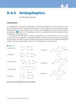
Pathology of the uterus
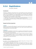
Figure 1 Fixed uterus showing a large anterior and right-sided diverticulum originating in a Cesarean
section scar. The specimen was sutured at operation, but placental villous tissue can be seen adjacent to the
suture
Figure 2 Anteroposterior section of uterus from Figure 1 showing anterior placenta creta
349
Z:\Sapiens Publishing\A5211 - Postpartum Hemorrhage\Make-up\Postpartum Hemorrhage - Voucher Proofs #T.vp
30 August 2006 14:23:58
Color profile: Generic CMYK printer profile
Composite Default screen
328
POSTPARTUM HEMORRHAGE
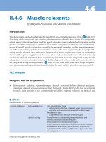
Figure 3 H/E section of lower uterine segment showing placenta creta and large vessels in thin
myometrium
Figure 4 Immunohistochemical stain for desmin accentuates the thin myometrial fibers in scar
350
Z:\Sapiens Publishing\A5211 - Postpartum Hemorrhage\Make-up\Postpartum Hemorrhage - Voucher Proofs #T.vp
30 August 2006 14:24:04
Color profile: Generic CMYK printer profile
Composite Default screen
329
Pathology of the uterus
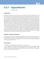
Figure 5 Right lateral endocervical tear at hysterectomy for postpartum hemorrhage
Figure 6 Elastin Van Geisson stain showing torn artery at apex of tear (×10). Arrow, torn elastic artery;
arrowhead, thin fibrin blood clot
351
Z:\Sapiens Publishing\A5211 - Postpartum Hemorrhage\Make-up\Postpartum Hemorrhage - Voucher Proofs #T.vp
30 August 2006 14:24:10
Color profile: Generic CMYK printer profile
Composite Default screen
CERVIX
Important pathologies in the cervix include
tears. Small shallow endocervical tears are
almost invariably found in the postpartum
uterus, and may be present even in those cases
where there has been a Cesarean section. Signif
-
icant and deep tears tend to be lateral in loca
-
tion. These tears may penetrate through to the
serosa, with or without hematoma formation,
and may extend up into the lower segment or
down the cervix into the vagina. Involvement
of large uterine arteries should be sought. It
is common to find meconium staining of the
mucus of the endocervix with fetal distress, and
meconium may contaminate the tear. A tear
may have severe consequences: an endocervical
tear may cause severe blood loss despite a fully
contracted uterus. Tears are associated with
amniotic fluid embolus or with amniotic
infusion and local defibrination. Bleeding
can extend into the broad ligament with
formation of a large hematoma. Suturing of the
tear may not prevent a deep hematoma from
forming and secondary rupture can result
in shock, despite cessation of external vaginal
hemorrhage.
In the dilated postpartum cervix, edema,
hemorrhage and fiber disarray may make it diffi
-
cult to identify tears on histologic examination.
Torn and contracted muscle fibers and torn
arteries with fibrin plugs and tense hematomas
provide corroboratory evidence of a tear. Histo
-
logic sampling should include blocks from
above the apex and from below the tear for deep
extension and for identification of large torn
vessels.
Examination of the uterus histologically
following amniotic fluid embolism will show no
evidence of intravascular disease in most cases.
Very occasionally, there may be fibrin clots
adherent to vascular endothelium and, rarely,
squames admixed with fibrin have been found
in vessels in the body of the uterus. In some
cases of postpartum hemorrhage, when there
have been no clinical features of amniotic infu
-
sion but bleeding and unexpected severe onset
of consumptive coagulopathy, histological
330
POSTPARTUM HEMORRHAGE
Figure 7 Amniotic debris in venules (arrows) of cervical stroma following a small endocervical tear in
labor. Postpartum hemorrhage and disseminated intravascular coagulopathy necessitated hysterectomy
(×20)
352
Z:\Sapiens Publishing\A5211 - Postpartum Hemorrhage\Make-up\Postpartum Hemorrhage - Voucher Proofs #T.vp
30 August 2006 14:24:13
Color profile: Generic CMYK printer profile
Composite Default screen
331
Pathology of the uterus
Figure 8 H/E comparison of (a) normal myometrial fibers and (b) myonecrosis in lower uterine segment
in hysterectomy specimen for postpartum hemorrhage following Cesarean section (×40). Long arrows,
normal viable cell nuclei; short arrows, non-viable necrotic cells
(a)
(b)
353
Z:\Sapiens Publishing\A5211 - Postpartum Hemorrhage\Make-up\Postpartum Hemorrhage - Voucher Proofs #T.vp
30 August 2006 14:24:19
Color profile: Generic CMYK printer profile
Composite Default screen
332
POSTPARTUM HEMORRHAGE
Figure 9 Desmin comparison of same myometrial fibers accentuates the necrosis. (a) Normal;
(b) myonecrosis (×40). Long arrow, normal myometrial cells with intercellular edema; short arrow, dense,
compacted necrotic myometrial cells at same magnification
(a)
(b)
354
Z:\Sapiens Publishing\A5211 - Postpartum Hemorrhage\Make-up\Postpartum Hemorrhage - Voucher Proofs #T.vp
06 September 2006 16:26:12
Color profile: Generic CMYK printer profile
Composite Default screen
sections of the endocervix will reveal localized
areas where amniotic debris fills and expands
venules and capillaries. This dramatic appear-
ance is present not just adjacent to the
endocervical surface and tears: its presence
deeper in the stroma distinguishs it from con
-
tamination of the surface mucosa by meconium
and amniotic fluid at delivery.
A subgroup of patients have a lesion of local
amniotic infusion associated with disseminated
intravascular coagulopathy and postpartum
hemorrhage without systemic collapse.
Squamous cells may be present in only one
or two sections taken from around the
circumference of the cervix. It is usually on
one side. Extensive sampling of the cervix
may be required to demonstrate amniotic
debris in cases of suspected amniotic fluid
embolism
2
. It is possible that ongoing blood loss
from a tear in this site may occur before the
onset of systemic disseminated intravascular
coagulopathy, because local thromboplastin
effect alone of the amniotic debris in the wound
may inhibit hemostasis.
LOWER UTERINE SEGMENT
Important pathologies here involve implanta-
tion on a previous Cesarean section scar,
with abnormal adherence or formation of a
diverticulum.
A Cesarean section results in chronic changes
in the lower uterine segment, including distor
-
tion and widening, inflammation, giant cell
reaction and adenomyosis
3
. In some cases, a
distinctive V-shaped defect of the anterior wall
(‘tenting’) may be present.
An important cause of weakening of a
Cesarean section scar is infection. Postoperative
wound infection is not uncommon following
Cesarean section, particularly emergency sec
-
tion. Prophylactic antibiotics can modify the
extent and rate of infection, as can the quality of
closure, the amount of local tissue trauma, the
technique used (one- or two-layer), swelling,
hematoma and the nature of the organisms
infecting the wound. There may be extensive
disruption and inflammation in the uterine wall
despite a normal healing appearance of the skin
wound. Conservative treatment of the wound
333
Pathology of the uterus
Figure 10 H/E section showing stitch material in uterine curettings following Cesarean section. Arrow,
absorbable suture; arrowhead, giant cell reaction to suture material (×40)
355
Z:\Sapiens Publishing\A5211 - Postpartum Hemorrhage\Make-up\Postpartum Hemorrhage - Voucher Proofs #T.vp
30 August 2006 14:24:28
Color profile: Generic CMYK printer profile
Composite Default screen
is normal, and surgical debridement the excep
-
tion. Accordingly, the consequences may be
only appreciated in a subsequent pregnancy. If
the patient does present before this, hemorrhage
and/or vaginal discharge may prompt internal
examination. A defect may be identified on pal
-
pation. Curettage may be undertaken and may
retrieve inflammatory exudate, degenerating
decidua, polypoid endometrium or fragments of
necrotic myometrium that have prolapsed into
the endocervical lumen from the internal edge
of the Cesarean section scar. Sometimes, quite
large pieces of myometrial tissue with edema
and coagulative necrosis are obtained. This
myonecrosis, or incisional necrosis, is caused by
local ischemia
4
. Remodelling of blood vessels
may influence implantation. Implantation on
either a normally healed or on a diseased scar
will not have the protective effect of the decidua
vera (see below), and so postpartum separation
is less likely to occur. A Cesarean section at
first birth is associated with increased risks
of placenta previa and abruption in second
pregnancies
5
.
Implantation in the lower segment (adjacent
to the defect) can cause expansion of the defect,
dehiscence of the wall and the formation of a
pulsion diverticulum which will further enlarge
and progress with growth of the placenta. If the
implantation is fundal, a fortuitous elective
section may reveal a thin, almost transparent
anterior lower segment wall. This should be
more easily resected at closure since the scar will
not be excessively vascular. If implantation is
in the lower segment or in the scar, then there
is a potential for catastrophic hemorrhage on
attempt at delivery of the placenta.
In examining a postpartum hysterectomy
specimen where there is a history of previous
Cesarean section, the points noted above should
be borne in mind. The recently sutured section
incision should be carefully reopened. Follow
-
ing photography, the edges and margins should
be inspected for thinning and scar tissue forma
-
tion. An enlarged, ragged and open defect of
the anterior lower uterine segment, now tightly
contracted and rigid with formalin fixation,
may be all that is left of a huge, thin-walled,
placenta-filled diverticulum, the result of scar
dehiscence and rupture. It is easy to destroy
this thin structure with precipitate dissection.
Examination of the lateral margins of the defect
may indicate left- or more often right-sided
extension of the bulging diverticulum into
parametrial soft tissue of the pelvis. A complete
section through the anterior lower uterine seg
-
ment can identify previous Cesarean section
scars with tenting defects and the shape and
edges of a recent section. Most importantly,
en-face examination of the lateral sides of the
lower segment will show the cavity and lateral
extension of a dehiscence diverticulum, fresh
tears and/or adherent placenta. The issue of
abnormal adherence is addressed below.
FUNDUS
Important pathologies include retained prod
-
ucts, placenta creta, and subinvolution. Pla
-
centa creta is the name given to abnormally
adherent or ingrowing placenta that does not
detach with full contraction of the uterus after
expulsion of the fetus. This term covers pla-
centa accreta (abnormal attachment to the
wall), increta (extension of villi into the myo-
metrium) and percreta (extension of villi
through to the serosa). The intimate relation-
ship of villous tissue to myometrium, without
intervening decidua, is the key to the diagnosis.
Descriptions of placenta percreta based on
illustrations or descriptions of chorionic villi
displaced between torn myometrial fibers
should be evaluated critically.
MRI may show the loss of zonation associ
-
ated with penetration rather than invasion of
chorionic villi.
Full-thickness anteroposterior sections of the
fundus make it easier to recognize the position
of the contracted placental site. It is surprisingly
difficult to identify the exact site on inspection
of the raw decidual surface that is seen if the
uterus is opened laterally.
Detachment of the placenta is dependent on
the presence of a normal spongy decidua vera,
where shearing of the placenta from the myo
-
metrium occurs. This soft compressible area is
not seen when the postpartum uterine lining is
examined histologically, because its many mucous
glands are disrupted to facilitate the normal
plane of cleavage. It is seen to its full extent in
the tragic case of maternal death prior to labor.
Either Alcian blue stain or diastase-PAS to
334
POSTPARTUM HEMORRHAGE
356
Z:\Sapiens Publishing\A5211 - Postpartum Hemorrhage\Make-up\Postpartum Hemorrhage - Voucher Proofs #T.vp
30 August 2006 14:24:28
Color profile: Generic CMYK printer profile
Composite Default screen
demonstrate mucopolysaccharides in the swol
-
len gland crypts can help to identify this layer.
Deficiency of this layer may be focal or, rarely,
complete. When it is absent, the thinned
Nitabuch’s layer with anchoring villi lies in close
proximity to muscle fiber bundles or interstitial
fibrous Cesarean section scar. An occasional
finding is the presence of abundant inter
-
mediate trophoblast infiltrating between muscle
fibers beneath a firmly adherent Nitabuch’s
layer. Histological examination of multiple
sections can show anchoring villi penetrating
Nitabuch’s fibrinoid and ghost villi in dense
fibrin adherent to muscle. The often described
appearance of chorionic villi infiltrating between
muscle fibers is characteristic only of invasive
mole; the key to placenta percreta is absence of
decidua. An increased number of implantation
site intermediate trophoblasts has been shown
in cases of placenta creta compared with
controls
6
. Retained placental fragments reflect
some degree of placenta creta and are more
common in women with a spectrum of changes
in previous pregnancies, such as pre-eclamptic
toxemia, growth restriction, spontaneous abor-
tion and retained placental fragments. It has
been hypothesized that these reflect abnormal
maternal–trophoblast interaction
7
.
Placenta creta is therefore due to a deficiency
of the decidua. The end result of penetration of
the placenta though a weakened part of the
uterine wall includes rupture and secondary
changes, including serosal peritoneal reaction.
Curette penetration may cause secondary infec
-
tion or hematoma formation and provide the
nidus for dehiscence into the adherent bladder
wall, if this had been injured at previous surgery.
Placenta creta is only part of the problem
of uncontrolled postpartum hemorrhage. The
thin myometrium, with little muscle, interstitial
fibrosis and increased intermediate trophoblast
will contain large dilated arteries of pregnancy
and often widespread extrauterine extension of
these changes into the parametrium, as
described on Doppler ultrasound. The degree
of constriction–contraction of the myometrium
is insufficient to close off these vessels. Where
there is severe thinning of the muscle of the
lower segment with diverticulum formation,
abnormal adhesion is not necessary to
sustain bleeding. Conversely, on histological
examination of the lining of the postpartum
uterus, the finding of chorionic villi in clefts in
the placental bed may be an artefact rubbed
in following clearance of uterine contents and
is of no diagnostic consequence. Smearing of
DNA due to crush artefact may be helpful in
distinguishing this from true extension.
RETAINED PRODUCTS OF
CONCEPTION
The failure of total expulsion of the placenta
may lead to postpartum hemorrhage. A frag
-
ment of placenta remains, assumes a polypoid
shape (‘placental polyp’), and undergoes com
-
pression and devitalization. Some viable cells
may remain in stem villi. Vessels below the
retained fragments may show persistent dilata
-
tion. There may be a plasma cell infiltrate in the
adjacent myometrium – this is not diagnostic of
(infective) endometritis in this context. The fre-
quency of detection of retained products varies
from 27 to 88%
7
, but much of this literature
is decades old. Retained placental fragments
are more common in women who have had
complications such as pre-eclampsia or growth
restriction in previous pregnancies. This has
been interpreted as indicative of an abnormal
maternal–trophoblast relationship
7
.
SUBINVOLUTION
Subinvolution of the blood vessels of the pla
-
cental bed, in the absence of retained placental
fragments, is an important and distinctive cause
of secondary postpartum hemorrhage.
Normal arterial involution involves a
decrease in the lumen size, disappearance of
trophoblast, thickening of the intima, re-growth
of endothelium and regeneration of internal
elastic lamina. These changes occur within
3 weeks of delivery. With subinvolution, arteries
remain distended and contain red cells or fresh
thrombus, and trophoblast persists in a peri
-
vascular location
8
. In some cases, endovascular
trophoblast may be present. Hemorrhage from
subinvolution is maximal in the second week
postpartum, although it may occur up to several
months later. It is commoner in older, multi
-
parous women and may recur in subsequent
deliveries.
335
Pathology of the uterus
357
Z:\Sapiens Publishing\A5211 - Postpartum Hemorrhage\Make-up\Postpartum Hemorrhage - Voucher Proofs #T.vp
30 August 2006 14:24:29
Color profile: Generic CMYK printer profile
Composite Default screen
Subinvolution is not related to the method
of delivery and may be regarded as a specific
entity, possibly due to an abnormal immuno
-
logic relationship between trophoblast and
the uterus
8
. Despite this, it did not show the
association with markers of such an abnormal
relationship seen with retained placental
fragments in another study
7
.
The changes may be recognized on curettage
specimens. The hysterectomy specimen will
show a uterus that is soft and larger than
expected
8
. As normally involuted vessels may be
present adjacent to subinvoluted ones, multiple
blocks of placental bed should be taken to
exclude this process.
ATONY
This is well-recognized obstetric phenomenon,
but there may be relatively little to report in
the way of pathology. The diagnosis is one of
exclusion. The uterus is enlarged, edematous
and soft, with edema and hemorrhage apparent
microscopically. The diagnosis will depend on
clinical information, combined with adequate
histologic sampling to exclude other causes.
ARTERIOVENOUS MALFORMATIONS
Uterine arteriovenous malformations (AVMs)
are rare and may present with profuse hemor
-
rhage, including hemorrhage in the postpartum
period. Congenital AVMs consist of multiple
small connections and may enlarge with preg
-
nancy. The more common acquired AVMs are
rare in nulliparous women, and are thought to
arise following uterine trauma: curettage, myo
-
mectomy or even previous uterine rupture
9,10
.
AVMs may co-exist with retained products
of conception or trophoblastic proliferation.
Pathologically, vessels of arterial and venous
caliber are present, along with large vessels of
indeterminate nature.
OTHER CAUSES
Lacerations of the inner myometrium have been
reported to cause postpartum hemorrhage
11
.
Women with leiomyomas are at an increased
risk of postpartum hemorrhage
12
. Less com
-
monly, endometrial carcinomas and congenital
anomalies may also result in reduced decidua
formation and subsequent postpartum hemor
-
rhage. Trophoblastic disease has also been
reported in this context.
ENDOMETRITIS
An acute endometritis is reported as a cause of
sepsis and postpartum hemorrhage. It is rela
-
tively uncommon in modern obstetric practice
in the West and may be due to a variety of
organisms. It accounted for < 5% of cases of
delayed postpartum hemorrhage in one series
7
.
PLACENTAL PATHOLOGY
The placenta should be examined in cases
of postpartum hemorrhage. Pre-eclampsia
may cause retroplacental hemorrhage: recent
and old hemorrhages and infarcts may be seen.
The characteristic changes of acute atherosis are
only present in 50% of cases of pre-eclamptic
toxemia. However, examination of the paren-
chyma will usually show so-called accelerated
villous maturation (distal villous hypoplasia) in
response to uteroplacental ischemia. Sampling
from the center of the disc is important to avoid
overinterpretation of physiologic changes
13
.
THE AUTOPSY IN POSTPARTUM
HEMORRHAGE
In data drawn from the Confidential Enquiries
into Maternal Deaths in the UK for the period
1970–90, approximately 10% of direct maternal
deaths are due to hemorrhage
14
. Roughly half
were antepartum and half postpartum. Excess
blood loss is more common in older women
(> 35 or 40 years, depending on the study)
15
.
Before beginning an autopsy in a case of
maternal death following postpartum or intra
-
partum hemorrhage, it is critical to plan the pro
-
cedure and the sequence of the autopsy in the
light of the information received and the sus
-
pected cause or causes and mode of death. The
autopsy must be unhurried and methodical; it is
a fundamental mistake to seek to demonstrate
immediately the proposed cause of death.
Members of the clinical team should be asked to
attend the autopsy, but it is unwise to have
everybody there during what will be a long
336
POSTPARTUM HEMORRHAGE
358
Z:\Sapiens Publishing\A5211 - Postpartum Hemorrhage\Make-up\Postpartum Hemorrhage - Voucher Proofs #T.vp
30 August 2006 14:24:29
Color profile: Generic CMYK printer profile
Composite Default screen
phase of inspection, measurement and initial
systematic dissection. When all is ready, the
procedure is stopped and members of the team
attend. In this way, the history can be reviewed,
pre-existing conditions or disease discussed and
demonstrated, e.g. chronic pyelonephritis, and
the dissection and demonstration of the focus of
main clinical interest can begin.
A fundamental aspect of good autopsy prac
-
tice is the confident exclusion of specific dis
-
eases and conditions in a systematic approach.
The understandable desire and pressure to skip
to the seat of disease must be resisted. The
parametrium, pelvic side-wall and vagina are as
important objects of attention as the uterus.
At the time of external inspection of the
body, the pathologist must consider in turn each
of the major causes of maternal death. Many
require modification of routine techniques,
e.g. air embolism, amniotic fluid embolism,
ruptured aneurysm, and these modifications are
detailed elsewhere
16
. Preparation and sampling
of blood and fluids for hematology, hemophilia,
toxicology and microbiology should be planned,
e.g. sample containers should be pre-labelled
and set out in sequence. Cardiopulmonary
resuscitation attempt most likely preceded
death and therefore the features and sequence
of sustained unsuccessful resuscitation must
be identified and complications and agonal
changes interpreted in this context. It is impor
-
tant from a medicolegal aspect not to allow such
artefact to be construed as a major factor in the
cause of death, e.g. liver or mesenteric tear,
blood in the abdomen, bone marrow embolus.
The traditional Y-shaped autopsy incision
should be extended to an abdominal inverted Y
with the incision continued to the inguinal
femoral triangle on each side. This allows better
examination of the ileofemoral vessels and
better exposure of the pelvis. Blood and blood
clots are removed from the abdomen and the
amounts measured. The relative size and posi
-
tion of the abdominopelvic organs are assessed.
The peritoneal lining of the pelvis is inspected,
noting color, texture and degree of congestion.
Patches of peritoneal decidual reaction of
pregnancy can be identified by their gelatinous
appearance.
In traditional autopsy practice, the state
of pregnancy can be suspected, even when the
uterus is still small, by the characteristic dilated
and congested appearance of retroperitoneal
veins. The degree of dilation and turgidity of the
pelvic veins should be noted at autopsy as they
will be dissected and examined in detail later.
Retroperitoneal hematoma and broad ligament
hematoma should be identified or excluded at
this stage as these may be less easily assessed
and measured following organ removal. The
uterus may be examined and opened in situ, but
it is better to remove adrenal, renal and pelvic
organs as one complete block.
The traditional method of blunt dissection
along the pelvic side-wall and pubis with
transection at the mid to upper vagina is
extended in the investigation of postpartum
hemorrhage. Following knife separation of the
symphysis pubis, the legs are externally rotated
and a knife cut is made along the lower edge of
the pubic bone. The pubic bones are forcefully
separated by 8–10 cm. This, together with the
inguinal femoral incisions, gives good exposure
of the paracervical and paravaginal soft tissues.
Lateral vaginal wall tears and hemorrhage can
be inspected and well demonstrated by this
modified technique. The ileofemoral vessels
are transected and inspected. The complete
urogenital block is placed on a dissection
board where it can be opened in layers, begin-
ning with the urethra and bladder, then the
vagina and cervix. Alternatively, the block can
be placed in formalin and later dissected after
short fixation.
The aorta is opened posteriorly and incision
is extended into the branches of the iliac arteries
for a short distance. The inferior vena cava
is opened from the anterior side, probed and
dissected into the right and left renal veins;
the ovarian veins are identified and opened
and dissection is continued into the branches of
the pelvic veins out to the limits of the excised
specimen. The intima is examined for evidence
of tear or abrasion and for adherent thrombus.
Pieces of tissue containing venous plexus from
the broad ligament and parametrium are
selected for formalin fixation and histological
examination.
When the patient has died of hemorrhage
and where there has been attempt to stem the
bleeding by hysterectomy and under-sewing of
bleeding sites and pedicles, it may be very
337
Pathology of the uterus
359
Z:\Sapiens Publishing\A5211 - Postpartum Hemorrhage\Make-up\Postpartum Hemorrhage - Voucher Proofs #T.vp
30 August 2006 14:24:29
Color profile: Generic CMYK printer profile
Composite Default screen
difficult to identify the exact sites of bleeding,
and ancillary techniques may be helpful. Prior
to pelvic dissection, an infusion of saline
through an intravenous infusion set and cannula
into the clamped abdominal aorta can identify a
bleeding point. With special preparation and
ligation of all peripheral vessels, post-mortem
specimen angiography may be very valuable in
selected cases.
The most useful of all techniques is the
histological examination of carefully selected
blocks of tissue demonstrating vital reaction to
injury and the presence or absence of conditions
predisposing to disease.
References
1. Schaaps JP, Tsatsaris V, Goffin F, et al. Shunting
the intervillous space: new concepts in human
uteroplacental vascularisation. Am J Obstet
Gynecol 2005;192:323–32
2. Cheung ANY, Luk SC. The importance of
extensive sampling and examination of cervix in
suspected cases of amniotic fluid embolism. Arch
Gynecol Obstet 1994;255:101–5
3. Morris H. Surgical pathology of the lower
uterine segment caesarean section scar: is the
scar a source of symptoms? Int J Gynecol Pathol
1995;14:16–20
4. Rivilin ME, Carroll CS, Morrison JC. Uterine
incisional necrosis complicating caesarean
section. J Reprod Med 2003;48:687–91
5. Getahun D, Oyelese Y, Salihu H, Anath CV.
Previous caesarean delivery and risks of placenta
previa and placental abruption. Obstet Gynecol
2006;107:771–8
6. Kim KR, Jun SY, Kim JY, Ro JY. Implantation
site intermediate trophoblasts in placenta cretas.
Mod Pathol 2004;17:1483–90
7. Khong TY, Khong TK. Delayed postpartum
hemorrhage: a morphologic study of causes and
their relation to other pregnancy disorders.
Obstet Gynecol 1993;82:17–22
8. Andrew AC, Bulmer JN, Wells M, Morrison L,
Buckley CH. Subinvolution of the uteroplacental
arteries in the human placental bed.
Histopathology 1989;15:395–405
9. Grivell RM, Reid KM, Mellor A. Uterine
arteriovenous malformations: a review of the
current literature. Obstet Gynecol Surv 2005;
60:761–7
10. Ciani S, Merino J, Vijayalakhsmi, Nogales FF.
Acquired uterine arteriovenous malformation
with massive endometrial stromal component
[letter]. Histopathology 2005;46:234–5
11. Hayashi M, Mori Y, Nogami K, Takagi Y,
Yaoi M, Ohkura T. A hypothesis to explain the
occurrence of inner myometrial laceration caus
-
ing massive postpartum hemorrhage. Acta Obstet
Gynecol Scand 2000;79:99–106
12. Qidwai GI, Caughey AB, Jacoby AF. Obstetric
outcomes in women with sonographically
identified uterine leiomyomata. Obstet Gynecol
2006;107:376–82
13. Mooney EE, Padfield J, Robboy SJ. Nidation
and placenta. In Robboy SJ, Anderson MC,
Russell P, eds. Pathology of the Female Repro-
ductive Tract. London: Churchill Livingstone
2002:721–57
14. Toner PG, Crane J. Pathology of death in preg-
nancy. In Anthony PP, MacSween RNM, eds.
Recent Advances in Histopathology, Vol 16. Edin
-
burgh: Churchill Livingstone 1994:189–212
15. Ohkuchi A, Onagawa T, Usui R, et al. Effect of
maternal age on blood loss during parturition:
a retrospective multivariate analysis of 10,053
cases. J Perinat Med 2003;31:209–15
16. Rushton DI, Dawson IMP. The maternal
autopsy. J Clin Pathol 1982;35:909–21
338
POSTPARTUM HEMORRHAGE
360
Z:\Sapiens Publishing\A5211 - Postpartum Hemorrhage\Make-up\Postpartum Hemorrhage - Voucher Proofs #T.vp
30 August 2006 14:24:29
Color profile: Generic CMYK printer profile
Composite Default screen
37
SEVERE ACUTE MATERNAL MORBIDITY
A. Vais and S. Bewley
INTRODUCTION
For every woman who dies of postpartum
hemorrhage, a host more suffer short- and
long-term consequences from postpartum
hemorrhages or their sequelae, even when
well-managed. During the 1990s, the concept
of severe adverse maternal morbidity (SAMM)
emerged in response to the need for a more
sensitive marker of quality of obstetric care
1,2
.
This term has the advantage over maternal
death of drawing attention to surviving
women’s reproductive health and lives and is
equally applicable in developing as well as
developed countries.
In developed countries, maternal death
from postpartum hemorrhage has become too
rare for adequate surveillance of services. For
example, the United Kingdom (UK) triennial
Confidential Enquiry into Maternal Deaths has
revealed that, over the past 50 years, the num
-
ber of maternal deaths from hemorrhage has
fallen from 40 to 3 per annum
3
. Currently, the
overall maternal mortality rate in the UK is
around 7 per 100 000 maternities
4
. However,
the same causes of death persist as in the 1950s,
with hypertensive disorders and hemorrhage as
the most common causes of direct obstetric
deaths
5
. Seventeen out of a total of 106 direct
obstetric deaths were attributed to hemorrhage
during 2000–2002 (i.e. 16%). Of these, ten
were due to postpartum hemorrhage
3
. Com
-
pared to the previous report (seven deaths
in 1997–1999)
6
, there was a slight rise in
incidence. Although this is not statistically
significant, it needs to be watched as a possible
trend alongside a rising Cesarean section rate.
A potentially far more worrying factor is that
substandard care was implicated in 80% of the
cases attributed to hemorrhage
3
.
The UK remains one of the few developed
countries in which every maternal death is
investigated. This was also the case in the
United States (US) after 1930, but the rapid
decline in maternal mortality in the latter part of
the 20th century diminished the vigor used to
investigate each individual case. It is not clear
how many developed countries have policies
similar to that of the UK. As a result of per
-
ceived racial discrepancies in maternal mortality
in the US, as well as evidence that not all mater-
nal deaths were reported to the National Vital
Statistics System (NVSS)
7
, a parallel, voluntary
system of reporting was introduced in 1983,
termed the Pregnancy-related Mortality Sur-
veillance System (PMSS)
8
. While the NVSS
collects information from death certificates
alone, the PMSS combines data from maternal
death certificates with fetal death certificates,
autopsy reports and reports produced by mater-
nal mortality review committees
8
. This has led
to better ascertainment of cause of death, and a
more accurate maternal mortality rate of 11.8
8,9
rather than 7.7
7,8
per 100 000 live births for the
period 1991–1999.
WHAT IS THE DIFFERENCE
BETWEEN A ‘NEAR-MISS’ AND A
SAMM?
A ‘near-miss’ used to be thought of as a
case where a woman had a near brush
with death; she would have died were good
fortune and medical care not on her side. This
characterization was also used for women with
severe organ dysfunction or organ failure who
survived
10,11
, that is, with intensive medical
intervention, a maternal death was avoided
and turned into a survival
12
. However, the term
339
361
Z:\Sapiens Publishing\A5211 - Postpartum Hemorrhage\Make-up\Postpartum Hemorrhage - Voucher Proofs #T.vp
30 August 2006 14:24:29
Color profile: Generic CMYK printer profile
Composite Default screen
‘near-miss’ is no longer used, as the ‘near-miss’
concept was originally derived from the aviation
industry and referred more to risk management
than the effect on the woman. In contrast,
SAMM refers to the morbidity a woman
actually suffers. Essentially, we can think of a
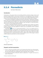
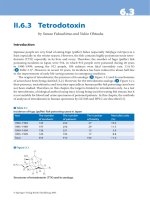
pyramid of disease in pregnancy (see Figure 1),
the base being the numerically larger general
pregnant population, the ‘tip of the iceberg’
being maternal death and much hidden
morbidity beneath the surface
9–11
. A clinical
insult may be followed by a systemic response
and subsequent organ dysfunction, which leads
to organ failure and eventual death
1,10
. Figure 1
illustrates both the severity continuum of
morbidity as well as factors that move a
woman up or down the pyramid. For example,
a faulty ambulance or wrongly cross-matched
blood might lead to an anemic woman dying of
hemorrhage unnecessarily (arrow A). If she is
well managed and treated promptly, there may
be no residual morbidity at all (arrow B). A
wrongly labelled blood bag that is spotted in
time still constitutes a ‘near-miss’ requiring
follow-up of the error, although the woman did
not experience a transfusion reaction. ‘Near-
miss’ now refers to avoidable risks whereas
SAMM retains the concept of the harmed or
damaged mother.
An agreed and accurate definition of SAMM
is not available, as studies in different parts of
the world use different criteria. The two main
definitions of SAMM to date are as follows:
(1) An organ system approach
10,11,13
, e.g.
shock from hemorrhage, severe pre-
eclampsia or specific organ failure; these are
best identified as they occur;
(2) Management, or process, based
12,14
, e.g.
admission to a high-dependency unit
(HDU) or intensive care unit (ICU) or
transfer to another health-care facility
(usually higher level); these are usually
retrospective studies.
A diligent unit is more likely to pick up cases via
the organ system approach and carefully record
them; this will translate into a disproportion
-
ately higher rate of SAMM
11
. On the other
hand, a poor-quality unit that does not recog-
nize and treat hemorrhage promptly may have
more severe sequelae as the natural history
progresses. Use of the management-based
approach relies on more easily agreed parame-
ters, but also on the availability of HDU/ICU
beds. Units have different policies and thresh-
olds for transfer. There may be an underestima-
tion of the true incidence of SAMM, especially
340
POSTPARTUM HEMORRHAGE
B
A
Poor antenatal
care
Antenatal
anemia
Patient factors
(social exclusion,
poor transport)
System factors Timely intervention
Skilled personnel
Good
maternity
services
System factors
(available drugs &
blood products)
Death
SAMM
Moderate morbidity
Mild morbidity
Clinical insult
General pregnant population
Figure 1 A representation of the morbidity–mortality continuum
362
Z:\Sapiens Publishing\A5211 - Postpartum Hemorrhage\Make-up\Postpartum Hemorrhage - Voucher Proofs #T.vp
30 August 2006 14:24:31
Color profile: Generic CMYK printer profile
Composite Default screen
in smaller, more isolated units and in
developing countries.
INCIDENCE OF SAMM
Quantifying SAMM is problematic as there
is no international definition and recording is
haphazard at best. Thus, there is a wide varia
-
tion in the estimate of incidence. Tables 1 and 2
summarize studies to date. Wide variations are
present in study settings, definitions and main
causes. Some studies use admission to ICU
14,15
,
others define the actual conditions responsible
for the morbidity
10,11
, and some list both
12
.
Two methods have been described to address
the relationship between severe morbidity and
mortality. These are the mortality-to-morbidity
ratio
1,13
and the mortality index
11,16
. The mortal
-
ity-to-morbidity ratio simply describes the num
-
ber of severe morbidity cases for each maternal
death
1,13
. The mortality index, on the other
hand, is defined as the number of maternal
deaths divided by the sum of women with
SAMM and maternal deaths, and is expressed
as a percentage
11,16
. Both can be expressed as
totals (all-cause) or by condition. They both
reflect the impact of a condition on severe
morbidity and mortality and identify those
conditions that are more or less amenable to
intervention. In general, the risk of mortality
depends on the prior health of the mother, the
severity of the particular condition, the access
to skilled help and the availability and quality
of medical intervention. Postpartum hemor
-
rhage is common, and has a very favorable
morbidity-to-mortality ratio (or low mortality
index)
1,11,13,16
. Stated another way, the condi
-
tion, at least in developed countries, is very
amenable to treatment. More women’s lives can
be saved with medical interventions than for a
comparable number of cases of infection or car
-
diac disease. Many lives can be, and indeed are
being, saved daily by the provision of adequate
maternity services world-wide. As hemorrhage
is so obviously both avoidable and treatable,
and because all parturients are at risk, it is tragic
that so many women die unnecessarily. Unfor
-
tunately, complacency in developed countries
about the daily marvels achieved in childbirth
4
has made any sudden unexpected threat to life
almost unbelievable and unbearable.
CAUSES OF SAMM
Most cases of SAMM fall into three major
categories:
(1) Hemorrhage;
(2) Hypertensive disorders of pregnancy
(including eclampsia and HELLP syn
-
drome);
(3) Sepsis.
Table 1 summarizes both the all-cause inci
-
dence of SAMM as well as the three major
causes. Rates in European countries are similar
for the three major causes of severe morbidity
despite the use of different definitions. Regard
-
less of geographical factors, hemorrhage is the
largest contributor, accounting for one-fifth
17
to one-half
10,18,19
of cases. Hypertensive disease
and its consequences account for 10%
18
to
45%
20
of cases of SAMM, whereas morbidity
secondary to sepsis is much lower (1.5%
18
to
20%
10
). Other rarer causes of severe morbidity
include uterine rupture, thromboembolic dis-
ease and psychiatric illness
5,21
. The Mothers’
Mortality and Severe Morbidity Survey was
conducted during the 1990s by an international
team which spanned 11 European countries.
There are two parts to the survey: MOMS-A
and MOMS-B
20
. MOMS-A collected and com-
pared data on maternal deaths, while MOMS-B
identified cases of severe morbidity
20
. The sur
-
vey established that, in European countries with
the highest SAMM rates, i.e. Belgium, Finland
and the UK, most of the difference was due
to higher incidence of hemorrhage. However,
maternal mortality was no higher than in other
European countries. This suggests either that
ascertainment of cases in these three countries
is more complete or that hemorrhage is not a
major cause of death; therefore the higher inci
-
dence of SAMM does not affect overall mortal
-
ity data. Alternatively, it may be that mortality
rates are associated more closely to the quality
of care than the prevalence of morbidity
20
. The
geographical areas chosen in different countries
had very different demographics, and this also
may have affected the rates of morbidity;
Belgium and the UK were represented by
Brussels and the South-East Thames region,
areas with significant inner-city and migrant
341
Severe acute maternal morbidity
363
Z:\Sapiens Publishing\A5211 - Postpartum Hemorrhage\Make-up\Postpartum Hemorrhage - Voucher Proofs #T.vp
30 August 2006 14:24:31
Color profile: Generic CMYK printer profile
Composite Default screen
342
POSTPARTUM HEMORRHAGE
Study, country and
year of publication
Incidence
of SAMM
(all causes)
Incidence of
hemorrhage
(% of total)
Incidence of
hypertension
(% of total)
Incidence of
severe sepsis
(% of total)
Additional comments
Stones
2
, UK, 1991 8.8 3.23
(36.8%)
2.77
(31.5%)
not available SAMM defined as ‘potentially life-threatening episodes’. Incidence for total
morbidity 267/1000. Incidence of all sepsis 30.5/1000 (severe sepsis not
separated out). Hemorrhage includes antepartum and postpartum if over
2000 ml and also 1 case of secondary PPH due to sepsis which required
hysterectomy
Bouvier-Colle
17
,
France, 1996
3.1 0.62
(20%).8
0.81
(26.2%)
0.14
(4.36%)
3rd highest cause of morbidity is embolic events at 0.38/1000. Hemorrhage
includes uterine rupture. Hypertensive disease includes cerebral hemorrhage
Bewley &
Creighton
12
,UK,
1997
4.97 2.3
(46.7%)
1.98
(40%).8
0.49
(10%)
SAMM = ITU admission. Total 30 cases of SAMM. 14 cases classed as
hemorrhage (blood loss > 2000 ml but a further 2 cases DIC/HELLP so
proportion due to hemorrhage could be > 50%)
Baskett & Sternadel
22
,
USA, 1998
0.72 0.16
(22%).8
0.18
(25%).8
0.1
(14.5%)
SAMM = ITU admission
Mantel
10
, South
Africa, 1998
10.9 6.1
(55.8%)
2.82
(25.8%)
2.16
(19.7%)
Sepsis incorporates septic abortion, chorioamnionitis and puerperal sepsis.
Hemorrhage incorporates antepartum and postpartum hemorrhage and
emergency hysterectomy; PPH alone is 1.8/1000
Prual
18
, West Africa,
2000
59.8 29.6
(49.5%)
6.15
(10.3%)
0.9
(1.5%)
Obstructed labor is significant cause for severe morbidity (20.5/1000 of
which 1.2/1000 uterine rupture)
Waterstone
13
(COSMO), UK, 2001
12.0 6.7
(55.7%)
4.6
(38%).8
0.35
(2.89%)
Clinically based definitions, not including management processes. Estimated
blood loss > 1500 ml picked up 55% of cases of SAMM due to hemorrhage
Brace
19
, Scotland,
2004
3.8 1.9
(50%).8
1.15
(30%).8
0.09
(3%).89
Septic shock is the only category for sepsis. Number of SAMM due to
hypertensive disease derived by adding the number of cases with eclampsia,
renal dysfunction and pulmonary edema. Only one-third of patients with
SAMM were admitted to ITU
Zhang
20
(MOMS-B),
Europe, 2005
9.48 4.6
(48.8%)
4.33
(45.7%)
0.8
(8.2%)
Multinational study, rates differing widely between countries. Range of
SAMM 6–14.7%, highest in Finland, Belgium and UK; lowest rates in Italy,
Ireland, France
DIC, disseminated intravascular coagulation; SAMM, severe adverse maternal morbidity; ITU = intensive therapy unit; HELLP, hemolysis, elevated liver
enzymes, low platelets; PPH, postpartum hemorrhage
Ta bl e 1
The incidence of each major cause of morbidity per 1000 deliveries
364
Z:\Sapiens Publishing\A5211 - Postpartum Hemorrhage\Make-up\Postpartum Hemorrhage - Voucher Proofs #T.vp
30 August 2006 14:24:32
Color profile: Generic CMYK printer profile
Composite Default screen
343
Severe acute maternal morbidity
Author,
country, year
of publication
Type of study,
type of unit
Number of
deliveries,
cases of
SAMM,
cases of
hemorrhage
Incidence
of SAMM
or ITU
(/1000
deliveries)
Incidence of
hemorrhage
(/1000
deliveries)
%of
SAMM
due to
hemorrhage
Definition of severe hemorrhage
(additional comments)
Perinatal outcome
Number
of maternal
deaths, MMR
for SAMM
overall,
mortality
per 100 000
Graham &
Luxton
41
,
UK, 1989
retrospective,
general ITU
21 983
23 (ITU)
1(5)*
1.04 0.05
(0.23)*
4.35%
(21.7%)*
1 case of hemorrhage counted but 5 cases
in total (3 abruptions: 1 was the hemorrhage
and 2 cases of DIC). 9 patients showed
some coagulopathy, 5 received > 4 units
transfusion
1 intrauterine death 2.4
11.5 : 1
9.1.4
Mabie &
Sibai
23
,US,
1990
retrospective, 3-bed
dedicated obstetric
ITU
22 651
200 (ITU)
21
8.82 0.93 10.5% massive hemorrhage not defined not collected not collected
Stones
2
,UK,
1991
retrospective, single
unit
2164
19
7
8.8 3.23 36.8% hemorrhage > 2000 ml or DIC or
hysterectomy
not collected 0.4
0.4
Killpatrick &
Matthay
14
,
US, 1992
retrospective,
general ITU
8000
32 (ITU)
4
4.82 0.5 12.5% hemorrhage/hemodynamic instability.
52% of postpartum admissions were for
hemodynamic instability
2 stillbirths delivered
on ITU. No neonatal/
fetal deaths after
admission to ITU
4.4
8:1
50.4
Monaco
15
,
US, 1993
retrospective, ITU
admissions
15 323
38 (ITU)
4
2.47 0.26 10.5% 2 cases following PPH and 2 cases of
hematologic dysfunction (local policy is
to admit only for ventilatory support or
pulmonaryarterycatheterization)
perinatal mortality
rate 12% (4 of 33
pregnancies followed
up)
7.4
5.4 : 1
45.7
Bouvier-Colle
17
,
France, 1996
retrospective,
2 French regions
140 323
435 (ITU)
87
3.1 0.62 20%.38 hemorrhage not defined but includes
uterine rupture
stillbirth rate collected
only 24.6% in
hypertension, 17.3%
in hemorrhage, 33.3%
in sepsis
22.4
19.8 : 1
15.7
Bewley &
Creighton
12
,
UK, 1997
retrospective,
general ITU
6039
30 (ITU)
14
4.97 2.3 46.7%.38 transfer to ITU for blood loss > 2000 ml not collected not collected
Continued
Ta bl e 2
The incidence of SAMM and hemorrhage and definitions used to ascertain cases
365
Z:\Sapiens Publishing\A5211 - Postpartum Hemorrhage\Make-up\Postpartum Hemorrhage - Voucher Proofs #T.vp
30 August 2006 14:24:33
Color profile: Generic CMYK printer profile
Composite Default screen
344
POSTPARTUM HEMORRHAGE
Author,
country, year
of publication
Type of study,
type of unit
Number of
deliveries,
cases of
SAMM,
cases of
hemorrhage
Incidence
of SAMM
or ITU
(/1000
deliveries)
Incidence of
hemorrhage
(/1000
deliveries)
%of
SAMM
due to
hemorrhage
Definition of severe hemorrhage
(additional comments)
Perinatal outcome
Number
of maternal
deaths, MMR
for SAMM
overall,
mortality
per 100 000
Lapinsky
27
,
Canada, 1997
retrospective, ITU
admissions in
tertiary hospital
25 000
65 (ITU)
11
2.6 0.44 17%.38 hemorrhage requiring ITU admission,
not defined. 52% of admissions involved
hemodynamic instability; 4 hysterectomies
perinatal mortality
rate 11%
0.4
0.4
Tang
25
,Hong
Kong, 1997
retrospective, single
center
39 354
49 (ITU)
26
1.24 0.66 53%.38 blood loss 1000–8500, mean 3500 ml.
Received blood transfusion (mean 12
units), platelet transfusion or FFP. DIC
in 34% and mild coagulopathy in 27%
of hemorrhage cases. Hysterectomy in
22 cases (84.6% of all hemorrhage)
perinatal mortality
rate 10.2%
2.4
24.5 : 1
5.1
Mantel
10
,South
Africa, 1998
prospective,
multicenter
13 429
147
82
10.9 6.1 55.8% severe hemorrhage = hypovolemia
requiring 5 or more units of blood for
resuscitation or emergency hysterectomy
not collected 30.4
4.9 : 1
223.4
Mahutte
28
,
Canada, 1999
retrospective,
2 tertiary care units
with general ITU
44 340
131 (ITU)
34
2.95 0.77 26%.38 hemorrhage causing admission due to
abnormal placentation, atony, lacerations,
RPOC, severe coagulopathy. 27 (79%)
received blood products and 12 (35%)
admitted after Cesarean hysterectomy
not collected 3.4
43.7 : 1
6.8
Waterstone
13,21
,
UK, 2001
prospective,
case–control,
multicenter
48 865
588
327
12.0 6.7 55.7% blood loss > 1500 ml/peripartum
hemoglobin drop ≥ 4g/dloracute
transfusion ≥ 4 units
perinatal mortality
rate 7.5%
5.4
.4118 : 1
10.2
Prual
18
,West
Africa, 2000
prospective,
6 countries
20 326
1215
601
59.8 29.6 49.5% hemorrhage requiring transfusion,
hospitalization > 4 days or hysterectomy
(only 2.8% of deaths were due to severe
hemorrhage)
not collected 38.4
32 : 1
187.4
Hazelgrove
24
,
UK, 2001
retrospective,
multi-unit
122 850
210 (ITU)
70
1.7 0.6 33.3% major hemorrhage not specified; 35%
were short admissions (< 2/7) and no
specific interventions. 7 patients required
transfer to specialist ITUs
fetal mortality rate
20% (includes fetal
losses < 24 weeks
gestation)
7.4
30 : 1
5.7
Ta bl e 2 Continued
366
Z:\Sapiens Publishing\A5211 - Postpartum Hemorrhage\Make-up\Postpartum Hemorrhage - Voucher Proofs #T.vp
30 August 2006 14:24:33
Color profile: Generic CMYK printer profile
Composite Default screen
345
Severe acute maternal morbidity
Murphy &
Charlett
29
,
US, 2002
retrospective cohort,
general ITU in
teaching hospital
51 576
50 (ITU)
12
0.97 0.23 24%.83 no definition/information on transfusion
given but cause of hemorrhage given;
7 hysterectomies
perinatal mortality
rate 14%.
3.4
16.7 : 1
5.8
Vandecruys
16
,
South Africa,
1997–1999
prospective, tertiary
center, phase 1
26 152
305
44
11.7 1.68 14.4% definitions not given. Data on hemorrhage
refer to PPH
not collected 59.4
5.2 : 1
225.6
Vandecruys
16
,
South Africa,
2002
prospective tertiary
center, phase 2
(re-audit)
13 854
121
23
8.7 1.66 19%.38 as above. SAMM and mortality declined
compared to the first audit due mainly to
reduction in abortion complications
not collected 26.4
4.7 : 1
188.4
Pattinson
11
,
South Africa,
2003
prospective,
3 geographic areas
(urban and rural)
NA
423
130
NA NA 30.7% condition-based definitions same as
Mantel10. Calculates mortality index but
cannot define incidence as number of
deliveries not given. Hemorrhage includes
antepartum and postpartum; PPH alone is
18%. PPH is second most common cause
of SAMM but 7th cause of death
not collected 128.4
3.3 : 1
NA
Brace
19
,
Scotland,
2004
prospective
observational,
(22 consultant-led
units in Scotland)
51 165
196
98
3.8 1.9 50%.38 major hemorrhage = cases transfused at
least 5 units during the acute episode of
hypovolemia (13 categories of morbidity
leading to ITU admission)
not collected 4.4
.449 : 1
7.8
Gandhi
26
,
South Africa,
2004
prospective, 4 rural
primary hospitals
5728
31
10
5.41 1.75 32%.38 Mantel’s definitions adapted for use in
primary hospital with limited resources.
Includes antepartum and postpartum
hemorrhage, DIC and hysterectomy
not collected not disclosed
Zhang
20
(MOMS-B),
Europe, 2005
Population based
questionnaire,
multi-unit, multi-
national
182 734
1734
847
9.48 4.6 48.8% blood loss > 1500 ml/blood loss requiring
plasma expanders and/or blood loss
> 2500 ml in 24 h/blood loss resulting in
maternal death. Incidence range 0.7–8.8
according to country
fetal death rate 4.8% 4.4
433.5 : 1
2.2
SAMM, severe adverse maternal morbidity; ITU, admissions to intensive therapy unit; (ITU), SAMM cases defined as ITU admissions; MMR, morbidity
to mortality ratio (calculated from rate of SAMM to mortality); PPH, postpartum hemorrhage; DIC, disseminated intravascular coagulation;
HD, high-dependency unit; RPOC, retained products of conception; NA, not available
*Data in parentheses are calculations as applied for five cases
367
Z:\Sapiens Publishing\A5211 - Postpartum Hemorrhage\Make-up\Postpartum Hemorrhage - Voucher Proofs #T.vp
30 August 2006 14:24:33
Color profile: Generic CMYK printer profile
Composite Default screen
populations, whilst the three regions in France
did not include major cities
20
.
In general, severe hemorrhage and hyper
-
tension have much higher incidence (range
0.6
17
–29.6
18
and 0.18
22
–6.15
18
per 1000
deliveries, respectively) than severe sepsis
(0.09
19
–2.16
10
per 1000). The same low rate for
sepsis is observed in West Africa, where the sec
-
ond greatest cause of SAMM after hemorrhage
is obstructed labor
18
. Uterine rupture has been
combined with data for obstructed labor in
one study
18
and with hemorrhage in another
17
.
Waterstone and colleagues (2001)
13
considered
uterine rupture as a separate entity; this is a
more accurate way of using the data unless we
have clear evidence of the blood loss associated
with each case
13
. Few studies have looked at
immediate moderate morbidity, although a
number of studies of the puerperium examine
moderate to minor morbidity
2,13
. For example,
Stones and colleagues (1991) included less
severe cases of morbidity in their analysis:
anesthetic complications (usually post-spinal
headache) 0.46%; urinary retention/inconti-
nence 1.2%; late perineal complications
0.65%
2
.
HEMORRHAGE AS A CAUSE FOR
SAMM: THE EVIDENCE
Most studies of SAMM to date report severe
hemorrhage as the largest single contributing
factor. Severe hemorrhage was defined by one
or a combination of factors:
(1) Estimated blood loss ≥ 1500 ml (or
≥ 2000 ml);
(2) Hemoglobin drop ≥ 40 g/dl;
(3) Transfusion of ≥ 4 units of blood.
Table 2 outlines the incidence of severe hemor
-
rhage in a variety of studies to date. The prob
-
lem of varied definitions is highlighted, making
comparisons between studies difficult. The pro
-
portion of SAMM due to hemorrhage is also
shown. This varies widely but tends to be lower
in studies that are management-based, as not all
cases require admissions to ICU. Local policies
and availability of obstetric HDU beds on labor
wards has a great influence on the management
of massive postpartum hemorrhage as it avoids
delays in treatment secondary to transfers and
also ensures continuity of obstetric care
23
.
Obstetric HDU beds are becoming more com
-
monplace in tertiary centers in the UK
12,13,24
and US
14,15,23
. Comparisons are more valid
between studies that have used agreed or similar
definitions for hemorrhage
10,12,18,19,25
. Many of
them are prospective
10,11,13,19,26
rather than ret
-
rospective
14,17,27–29
, and they tend to find higher
proportions of hemorrhage: 30–50% rather
than 10–30%, although there is some over
-
lap
12,18
.The higher rate from prospective
studies is likely to be due to better case
ascertainment. Rates appear to be very
similar in developed
13,19,20
and developing
10,18
countries when comparable definitions are
used.
Emergency obstetric hysterectomy provides
another means of examining SAMM caused by
postpartum hemorrhage. It has the advantage
of being relatively clearly defined, and is rare
enough to be easy to collect data. The tradi-
tional advice is to perform hysterectomy early to
avoid mortality
6
. The threshold for performing
hysterectomy clearly varies with operator and
unit, but evidence exists that early hysterectomy
decreases morbidity
30
. The new United King-
dom Obstetric Surveillance System (UKOSS)
requires units to report cases of specified rare
conditions on a monthly basis. Hysterectomy
has been chosen as the exemplar obstetric
morbidity, and this large national surveillance
should provide further information about best
practice in the future.
RISK FACTORS FOR SAMM AS
APPLIED TO HEMORRHAGE
Although it is challenging to define the size of
the problem (i.e. the incidence of SAMM as a
result of hemorrhage), it is necessary to under
-
stand the factors that increase the risk of severe
hemorrhage. Table 3 has been adapted from
the findings of a multicenter case–control study
in the South East Thames region of the UK
(COSMO)
13
and outlines the odds ratios of
having a severe hemorrhage (as defined by
blood loss ≥ 1500 ml, hemoglobin drop
≥ 40 g/dl, or blood transfusion ≥ 4 units),
depending on a wide range of risk factors. The
main predictors are:
346
POSTPARTUM HEMORRHAGE
368
Z:\Sapiens Publishing\A5211 - Postpartum Hemorrhage\Make-up\Postpartum Hemorrhage - Voucher Proofs #T.vp
30 August 2006 14:24:34
Color profile: Generic CMYK printer profile
Composite Default screen
(1) Demographic: age ≥ 35 years, non-white
race, social exclusion (this was a composite
measure of a woman’s social deprivation
beyond the use of her marital or partner’s
employment status. It included concealed
pregnancy, age < 16 years, poor housing,
‘on income support’ in the notes, previous
minor or child in local authority or state
care (currently or in the past), in trouble
with the law (currently or previously), living
alone, unbooked, unwanted pregnancy,
currently or previously in foster care, care
order being considered on potential child,
social worker involved, or drug or alcohol
dependency
21
);
(2) General medical factors: anemia, depression,
diabetes, hypertension, epilepsy;
(3) Obstetric factors: previous postpartum
hemorrhage, multiple pregnancy, ante
-
natal admissions, obstetric interventions
(augmentation with oxytocin, manual
removal of placenta, emergency Cesarean
section).
Other studies
31,32
find the same trend, with
death and severe morbidity being more com-
mon in black women, multiparae and women
of ‘low status’
32
as defined by poor education,
poverty, low antenatal care attendance, low
contraceptive ever-use and little power to make
decisions regarding access to health care. In
Geller’s study (2004)
31
, the peak of mortality
and SAMM occurred in the 20–34-year age
group, with three times fewer women aged > 35
years in all categories of the morbidity-to-
mortality continuum
31
. This is more likely
to reflect the age distribution of the study
population rather than a true difference
between the USA and the UK, and emphasizes
the need for the use of multiple logistic
regression to tease out risk factors. Manual
removal of placenta had the biggest impact
13
,
in keeping with abnormally adherent placentas
being a major cause of postpartum hemorrhage.
Antenatal anemia, Cesarean section, and the
use of antidepressants or antiepileptics at
booking also appear to have significant impacts,
though their relative importance is difficult
347
Severe acute maternal morbidity
Risk factors
Odds ratios for SAMM
(all causes)
Odds ratios for SAMM
due to hemorrhage
Age > 35 years
Blood pressure at booking
Black
Other race
Social exclusion
Smoker
Previous postpartum hemorrhage
Hypertension
Diabetes
Multiple pregnancy
Antenatal admission
Taking iron at booking
Taking antidepressants at booking
Taking antiepileptics at booking
IOL because overdue
IOL on medical grounds
Oxytocin augmentation
Manual removal of placenta
Emergency Cesarean
1.46 (1.11–1.92)
1.23 (1.12–1.34)
1.16 (0.85–1.58)
1.93 (1.24–2.99)
2.64 (1.69–4.11)
0.68 (0.49–0.93)
2.41 (1.53–3.77)
1.10 (0.63–1.95)
1.76 (0.43–7.20)
2.21 (1.24–3.96)
1.75 (1.37–2.23)
5.53 (2.28–13.41)
4.3 (0.91–1.88)
5.31 (1.4–20.13)
1.36 (0.99–1.88)
2.45 (1.68–3.57)
0.99 (0.76–1.28)
9.60 (5.67–16.28)
4.31 (3.39–5.49)
1.41 (1.03–1.95)
1.18 (1.06–1.31)
0.97 (0.66–1.42)
1.82 (1.09–3.03)
2.91 (1.76–4.82)
0.65 (0.44–0.96)
2.74 (1.69–4.44)
0.82 (0.37–1.80)
1.85 (0.38–9.14)
2.29 (1.2–4.37)
1.85 (1.39–2.47)
5.98 (2.28–15.65)
10.55 (2.19–50.71)
5.75 (1.28–25.72)
1.38 (0.95–1.99)
1.33 (0.87–1.07)
1.61 (1.2–2.15)
13.12 (7.72–22.30)
3.09 (2.29–4.17)
SAMM, severe adverse maternal mortality; IOL, induction of labor
Ta bl e 3
Risk factors for having a severe adverse maternal morbidity, or severe hemorrhage (from
Waterstone et al., 2001)
8
. Figures are odds ratios (95% confidence interval)
369
Z:\Sapiens Publishing\A5211 - Postpartum Hemorrhage\Make-up\Postpartum Hemorrhage - Voucher Proofs #T.vp
30 August 2006 14:24:34
Color profile: Generic CMYK printer profile
Composite Default screen