Báo cáo y học: "Successful tumour necrosis factor (TNF) blocking therapy suppresses oxidative stress and hypoxiainduced mitochondrial mutagenesis in inflammatory arthritis" pot
Bạn đang xem bản rút gọn của tài liệu. Xem và tải ngay bản đầy đủ của tài liệu tại đây (935.01 KB, 9 trang )
RESEARCH ARTICLE Open Access
Successful tumour necrosis factor (TNF) blocking
therapy suppresses oxidative stress and hypoxia-
induced mitochondrial mutagenesis in
inflammatory arthritis
Monika Biniecka
1
, Aisling Kennedy
1
, Chin T Ng
1
, Ting C Chang
1
, Emese Balogh
1
, Edward Fox
2
, Douglas J Veale
1
,
Ursula Fearon
1
and Jacintha N O’Sullivan
3*
Abstract
Introduction: To examine the effects of tumour necrosis factor (TNF) blocking the rapy on the levels of early
mitochondrial genome alterations and oxidative stress.
Methods: Eighteen inflammatory arthritis patients underwent synovial tissue oxygen (tpO
2
) measurements and
clinical assessment of disease activity (DAS28-CRP) at baseline (T0) and three mont hs (T3) after starting biologic
therapy. Synovial tissue lipid peroxidation (4-HNE), T and B cell specific markers and synovial vascular endothelial
growth factor (VEGF) were quantified by immunohistochemistry. Synovial levels of random mitochondrial DNA
(mtDNA) mutations were assessed using Random Mutation Capture (RMC) assay.
Results: 4-HNE levels pre/post anti TNF-a therapy were inversely correlated with in vivo tpO
2
(P < 0.008; r = -0.60).
Biologic therapy responders showed a significantly reduced 4-HNE expression (P < 0.05). High 4-HNE expression
correlated with high DAS28-CRP (P = 0.02; r = 0.53), tender joint count for 28 joints (TJC-28) ( P = 0.03; r = 0.49),
swollen joint count for 28 joints (SJC-28) (P = 0.03; r = 0.50) and visual analogue scale (VAS) (P = 0.04; r = 0.48).
Strong positive association was found between the number of 4-HNE positive cells and CD4+ cells (P = 0.04; r =
0.60), CD8+ cells (P = 0.001; r = 0.70), CD20+ cells (P = 0.04; r = 0.68), CD68+ cells (P = 0.04; r = 0.47) and synovial
VEGF expression (P = 0.01; r = 063). In patients whose in vivo tpO
2
levels improved post treatment, significant
reduction in mtDNA mutations and DAS28-CRP was observed (P < 0.05). In contrast in those patients whose tpO
2
levels remained the same or reduced at T3, no significant changes for mtDNA mutations and DAS28-CRP were
found.
Conclusions: High levels of synovial oxidative stress and mitochondrial mutation burden are strongly associated
with low in vivo oxygen tension and synovial inflammation. Furthermore these significant mitochondrial genome
alterations are rescued following successful anti TNF-a treatment.
Introduction
Mitochondria produce ATP through oxidat ive metabo-
lism to provide cells with energy under p hysiological
conditions. The mitochondrial electron transport chain
(ETC) i s also a major cellular source of reactive oxygen
species (ROS) as some of the electrons passing to
molecular oxygen are prone to leakage from the chain
and get trapped by oxygen, which converts to superox-
ide [1]. Hypoxia characterised by an inadequate supply
of molecular oxygen, can trigger mitochondria dysfunc-
tion through ineffective functioning of respiratory com-
plexes of ETC [2,3].
Free oxygen radicals are highly active molecules and
increased mitochondrial ROS generation promotes cel-
lular oxidative stress resulting in oxidative mitochondrial
DNA (mtDNA) damage and lipid peroxidation.
* Correspondence:
3
Department of Surgery, Institute of Molecular Medicine, Trinity Centre for
Health Sciences, St James’s Hospital, St James’s Hospital, St James’s Street,
Dublin 8, Ireland
Full list of author information is available at the end of the article
Biniecka et al. Arthritis Research & Therapy 2011, 13:R121
/>© 2011 Binie cka et al.; licensee BioMed Central Ltd. This is an open access article distributed under the terms of the Creative Commons
Attribution License (htt p://creativecommons.org/licenses /by/2.0), which permits unrestricted use, distribution, and reproduction in
any medium, provided the original work is properly cited.
Moreover, ROS mediate the stress signalling pathways
involving nuclear factor-kappa B (NF-B) [4]. mtDNA is
in the proximity of ROS generation site and has rela-
tively limit ed repair capacity, which makes it vulnerable
to high mutation rates [5]. Mutations and deletions of
the mitochondrial genome in genes encoding proteins
for subunits of mitochondrial respiratory chain com-
plexes I-V, rRNA and tRNA have been linked to a vari-
ety of degenerative human diseases and high levels of
mtDNA mutations have been also found in many
tumours and cancer cells [5,6].
Oxidative stress, which arises from an imbalance
between ROS production and ant ioxidant defences,
results also in lipid peroxidation of cell membrane poly-
unsaturated fatty acids [7]. The primary products of free-
radical attack of biological membranes are lipid hydro-
peroxides, which can decompose to highly reactive, cyto-
toxic secondary end products, such as 4-hydroxy-2-
nonenal (4-HNE) [8]. 4-HNE is an endogenously gener-
ated a,b unsaturated aldehyde, which is not only a mar-
ker of extensive oxidative stress but also can modulate
cellular metabolism, inflammatory responses and apopto-
sis via its effects on transcriptional regulation and protein
modification [9]. 4-HNE-induced mitochondrial protein
modifications include those involved in the ETC, cellular
respiration and Krebs cycle [10]. Moreover, 4-HNE can
form adducts on DNA bases and modifies mtDNA thus
measurement of such modifications may reflect the level
of mitochondrial alterations [11].
Inflammatory arthritis (IA) is a chronic, progressive
disorder associated with joint inflammation, synovial tis-
sue hypertrophy, joint effusions and degradation of
articular cartilage and bone. The normal synovial tissue
is a relatively acellular structure with a lining layer (one
to two cells thick) comprised of macrophages and fibro-
blasts. The morphology of IA synovium is strikingly dif-
ferent. There is a significant increase in the number of
blood vessels that are associated with differential vascu-
lar morphology. Furthermore, the early vascular changes
are accompanied by increased recruitment of macro-
phages and synovial fibroblast cells in the lining layer,
along with infiltration of T, B and plasma cells. The pre-
cise mechanisms involved in regulation of persistent
synovial infiltration and invasion are unclear, but high
levels of TNF-a may be crucial in mediating the patho-
genesis of IA. TNF-a is a proinflammatory cytokine,
activating the NF-B pathway, leading to a downstream
cascade of other proinflammatory cytokines [12,13].
Moreover, it is known to increase mitochondrial ROS
production [14,15] and induce the formation of lipid-
derived aldehydes [16]; however TNF-a-induced mito-
chondrial mutagenesis has not yet been examined in
patients with IA. Current targeted biologic therapies,
including anti-TNF-a inhibitors result in greater disease
improvement and prevention of joint erosion, although
clinical studies on the efficacy of TNF-a blocking agents
clearly show that ab out 40% of patients receiving this
therapy are non-responders.
Recently, we demonstrated that successful biologic
therapy significantly improves in vivo synovial hypoxia
and i t is strongly associated with improvement of joint
inflammation [17]. In this study we investigate if suc-
cessful anti-TNF-a treatment alters the levels of early
mitochondrial genome altera tions, which can play a cru-
cial role in governing clinical response or resistance.
Furthermore, we determine if TNF-a blocking therapy
changes the levels of synovial 4-HNE, further confirming
the relation between hypoxia, oxidative damage and
mitochondrial mutagenesis.
Materials and methods
Patient recruitment
All research was carried out in accordance with the
Dec laration of Helsinki, and approval for this study was
granted by the St. Vincent’s University Hospital Medical
Research and Ethics Committee. Eighteen patients with
active IA (rheumatoid arthritis (RA) n = 14 and psoriatic
arthritis (PsA) n = 4) were recruited from outpatient
clinics at Department of Rheumatology, St. Vincent’s
University Hospital. All patients fulfilled the diagnostic
criteria for RA and PsA [18,19]. All patients provided
fully informed consent and underwent arthroscopy at
baseline (T0) and three months after commencement of
TNF blocking therapy (T3). At baseline, 50% of patients
were naive for disease-modifying anti-rheumatic drugs
(DMARDs) and corticosteroids; however, all patients
including those on DMARDs (methotrexate (MTX)
alone 35%, MTX + salazopyrine 10%, and plaquenil
alone 5%) were biologic naive, had active disease, had at
least one inflamed knee joint and were due to com-
mence biologic therapy. Clinical and laboratory assess-
ment was performed using standard measures of 28
tender and swollen joint count (DAS28), rheumatoid
factor, anti-cyclic citrullinated peptide antibody, erythro-
cyte sedimentation rate (ESR), C-reactive protein (CRP)
and global health visual analogue scale (VAS). All mea-
surements were obtained on the same day prior to base-
line and three months after anti TNF-a treatment
arthroscopy.
Arthroscopy, measurement of in vivo tpO
2
and sample
collection
Under local anaesthetic, patients (n =18)underwent
arthroscopy at baseline and three months after com-
mencement of TNF blocking therapy. Arthroscopy of
the inflamed knee was perfo rmed using a Wolf 2.7 mm
needle arthros cope. Macroscopic synovitis and v ascular-
ity were scored on a VAS (0-100 mm). A LICOX
®
Biniecka et al. Arthritis Research & Therapy 2011, 13:R121
/>Page 2 of 9
combined pO
2
and temperature probe (Integra Life
Sciences Corporation, New Jersey, USA) was used to
obtain synovial tissue oxygen partial pressure as pre-
viously described [20]. Synovial membrane biopsies were
obtained from the site of the oxygen tension measure-
ment and immediately embedded in mounting media
for immunohistochemical analysis or snap frozen in
liquid nitrogen for mitochondrial mutagenesis analysis.
Immunohistochemistry and scoring
Immunohistochemistry was performed using 7 μm cryo-
stat synovial tissue sections and the DAKO ChemMate
Envision Kit (DAKO, Glostrup, Denmark). Sections
were defrosted at room temperature for 20 minutes,
fixed in acetone for 10 minutes and washed in PBS for
5 minutes. Non-specific binding was blocked using 10%
casein in PBS for 20 minutes. The sections were incu-
bated with primary antibodies against human 4-HNE
(Genox, Baltimore, MD, USA), CD4, CD8, CD20, CD68
(all from DAKO, Glostrup, Denmark) and vascular
endothelial growth factor ( VEGF) (Santa Cruz Biotech-
nology, Inc., Santa Cruz, CA, USA). IgG control antibo-
dies were used as negative controls. Following primary
antibody incuba tion endogenous peroxidase activity was
blocked using 0.3% hydrogen peroxide for seven minutes
at room temperature. Slides wer e incubated with sec-
ondary antibody/HRP (DAKO, Glostrup, Denmark).
DAB (1:50) was used to visualise staining, and Mayer’ s
haematoxylin (BDH Laboratories, Poole, UK) was incu-
bated for 30 seconds as a counterstain prior to mount-
ing in Pertex mounting media. Images were captured
using Olympus DP50 light microscope and AnalySIS
software (Soft Imaging System Corporation, Lakewood,
CO, USA). Slides were scored separately for lining and
sublining layers using well established and validated
semi -quantitative scoring method, where the percentage
of cells that were positive for a specific marker was
compared with the percentage of cells that were nega-
tive [21]. Percentage positivity was graded using 0 to 4
scale, where 0 represented no stained cells, 1 was 1 to
25% stained cell s, 2 was 25 to 50% stained cells, 3 was
50 to 75% stained cells, and 4 was 75 to 100% stained
cells.
Mitochondrial random mutation capture assay
A sub-group of eight patients were selected from the
initial cohort to quantify the levels of mitochondrial
point mutations before and after treatment. Levels of
mitochondrial point mutations in snap frozen synovial
biopsies were analysed in a blinded fashion using Mito-
chondrial Random Mutation Capture assay as described
previously [22]. Biopsies were homogenised (Precellys
24, Stretton Scientific Ltd., Stretton, Derbyshire, United
Kingdom) in 10 mM Tris-HCl, pH 8.0, 150 mM NaCl,
20 mM EDTA, 0.5% SDS buffer and digested with Pro-
teinase K (Sigma-Aldrich, Dublin, Ireland) at a final con-
centration o f 0.2 mg/ml and incubated overnight at 56°
C. The mtDNA was extracted using phenol-chloroform-
isoamyl alcohol (25:24:1 by volume, Sigma-Aldrich,
Dublin, Ireland) added in a 1:1 ratio with the lysed tis-
sue, mixed thoroughly by shaking, and centrifuged at
more than 12,000 × g for 10 minutes. The aqueous
phase was gently removed from the top of the solution,
without disturbing the interphas e. The aqueous solution
was again mixed with phenol-chloroform-i soamyl alco-
hol in a 1:1 ratio and r e-extracted. One-tenth volume o f
3 M sodium acetate was added, and the samples were
precipitated with 2 to 2.5 volumes of ethanol. The DNA
samples were resuspe nded in 50 μl10mMTrisCl.Ten
micrograms of mtDNA were digested with 100 units of
TaqaI restriction enzyme (New England BioLabs, Herts,
United Kingdom), 1 × BSA and a TaqaI-specific diges-
tion buffer (10 mM Tris-HCl, 10 mM MgCl
2
, 100 mM
NaCl, pH 8.4) for 10 hours; 100 units of TaqaIbeing
added to the reaction mixture every hour.
PCR amplification was performed in 25 μl reactions,
containing 12.5 μl 2 × SYBR Green Brilliant Mastermix
(Strata gene, Agilent Technologies, Inc., Santa Clara, CA,
USA), 0.1 μl UDG (New England Biosciences, Herts, Uni-
ted Kingdom), 0.7 μlof10pM/μl forward and reverse pri-
mers (Integrated DNA Technologies, Inc., San Diego, CA,
USA), and 6.7 μl water. The samples were amplified using
a Roche Lightcycler 480 using the following protocol: 37°C
for 10 minutes and 95°C for 10 minutes followed by 45
cycles of 95°C for 15 seconds, 60°C for 1 minute. Samples
were held at 72°C for 7 minutes and, following melt curve
analysis, immediately stored at -80°C. The primer
sequences used were as follows: for mtDNA copy number:
5’ACAGTTTATGTAGCTTACCTCC-3’ (forward) and 5’-
TTGCTGCGTGCTTGATGCTTGT-3’ (reverse); for ran-
dom mutations: 5’ -CCTCAACAGTTAAATCAA-
CAAAACTGC-3’ (forward) and 5’ -GCGCTTACTT T
GTAGCCTTCA-3’ (reverse).
Statistical analysis
Data are presented as medians and interquartile rang es.
Data were assessed using Wilcoxon’s signed-rank test or
Spearman’ s rank correlation coefficient as appropriate
using the Statistical Package for the Social Sciences
(SPSS, Chicago, IL, USA). All P values are two-sided
and P values less than 0.05 were considered statistically
significant.
Results
In vivo changes of oxidative stress pre/post anti TNF-a
therapy
Eighteen IA patients underwent synovial tissue oxygen
tension (tpO
2
) measurements and clinical assessment of
Biniecka et al. Arthritis Research & Therapy 2011, 13:R121
/>Page 3 of 9
disease activity (28-joint coun t disease activity score
using C-reactive protein (DAS28-CRP)) at baseline and
three months after start ing biologic therapy. At T3
patients were categorised according to remission criteria
using the DAS28 cut-off less than or more than 2.6.
Patients with DAS28-CRP less than 2.6 were defined as
responders (n = 7) and patients with DAS28-CRP more
than 2.6 were defined as non-responders (n =11).In
responders, the median baseline pO
2
in the synovial tis-
sue was 18.07 mmHg (range 4.3 to 42.2 mmHg), and
was lower than in those patients at T3 (median tpO
2
39.25 mmHg (range 24.7 to 68.2 mmHg)). Of clinical
responders, 86% had a corresponding increase in their
synovial tpO
2
measurements. In non-responders the
median baseline pO
2
was 23.75 mmHg (range 6.8 to
46.4 mmHg), and their median pO
2
level after biologic
therapy was 19.78 mmHg (range 10.5 to 39.6 mmHg).
In clinical non-responders, 64% patients showed
decrease in their synovial tpO
2
levels at T3. Further-
more, tpO
2
levels did not differ significantly between
baseline patients with RA and those with PsA (n =14
RA, n = 4 PsA). The median oxygen tension for RA was
23.5 mmHg and for PsA was 14.5 mmHg (P = 0.3).
To determine whether biologic trea tment changes the
levels of synovial oxidative damage, the number of 4-
HNE positive cells was assessed in both lining and sub-
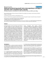
lining layers of synovial tissue. Figures 1a and 1b show
representative images of 4-HNE expression levels in
responders at T0 and T3, respectively. Figure 1c graphi-
cally illustra tes significantly reduced cytoplasmic 4-HNE
expression in sublining layer in patients who successfully
responded to anti-TNF-a therapy (P < 0.05). No signifi-
cant differences in the levels of cytoplasmic 4-HNE
expression pre/post therapy were found in non-respon-
ders (Figures 1d to 1f). In addition, the levels of 4-HNE
did not differ significantly between baseline patients
with RA and those with PsA (P = 0.6).
Previously, we demonstrated significant baseline
inverse c orrelation between tpO
2
measurements and 4-
HNE expression [20] . In this study we extend these
findings and demonstrate that change in tpO
2
is also
significantly and inversely correlated with changes in 4-
HNE levels pre/post biologic therapy (P <0.008;r=
-0.60; Table 1). It suggests that as synovial tissue
becomes less hypoxic oxidative stress is decreased.
Furthermore, when patients were categorised according
Figure 1 Representativ e pre/post immun ohistochemical images of 4-HNE expression and their graphical representation . (a to c)
Responders. (d to f) Non-responders. T0 is time at baseline; T3 is three months after anti-TNF-a treatment. (a to b) Biologic therapy responders
showed lower synovial 4-hydroxy-2-nonenal (4-HNE) expression at (b) T3 compared with their (a) T0 levels. (c) Graphical illustration of synovial
4-HNE levels at T0 and T3 (P < 0.05). (d to e) No significant 4-HNE changes were seen between (d) T0 and (e) T3 in patients who did not
respond to therapy. (f) Graphical representation of synovial 4-HNE levels in non-responders at T0 and T3.
Biniecka et al. Arthritis Research & Therapy 2011, 13:R121
/>Page 4 of 9
to their changes in tpO
2
before and after therapy, a sig-
nificant redu ction in the number of 4-HNE p ositive
cell s was observed only in patients who had higher oxy-
gen levels at T3 compared with T0 (data not shown).
Synovial oxidative stress and clinical markers
The relation of oxidative st ress marker and clinical mar-
kers pre/post anti-TNF-a therapy is shown in Table 1.
We found significant positive correlations between levels
of 4-HNE and DAS28-CRP (P = 0.02; r = 0.53), 4-HNE
and t ender joint count ( TJC)-28 (P = 0.03; r = 0.49), 4-
HNE and swollen joint count (SJC)-28 (P =0.03;r=
0.50), 4-HNE and VAS (P = 0.04; r = 0.48). These
results demonstrate a link between oxidative stress and
clinical parameters of disease activity and suggest that
microscopically assessed levels of 4-HNE may closely
reflect clinical scores of IA.
Synovial levels of oxidative stress, inflammation and
angiogenesis pre/post biologic therapy
Levels of lipid peroxidation were correlated with specific
markers of T-cells (CD4 and CD8), B-cells (CD20), and
macrophages (CD68). Table 2 demonstrates significant
positive associations between the number of 4-HNE
positive cells and CD4
+
cells ( P = 0.04; r = 0.60), CD8
+
cells (P = 0.001; r = 0.70), CD20
+
cells (P = 0.04; r =
0.68) and CD68
+
cells (P = 0.04; r = 0.47). Furthermor e,
high 4-HNE expression correlates with high level of
VEGF angiogenic marker (P = 0.01; r = 0.63; Table 2).
We have also performed the colocalisation staining
between synovial 4-HNE and all cellular specific mar-
kers and observed 4-HNE expression in T-cells, B-cells,
macrophages and cells of blood vessels.
As higher levels of 4-HNE are strongly associated with
high VEGF expression and the number of inflammatory
cells pre/post therapy, it maysuggestakeyroleofoxi-
dative stress in driving inflammation and angiogenesis,
two crucial processes involved in progression of IA.
Effect of biologic therapy on mitochondrial mutagenesis
To determine whether biologic therapy alters mitochon-
drial genome instability , random mutation capture assay
was performed at b aseline and three months after treat-
ment in a sub-group of eight patients. Patients were
categorised into two groups, those whose tpO
2
levels
improved after treatment (n =4)andthosewhosein
vivo oxygen level remained the same or reduced after
three months therapy (n = 4). Figure 2a shows pre/post
tpO
2
changes in patients who had a significant increase
in in vivo oxygen measurements after treatment in com-
parison with their baseline levels (P <0.05).Thiswas
ass ociated with significantly reduced freque ncy of mito-
chondrial point mutations in comparison with baseline
levels (P < 0.05; Figure 2b) and with significan tly lower
DAS28-CRP scores at T 3 than before treatment (P <
0.05; Figure 2c). In contrast, no significant changes in
the pre/post levels of mtDNA mutations (Figure 2e) and
DAS28-CRP (Figure 2f) were observed in patients who
showed no improvement in in vivo tpO
2
levels post
treatment (P < 0.05; Figure 2d). This data may suggest
mitochondrial genome alterations as a consequence of
elevated synovial hypoxia. In addition, we found that
hypoxia-induced mitochondrial mutagenesis was posi-
tively correlated with clinical markers of IA. As shown
in Table 3 we found significant associations between the
levels of mitochondrial point mutations and DAS28-
CRP (P = 0.01; r = 0.83), CRP (P = 0.02; r = 0.77) and
ESR (P = 0.04; r = 0.73).
Discussion
Chronic inflammatory arthropathies, such as RA and
PsA, are characterised by complex chronic inflammatory
processes. Oxygen metabolism is important in synovitis
and joint destruction [23]. ROS stimulates synovial
fib roblasts to secr ete matrix metalloprote inases, inhibits
cartilage proteoglycan synthesis and accelerates bone
resorption [24,25]. Previously, we have dem onstrated
profoundly hypoxic synovial environment of the
inflamed joint (approximately 3%) [26]. Furthermore, we
have shown that biologic anti-TN F-a therapy signifi-
cantly increased the synovial in vivo tpO
2
levels only in
those patients who respond to anti-TNF-a therapy [17].
Table 1 Spearman’s rank test correlations of 4-HNE
microscopic scores in synovial tissue pre/post anti TNF-a
therapy with clinical parameters
4-HNE r-value P value
tpO
2
-0.60 0.008
DAS28-CRP 0.53 0.02
TJC-28 0.49 0.03
SJC-28 0.50 0.03
VAS 0.48 0.04
DAS28-CRP, 28-joint count disease activity score using C-reactive protein; 4-
HNE, 4-hydroxy-2-nonenal; SJC-28, swollen joint co unt for 28 joints; TJC-28,
tender joint count for 28 joints; tpO
2
, in vivo tissue oxygen tension; VAS, visual
analogue scale.
Table 2 Spearman’s rank test correlations of 4-HNE
synovial tissue pre/post anti TNF-a therapy with synovial
inflammation and angiogenesis
4-HNE r-value P value
CD4 ll 0.60 0.04
CD8 sl 0.70 0.001
CD20 sl 0.68 0.04
CD68 ll 0.47 0.04
VEGF bv 0.63 0.01
bv, blood vessel; CD4 and CD8, T-cell markers; CD20, B-cell marker; CD68,
marker of macrophages; 4-HNE, 4-hydroxy-2-nonenal; ll, lining layer; sl,
sublining layer; VEGF, vascular endothelial growth factor.
Biniecka et al. Arthritis Research & Therapy 2011, 13:R121
/>Page 5 of 9
In this study we examine the effect of TNF-blocking
therapy on mitochondrial mutagenesis and synovial oxi-
dative stress pro files. We repor t for the first time that
theincreaseintpO
2
levels observed in responders is
associated with significant decrease and strong inverse
correlation of synovial lipid peroxidation. In addition,
increases in tpO
2
significantly reduces the levels of
random mitochondrial mutations, presumably as a result
of decreased oxidative stress profile.
TNF-a affects many cellular processes, such as acti-
vation of phospholip ases [27], proteases [28] and DNA
damage [29]. Mitochondrially derived R OS are strongly
implicated in TNF-a cytotoxicity and may mediate the
activation of transcriptional factor NF-B, which in
turn can stimulate mitochondrial NADPH oxidase
[15,30]. Inhibition of ETC complex III by antimycin A
increases ROS and inhibits TNF-a triggered NF-B
activation, highlighting the importance of the ETC in
TNF-a cytotoxicity [31]. Recently, we have shown that
hypoxia is a n important stimulus of TNF-a secretion,
where higher levels of synovial fluid TNF-a were
detected in patients with synovial tpO
2
less than 20
mmHg than in those with tpO
2
more than 20 mmHg
[26].
Figure 2 Effects of anti TNF-a therapy on the levels of mitochondrial point mutation and disease activity (DAS28-CRP). Patients were
categorised into two groups according to their in vivo tissue oxygen tension (tpO
2
) changes from baseline (T0 - white boxes) to three months
after anti TNF-a therapy (T3 - grey boxes). (a) Group 1 represents patients whose tpO
2
levels improved at T3 in comparison with T0 (n =4;P <
0.05). (b) Increase in tpO
2
was associated with significantly reduced frequency of mitochondrial point mutations at T3 in comparison with
baseline levels (P < 0.05). (c) It was also associated with significantly lower DAS28-CRP scores at T3 than at T0 (P < 0.05). (d) Group 2 represents
patients whose in vivo oxygen levels remained the same or reduced at T3 in comparison with T0 (n =4;P < 0.05). (e) No significant changes in
the pre/post levels of mtDNA mutations were observed in patients having more hypoxic synovium at T3 than at T0 (NS). (f) No significant
changes in the pre/post levels of DAS28-CRP were found in patients who were more hypoxic at T3 than at T0 (NS). Boxes represent the 25th to
75th percentiles, lines within the boxes represent the median, and lines outside the boxes represent the 10th and 90th percentiles.
Table 3 Spearman’s rank test correlations of
mitochondrial point mutations pre/post anti TNF-a
therapy with clinical parameters
Mitochondrial point mutations r-value P value
DAS28-CRP 0.83 0.01
CRP (mmg/L) 0.77 0.02
ESR (mm/hr) 0.73 0.04
CRP, C-reactive protein; DAS28-CRP, 28-joint count disease activity score using
C-reactive protein; ESR, erythrocyte sedimentation rate.
Biniecka et al. Arthritis Research & Therapy 2011, 13:R121
/>Page 6 of 9
Oxidative stress arising from overproduction of ROS
leads to formation of reactive aldehydes such as 4-HNE.
Mitochondrial are primed for attack by 4-HNE and for-
mation of adducts between 4-HNE and mitochondrial
components. Detection of 4-HNE-mitochondrial protein
adducts can reflect mitochondrial dysfunction and oxi-
dative stress [32]. We have previously assessed the
expression of synovial lipid peroxidation in IA patients
and demonstrated a significant inverse correlation
between 4-HNE expression and oxygen tension of the
inflamed join, p robably reflecting mitochondrial damage
[20]. Mitochondrial memb rane components ar e targets
for 4-HNE modification and the adenine nucleotide
translocator in the inner mitochondrial membrane is
affected by lipid peroxidation [33]. This study in the
first to show that patients who respond to TNF-blocking
therapy show a significant i ncrease in tpO
2
and this is
associated with reduced 4-HNE levels. In contrast, in
non-responders there is no change in in vivo oxygen
levels and subsequently no change in 4-HNE levels.
These data suggest that as the joint tissue becomes less
hypoxic, a corresponding reductio n in oxidative stress is
affected. Previous studies have demonstrated positive
effects of anti-TNF-a treatment on oxidative damage in
RA, where urinary levels of oxidative DNA damage and
lipid peroxidation were significantly reduced at three
months therapy [34]. However, our study considerably
extends the above reports and shows direct evidence of
a significant reduction of oxidative stress in relation to
in vivo hypoxia measurements.
We have recently demonstrated that increased tpO
2
levels after successful anti-TNF biologic therapy is asso-
ciated with reduced disease activity and macroscopic
vascularity [17]. Furthermore, we have also reported
that high synovial 4-HNE levels positively correlated
with clinical disease activity scores in patients prior to
receiving TNF-a blocking therapy [20]. In this study the
same parameters were assessed in patients after anti-
TNF-a treatment and we found significant positive asso-
ciation between synovial 4-HNE expression and clinical
measures of arthritis.
Several cellular and environmental sources of synovial
oxidative stress have been proposed, including activated
neutrophils, monocytes and macrophages, hypoxia and
vascular changes. Furthermore, studies by Remans et al.
indicated synovial T lymphocytes as the main generators
of intracellular free radicals in RA patients [35]. We
demonstrate a correlation between oxidative stress,
inflammation and angiogenesis, where increase in t pO
2
and reduce oxidative stress observed in responders is
associated with lower microscopic scores of T-cells
(CD4 and CD8), B-cells (CD20), macrophages (CD68)
and angiogenesis (VEGF). Experime nts using 4-HNE-
modified antigens of T and B cells showed rapid
autoimmune response, suggesting that B and T cell
modification by 4-HNE may result in the onset of auto-
immune reactions or even autoimmune disease pro-
cesses [36]. The link between oxidative lipid
modifications and activation of the inflammatory poten-
tial of macrophages has been also suggested [37]. In
human osteoarthritic chondrocytes 4-HNE induces pros-
taglandin E release and cyclooxygenase-2 (COX-2)
expressi on, providing evidence for the role of 4- HNE as
redox-sensitive signalling mechanisms of inflammatory
response [38]. Furthermore, 4-HNE elevated VEGF
secretion has been shown in retinal pigment epithelial
cells [39] and vascular smooth muscle cel ls [40]. This
correlation of VEGF expression and 4-HNE supports
our current findings.
RA has many features of autoimmune disease; how-
ever, some studies suggest inflammation-independent
joint destruction [41]. It h as been shown that elevated
production of ROS at t he sites of chronic inflammation
has genotoxic effects and increases the likelihood of
mutagenic events. In RA, local exposure to oxidative
stress was found to induce genetic changes and was pro-
posed as a mechanism that permanently alters and
imprints synovial cells [42,43]. Furthermore, oxidative
stress can suppress expression of DNA repair enzymes
in inflamed synovium such as DNA mismatch repair
system that might potentially limit the accumulation of
mutations [44]. Other ext ensive studies demonstrat ed
synovial p53 mutations, which are characteristic DNA
damage caused by oxidative stress. High expression of
p53 was found in synovial tissue from longstanding RA
patients and lower in early RA patients, osteoarthritis
(OA) and reactive arthritis patients [45]. This oxidative
DNA damage of p53 gene is likely to promote neoplastic
transformation of synovial cells that may subsequently
contribute to disease progression and joint destruction.
Oxidative stress may also contribute to somatic
mtDNA mutation. mtDNA mutations were known to
have a key role in ageing-related diseases and carcino-
genesis. Currently, there is a growing body of evidence
suggesting the role of mitochondrial alterations in rheu-
matoid disorders [46]. Recent studies showed h igher
accumulation of mtDNA damage in chondrocytes from
OA patients compared with those from normal donors
[47]. Higher incidence of mtDNA somatic mutations
has also been detected in synoviocytes and synovial tis-
sue of RA th an OA controls [48]; however, the fre-
quency of mitochondrial mutations has not been
examined. Recently, using synovial tissue of baseline IA
patients, we have screened a large number of mtDNA
molecules for the presence of un expanded random
mutations, which may be crucial in drivi ng di sease pro-
gression. We demonstrated, for the first time t hat
greater levels of mtDNA point mutations were
Biniecka et al. Arthritis Research & Therapy 2011, 13:R121
/>Page 7 of 9
significantly associated with higher hypoxia in vivo, oxi-
dative stress and disease activity [49].
TNF-a was demonstrated to induce in vi tro mito-
chondrial ROS release and DNA damage in human
chondrocyt es and overexpression of the DNA repair
enzyme prevents mtDNA alterations following TNF-a
exposure [50]. In this study, we determined whether
TNF therapy affect the levels of mtDNA mutations.
We observed that the increase in tpO
2
after treatment
was associated with significant decrease in the levels of
mtDNA mutations and reduction of disease activit y
scores DAS28-CRP. Contrary, n o significant improve-
ments in the levels of mtDNA mutations and DAS28-
CRP were found in patients who had more hypoxic
synovium after receiving TNF blocking treatment. Our
findings strongly support the hypothesis that an
increase in mutation freque ncy is a consequence of
elevated hypoxia and oxidative damage to the mito-
chondrial genome. Furthermore, our results are in
agreement with another report indicating the role of
oxidative stress and dimini shed mtDNA integrity in
the progression of OA, where high levels of mutagen-
esis following exposure to ROS were associated with
reduced mtDNA capacity and cell viability [47]. In
addition, our study is the first to show that successful
anti-TNF-a therapy reduces the frequency of mito-
chondrial synovial mutagenesis in IA. It may suggest a
central role of mitochondrial mutagenesis in the cellu-
lar mechanism of anti-TNF-a response or resistance to
the treatment
Conclusions
We have clearly demonstrated a close association
between oxidative stress, mitochondrial mutagenesis and
clinical responses to TNF-blocking therapy in IA
patients. The greater mitochondrial mutation burden in
synovial tissue is associated with higher hypoxia levels
in vivo and t hese significant mitochondrial genome
alterations are rescued following successful anti-TNF
treatment.
Abbreviations
4-HNE: 4-hydroxy-2-nonenal; CRP: C-reactive protein; DAS28-CRP: 28-joint
count disease activity score using C-reactive protein; DMARDs: disease-
modifying anti-rheumatic drug; ESR: erythrocyte sedimentation rate; ETC:
electron transport chain; IA: inflammatory arthritis; mtDNA: mitochondrial
DNA; MTX: methotrexate; NF-κB: nuclear factor-kappa B; OA: osteoarthritis;
PBS: phosphate-buffered saline; PsA: psoriatic arthritis; RA: rheumatoid
arthritis; ROS: reactive oxygen species; SJC-28: swollen joint count for 28
joints; T0: timepoint 0 or baseline; T3: timepoint three months after starting
therapy; TJC-28: tender joint count for 28 joints; TNF-α: tumour necrosis
factor alpha; tpO
2
: tissue oxygen partial pressure; VAS: visual analogue scale;
VEGF: vascular endothelial growth factor.
Acknowledgements
This work was funded by the Health Research Board of Ireland (R10238 and
JRFC-05-01).
Author details
1
Translation Rheumatology Research Group, Dublin Academic Medical
Centre, The Conway Institute of Biomolecular and Biomedical Research, St.
Vincent’s University Hospital, Elm Park, Dublin 4, Ireland.
2
Department of
Pathology, University of Washington, 1959 NE Pacific St, HSB k056, Seattle,
WA 98195, USA.
3
Department of Surgery, Institute of Molecular Medicine,
Trinity Centre for Health Sciences, St James’s Hospital, St James’s Hospital, St
James’s Street, Dublin 8, Ireland.
Authors’ contributions
MB conducted most of the experiments and analysis of data. AK, CTN, TCC,
EB, EF and UF performed some of the experiments. JNO, UF, DV and MB
participated in the data analysis and manuscript preparation and final
approval of the version to be published. JNO, UF and DV participated in the
study design and supervised the research. DV and CTN recruited all patients,
performed the arthroscopies and oxygen measurements and provided all
clinical information. All authors read and approved the final manuscript.
Competing interests
The authors declare that they have no competing interests.
Received: 16 March 2011 Revised: 2 June 2011 Accepted: 25 July 2011
Published: 25 July 2011
References
1. Hamanaka RB, Chandel NS: Mitochondrial reactive oxygen species
regulate hypoxic signaling. Curr Opin Cell Biol 2009, 21:894-899.
2. Ralph SJ, Rodriguez-Enriquez S, Neuzil J, Saavedra E, Moreno-Sanchez R: The
causes of cancer revisited: “mitochondrial malignancy” and ROS-induced
oncogenic transformation - why mitochondria are targets for cancer
therapy. Mol Aspects Med 2010, 31:145-170.
3. Poyton RO, Castello PR, Ball KA, Woo DK, Pan N: Mitochondria and hypoxic
signaling: a new view. Ann N Y Acad Sci 2009, 1177:48-56.
4. Reuter S, Gupta SC, Chaturvedi MM, Aggarwal BB: Oxidative stress,
inflammation, and cancer: how are they linked? Free Radic Biol Med 2010,
49:1603-1616.
5. Wallace DC: The mitochondrial genome in human adaptive radiation
and disease: on the road to therapeutics and performance
enhancement. Gene 2005, 354:169-180.
6. Taylor RW, Turnbull DM: Mitochondrial DNA mutations in human disease.
Nat Rev Genet 2005, 6:389-402.
7. Catala A: Lipid peroxidation of membrane phospholipids generates
hydroxy-alkenals and oxidized phospholipids active in physiological
and/or pathological conditions. Chem Phys Lipids 2009, 157:1-11.
8. Yang Y, Sharma R, Sharma A, Awasthi S, Awasthi YC: Lipid peroxidation
and cell cycle signaling: 4-hydroxynonenal, a key molecule in stress
mediated signaling. Acta Biochim Pol 2003, 50:319-336.
9. Poli G, Biasi F, Leonarduzzi G: 4-Hydroxynonenal-protein adducts: A
reliable biomarker of lipid oxidation in liver diseases. Mol Aspects Med
2008, 29:67-71.
10. Roede JR, Jones DP: Reactive species and mitochondrial dysfunction:
mechanistic significance of 4-hydroxynonenal. Environ Mol Mutagen 2010,
51:380-390.
11. Nair U, Bartsch H, Nair J: Lipid peroxidation-induced DNA damage in
cancer-prone inflammatory diseases: a review of published adduct types
and levels in humans. Free Radic Biol Med 2007, 43:1109-1120.
12. Karouzakis E, Neidhart M, Gay RE, Gay S: Molecular and cellular basis of
rheumatoid joint destruction. Immunol Lett 2006, 106:8-13.
13. Tak PP, Firestein GS: NF-kappaB: a key role in inflammatory diseases. J
Clin Invest 2001, 107:7-11.
14. Woo CH, Eom YW, Yoo MH, You HJ, Han HJ, Song WK, Yoo YJ, Chun JS,
Kim JH: Tumor necrosis factor-alpha generates reactive oxygen species
via a cytosolic phospholipase A2-linked cascade. J Biol Chem
2000,
275:32357-32362.
15.
Sakon S, Xue X, Takekawa M, Sasazuki T, Okazaki T, Kojima Y, Piao JH,
Yagita H, Okumura K, Doi T, Nakano H: NF-kappaB inhibits TNF-induced
accumulation of ROS that mediate prolonged MAPK activation and
necrotic cell death. EMBO J 2003, 22:3898-3909.
16. Kageyama Y, Takahashi M, Ichikawa T, Torikai E, Nagano A: Reduction of
oxidative stress marker levels by anti-TNF-alpha antibody, infliximab, in
patients with rheumatoid arthritis. Clin Exp Rheumatol 2008, 26:73-80.
Biniecka et al. Arthritis Research & Therapy 2011, 13:R121
/>Page 8 of 9
17. Kennedy A, Ng CT, Chang TC, Biniecka M, O’Sullivan J N, Heffernan E,
Fearon U, Veale DJ: TNF blocking therapy alters joint inflammation and
hypoxia. Arthritis Rheum 2011, 63:923-932.
18. Veale D, Rogers S, Fitzgerald O: Classification of clinical subsets in
psoriatic arthritis. Br J Rheumatol 1994, 33:133-138.
19. Arnett FC, Edworthy SM, Bloch DA, McShane DJ, Fries JF, Cooper NS,
Healey LA, Kaplan SR, Liang MH, Luthra HS: The American Rheumatism
Association 1987 revised criteria for the classification of rheumatoid
arthritis. Arthritis Rheum 1988, 31:315-324.
20. Biniecka M, Kennedy A, Fearon U, Ng CT, Veale DJ, O’Sullivan JN: Oxidative
damage in synovial tissue is associated with in vivo hypoxic status in
the arthritic joint. Ann Rheum Dis 2010, 69:1172-1178.
21. Youssef PP, Kraan M, Breedveld F, Bresnihan B, Cassidy N, Cunnane G,
Emery P, Fitzgerald O, Kane D, Lindblad S, Reece R, Veale D, Tak PP:
Quantitative microscopic analysis of inflammation in rheumatoid
arthritis synovial membrane samples selected at arthroscopy compared
with samples obtained blindly by needle biopsy. Arthritis Rheum 1998,
41:663-669.
22. Vermulst M, Bielas JH, Loeb LA: Quantification of random mutations in
the mitochondrial genome. Methods 2008, 46:263-268.
23. Mirshafiey A, Mohsenzadegan M: The role of reactive oxygen species in
immunopathogenesis of rheumatoid arthritis. Iran J Allergy Asthma
Immunol 2008, 7:195-202.
24. Henrotin YE, Bruckner P, Pujol JP: The role of reactive oxygen species in
homeostasis and degradation of cartilage. Osteoarthritis Cartilage 2003,
11:747-755.
25. Tsay J, Yang Z, Ross FP, Cunningham-Rundles S, Lin H, Coleman R, Mayer-
Kuckuk P, Doty SB, Grady RW, Giardina PJ, Boskey AL, Vogiatzi MG: Bone
loss caused by iron overload in a murine model: importance of
oxidative stress. Blood 2010, 116:2582-2589.
26. Ng CT, Biniecka M, Kennedy A, McCormick J, Fitzgerald O, Bresnihan B,
Buggy D, Taylor CT, O’Sullivan J, Fearon U, Veale DJ: Synovial tissue
hypoxia and inflammation in vivo. Ann Rheum Dis 2010, 69:1389-1395.
27. Suffys P, Beyaert R, De Valck D, Vanhaesebroeck B, Van Roy F, Fiers W:
Tumour-necrosis-factor-mediated cytotoxicity is correlated with
phospholipase-A2 activity, but not with arachidonic acid release per se.
Eur J Biochem 1991, 195:465-475.
28. Ruggiero V, Johnson SE, Baglioni C: Protection from tumor necrosis factor
cytotoxicity by protease inhibitors. Cell Immunol 1987, 107:317-325.
29. Dealtry GB, Naylor MS, Fiers W, Balkwill FR: DNA fragmentation and
cytotoxicity caused by tumor necrosis factor is enhanced by interferon-
gamma. Eur J Immunol 1987, 17:689-693.
30. Miesel R, Murphy MP, Kroger H:
Enhanced mitochondrial radical
production in patients which rheumatoid arthritis correlates with
elevated levels of tumor necrosis factor alpha in plasma. Free Radic Res
1996, 25:161-169.
31. Schulze-Osthoff K, Beyaert R, Vandevoorde V, Haegeman G, Fiers W:
Depletion of the mitochondrial electron transport abrogates the
cytotoxic and gene-inductive effects of TNF. EMBO J 1993, 12:3095-3104.
32. Echtay KS, Esteves TC, Pakay JL, Jekabsons MB, Lambert AJ, Portero-Otin M,
Pamplona R, Vidal-Puig AJ, Wang S, Roebuck SJ, Brand MD: A signalling
role for 4-hydroxy-2-nonenal in regulation of mitochondrial uncoupling.
EMBO J 2003, 22:4103-4110.
33. Chen JJ, Bertrand H, Yu BP: Inhibition of adenine nucleotide translocator
by lipid peroxidation products. Free Radic Biol Med 1995, 19:583-590.
34. Kageyama Y, Takahashi M, Nagafusa T, Torikai E, Nagano A: Etanercept
reduces the oxidative stress marker levels in patients with rheumatoid
arthritis. Rheumatol Int 2008, 28:245-251.
35. Remans PH, van Oosterhout M, Smeets TJ, Sanders M, Frederiks WM,
Reedquist KA, Tak PP, Breedveld FC, van Laar JM: Intracellular free radical
production in synovial T lymphocytes from patients with rheumatoid
arthritis. Arthritis Rheum 2005, 52:2003-2009.
36. Kurien BT, Scofield RH: Autoimmunity and oxidatively modified
autoantigens. Autoimmun Rev 2008, 7:567-573.
37. Kumagai T, Matsukawa N, Kaneko Y, Kusumi Y, Mitsumata M, Uchida K: A
lipid peroxidation-derived inflammatory mediator: identification of 4-
hydroxy-2-nonenal as a potential inducer of cyclooxygenase-2 in
macrophages. J Biol Chem 2004, 279:48389-48396.
38. Shi Q, Vaillancourt F, Cote V, Fahmi H, Lavigne P, Afif H, Di Battista JA,
Fernandes JC, Benderdour M: Alterations of metabolic activity in human
osteoarthritic osteoblasts by lipid peroxidation end product 4-
hydroxynonenal. Arthritis Res Ther 2006, 8:R159.
39. Ayalasomayajula SP, Kompella UB: Induction of vascular endothelial
growth factor by 4-hydroxynonenal and its prevention by glutathione
precursors in retinal pigment epithelial cells. Eur J Pharmacol 2002,
449:213-220.
40. Ruef J, Hu ZY, Yin LY, Wu Y, Hanson SR, Kelly AB, Harker LA, Rao GN,
Runge MS, Patterson C: Induction of vascular endothelial growth factor in
balloon-injured baboon arteries. A novel role for reactive oxygen species
in atherosclerosis. Circ Res 1997, 81:24-33.
41. Mulherin D, Fitzgerald O, Bresnihan B: Clinical improvement and
radiological deterioration in rheumatoid arthritis: evidence that the
pathogenesis of synovial inflammation and articular erosion may differ.
Br J Rheumatol 1996, 35:1263-1268.
42. Firestein GS, Echeverri F, Yeo M, Zvaifler NJ, Green DR: Somatic mutations
in the p53 tumor suppressor gene in rheumatoid arthritis synovium.
Proc Natl Acad Sci USA 1997, 94:10895-10900.
43. Tak PP, Zvaifler NJ, Green DR, Firestein GS: Rheumatoid arthritis and p53:
how oxidative stress might alter the course of inflammatory diseases.
Immunol Today 2000, 21:78-82.
44. Lee SH, Chang DK, Goel A, Boland CR, Bugbee W, Boyle DL, Firestein GS:
Microsatellite instability and suppressed DNA repair enzyme expression
in rheumatoid arthritis. J Immunol 2003, 170:2214-2220.
45. Tak PP, Smeets TJ, Boyle DL, Kraan MC, Shi Y, Zhuang S, Zvaifler NJ,
Breedveld FC, Firestein GS: p53 overexpression in synovial tissue from
patients with early and longstanding rheumatoid arthritis compared
with patients with reactive arthritis and osteoarthritis. Arthritis Rheum
1999, 42:948-953.
46. Ospelt C, Gay S: Somatic mutations in mitochondria: the chicken or the
egg? Arthritis Res Ther 2005, 7:179-180.
47. Grishko VI, Ho R, Wilson GL, Pearsall AWt: Diminished mitochondrial DNA
integrity and repair capacity in OA chondrocytes. Osteoarthritis Cartilage
2009, 17:107-113.
48. Da Sylva TR, Connor A, Mburu Y, Keystone E, Wu GE: Somatic mutations in
the mitochondria of rheumatoid arthritis synoviocytes. Arthritis Res Ther
2005, 7:R844-851.
49. Biniecka M, Fox E, Gao W, Ng CT, Veale DJ, Fearon U, O’Sullivan J: Hypoxia
induces mitochondrial mutagenesis and dysfunction in inflammatory
arthritis. Arthritis Rheum 2011.
50. Kim J, Xu M, Xo R, Mates A, Wilson GL, Pearsall AWt, Grishko V:
Mitochondrial DNA damage is involved in apoptosis caused by pro-
inflammatory cytokines in human OA chondrocytes. Osteoarthritis
Cartilage 2010, 18:424-432.
doi:10.1186/ar3424
Cite this article as: Biniecka et al.: Successful tumour necrosis factor
(TNF) blocking therapy suppresses oxidative stress and hypoxia-induced
mitochondrial mutagenesis in inflammatory arthritis. Arthritis Research &
Therapy 2011 13:R121.
Submit your next manuscript to BioMed Central
and take full advantage of:
• Convenient online submission
• Thorough peer review
• No space constraints or color figure charges
• Immediate publication on acceptance
• Inclusion in PubMed, CAS, Scopus and Google Scholar
• Research which is freely available for redistribution
Submit your manuscript at
www.biomedcentral.com/submit
Biniecka et al. Arthritis Research & Therapy 2011, 13:R121
/>Page 9 of 9