Báo cáo y học: "Do pediatric intensivists and radiologists concur on the interpretation of chest radiographs" docx
Bạn đang xem bản rút gọn của tài liệu. Xem và tải ngay bản đầy đủ của tài liệu tại đây (326.3 KB, 6 trang )
Available online />Page 1 of 6
(page number not for citation purposes)
/>Research
Do pediatric intensivists and radiologists concur on the
interpretation of chest radiographs?
C Robert Chambliss
1
, Toni Petrillo
1
, Burton L Lesnick
2
and Kevin Sullivan
3
1
Egleston Children's Hospital, 1405 Clifton Road, NE, Atlanta, Georgia 30322, USA.
2
Department of Pediatrics, Emory University, 2040 Ridgewood Drive, Atlanta, Georgia 30322, USA.
3
Emory University, 448 Grace C Rollins Building, 1559 Clifton Road, Atlanta, Georgia 30322, USA.
Abstract
Background: Therapeutic decisions in the pediatric intensive care unit are made by pediatric
intensivists (PI) based on their interpretation of chest radiographs before the formal interpretation by a
pediatric radiologist (PR). This study was designed to determine the adequacy of chest radiograph
interpretations by pediatric intensivists and the effects on patient care. The PI recorded their chest
radiograph interpretations, documenting support devices and thoracic abnormalities. Concordance
and discordance were determined by the pediatric pulmonologist who was not involved in the care of
the patient by comparing the interpretations of the PI and PR. Clinically significant discordance was
defined as interpretations by the radiologist that differed to those from the PI that may have required
therapeutic intervention.
Results: The evaluation of 291 chest radiographs demonstrated an overall concordance rate of 82.5%
(240 out of 291; P < 0.05). There was no significant difference in the ability of critical care medicine
physicians to identify atelectasis, infiltrates, pleural effusions, or airleaks (P > 0.05). Support devices
were correctly identified in 100% of the cases. Discordant interpretations included 20 that were
clinically significant, 17 insignificant findings and 14 films over-interpreted by the PI. A chart review of
the patients with discordant findings revealed only one finding that required an alteration in therapy.
Conclusions: These findings demonstrate significant agreement between the interpretation of chest
radiographs by PI and PR in selected clinical situations. These data support the current practice of the
PI making therapeutic decisions based on their interpretations of chest radiographs.
Keywords: children, critical care, radiographic interpretation
Introduction
Chest radiographs are obtained in the pediatric intensive
care unit to assess cardiopulmonary abnormalities, evalu-
ate acute clinical deterioration, and to determine the posi-
tion of invasive life support devices such as central venous
catheters and endotracheal tubes. Immediate interpretation
of these chest radiographs is often necessary to assess
whether further diagnostic or therapeutic interventions are
necessary and to determine proper position of invasive
devices. The pediatric intensivists (PI) at the bedside are
often the first physicians to interpret a radiograph and fre-
quently base diagnostic and therapeutic interventions on
their interpretations. With fewer than 30% of hospitals hav-
ing a radiologist available in the hospitals having a radiolo-
gist available in the hospital 24 h a day [1], a formal
interpretation by the radiologist is not readily available until
after most acute interventions have occurred. Accurate
interpretation of chest radiographs by a PI when a radiolo-
gist is not immediately available is crucial for optimum
patient care. Few centers have mechanisms to determine if
discrepancies exist between the radiologist and the
treating physician or whether these discrepancies lead to
inappropriate changes in therapy.
To our knowledge no previous studies have evaluated the
accuracy with which board-certified PI interpret chest radi-
Received: 12 February 1998
Revisions requested: 17 April 1998
Revisions received: 8 May 1998
Accepted: 12 May 1998
Published: 22 May 1998
Crit Care 1998, 2:67
© 1998 Current Science Ltd
(Print ISSN 1364-8535; Online ISSN 1466-609X)
Critical Care Vol 2 No 2 Chambliss et al.
ographs. This study was undertaken to determine the con-
cordance of chest radiograph interpretation between PI
and pediatric radiologists (PR) and to determine whether
discordant interpretations resulted in adverse patient
outcomes.
Methods
Population
All patients admitted to the pediatric intensive care unit
(PICU) at Egleston Children's Hospital who received a
chest radiograph from August 1995 to July 1996 were eli-
gible for the study. Egleston is a 235 bed tertiary care chil-
dren's hospital affiliated with an academic medical center
(Emory University School of Medicine) admitting 1200
patients to the PICU per year. Following each chest radio-
graph, the patient's medical record number, age, diagnosis
and the indication for he radiograph were recorded by a
board-certified PI responsible for the patient's care. The PI
reviewed each chest radiograph and documented in a log
book any abnormal findings (eg atelectasis, infiltrates, effu-
sions and airleaks), the position of invasive support devices
(eg endotracheal tubes and central venous catheters) and
any therapeutic interventions performed. Formal review and
interpretation of all chest radiographs were independently
performed daily by a board-certified or eligible staff PR who
was not involved in the study. The PR also did not have
access to any preliminary reports, but was provided with a
routine description of the clinical situation and the indica-
tion for the chest radiograph. The formal interpretation of
the PR was retrospectively obtained from the patient med-
ical record and served as the `gold standard' for statistical
analysis. The paired interpretations by the PI and PR were
reviewed by a board-certified pediatric pulmonolgist (PP).
The PP was not involved in the care of the patient and was
responsible for categorizing the two interpretations as con-
cordant or discordant. Discordant interpretations were fur-
ther assessed by retrospective chart review to determine if
subsequent alterations in patient management were made
by PI on the basis of the official interpretation of radiograph.
An interpretation was considered concordant if the PI read-
ing was consistent with the formal PR interpretation; con-
cordance was the correctly identified presence and
location of atelectasis, infiltrates, invasive support devices
or other clinically significant findings by the PI. An interpre-
tation was discordant if, in the opinion of the PP, the inter-
pretation of the PI did not coincide with the formal
interpretation by the PR. Discordant interpretations were
further subdivided by the PP into clinically significant (those
that may have altered therapy) or clinically significant (those
that may have altered therapy) or clinically insignificant
(those requiring no change in patient management). Using
the formal interpretation by the PR to determine accuracy,
the data were analyzed for sensitivity, specificity and posi-
tive-predictive value (PPV). Sensitivity was taken as the pro-
portion of positive (true-positive) findings identified by the
PI in relation to the total number of positive findings identi-
fied by the PR (true-positives + false-negatives). Specificity
was defined as the proportion of negative findings by the PI
(true-negative) to the total number of negative films read by
the Pr (true-negatives + false-positives). Finally, the propor-
tion of positive findings by the PI (true-positive) to true radi-
ographic findings by the radiologist (true-positive + false-
positive) was taken as the PPV.
Data and variables of the two groups were compared dur-
ing the two-sample test of binomial proportions for
matched pair data (McNemar's Test). A value of P < 0.05
was considered statistically significant.
Results
Nine hundred and thirty-nine chest radiographs were
obtained for the 958 admissions to the PICU during the
study period. A convenience sample of 291 chest radio-
graphs was obtained from 161 patients with a median age
of 3.5 years. Primary indications for obtaining a chest radi-
ograph are listed in Table 1.
Overall interpretation
These was concordance in 240 out of 291 (82.5%) of the
chest radiographs interpreted by the PI (P < 0.05; Table 2).
Twenty of the discordant interpretations were assessed as
clinically significant and 17 as clinically insignificant; 14
were assessed as over-reads by the PI (Table 2). Using the
radiologist interpretation as the `gold standard', the overall
sensitivity, specificity, and PPV were 83.3%, 79.7% and
93.0%, respectively (Table 2).
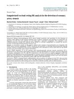
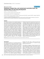
Assessment of abnormal findings
There was no statistical difference in the ability of the PI to
identify infiltrates, atelectasis, effusions or air leak syn-
drome (Fig 1). Considering each abnormal finding sepa-
rately, the PI was able to accurately identify the presence of
pulmonary infiltrates (sensitivity 85.1%, specificity 96.1%,
PPV 96.6%) and atelectasis (sensitivity 84.6% specificity
97%, PPV 88.7%). The identification of airleaks had a sen-
sitivity of 64.3% with 100% specificity and 100% PPV and
pleural effusions had a sensitivity of 66.6%, specificity was
99.2%, and PPV was 90.9%.
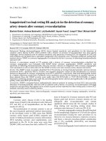
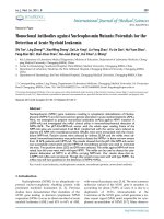
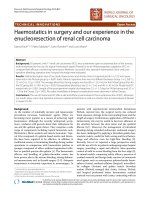
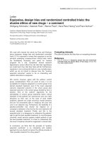
Finally we analyzed the ability of the PI to accurately deter-
mine the correct position of life-support devices. There was
100% concordance between the interpretations of the PI
and PR regarding the appropriate or inappropriate position
of endotracheal tubes and central venous catheters (sensi-
tivity 100%, specificity 100% and PPV 100%; Figs 2 and
3).
Available online />Page 3 of 6
(page number not for citation purposes)
Discordant findings
Fifty-one discordant findings were identified. Seventeen
were classified as clinically insignificant and 20 were clas-
sified as clinically significant by the PP. Clinically significant
discordant chest radiograph findings are listed in Table 3.
On review of the medical records of the patients with clini-
cally significant discordant chest radiograph findings, ther-
apy was found to be altered in one case. In this patient, the
reaccumulation of a pneumothorax was not recognized by
the PI. Identification of the pneumothorax by the PR and
subsequent communication with the PI resulted in the
patient's thoracotomy tube being returned to suction
although the patient was unchanged clinically.
In the other clinically significant and insignificant (Table 4)
discordant cases, changes in therapy were felt to be not
indicated based on the interpretation of the PR and based
on the patient's clinical condition. Of the 14 radiograph
over-reads by the PI, seven had clinically insignificant areas
of atelectasis that did not receive additional therapy. Addi-
tionally, five films were interpreted to have an infiltrate, but
other clinical indicators were used to assist with therapeu-
tic intervention (ie, fever, leukocytes and physical findings).
Finally, the PI assessed two radiographs as having small
pleural effusions that did not require intervention.
Figure 1
Identification of infiltrates, atelectasis, pleural effusions and leaks. ■,
Radiologists' positive findings; ᮀ, intensivists' concordant findings
(those that were in agreement with the radiologist); І, intensivists' dis-
cordant films, and ■, intensivists' over-reads (a subgroup of the dis-
cordant films). There was no statistical significance (P < 0.05) between
the two groups.
Figure 2
Comparison of radiologists' and intensivists' readings of line placement.
■, Radiologist reading; ᮀ, intensivist readings. There was 100% con-
cordance between the two groups.
Figure 3
Comparison of radiologists' and intensivists' readings of endotracheal
tube placement. ■, Radiologist readings; ᮀ, intensivist readings. There
was 100% concordance between the two groups.
Table 1
Indications for obtaining chest film
Indication Number
Respiratory distress 65
R/O pneumonia 16
Cystic fibrosis 3
R/O acute chest syndrome 1
R/O congestive heart failure 1
R/O pleural effusion or pneumothorax 2
Follow-up 144
Procedures
Intubation 39
Line placement 18
Chest tube placement 1
Thoracentesis 3
R/O, rule out.
Critical Care Vol 2 No 2 Chambliss et al.
Discussion
These findings demonstrate agreement between PI and PR
at our institution in greater than 80% of the chest radio-
graphs reviewed. Our findings are consistent with other
studies reporting radiographic interpretation concordance
rates for non-radiologists of 77-97% [2,3]. Reviews of pedi-
atric radiographic interpretation typically have higher dis-
cordance rates (8.9-16.4%) due to the inherent difficulty in
interpreting these radiographs given the wide variations in
patient size, physical development and the variety of
disease processes. Based on our data, PI and PR at our
institution showed similar abilities to determine the proper
position of central venous lines and endotracheal tubes on
radiographs. As shown in Fig 1, PI appeared to have diffi-
culty identifying small pneumothoraces and pleural
effusions. There were very few patients in our population
who developed airleaks making it difficult to draw any sta-
tistical conclusions from the low concordance rate pre-
sented. Although we found a statistically significant number
of discordant interpretations, discordance produced a
change in therapy in < 1% (1 out of 161) of the enrolled
patients. Discordance did not affect PICU length of stay,
significantly alter clinical care of result in any adverse
patient outcomes in this series.
Three potential reasons that may have contributed to the
discordance between PI and PR in the study include: (1)
interobserver variability; (2) physician bias; and (3) the abil-
ity to correlate clinical findings with radiographic findings.
Variability between multiple observers reviewing a radio-
graph is recognized as a significant cause of discrepancy
[3–5]. Studies have shown discrepancy rates as high as
30% amongst radiologists reviewing the same radiograph
[3]. Interobserver variability should be considered when
discrepancies occur between radiologists and nonradiolo-
gists. Fleshier et al[6] demonstrated that interobserver var-
iability contributed to the discrepancies in radiograph
interpretation seen between the emergency room pediatri-
cians and radiologists. These discrepancies frequently
occurred between the reviewers while attempting to distin-
guish normal pulmonary markings from peribronchial thick-
ening or atelectasis from small infiltrates [6]. Though
interobserver variability was not specifically addressed in
this study, it may account for some of the discordant
interpretations.
PR Usually receive limited information about the patient's
clinical condition and their interpretation is based primarily
on the radiographic findings present. Due to this limited
information, radiologists tend to over-read radio-graphs to
minimize errors, to meet community standards, and to avoid
potential malpractice litigation [3]. In teaching institutions,
as many as 11% of radiographs may be over-read by staff
radiologists [3]. Furthermore, when radiologists have
access to the treating physician's interpretation they are
more likely to be biased by the treating physician's interpre-
tation. Kramer et al[7] reported that radiologists are more
likely to overdiagnosis pneumonias in febrile children when
they knew the treating physician's interpretation of the
chest radiograph. This bias was eliminated when the radiol-
ogist was only supplied with information on vital signs or
physical findings. Although the effects of overinterpretation
on patient outcome have not been determined, routine
overinterpretation has the potential to result in inappropri-
ate therapies and increased hospitalizations and proce-
dures. Because we used the PR as the `gold standard' we
could not determine how frequently over-interpretation by
the PR occurred and how that may have impacted on
patient care and outcome.
Finally, we theorize that discordance could be secondary to
the treating PI having first-hand knowledge of both the
patient's clinical and radiographic findings. The ability of the
treating physician to correlate the patient's clinical findings
and radiographic findings may have resulted in the under-
or over-interpretation of the radiograph compared with that
of the PR, leading to a higher disordance rate.
The PR were not aware of this study at the time of perform-
ance; all of the PI were involved in the study. Therefore, it is
possible that the PI tended to be more attentive to detailed
chest radiograph findings that in a typical ICU setting,
potentially improving concordance rates in an unrealistic
fashion.
These results suggest that board-certified PI in our institu-
tion are capable of making appropriate therapeutic deci-
sions based on their interpretations of chest radiographs
obtained in the PICU. Thus, the appropriateness of acute
interventions based on the radiographic findings in our
PICU is the result of the interpretation of the chest radio-
Table 2
Overall concordance of the interpretation of pediatric intensivists
(PI) compared to pediatric radiologists (PR)
Concordant 240
PR and PI positive findings concordant 185
PR and PI negative findings concordant 55
Discordant 51
PR ≠ PI clinically significant 20
PR ≠ PI clinically insignificant 17
Over-read PR Ň PI ⊕ 14
Sensitivity 83.3
Specificity 79.7
PPV 93.0
Available online />Page 5 of 6
(page number not for citation purposes)
graph and assessment of the patient's clinical findings by
the PI. Further studies in other PICU will be necessary to
validate our findings, as results may depend on the specific
abilities and experience of the PI.
Future implications
In today's healthcare environment, capitation is stimulating
the move towards reduced cost and eliminate the duplica-
tion of services. Is the routine review of all radiographs by a
radiologist therefore still cost-effective or clinically neces-
sary? In a study comparing the interpretation of plain ortho-
pedic films, Turen et al[8] found no difference in
interpretations between radiologists and orthopedists.
They concluded that the review of orthopedic films by the
radiologist did not influence patient care and that concur-
rent review by both radiologists and orthopedists was
redundant, resulting in unnecessary expense to the patient
[8]. Simon et al[9], in another study comparing radio-
graphic interpretations by pediatric emergency room physi-
cians and radiologists from our institution, concluded that a
substantial cost saving could be obtained by eliminating
the radiologist's routine interpretation of all radiographs
and consulting the radiologist with difficult or high risk
cases.
From the data that we have presented, it could be con-
cluded that the radiologist may not need to review all chest
radiographs ordered in the PICU. Radiographs that are
Table 3
Clinically significant discordant chest film interpretations
PI interpretation Number of patients PR interpretation Number of patients Management
Normal or clear lung fields 8 Pneumonia/infiltrate 3 No change in therapy
Atelectasis 1 No change in therapy
Pleural effusion 1 No change in therapy
Airleak 2 One chest tube placed to suction
Fracture 1 No change in therapy
Pneumonia or infiltrate 6 (+) Airleak 3 No change in therapy
Multilobe infiltrates 3 No change in therapy
Pleural effusion 2 (+) Atelectasis 1 No change in therapy
(+) Small pneumothorax 1 No change in therapy
Atelectasis 3 Pleural effusion 1 No change in therapy
Multilobed 1 No change in therapy
(+) Infiltrate 1 No change in therapy
Hyperinflation 1 (+) Atelectasis 1 No change in therapy
PI, pediatric intensivist; PR, pediatric radiologist; (+), PI interpretation plus an additional finding.
Table 4
Clinically insignificant discordant chest film interpretations
PI interpretation Number of patients PR interpretation Number of patients Management
Normal or no change 7 Increasing infiltrate 3 No change in therapy
Small pleural effusion 1 No change in therapy
RUQ density R/O gall stones 1 No change in therapy
Mild cardiomegaly 1 No change in therapy
Small patchy RUL density 1 No change in therapy
Infiltrate 4 (+) Pleural effusion 1 No change in therapy
Bilateral infiltrates 1 No change in therapy
(+) Healing clavicle fracture 1 No change in therapy
RUL and RML infiltrates 1 No change in therapy
Resolved pleural effusion 3 Small pleural effusion 2 No change in therapy
(+) Atelectasis 1 No change in therapy
Residual pneumothorax 1 No pneumothorax 1 No change in therapy
Atelectasis LLL 1 LLL and RUL 1 No change in therapy
RUL 1 RUL alveolar density 1 No change in therapy
PI, pediatric intensivisit; PR, pediatric radiologist; RUQ, right upper quadrant; R/O, rule out; RUL, right upper lobe; (+), PI interpretation plus an
additional finding; RML, right middle lobe; LLL, left lower lobe.
Critical Care Vol 2 No 2 Chambliss et al.
obtained solely for determining central venous line or
endotracheal tube position may only need to be reviewed
by the PI for the accuracy of placement. The charges asso-
ciated with interpreting the radiograph could be bundled
with the charges associated with the procedure. If other
studies show similar results, perhaps the current policies of
requiring radiologists' review of all chest radiographs, as in
our institution, can be reconsidered. Additional cost reduc-
tions would be generated. Additional cost reductions
would be generated if, for example, radiologists were
required to review the initial chest radiograph upon patient
admission to the PICU and were then consulted for specific
clinical questions by the PI on subsequent radiographs. For
this change to truly be effective, it will require additional
emphasis during the training for PI to aid in their interpreta-
tion of radiography. Future studies will be required to more
accurately determine clinical predictors that could help cli-
nicians determine which radiographs would need further
evaluation by a radiologist.
Acknowledgements
The authors would like to thank Jean Wright, MD, MBA, KS Anand,
MBBS, James Fortenberry, MD, Robert Pettignano, MD, Jana Stockwell,
MD, Critical Care, Engleston Pediatric Group, and Turner Ball, Depart-
ment of Radiology, Emory University, for their assistance with the prep-
aration of this manuscript.
References
1. O'Leary MR, Smith MS, O'Leary DS: Application of clinical indi-
cators in the emergency department. JAMA 1989, 262:3444-
3447.
2. Walter RS: Radiology practices in emergency departments
associated with pediatric residency training programs. Pediatr
Emerg Care 1995, 11:78-81.
3. Warren JS, Lara K, Connor PD, Cantrell J, Hahn RG: Correlation
of emergency department radiographs: results of a quality
assurance review in an urban community hospital setting. J Am
Board Fam Pract 1993, 6:255-259.
4. Zir LM, Miller SW, Dinsmore RE: Interobserver variability in cor-
onary angiography. Circulation 1976, 53:627-632.
5. Mata AG, Rosengant RM: Interobserver variability in the radio-
graphic diagnosis of necrotizing enterocolitis. Pediatrics 1980,
66:69-71.
6. Fleisher G, Ludwig S, McSorley M: Interpretation of pediatric X-
ray films by emergency department pediatricians. Ann Emerg
Med 1983, 12:153-158.
7. Kramer MS, Roberts-Brauer R, Williams RL: Bias and `overcall' in
interpreting chest radiographs in young febrile children. Pedi-
atrics 1992, 90:11-13.
8. Turen CH, Mark JB, Bozman R: Comparative analysis of radio-
graphic interpretation of orthopedic films: is there
redundancy? J Trauma 1995, 39:720-721.
9. Simon HK, Khan NS, Nordenberg DF, Wright JA: Pediatric emer-
gency physician interpretation of plain radiographs: is routine
review by a radiologist necessary and cost-effective? Ann
Emerg Med 1996, 27:295-298.