Báo cáo y học: "Heliox improves pulmonary mechanics in a pediatric porcine model of induced severe bronchospasm and independent lung mechanical ventilation" pdf
Bạn đang xem bản rút gọn của tài liệu. Xem và tải ngay bản đầy đủ của tài liệu tại đây (161.49 KB, 6 trang )
Heliox improves pulmonary mechanics in a pediatric porcine
model of induced severe bronchospasm and independent lung
mechanical ventilation
Anthony J. Orsini*, John L. Stefano
†‡
, Kathleen H. Leef
‡
, Melinda Jasani
§
,
Andrew Ginn
‡
, Lisa Tice
¶
and Vinay M. Nadkarni
†#
Background: A helium–oxygen gas mixture (heliox) has low gas density and low
turbulence and resistance through narrowed airways. The effects of heliox on
pulmonary mechanics following severe methacholine-induced bronchospasm
were investigated and compared to those of a nitrogen–oxygen gas mixture
(nitrox) in an innovative pediatric porcine, independent lung, mechanical
ventilation model.
Results: All of the lungs showed evidence of severe bronchospasm after
methacholine challenge. Prospective definition of ‘heliox response’ was a 15% or
greater improvement in lung function in the lung receiving heliox compared with
the matched lung receiving nitrox. Seven out of 10 pigs responded to heliox
therapy with respect to resistance and eight out of 10 pigs responded to heliox
therapy with respect to compliance and tidal volume (P<0.03). After crossover
from nitrox to heliox, eight out of eight lungs significantly improved with respect
to tidal volume, resistance and compliance (P<0.001). After crossover from
heliox to nitrox all eight lungs showed a significant increase in resistance and a
significant decrease in tidal volume (P<0.001).
Conclusions: In a pediatric porcine model of acute, severe methacholine-
induced bronchospasm and independent lung mechanical ventilation,
administration of heliox improves pulmonary mechanics, gas flow, and ventilation.
Administration of heliox should be considered for support of pediatric patients
with acute, severe bronchospasm requiring mechanical ventilation through small
artificial airways.
Addresses: *Department of Neonatology, New
York University School of Medicine, Albany, New
York, USA,
†
Jefferson Medical College,
Philadelphia, Pennsylvania, USA,
‡
Christiana Care
Health Center, Newark, Delaware, USA,
§
Department of Emergency Medicine, St.
Christopher’s Hospital For Children, Philadelphia,
Pennsylvania, USA and
¶
Department of Research
and
#
Department of Anesthesia and Critical Care,
duPont Hospital For Children, Wilmington,
Delaware, USA
Sponsorship: Research funded by the Nemours
Foundation, Alfred I. duPont Hospital for Children.
Correspondence: Vinay Nadkarni MD, Department
of Anesthesia and Critical Care, duPont Hospital
for Children, P.O. Box 269, 1600 Rockland Road,
Wilmington, Delaware, USA, 19899. Tel: 302-
651-5159; fax: 302-651-6410; e-mail: vnadkar@
nemours.org
Keywords: asthma, bronchospasm, heliox, helium,
mechanical ventilation, independent lung
ventilation
Received: 1 September 1997
Revisions requested: 10 April 1999
Revisions received: 17 April 1999
Accepted: 23 April 1999
Published: 18 May 1999
Crit Care 1999, 3:65–70
The original version of this paper is the electronic
version which can be seen on the Internet
(). The electronic version may
contain additional information to that appearing in
the paper version.
© Current Science Ltd ISSN 1364-8535
Research paper 65
Introduction
In 1935, Barach first advocated helium–oxygen gas mix-
tures (heliox) as a therapy for obstructive lesions of the
airway [1]. Since then, heliox has been shown to be effica-
cious in the treatment of various disease entities involving
narrowed airways [2–8]. Its safety has been demonstrated
in both mechanically ventilated and spontaneously
breathing patients [7]. Combining 70% helium and 30%
oxygen results in a gas which is much less dense than a
nitrogen–oxygen gas mixture (nitrox) and has approxi-
mately the same viscosity [1]. The therapeutic effects of
heliox gas mixtures are believed to relate to its ability to
deliver oxygen and gas flow with less turbulence and
resistance through narrowed airways. Since airway resis-
tance is directly proportional to the density of the gas, the
administration of heliox is expected to improve ventila-
tion by decreasing resistance, reducing turbulence and
promoting laminar gas flow.
Although there have been advancements in the treatment
of asthma since Barach first studied heliox in 1935, mortal-
ity continues to increase [9]. The use of bronchodilators
heliox = Helium–oxygen gas mixture; nitrox = nitrogen–oxygen gas mixture; ECG = electrocardiogram; PIP = peak inspiratory pressure;
PEEP = positive end-expiratory pressure; I/E = inspiratory to expiratory ratio; F
i
O
2
= fractional inspiratory oxygen concentration; PFT = pulmonary
function test
cc032.qxd 14/05/99 07:00 Page 65
and anti-inflammatory agents have become the standard of
care for reactive airway disease and asthma. However,
some patients fail to respond to aggressive therapy and
require mechanical ventilation. Mechanical ventilation
may result in turbulent gas flow secondary to high gas
velocity which may cause additional difficulty achieving
adequate ventilation. Heliox may, therefore, be most
effective in intubated patients with severe bronchospasm
and small diameter airways by decreasing turbulent flow,
improving ventilation and limiting barotrauma while ther-
apies targeted to the underlying etiology of the bron-
chospasm are given time to achieve their effect.
Several animal and human studies have investigated the
effects of heliox on pulmonary function [10–14]. Although
results from these studies have been promising, the wide
variation between each patient’s biological response to
bronchospasm make many of these results difficult to
interpret. We have developed a pediatric porcine, inde-
pendent lung ventilation model of severe bronchospasm
which allows one of the animal’s lungs to act as a simulta-
neous control for the contralateral lung. This unique
model allows each subject to act as its own control during
the same bronchospastic event, thereby minimizing influ-
ence from various systemic variable biological responses to
acute stress and eliminating the need to compare matched
control subjects or different bronchospastic events in the
same animal. Our hypothesis is that, during mechanical
ventilation, the low density heliox gas mixture will
increase flow through constricted airways and improve
pulmonary mechanics in the lung receiving heliox com-
pared to the lung receiving nitrox.
Methods and materials
This study was approved by the Institutional Review
Board at the Alfred I. duPont Institute of the Nemours
foundation. Thirteen pre-adolescent Yucatan swine
(9.0±1.7kg) were pre-anesthetized with 500mg pentobar-
bital intramuscularly. Peripheral hydration was maintained
with 10% Dextrose in water at 4ml/kg perh. Following
placement of continuous monitors for heart rate, electro-
cardiogram (ECG), respirations, and oxygen saturation,
150mg (15mg/kg) pentobarbital was given intravenously.
Each pig was initially intubated with a 5.0mm cuffed
endotracheal tube and mechanically ventilated with a time
cycled, pressure limited ventilator [peak inspiratory pres-
sure (PIP) 18cmH
2
O, positive end-expiratory pressure
(PEEP) 5cmH
2
O, rate 30breaths/min, inspiratory to expi-
ratory ratio (I/E) 1:1, fractional inspiratory oxygen concen-
tration (FiO
2
) 30%]. Central arterial and venous catheters
were placed by femoral cutdown for continuous monitor-
ing of heart rate, blood pressure and blood sampling. A tra-
cheostomy was then performed and, immediately after
removal of the initial endotracheal tube, separate cuffed
3.0 endotracheal tubes (16cm in length) were placed
through the tracheostomy stoma into the right and left
mainstem bronchi. Correct placement of each endotra-
cheal tube was verified by auscultation during indepen-
dent ventilation and later confirmed by bronchoscopy and/or
autopsy. Additional doses of pentobarbital were given to
ensure adequate anesthesia (titrated to achieve a heart
rate of <160beats/min, systolic blood pressure
≤140mmHg, and absence of withdrawal to painful
stimuli).
Each lung was independently mechanically ventilated (BP
200, Bear Medical Systems, Riverside, California, USA)
simultaneously with identical settings. Pulmonary func-
tion tests (PFTs) were recorded for each lung at baseline
while on heliox and nitrox using an infant/pediatric pul-
monary function computer (PeDS, Medical Associated
Services, Inc., Hatfield, Pennsylvania, USA), calibrated for
the gas mixture being delivered to derive tidal volume,
resistance and compliance. Airflow data were obtained by
a Fleisch 0 (Pediatric) tachometer (OEM Medical, Rich-
mond, Virginia, USA). Transpulmonic pressure was mea-
sured by a differential pressure transducer with an
esophageal balloon. While ventilating both lungs with
nitrox, methacholine (10mg/ml×1.5ml diluted to 3ml
with buffer) was aerosolized continuously to both lungs
simultaneously over 3 min until airway resistance of each
lung at least doubled from baseline. One lung was then
randomized to receive nitrox and the other to receive
heliox. FiO
2
was not adjusted to either lung, but remained
at 30% throughout the experiment. Pulmonary function
testing was performed every 2min, alternating lungs until
the resistance of one lung returned to within 15% of base-
line or until 16 min had elapsed. As approximately 2min
was required to complete each pulmonary function assess-
ment and because of differences in calibration between
the two gases used, right and left lung PFTs were not
obtained simultaneously. Therefore, the order for each
lung to be tested was determined randomly and data were
compared by pulmonary function assessment number.
Results of PFTs obtained at 2min and 4min are reported
as assessment number 1; the results of PFTs performed at
6min and 8min are reported as assessment number 2; the
results of PFTs performed at 10 and 12min as assessment
number 3; and the results of PFTs performed at 14 and
16min as assessment number 4. The maximum number of
PFTs performed for each lung was four per lung. In eight
out of 10 piglets, the lung gases were then switched and
PFTs were measured in each lung after 2min. (The proto-
col was expanded to include the crossover after the first
two piglets had been studied, and three piglets could not
be evaluated due to severe cardiopulmonary compromise
requiring medication intervention during induction of
bronchospasm.) To correct for differences between right
and left lungs in absolute values of tidal volume, resis-
tance and compliance following induced bronchospasm,
outcome variables are expressed as % improvement from
66 Critical Care 1999, Vol 3 No 2
cc032.qxd 14/05/99 07:00 Page 66
the parameters recorded immediately after bronchospasm.
A deterioration of lung function was assigned a negative
number. Percentage improvement in tidal volume, resis-
tance and compliance after crossover were compared to
the measurement immediately before crossover of gases
(heliox to nitrox or nitrox to heliox). Upon completion of
the study, animals were humanely euthanized using intra-
venous pentobarbital, 15mg/kg and KCl, 2mEq/kg (see
timeline, Fig. 1).
Statistical analysis
A Student t test was used to compare lung resistance, com-
pliance and tidal volume at baseline (pre-bronchospasm),
after methacholine induction of bronchoscopy and after
crossover. In addition, ‘heliox response’ was prospectively
defined as a greater than 15% improvement in resistance
of the heliox lung compared to the nitrox lung. Using this
definition, if there were no effect of heliox on resistance,
we would expect no ‘heliox responders’. Fisher’s exact
test was used to compare responders versus non-respon-
ders. A P value <0.05 was considered to be significant.
Measured outcome variables were resistance, dynamic
compliance and tidal volume.
Results
Thirteen swine were anesthetized and enrolled. Ten out
of 13 pigs completed the study without deviation from the
protocol. Two pigs became severely hypoxemic and dys-
rhythmic and required resuscitation before administration
of methacholine was completed and data from one pig was
eliminated secondary to failure of the pulmonary function
apparatus. There were no significant differences found
between the two groups with respect to pulmonary func-
tion at baseline before bronchospasm or immediately fol-
lowing methacholine challenge. Successful methacholine-
induced bronchospasm was documented by a significant
deterioration of all pulmonary function parameters studied
(tidal volume, resistance and compliance). The mean±SD
resistance measured after methacholine challenge was
425±234cm/H
2
O/l/s for the lungs randomized to receive
heliox and 305 ± 199 cmH
2
O/l/s for the lungs randomized
to receive nitrox (difference not significant). The
mean±SD compliance measured after induction of bron-
chospasm was 0.15±0.14ml/cmH
2
O/kg for the heliox
group and 0.23±0.14ml/cmH
2
O/kg for the nitrox group
(difference not significant). The mean±SD tidal volume
measured after induction of bronchospasm was
1.9±1.8ml/kg for the heliox group and 2.8±1.8ml/kg for
the nitrox group (difference not significant).
Table 1 demonstrates the number of heliox ‘responders’
at each time point measured, including after each lung
was crossed over from nitrox to heliox. Figure 2 shows the
Research paper Heliox for experimental bronchospasm Orsini et al 67
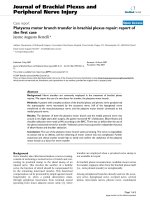
Figure 1
Timeline for study. Following documentation of severe bronchospasm,
lungs were randomized to receive either a helium–oxygen gas mixture
(heliox) or a nitrogen–oxygen gas mixture (nitrox). The order in which
each lung received pulmonary function tests (PFTs) was also
randomized.
Anesthesia
Tracheostomy
Randomization and
independent ventilation
Lung 1
Lung 2
Methacholine
Document Bronchospasm
Heliox
Nitrox
PFTs
Switch Gases
Nitrox
Heliox
PFT
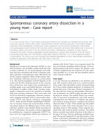
Figure 2
Percentage improvement of resistance, tidal volume and compliance
after lungs were crossed over from a nitrogen–oxygen gas mixture
(nitrox) to a helium–oxygen gas mixture (heliox) and from heliox to
nitrox. A negative percentage improvement indicates a deterioration of
lung function, i.e. an increase in resistance is depicted as a negative
percentage improvement.
–50
–40
–30
–20
–10
0
10
20
30
40
50
60
70
80
Nitrox to
Heliox
Heliox to
Nitrox
Resistance Tidal Volume Compliance
% Improvement
* < 0.01P
**P < 0.02
*
*
**
Table 1
The number of helium–oxygen gas mixture (heliox)
‘responders’ for tidal volume, compliance and resistance for
each performed including pulmonary function tests after
crossover.
Assessment
12 3 4CO
Tidal Volume 8/10* 8/10* 8/10* 8/10* 8/8*
Resistance 7/10* 6/10 5/10 8/10* 8/8*
Compliance 8/10* 7/10* 7/10* 2/10 7/8*
CO, positive response after crossover from a nitrogen–oxygen gas
mixture (nitrox) to heliox. Note only eight piglets were crossed over.
*P <0.05.
cc032.qxd 14/05/99 07:00 Page 67
percent improvement of each parameter measured after
the gases were switched from heliox to nitrox and from
nitrox to heliox. Eight out of 10 subjects had pulmonary
function parameters recorded after the gases delivered to
each lung were crossed over. All eight subjects showed an
improvement in resistance of greater than 15% after
crossover from nitrox to heliox. In addition, all lungs
crossed over from heliox to nitrox showed a deterioration
of resistance and tidal volume of greater than 15%. The
mean±SD improvement in resistance after crossover from
nitrox to heliox was 32.6±14.4% compared with
–19.8±20.3% after crossover from heliox to nitrox
(P<0.001). Eight out of eight pigs met prospectively
defined criteria for a positive ‘response’ to heliox therapy
with respect to tidal volume and seven out of eight pigs
met prospectively defined criteria for a positive ‘response’
with respect to compliance after crossover from nitrox to
heliox. The mean±SD compliance and tidal volume
change after crossover from nitrox to heliox was
36.2±20.3% and 65.2±19.1%, respectively, compared with
only 3.4±20.3% and –18.4±14.5%, respectively, after
crossover from heliox to nitrox (P<0.001).
Discussion
Since Barach first described heliox as an effective treatment
for diseases involving airway obstruction, there have been
many studies performed in both animals and humans exam-
ining its effectiveness [10–16]. Although heliox has been
used safely for many years in the pediatric population for
the treatment of severe croup and upper airway obstruction
[2–7], it has been an uncommon treatment for severe bron-
chospasm. The success of bronchodilators and anti-inflam-
matory agents as well as inconsistent results in clinical
studies have resulted in limited application of heliox in the
mechanically ventilated critically ill child. The complex
pathophysiology of asthma and the variability of disease
between patients and their response to therapy makes the
study of a single agent during acute, severe bronchospasm
difficult to extrapolate to the clinical setting.
Studies have shown a variable response to heliox therapy
in spontaneously breathing patients with severe bron-
chospasm. It has been suggested that this variability may
be due to the greater effectiveness of heliox in patients
with predominately large airway disease [10–12,14,17–19].
Studies of heliox involving mechanically ventilated
patients with severe bronchospasm are promising
[8,15,16]. The beneficial effects demonstrated in these
studies may be due to the decreasing turbulence of bulk
gas flow with heliox during mechanical ventilation.
In mechanically ventilated patients with severe bron-
chospasm, the improvement in ventilation during heliox
therapy may be due to the mechanism by which low
density gases affect ventilation. Heliox and other low
density gases decrease turbulent gas flow by lowering the
Reynolds number. The Reynolds number is measured by
the product of the gas velocity, airway diameter, and gas
density divided by viscosity [16]. It is a unitless number
that predicts whether flow is turbulent or laminar. For a
given set of airway dimensions, turbulent flow results in a
higher resistance than laminar flow. In addition, mechani-
cal ventilation may further complicate the management of
acute severe asthma by delivering a gas with increased
velocity through a narrow endotracheal tube, particularly
in pediatric and neonatal patients. This increases the
Reynolds number, which indicates greater turbulent flow
and airway resistance. Adequate ventilation in mechani-
cally ventilated patients with severe bronchospasm may
be more dependent on the density of the gas than in spon-
taneously breathing patients.
Several studies have examined the efficacy of heliox in
mechanically ventilated patients with severe bronchospasm
or other diseases involving narrowed airways [8,15,16].
Although these studies are small and have not included
children, the results have been promising. In 1990, Gluck et
al. [15] reported an immediate and significant improvement
in seven intubated patients with severe bronchospasm and
respiratory acidosis. All seven patients showed a significant
improvement in pCO
2
within 20min and six out of the
seven patients showed a significant decrease in mean
airway pressure during volume-limited ventilation.
The independent lung ventilation model of acute, severe
bronchospasm used in this study is unique in that it allows
each animal’s contralateral lung to represent its own
control. It eliminates the need to monitor systemic arterial
blood gases, global circulating mediator or hormone levels
and assures that the systemic milieu is identical for com-
parison of gross outcome measures. It is recognized that
the model is limited in its ability to monitor and control
local microcirculation. This model controls for the variable
macrocirculatory responses to methacholine (e.g. hemody-
namic status: heart rate, blood pressure, temperature, cir-
culating epinephrine level) between subjects and allows
comparisons of pulmonary mechanics on heliox versus
nitrox gas mixtures within the same animal and during the
same bronchospastic event. This model allows for a clear
determination of response to heliox without the variable
biological responses which may affect studies involving
separate subjects or different bronchospastic events within
the same subject as controls. It uses the same small (3.0)
sized endotracheal tubes that might be expected to clini-
cally increase resistance to gas flow in small infants. A 15%
difference in pulmonary function between the lung
receiving heliox and the lung receiving nitrox (control)
was prospectively selected as the primary outcome vari-
able suggesting a favorable response to heliox versus
nitrox. It is recognized that lung function measurements
in human subjects can be very variable and affected by
many factors. Although the coefficient of variation is
68 Critical Care 1999, Vol 3 No 2
cc032.qxd 14/05/99 07:00 Page 68
extremely small when calibrating the PFT machine
(Fleisch pneumotach) using known standards within the
physiologic ranges encountered in this study, patient
factors can introduce intra- and intersubject variability
[20]. For this reason, the calibrated PFT computer (cali-
brated both to 70%N/30% O and 70%He/30% O) was
applied serially over a relatively short time span (30min)
and relative improvement/deterioration rather than
absolute raw numbers were selected as the primary
outcome measures to be compared. In addition, a 15%
improvement in PFTs is generally accepted as clinically
significant and is well beyond the coefficient of variance
for the PFT computer and pneumotachometer when cali-
brated to a known standard on nitrox or heliox gas mixture.
Of particular interest was the dramatic improvement in
resistance and tidal volume in all lungs after crossover
from nitrox to heliox. Conversely, there was a statistically
significant deterioration in PFTs for all parameters
studied after crossover from heliox to nitrox. Even the
subjects who did not appear to be responding to heliox
therapy still showed a significant and immediate deteriora-
tion in pulmonary function when switched to nitrox.
The results of this study suggest that heliox may be effec-
tive in improving pulmonary mechanics in patients with
small endotracheal tubes being mechanically ventilated
for severe bronchospasm. These results also indicate that
the response to heliox is potentially rapid and persistent
during heliox ventilation.
Although the pediatric porcine model of independent
mechanical ventilation and methacholine-induced bron-
chospasm used in this study is unique and offers many
strengths, we acknowledge the limitations of this study.
Limitations include the small number of subjects, wide
variability in lung response to methacholine challenge and
inability to accurately discriminate between the effect of a
lower density gas on the resistance generated by the endo-
tracheal tube, large and small airways. In addition, anes-
thetic agents may effect pulmonary function.
Pentobarbital was chosen for this study because of its
minimal effects on pulmonary mechanics compared to
inhalation or alternative intravenous agents. No arterial
blood gases were reported because heliox and nitrox gas
mixtures were given to separate lungs simultaneously and
therefore systemic arterial blood gases would not reflect
unilateral lung function or microenvironment. The assess-
ment of right and left independent pulmonary venous
blood gases, although potentially useful, was beyond the
scope of this pilot protocol. However, documentation of
the severity of bronchospasm was confirmed by at least a
50% increase in total lung resistance in each lung, prior to
the start of the experimental therapy. Percent improve-
ment from baseline after bronchospasm was prospectively
selected for outcome analysis instead of comparison of raw
values for lung resistance and compliance because of
recognition during pilot studies of wide variability
between individual piglets right and left lung baseline
lung resistance values after methacholine challenge. The
crossover technique and the desire to use the fewest
piglets possible to demonstrate a treatment effect dictated
prospective use of the percentage improvement compared
to baseline bronchospasm.
Conclusion
In a pediatric porcine model of independent lung mechan-
ical ventilation and severe methacholine-induced bron-
chospasm, heliox improved pulmonary mechanics when
compared to a nitrogen–oxygen gas mixture during
mechanical ventilation at identical ventilator settings.
This study also indicates that most subjects responded to
heliox within the first 4min of therapy and that this
response was sustained for at least 20min. The authors
speculate that heliox may be beneficial to critically ill chil-
dren requiring mechanical ventilation with small endotra-
cheal tubes secondary to severe bronchospasm and high
airway resistance with low compliance. In these patients,
heliox may be expected to improve tidal volume, lung
compliance and resistance and decrease potential ventila-
tor barotrauma while waiting for etiologic targeted thera-
pies to take effect.
Acknowledgments
The authors would like to thank Susan Buck, Behzad Taghizadeh, Bill
Hofmann, Patty Resnik, Tina Hurst, David Corddry, Ellen Deutsch, Brett
Goudie, and Ilene Sivakoff for their assistance and support in completing
this project.
References
1. Barach A: The use of helium in the treatment of asthma and
obstructive lesions in the larynx and trachea. Ann Int Med 1935,
9:739–765.
2. Jordan W, Graves C, Elwyn R: New therapy for postintubation
laryngeal edema and tracheitis in children. JAMA 1970, 212:585–
588.
3. Ishikawa S, Segal M: Re-appraisal of helium–oxygen therapy on
patients with chronic lung disease. Ann Allergy 1973, 32:536–542.
4. Duncan P: Efficacy of helium–oxygen gas mixture in the treatment
of severe viral and post-extubation croup. Can Anaesth Soc J
1979, 26:206–212.
5. Houck J, Keamy M, McDonough J: Effect of helium concentration on
experimental upper airway obstruction. Ann Otol Rhinol Laryngol
1990, 99:556–611.
6. Kemper K, Ritz R, Benson M, Bishop M: Helium–oxygen mixture in
the treatment of post-extubation stridor in pediatric trauma
patients. Crit Care Med 1991, 19:356–359.
7. Wolfson MR, Bhutani VK, Shaffer TH, Bowen FW: Mechanics and
energetics of breathing helium in infants with bronchopulmonary
dysplasia. J Pediatr 1984, 104:752–757.
8. Kass JE, Castriotta RJ: Heliox therapy in acute severe asthma.
Chest 1995, 107:757–760.
9. Robin E: Death from bronchial asthma. Chest 1988, 93:614–618.
10. Chan-Yeung M, Abboud R, Ysao MS, Maclean L: Effect of helium on
maximal expiratory flow in patients with asthma before and during
induced bronchoconstriction. Am Rev Respir Dis 1976, 106:433–
443.
11. Mink SN, Wood LDH: How does HeO
2
increase maximum expira-
tory flow in human lungs? J Clin Invest 1980, 66:720–729.
12. Weiss J, McFadden E, Ingram R: Bronchodilation, lung recoil, and
density dependence of maximal expiratory flow. J Applied Physiol
1982, 52:874–878.
Research paper Heliox for experimental bronchospasm Orsini et al 69
cc032.qxd 14/05/99 07:00 Page 69
13. Christopherson S, Hlastala M: Pulmonary gas exchange during
altered density gas breathing. J Appl Physiol 1983, 40:221–225.
14. Eliason O, Zuwallack RL: Density dependence of maximal expira-
tory air flow in asthmatics with exacerbation of their disease. Am
Rev Respir Dis 1986, 135:573–578.
15. Gluck EH, Onaorato DJ, Castriotta R: Helium–oxygen mixtures in
intubated patients with status asthmaticus and respiratory acido-
sis. Chest 1990, 98:693–698.
16. Shiue S-T, Gluck E: The use of helium–oxygen mixture in the
support of patients with status asthmaticus and respiratory acido-
sis. J Asthma 1989, 26:177–180.
17. Madison JM, Irwin RS: Heliox for asthma: a trial balloon. Chest 107:
597–600.
18. Metzger WJ, Nugent K, Richerson HB: Site of airflow obstruction
during early and late phase asthmatic responses to allergen bron-
choprovocation. Chest 1985, 8:369–375.
19. Macnee W, Power J, Innes A, Douglas NJ, Sudlow MF: The depen-
dence of maximal flow in man on the airway gas physical proper-
ties. Clin Sci 1983, 65:273–279.
20. Clayton RG, Leef KM, Stefano JL: Determination of coefficient of
variance of pulmonary function tests in infants with bronchopul-
monary dysplasia [abstract]. Pediatr Res 1992, 31:303A.
70 Critical Care 1999, Vol 3 No 2
cc032.qxd 14/05/99 07:00 Page 70