Báo cáo y học: " Multiple P2Y receptors couple to calcium-dependent, chloride channels in smooth muscle cells of the rat pulmonary artery" ppsx
Bạn đang xem bản rút gọn của tài liệu. Xem và tải ngay bản đầy đủ của tài liệu tại đây (637.59 KB, 10 trang )
BioMed Central
Page 1 of 10
(page number not for citation purposes)
Respiratory Research
Open Access
Research
Multiple P2Y receptors couple to calcium-dependent, chloride
channels in smooth muscle cells of the rat pulmonary artery
Krongkarn Chootip
1,2
, Alison M Gurney
1
and Charles Kennedy*
1
Address:
1
Department of Physiology and Pharmacology, University of Strathclyde, Strathclyde Institute for Biomedical Sciences, John Arbuthnott
Building, 27 Taylor Street, Glasgow G4 ONR, UK and
2
Department of Physiology, Faculty of Medical Science, Naresuan University, Phitsanulok
65000, Thailand
Email: Krongkarn Chootip - ; Alison M Gurney - ;
Charles Kennedy* -
* Corresponding author
Abstract
Background: Uridine 5'-triphosphate (UTP) and uridine 5'-diphosphate (UDP) act via P2Y
receptors to evoke contraction of rat pulmonary arteries, whilst adenosine 5'-triphosphate (ATP)
acts via P2X and P2Y receptors. Pharmacological characterisation of these receptors in intact
arteries is complicated by release and extracellular metabolism of nucleotides, so the aim of this
study was to characterise the P2Y receptors under conditions that minimise these problems.
Methods: The perforated-patch clamp technique was used to record the Ca
2+
-dependent, Cl
-
current (I
Cl,Ca
) activated by P2Y receptor agonists in acutely dissociated smooth muscle cells of rat
small (SPA) and large (LPA) intrapulmonary arteries, held at -50 mV. Contractions to ATP were
measured in isolated muscle rings. Data were compared by Student's t test or one way ANOVA.
Results: ATP, UTP and UDP (10
-4
M) evoked oscillating, inward currents (peak = 13–727 pA) in
71–93% of cells. The first current was usually the largest and in the SPA the response to ATP was
significantly greater than those to UTP or UDP (P < 0.05). Subsequent currents tended to decrease
in amplitude, with a variable time-course, to a level that was significantly smaller for ATP (P < 0.05),
UTP (P < 0.001) and UDP (P < 0.05) in the SPA. The frequency of oscillations was similar for each
agonist (mean≈6–11.min
-1
) and changed little during agonist application. The non-selective P2
receptor antagonist suramin (10
-4
M) abolished currents evoked by ATP in SPA (n = 4) and LPA (n
= 4), but pyridoxalphosphate-6-azophenyl-2',4'-disulphonic acid (PPADS) (10
-4
M), also a non-
selective P2 antagonist, had no effect (n = 4, 5 respectively). Currents elicited by UTP (n = 37) or
UDP (n = 14) were unaffected by either antagonist. Contractions of SPA evoked by ATP were
partially inhibited by PPADS (n = 4) and abolished by suramin (n = 5). Both antagonists abolished
the contractions in LPA.
Conclusion: At least two P2Y subtypes couple to I
Cl,Ca
in smooth muscle cells of rat SPA and LPA,
with no apparent regional variation in their distribution. The suramin-sensitive, PPADS-resistant
site activated by ATP most resembles the P2Y
11
receptor. However, the suramin- and PPADS-
insensitive receptor activated by UTP and UDP does not correspond to any of the known P2Y
subtypes. These receptors likely play a significant role in nucleotide-induced vasoconstriction.
Published: 26 October 2005
Respiratory Research 2005, 6:124 doi:10.1186/1465-9921-6-124
Received: 20 July 2005
Accepted: 26 October 2005
This article is available from: />© 2005 Chootip et al; licensee BioMed Central Ltd.
This is an Open Access article distributed under the terms of the Creative Commons Attribution License ( />),
which permits unrestricted use, distribution, and reproduction in any medium, provided the original work is properly cited.
Respiratory Research 2005, 6:124 />Page 2 of 10
(page number not for citation purposes)
Background
Uridine 5'-triphosphate (UTP) and uridine 5'-diphos-
phate (UDP) act via P2Y receptors, whilst adenosine 5'-tri-
phosphate (ATP) acts via P2X as well as P2Y receptors, to
modulate vascular tone [1-3]. P2X receptors are ligand-
gated cation channels and the ability of the P2X
1
subtype
to mediate rapid, transient inward currents in pulmonary
artery smooth muscle cells [4,5] and induce constriction
of the pulmonary vasculature (see [6] and references
therein) has been characterised in some depth. P2Y recep-
tors are G protein-coupled receptors and P2Y agonists act
at smooth muscle receptors to evoke vasoconstriction in
the rat perfused lung at resting tone, but induce vasodila-
tion via endothelial receptors if muscle tone is first raised
[7-10]. Similarly, P2Y agonists are contractile at resting
tone and relaxant at raised tone in isolated branches of rat
intrapulmonary arteries [11-13]. Compared with P2X
receptors much less is known about which of the eight
mammalian P2Y subtypes (P2Y
1,2,4,6,11,12,13,14
) [14,15] are
expressed in pulmonary vascular smooth muscle or about
the signalling pathways through which they act.
In a previous study [6] we showed that UTP and UDP
both act via two P2Y receptors to evoke contraction of rat
isolated pulmonary arteries. For each agonist one site was
insensitive to the antagonists suramin and pyridoxalphos-
phate-6-azophenyl-2',4'-disulphonic acid (PPADS),
whilst the other was inhibited by suramin, but not
PPADS. UTP is a potent agonist at the P2Y
2
and P2Y
4
receptors and a weaker agonist at the P2Y
6
subtype
[16,17]. Of these three receptors, only the P2Y
2
is
suramin-sensitive and PPADS-insensitive [18], so this is
likely to be one of the sites of action of UTP. The molecu-
lar identity of the suramin-and PPADS-insensitive site of
action of UTP is unclear as the P2Y
4
and P2Y
6
subtypes are
both reported to be antagonised by PPADS, but not
suramin [18-20]. UDP is a potent agonist at the P2Y
6
receptor only [16,17]. mRNA for this subtype and
suramin-insensitive contractions to UDP in pulmonary
arteries have been demonstrated [12], but the lack of
effect of PPADS against the contractions evoked by UDP
in our previous study are inconsistent with the P2Y
6
recep-
tor.
A number of factors that can complicate the characterisa-
tion of P2Y receptors may have prevented the clear identi-
fication of the P2Y receptors mediating the contractions
seen in previous studies. These include the release of
nucleotides from cells, their breakdown by ecto-nucleoti-
dases and their bioconversion by ecto-nucleoside diphos-
phokinase (eNDPK) [16,21-24]. Thus, as well as a direct
action at the P2Y
6
receptor, UTP can also act indirectly,
after dephosphorylation to UDP. Likewise, UDP can be
converted to UTP by eNDPK and so act indirectly at P2Y
2
and P2Y
4
receptors. At present, potent and selective inhib-
itors of the ecto-enzymes are not available, but one way to
minimise these metabolic problems is to apply the ago-
nists to rapidly perfused, dissociated cells.
The aim of the present study was to extend the pharama-
cological characterisation of the P2Y receptors mediating
pulmonary vasoconstriction, in conditions that minimise
the influence of the release and extracellular metabolism
of nucleotides. We used the perforated-patch clamp tech-
nique to record the Ca
2+
-dependent, Cl
-
current (I
Cl,Ca
)
induced by nucleotides in single, acutely dissociated pul-
monary artery smooth muscle cells [4,5]. The pulmonary
vascular bed has well characterised regional differences in
receptor and ion channel distribution, including I
Cl,Ca
[8,25], so the responses of cells isolated from large and
small pulmonary arteries were compared. In addition to
UTP and UDP, we also applied ATP, an agonist at the
P2Y
1,2,4,11 & 12
subtypes [14] and determined the ability of
the P2 antagonists suramin and PPADS to inhibit the
responses evoked by each of the agonists. We found that
at least two P2Y subtypes couple to I
Cl,Ca
, with no appar-
ent regional variation in their distribution. The suramin-
sensitive, PPADS-resistant site activated by ATP most
resembles the P2Y
11
receptor. However, the suramin- and
PPADS-insensitive receptor activated by UTP and UDP
does not correspond to any of the known P2Y subtypes.
Methods
Isolated cell preparation
Male Sprague-Dawley rats (150 – 250 g) were killed by the
approved Schedule 1 method of cervical dislocation and
exsanguination. After thoracotomy, the heart and lungs
were removed en bloc, the lungs separated and small (SPA,
200–500 µm id) and large (LPA, 1.0–1.5 mm id) intrapul-
monary arteries dissected out. The arteries were cut open
longitudinally and strips of smooth muscle bathed in a
dissociation medium (DM) composed of (mM); NaCl
110; KCl 5; KH
2
PO
4
0.5; NaH
2
PO
4
0.5; NaHCO
3
10; N-[2-
hydroxyethyl]piperazine-N'-[2-ethane-sulfonic acid]
(HEPES)10; phenol red 0.03; taurine 10; ethylenediami-
netetraacetic acid (EDTA) 0.5; MgCl
2
2; glucose 10 and
CaCl
2
0.16, titrated to pH 7.0 with KOH. After incubation
in DM containing 0.6 – 0.8 mg.ml
-1
papain, 0.04% BSA
and 0.4 mM dithiothreitol at 37°C (15 min for LPA, 10
min for SPA), collagenase (0.6 – 0.8 mg.ml
-1
; type IA) was
added and the tissues incubated for a further 10 (LPA) or
5 (SPA) min. Cells were then dispersed by mild trituration
in enzyme-free solution and used within 7 hours.
Electrophysiological recording
Cells were placed in a 50 µl chamber and superfused at
room temperature with physiological salt solution (PSS)
composed of (mM): NaCl 122; KCl 5; HEPES 10; KH
2
PO
4
0.5; NaH
2
PO
4
0.5; MgCl
2
1; glucose 11; CaCl
2
1.8; titrated
to pH 7.3 with NaOH. Electrophysiological responses of
Respiratory Research 2005, 6:124 />Page 3 of 10
(page number not for citation purposes)
isolated smooth muscle cells were studied in the whole-
cell, perforated-patch mode with amphotericin B (150
µg.ml
-1
) added to a pipette solution of the following com-
position (mM): KCl 125; MgCl
2
4; HEPES 10; ethylene
glycol-bis(2-aminoethylether)-N,N,N',N'-tetraacetic acid
(EGTA) 0.02, titrated to pH 7.3 with KOH. Pipette resist-
ance was 4–8 MΩ. The cells were voltage-clamped at -50
mV using an Axopatch 200A amplifier (Axon Instru-
ments). Data were recorded and analysed with a personal
computer interfaced with a Digidata 1200 A/D converter
(Axon Instruments) using Axotape and pClamp (V5) soft-
ware (Axon Instruments). Current responses to -10 mV
hyperpolarizing steps were used to measure cell
capacitance.
We have reported previously that 10
-4
M ATP, UTP and
UDP each evoked pronounced contractions of rat isolated
SPA and LPA and that 10
-4
M suramin and PPADS pro-
duced maximum inhibition of these responses [6]. There-
fore, this concentration of these drugs was used here. All
were applied to the cells using a gravity-feed perfusion sys-
tem, for which the time for complete solution exchange
was less than 2 s. Only one agonist was applied to each
cell. P2Y receptor-mediated contractions develop slowly
and take 5–10 min to reach a steady-state plateau, there-
fore, in most cases the agonists were applied to the cells
for 5 min or more.
Electrophysiological analysis
The rat pulmonary artery is a relatively short vessel, with
only a thin layer of smooth muscle cells. Enzymatic disso-
ciation produces a lower yield of cells that are smaller and
often less robust than those from systemic blood vessels.
Consequently, although oscillating inward currents were
observed in response to P2Y receptor agonists in the
majority of cells studied, quantitative analysis was ham-
pered by the short period of time that many cells could be
maintained in the perforated-patch configuration or by
the disappearance of the response during the recording.
Quantitative analysis was applied only to cells that could
be held for 5 min or more and in which the oscillating cur-
rents lasted more than 4 min. In these cells the following
parameters were measured: a) the peak amplitude of each
current (pA), which was normalised against the cell capac-
itance (pF) to control for variations in cell size; b) the rise
time (ms) of the current at each oscillation from baseline
holding current to peak; c) the width of each oscillation
(ms) at the point where it reached 50% of its peak ampli-
tude. For each, the average value during successive 30 s
intervals over a 4 min period was calculated and com-
pared. Finally, the frequency of oscillations (peak.min
-1
)
was measured as the number of transient currents occur-
ring during successive 1 min intervals over a 4 min period.
To investigate the effects of P2 receptor antagonists on the
currents, some cells were preincubated with antagonist for
5 min before adding an agonist, but in most cases an ago-
nist was applied for 2 min and then suramin or PPADS
were co-applied for a further 2–3 min. The current ampli-
tude and frequency were measured and average values
compared for the 1 min periods immediately before and
after antagonist addition. The data were compared with
control cells where agonist alone was added and the cur-
rents measured over the same time course.
Tension recording
Rat SPA and LPA were dissected out as described above,
cut into rings 5 mm long and mounted horizontally in 1
ml baths on a pair of intraluminal wires [6]. Tissues were
allowed to equilibrate under a resting tension of 0.5 g
(SPA) and 1.0 g (LPA) for 60 min at 37°C in PSS. Tension
was recorded with Grass FT03 isometric force transducers
connected to a MacLab/4e system, using Chart 3.3 soft-
ware (AD Instruments). Cumulative concentration-
response curves to ATP were obtained in rings in the
absence of antagonist (control) or in the presence of a sin-
gle concentration (3 × 10
-5
, 10
-4
or 3 × 10
-4
M) of suramin
or PPADS. Contractions generally took 1–4 min to reach
a plateau and are expressed as a percentage of the contrac-
tion induced in the same preparation by 4 × 10
-2
M KCl,
which was applied by replacement of the PSS solution
with PSS in which the KCl concentration was raised by
equimolar substitution for NaCl.
Data analysis
Values in the text and figures refer to mean ± S.E.M Data
were compared by paired and unpaired t-tests, or one-way
analysis of variance and Tukey's comparison as appropri-
ate. Differences were considered significant when P <
0.05.
Drugs and solutions
ATP (magnesium salt), UDP (sodium salt), UTP (sodium
salt), suramin hexasodium and PPADS tetrasodium
(Sigma/RBI, UK) were dissolved in deionised water as 100
mM stock solutions and diluted in PSS before application
to the cells.
Results
P2Y receptor agonists induce oscillating inward currents
ATP, UTP and UDP (10
-4
M) each evoked inward currents
(peak amplitude = 13 – 727 pA) in most SPA (n = 118)
and LPA (n = 117) smooth muscle cells held at -50 mV
(ATP-91%/88%, UTP-91%/93%, UDP-71%/81%, SPA/
LPA respectively). Outward currents or no response were
evoked in the remaining cells, which were not studied fur-
ther. In most cells the inward currents activated in an
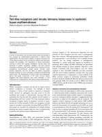
oscillating manner (Figure 1). The first current was usually
the largest and subsequent currents decreased in
Respiratory Research 2005, 6:124 />Page 4 of 10
(page number not for citation purposes)
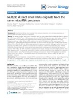
amplitude, with a variable time-course. The peak of the
first current appeared to be larger for ATP than UTP or
UDP (Figure 2), but this was significant only in the SPA (P
< 0.05). For each agonist there was no significant differ-
ence in the amplitude of the first response between the
small and large vessels. The initial current often had a "W-
shaped" profile (Figure 1), but in most cells (85%) the
biphasic profile disappeared by the second, third or
fourth oscillation, such that subsequent currents were
monophasic. ATP, UTP and UDP each evoked this profile
of responses in a similar proportion of cells.
Quantitative analysis of oscillating currents
The decrease in the amplitude of the oscillating currents
during agonist application complicated the quantification
of the effects of P2Y antagonists, so it was necessary to first
quantify the time-course of the currents. In order to be
able to study the effects of both an agonist and antagonist
on the same cell, the analysis was limited to a subpopula-
tion of cells that were maintained under voltage-clamp for
more than 5 min and in which the oscillating currents
lasted for 4 min or more.
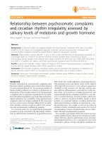
In both SPA (Figure 3a) and LPA (not shown), the ampli-
tude of the oscillating currents tended to decrease over
successive 30 s intervals, particularly within the first 2
min, and this was significant for ATP (P < 0.05), UTP (P <
0.01) and UDP (P < 0.05) in the SPA. The oscillations
induced by ATP, UTP and UDP had similar frequencies
(mean≈6–11.min
-1
), both in SPA (Figure 3b) and LPA
(not shown) and showed no significant change over 4
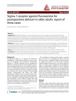
Oscillating currents induced by P2Y receptor agonistsFigure 1
Oscillating currents induced by P2Y receptor ago-
nists. (a) ATP, (b) UTP and (c) UDP (all 10
-4
M), added as
indicated by the horizontal bars, evoked oscillating inward
currents in smooth muscle cells isolated from SPA (a & c)
and LPA (b) and voltage-clamped at -50 mV. The insets show
W-shape currents induced by (a) ATP and (b) UTP. The
arrow on the left-hand side of each trace indicates zero hold-
ing current.
50 pA
1 min
50 pA
ATP
(
10
-
4
M
)
5 s
100
pA
UTP
(
10
-
4
M
)
30 s
5 s
50 pA
UDP
(
10
-
4
M
)
30 pA
30 s
b.
c.
a.
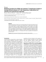
Amplitude of the first inward current induced by P2Y recep-tor agonistsFigure 2
Amplitude of the first inward current induced by P2Y
receptor agonists. The mean ± s.e.mean of the peak ampli-
tude of the initial inward currents evoked by 10
-4
M ATP,
UTP and UDP in SPA and LPA isolated smooth muscle cells,
voltage-clamped at -50 mV, are shown. The number of cells
for each is indicated in parentheses. *P < 0.05 for responses
to ATP versus UTP and UDP in SPA.
ATP UTP UDP ATP UTP UDP
0
10
20
30
(52)
(17)
(29)
(41)
(12)
(37)
*
SPA LPA
Peak current (pA.pF
-1
)
Respiratory Research 2005, 6:124 />Page 5 of 10
(page number not for citation purposes)
min, apart from a small increase in the LPA between the
first and second min after UTP application (P < 0.05). The
currents evoked by ATP in the first 30 s had a rise time of
1.7 ± 0.3 s and width at 50% peak of 2.9 ± 0.4 s (n = 4) in
SPA and 1.5 ± 0.6 s and 2.2 ± 0.3 s (n = 5) respectively in
LPA. The rise time and width at 50% peak then decreased
significantly (P < 0.01) over the next 60–90 s to a steady
state of around 0.8 s in both SPA and LPA. The width at
50% peak of currents evoked by UTP in the first 30 s was
2.1 ± 0.5 s (n = 5) in SPA and 2.1 ± 0.5 s (n = 5) in LPA
and both decreased significantly (P < 0.01) over the next
60–90 s to a steady state of also about 0.8 s. In contrast,
currents evoked by UTP and UDP in SPA and LPA showed
no significant change in rise time and those to UDP in SPA
and LPA showed no significant change in width at 50%
peak, all having a steady-state value of about 0.8 s.
Effects of P2 receptor antagonists
Having quantified the time-course of the agonist-induced
oscillating currents, we then determined the effects of the
P2Y antagonists suramin and PPADS (10
-4
M). In most
cells currents were initiated by an agonist, the antagonist
was then coapplied and the currents compared for 1 min
before and after antagonist addition. To take into account
the decline in current amplitude normally seen over this
time-course (~20–40%), the % decrease in amplitude
over the 2 analysis periods was calculated and compared
with that in control cells where agonist alone was added.
Suramin rapidly and reversibly abolished the currents
evoked by ATP in SPA (Figure 4a, 5) and LPA (not shown).
Additionally, ATP did not elicit currents if cells were pre-
incubated with suramin for 5 min (n = 2, not shown). In
contrast, PPADS (10
-4
M) had no effect on the amplitude
or frequency of the ATP-induced currents in SPA (Figure
4b, 5) or LPA (not shown). The rise-time and width at
50% peak were also unaffected (not shown). PPADS was
also ineffective if applied for 5 min prior to ATP (n = 2).
Neither suramin nor PPADS had any effect on the ampli-
tude or frequency of the oscillating currents elicited by
UTP or UDP in either SPA or LPA (Figure 4c, 5). The rise-
time and width at 50% peak were also unaffected (not
shown). PPADS (n = 12) and suramin (n = 7) (Figure 4d)
were also ineffective if applied for 5 min before UTP.
Effects of suramin and PPADS on contractions evoked by
ATP
We have reported previously the effects of suramin and
PPADS on contractions of rat pulmonary arteries induced
by UTP and UDP [6]. Since suramin abolished current
oscillations induced by ATP, but not UTP or UDP, we
investigated if nucleotide-induced contractions showed
the same differential sensitivity. We report that ATP (10
-7
- 3 × 10
-4
M) evoked concentration-dependent contrac-
tions of the rat SPA (Figure 6). Suramin (3 × 10
-5
- 10
-4
M)
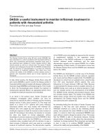
The amplitude and frequency of oscillating currents induced by P2Y receptor agonistsFigure 3
The amplitude and frequency of oscillating currents
induced by P2Y receptor agonists. (a) The mean ampli-
tude of oscillating currents induced by ATP, UTP and UDP
(10
-4
M) was measured over successive 30 s intervals for 4
min in smooth muscle cells isolated from SPA and voltage-
clamped at -50 mV. 0.5 = the first 30 s of agonist application,
1 = 30 – 60 s, and so on. (b) The frequency of the oscillations
in the same cells was measured over successive 1 min inter-
vals. 1 = the first min of agonist application, 2 = the second
min, and so on. Vertical bars represent s.e.mean. The
number of cells for each agonist is shown in parentheses. * P
< 0.05 for amplitude of peak current at 30 s versus that at
2:30, 3:00, 3:30 and 4:00 min; ** P < 0.01 for 30 s versus 3:30
and 4:00 min.
0
10
20
30
ATP (4) UTP (5) UDP (3)
a.
0.5 1 1.5 2 2.5 3 3.5 4 0.5 1 1.5 2 2.5 3 3.5 4 0.5 1 1.5 2 2.5 3 3.5 4
*
**
*
Peak current (pA.pF
-1
)
1234 1234 1234
0
2
4
6
8
10
12
14
ATP (4) UTP (5) UDP (3)
b.
Oscillation frequency (peak.min
-1
)
Respiratory Research 2005, 6:124 />Page 6 of 10
(page number not for citation purposes)
The effects of P2 receptor antagonists on oscillating currentsFigure 4
The effects of P2 receptor antagonists on oscillating
currents. The P2 receptor antagonists suramin and PPADS
(10
-4
M) were applied either 2 min after oscillations were
induced by continuous application of 10
-4
M (a, b) ATP or (c)
UDP or (d) 5 min before application of 10
-4
M UTP to SPA (a,
b) or LPA (c, d) dissociated smooth muscle cells voltage-
clamped at -50 mV. The horizontal bars indicate agonist and
antagonist applications. The arrow on the left-hand side of
each trace indicates zero holding current.
The effects of P2 receptor antagonists on oscillating current amplitude and frequencyFigure 5
The effects of P2 receptor antagonists on oscillating
current amplitude and frequency. The effects of suramin
and PPADS (10
-4
M) on (a) the amplitude and (b) the fre-
quency of oscillating inward currents induced by ATP, UTP
and UDP (10
-4
M) in SPA dissociated smooth muscle cells,
voltage-clamped at -50 mV, are shown. The agonist was
applied for 2 min and then suramin or PPADS was coapplied
for a further 2–3 min. The current amplitude was measured
for 1 min immediately before and after antagonist addition
and average values calculated. The % decrease in amplitude
was then calculated as the difference in the 2 average values.
The control data were obtained over the same time-course
in cells where agonist alone was added. The average fre-
quency of oscillations was also measured for 1 min immedi-
ately before and after antagonist addition and compared
directly. Vertical lines show s.e.mean. The number of cells is
shown in parentheses.
0
20
40
60
80
100
contr ol
+suramin(10
-4
M)
+PPADS(10
-4
M)
ATP UTP UDP
(3)
(4)
(3)
(4)
(3)
(6)
(4)
(4)
(4)
a.
% Decrease current amplitude
0
2
4
6
8
10
12
14
16
co n t r o l
+ antagonist (10
-4
M)
ATP
(4)
UTP
(3)
UDP
(4)
ATP
(4)
UTP
(4)
UDP
(3)
±
±±
±suramin ±
±±
±PPADS
b.
Oscillation frequency (peak.min
-1
)
Respiratory Research 2005, 6:124 />Page 7 of 10
(page number not for citation purposes)
caused a progressive rightward shift of the ATP concentra-
tion-response curve and the responses were abolished by
the highest concentration of the antagonist (Figure 6a).
PPADS (3 × 10
-5
M) also shifted the ATP concentration-
response curve to the right, but increasing its concentra-
tion to 10
-4
M and 3 × 10
-4
M) produced no further inhi-
bition (Figure 6b). In rat LPA ATP induced contractions
only at 10
-4
M and above [6] and these small contractions
were abolished by 3 × 10
-4
M suramin or PPADS (not
shown).
Discussion
The present study shows that ATP, UTP and UDP induce
oscillating inward currents with similar amplitudes and
frequencies in smooth muscle cells of rat pulmonary arter-
ies. Such Cl
-
currents have been reported previously in
these cells and are dependent upon nucleotide-evoked
release of Ca
2+
from sarcoplasmic reticulum stores
[4,5,26]. The P2Y
1
, P2Y
2
, P2Y
4
, P2Y
6
and P2Y
11
receptors
all couple to the G
q/11
G proteins, leading to the release of
IP
3
-sensitive Ca
2+
stores [14] and so could, if present in
the tissue, mediate activation of I
Cl,Ca
. UTP and UDP both
acted at a site that was insensitive to the antagonists
suramin and PPADS, which may be the P2Y
6
receptor or
perhaps a novel receptor. ATP clearly acted via a different
subtype, which most resembles the P2Y
11
receptor. There
were few differences apparent between the SPA and LPA,
consistent with our previous conclusion from contractile
studies that there is no regional variation in the P2Y
subtype distribution. Thus, multiple subtypes of P2Y
receptor are widely expressed in pulmonary artery smooth
muscle and are likely to play a role in nucleotide-induced
vasoconstriction.
P2Y receptors in SPA and LPA
In these experiments, the currents evoked by ATP in cells
from the SPA and LPA were abolished by suramin, but
unaffected by PPADS. ATP is an agonist at the P2Y
1,2,4 & 11
receptors [15] and the P2Y
1
and P2Y
4
subtypes can be
ruled out, because PPADS antagonises both of these [4].
The P2Y
2
receptor can also be discounted as responses to
the P2Y
2
agonist UTP were not inhibited by suramin. The
remaining P2Y
11
receptor is antagonised by suramin, but
not PPADS [27], consistent with a role in mediating the
ATP-induced I
Cl,Ca
. This is problematic, however, as the rat
P2Y
11
receptor has yet to be cloned. Indeed, it is not clear
that it is present in the rodent genome, although a previ-
ous pharmacological study is also consistent with its
expression in rat blood vessels [28]. Further studies are
required to address this issue. Note that ATP has been
reported to be an agonist at the P2Y
12
receptor [29] and
that a contractile P2Y
12
receptor was recently reported in
human blood vessels [30]. However, the agonist action of
ATP has been questioned [31] and the P2Y
12
receptor cou-
ples to G
i
and so is unlikely to induce the release of Ca
2+
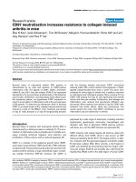
Effects of suramin and PPADS on contractions evoked by ATPFigure 6
Effects of suramin and PPADS on contractions
evoked by ATP. The effects of (a) suramin and (b) PPADS
on contractions of rat isolated SPA induced by ATP are
shown. Cumulative concentration-response curves to ATP
(10
-7
- 3 × 10
-4
M) were obtained in rings in the absence of
antagonist (control) or in the presence of 3 × 10
-5
, 10
-4
or 3
× 10
-4
M of antagonist. Contractions are expressed as a per-
centage of the contraction induced by 4 × 10
-2
M KCl. Verti-
cal lines show s.e.mean. n = 5 for suramin and 4 for PPADS.
-7 -6 -5 -4 -3
0
20
40
60
80
100
control
+Suramin(3x10
-5
M)
+Suramin(10
-4
M)
+Suramin(3x10
-4
M)
a.
[ATP] (log M)
% contraction to KCl (4 X 10
-2
M)
-7 -6 -5 -4 -3
0
20
40
60
80
100
control
+ PPADS (3 x 10
-5
M)
+ PPADS (1 0
-4
M)
+ PPADS (3 x 10
-4
M)
b.
[ATP] (log M)
% co ntra c tion to KCl (4 X 10
-2
M)
Respiratory Research 2005, 6:124 />Page 8 of 10
(page number not for citation purposes)
stores needed to activate I
Cl,Ca
in rat pulmonary arteries
[5].
UTP and UDP also activated I
Cl,Ca
, but the responses were
unaffected by suramin or PPADS. This is consistent with
the lack of effect of the antagonists on UTP- and UDP-
induced vasoconstriction in the rat perfused pulmonary
vascular bed [10] and with the antagonist-insensitive
component of UTP and UDP contraction of isolated pul-
monary artery [6], but contrasts with the abolition by
suramin of UTP-elicited oscillating currents seen
previously in single cells [5,12]. The reason for these dif-
ferences in suramin activity is not clear. Which receptor(s)
mediated the actions of UTP and UDP in the present study
is also unclear. UTP is an agonist at the P2Y
2
,
4
&
6
subtypes,
whilst UDP is only active at the P2Y
6
receptor [2,15].
Detailed studies show clearly that the P2Y
2
receptor is
antagonised by suramin and the P2Y
4
receptor by PPADS
(2,20). So, these subtypes do not mediate the effects of
UTP (or UDP) seen here.
If the P2Y
2
and P2Y
4
receptors are ruled out, then the P2Y
6
receptor is the prime candidate for the site of action of
UTP and UDP. Indeed, its mRNA is present in rat
pulmonary artery smooth muscle and it has been pro-
posed to underlie the UDP-induced I
Cl,Ca
[12]. However,
the effects of suramin and PPADS at this site are not well
characterised. In the only study on the cloned rat P2Y
6
receptor, 10
-4
M suramin (the same concentration used in
the present study) depressed the agonist response by 20%
[32]. Similar inhibition (27%) was seen at the cloned
human P2Y
6
receptor [18]. PPADS was not tested at the rat
receptor, but at 10
-4
M it inhibited the response to UDP at
the human subtype by 69%. This pronounced effect of
PPADS is inconsistent with the P2Y
6
receptor being the
receptor through which UTP and UDP activated I
Cl,Ca
in
the present study. Further characterisation of the effects of
suramin and PPADS at the recombinant rat P2Y
6
receptor
is, however, needed to substantiate this conclusion.
If the P2Y
2
, P2Y
4
and P2Y
6
receptors are not the site(s) of
action of UTP and UDP, then what is? One possibility is
that UTP and UDP activated I
Cl,Ca
in rat SPA and LPA
smooth muscle via a novel, as yet uncloned P2Y receptor
or another, non-P2Y receptor. For example, UDP has been
proposed to interact with cysteinyl leukotriene receptors
in human mast cells [33,34]. Alternatively, one of the
known P2Y receptors may interact with another P2Y sub-
type, or with a non-P2Y receptor, to form a dimer with
novel pharmacological properties. Indeed, the P2Y
1
and
P2Y
2
receptors both appear to form dimers with the A1
adenosine receptor [35]. Further studies are needed to
investigate these possibilities.
P2X receptors in SPA and LPA
In this study, the currents evoked by ATP, UTP and UDP
had similar time-courses, as measured by rise time and
width at 50% peak. This may appear surprising as ATP,
but not UTP or UDP, is also an agonist at the P2X
1
recep-
tor and so might be expected to activate an initial, rapid,
transient inward current, in addition to the slower, longer
lasting, P2Y-mediated oscillations, as has been reported
previously in rat pulmonary artery smooth muscle cells
[4,5]. The apparent absence of the transient response may
be due to the relatively slow speed of application of ATP
used here. The P2X
1
receptor desensitizes rapidly and slow
agonist administration elicits much slower and smaller
currents in vascular smooth muscle cells [36]. Although
this would be disadvantageous if studying P2X receptors,
by minimizing the P2X response it is in fact an advantage
when P2Y receptors are under study. The initial current
evoked by ATP in SPA and LPA may well be a mixture of
P2X
1
and P2Y receptor-induced responses, which would
explain the larger amplitude of the initial ATP-induced
current, compared with UTP and UDP. Nevertheless, any
P2X
1
component appears to play a relatively minor role
and would not contribute to the sustained phase of
oscillations.
Contribution of P2Y subtypes to contractions
Although the receptors that mediate activation of I
Cl,Ca
by
nucleotides in the rat pulmonary artery have not been
identified unequivocally, we can still consider their role in
vasoconstriction of the rat pulmonary vascular bed [10]
and isolated arteries [6,12,13]. In this study, contractions
of the SPA elicited by ATP were abolished by suramin, but
only partially inhibited by PPADS. The PPADS-resistant
contractions likely reflect release of Ca
2+
stores, causing
the I
Cl,Ca
recorded here. They may also involve Ca
2+
influx
via L-type Ca
2+
channels, opened by depolarisation due to
I
Cl,Ca
. Further experiments using channel blockers are
needed to confirm this. The P2X
1
receptor in SPA smooth
muscle [6] is most likely to underlie the remaining
suramin- and PPADS-sensitive component. Interestingly,
contractions of the rat LPA were abolished by both
suramin and PPADS, suggesting that only one receptor,
probably the P2X
1
receptor, mediates the contractile
actions of ATP here. This is consistent with the much
lower contractile potency of ATP in LPA [6], but it suggests
that the similar suramin- and PPADS-insensitive I
Cl,Ca
observed in response to ATP in LPA and SPA may serve
different functions. In our previous study [6] contractions
of rat SPA induced by UTP and UDP were not inhibited by
PPADS and were only partially suppressed by suramin.
These antagonist-resistant contractions again likely reflect
release of Ca
2+
stores and activation of I
Cl,Ca
. The identity
of the suramin-sensitive receptors remains to be
determined.
Respiratory Research 2005, 6:124 />Page 9 of 10
(page number not for citation purposes)
Advantages of the patch clamp technique
This study shows that recording ion currents in single cells
can be useful in characterising the receptors expressed in
tissues where multiple subtypes are present. A particular
problem with P2Y receptors is ecto-nucleotidases, which
are inhibited by PPADS in smooth muscle [37] and other
tissues [38,39]. Recording from rapidly perfused, single
cells minimises the problems created by extracellular
metabolism in whole tissues, which may explain why
PPADS potentiated contractions to UTP and UDP in the
intact artery [6], but had no effect on activation of I
Cl,Ca
in
single cells. Such studies also allow the regional variation
in ion channel expression to be studied. Interestingly, we
recorded I
Cl,Ca
in a similar proportion of rat SPA and LPA
smooth muscle cells, whereas in rabbits it is more
predominant in smaller pulmonary arteries [25]. Limita-
tions of the patch clamp technique encountered here were
short recording times, wide variation in current amplitude
between cells and a decline in the amplitude of I
Cl,Ca
over
the recording period, all of which hampered quantitative
analysis of antagonist action. It is not clear why rundown
occurred, as loss of diffusible cytosolic factors into the
recording pipette should have been minimised with the
perforated-patch technique. Similar rundown was seen in
previous patch clamp studies in these cells [4,5] and with
ATP- and UTP-induced oscillations in cytosolic [Ca
2+
]
[26]. Thus, the decline in I
Cl,Ca
may in fact reflect a physi-
ological mechanism of signalling whereby the P2Y recep-
tors become desensitised and/or intracellular stores
release progressively less Ca
2+
during maintained activa-
tion of P2Y receptors.
Conclusion
The results of the present study indicate the presence of at
least two different subtypes of P2Y receptors mediating
oscillating inward currents in rat SPA and LPA smooth
muscle cells. ATP acts via a suramin-sensitive, PPADS-
insensitive site, which most resembles the P2Y
11
receptor.
The site of action of UTP and UDP is less clear. Its phar-
macology is inconsistent with our present understanding
of P2Y
2,4 & 6
receptors, so a novel receptor or receptor com-
plex may be involved. These different P2Y receptors are
likely to play a significant role in nucleotide-induced pul-
monary vasoconstriction as ATP, UTP and UDP each
induce contractions of the rat pulmonary artery with
matching pharmacological profiles.
Competing interests
The author(s) declare that they have no competing
interests.
Authors' contributions
KC was involved in the planning of the experiments
described and carried them out. She also analysed the
data, drafted the manuscript and was involved in its revi-
sion. AMG was involved in the planning of the experi-
ments and revision of the manuscript. CK was involved in
the planning of the experiments, the analysis of the data
and revision of the manuscript. All authors read and
approved the final manuscript.
Acknowledgements
This work was supported by funding from the Faculty of Medical Science,
Naresuan University, Thailand and The Royal Society of Edinburgh.
References
1. Burnstock G, Kennedy C: A dual function for adenosine triphos-
phate in the regulation of vascular tone: excitatory cotrans-
mitter with noradrenaline from perivascular nerves and
locally released inhibitory intravascular agent. Circ Res 1986,
58:319-330.
2. Boarder MR, Hourani MO: The regulation of vascular function
by P2 receptors: multiple sites and multiple receptors. Trends
Pharmacol Sci 1998, 19:99-107.
3. Ralevic V, Burnstock G: Receptors for purines and pyrimidines.
Pharmacol Rev 1998, 50:413-491.
4. Bakhramov A, Hartley SA, Salter KJ, Kozlowski RZ: Contractile
agonists preferentially activate Cl
-
over K
+
currents in arte-
rial myocytes. Biochem Biophys Res Comm 1996, 227:168-175.
5. Hartley SA, Kozlowski RZ: Electrophysiological consequences
of purinergic receptor stimulation in isolated rat pulmonary
arterial myocytes. Circ Res 1997, 80:170-178.
6. Chootip K, Ness KF, Wang Y, Gurney AM, Kennedy C: Regional
variation in P2 receptor expression in the rat pulmonary
arterial circulation. Br J Pharmacol 2002, 137:637-646.
7. Mc Cormack DG, Barnes PJ, Evans TW: Purinoceptors in the pul-
monary circulation of the rat and their role in hypoxic
vasoconstriction. Br J Pharmacol 1989, 98:367-372.
8. Barnes PJ, Liu SF: Regulation of pulmonary vascular tone. Phar-
macol Rev 1995, 47:87-131.
9. Hasséssian H, Burnstock G: Interacting roles of nitric oxide and
ATP in the pulmonary circulation of the rat. Br J Pharmacol
1995, 114:846-850.
10. Rubino A, Burnstock G: Evidence for a P
2
purinoceptor media-
tor mediation vasoconstriction by UTP, ATP and related
nucleotides in the isolated pulmonary vascular bed of the
rat. Br J Pharmacol 1996, 118:1415-1420.
11. Liu SF, Mc Cormack DG, Evans TW, Barnes PJ: Characterization
and distribution of P
2
-purinoceptor subtypes in rat pulmo-
nary vessels. J Pharmacol Exp Ther 1989, 251:1204-1210.
12. Hartley SA, Kato K, Salter KJ, Kozlowski RZ: Functional evidence
for a novel suramin-insensitive pyrimidine receptor in rat
small pulmonary arteries. Circ Res 1998, 83:940-946.
13. Rubino A, Ziabary L, Burnstock G: Regulation of vascular tone by
UTP and UDP in isolated rat intrapulmonary arteries. Eur J
Pharmacol 1999, 370:139-143.
14. Boarder MR, Webb TE: P2Y receptors: structure and function.
In Handbook of Experimental Pharmacology 151/1; Purinergic and Pyrimi-
dinergic Signalling I: Molecular, Nervous and Urogenitary System Function
Edited by: Abbracchio MP, Williams M. Springer; 2001:65-88.
15. Abbracchio MP, Boeynaems JM, Barnard EA, Boyer JL, Kennedy C,
Miras-Portugal M, King BF, Gachet C, Jacobson KA, Weisman GA,
Burnstock G: Characterization of the UDP-glucose receptor
(re-named here the P2Y
14
receptor) adds diversity to the
P2Y receptor family. Trends Pharmacol Sci 2003, 24:52-55.
16. Nicholas RA, Watt WC, Lazarowski ER, Li Q, Harden TK: Uridine
nucleotide selectivity of three phospholipase C-activating P2
receptors: Identification of a UDP-selective, a UTP-selec-
tive, and an ATP- and UTP-specific receptor. Mol Pharmacol
1996, 50:224-229.
17. Filippov AK, Webb TE, Barnard EA, Brown DA: Dual coupling of
heterologously-expressed rat P2Y
6
nucleotide receptors to
N-type Ca
2+
and M-type K
+
currents in rat sympathetic
neurones. Br J Pharmacol 1999, 126:1009-1017.
18. Robaye B, Boeynaems JM, Communi D: Slow desensitization of
the human P2Y
6
receptor. Eur J Pharmacol 1997, 329:231-236.
Publish with BioMed Central and every
scientist can read your work free of charge
"BioMed Central will be the most significant development for
disseminating the results of biomedical research in our lifetime."
Sir Paul Nurse, Cancer Research UK
Your research papers will be:
available free of charge to the entire biomedical community
peer reviewed and published immediately upon acceptance
cited in PubMed and archived on PubMed Central
yours — you keep the copyright
Submit your manuscript here:
/>BioMedcentral
Respiratory Research 2005, 6:124 />Page 10 of 10
(page number not for citation purposes)
19. Bogdanov YD, Wildman SS, Clements MP, King BF, Burnstock G:
Molecular cloning and characterisation of rat P2Y
4
nucle-
otide receptor. Br J Pharmacol 1988, 124:428-439.
20. Suarez-Huerta N, Pouillon V, Boeynaems JM, Robaye B: Molecular
cloning and characterization of the mouse P2Y
4
nucleotide
receptor. Eur J Pharmacol 2001, 416:197-202.
21. Kennedy C, Leff P: How should P2X-purinoceptors be charac-
terised pharmacologically? Trends Pharmacol Sci 1995,
16:168-174.
22. Lazarowski ER, Boucher RC, Harden TK: Constitutive release of
ATP and evidence for major contribution of ecto-nucleotide
pyrophosphatase and nucleoside diphosphokinase to extra-
cellular nucleotide concentrations. J Biol Chem 2000,
275:31061-31068.
23. Lazarowski ER, Boucher RC, Harden TK: Mechanisms of release
of nucleotides and integration of their actions as P2X- and
P2Y-receptor activating molecules. Mol Pharmacol 2003,
64:785-795.
24. Zimmermann H: Extracellular metabolism of ATP and other
nucleotides. Naunyn-Schmied Arch Pharmacol 2000, 362:299-309.
25. Smani T, Iwabuchi S, López-Barneo J, Ureña J: Differential segmen-
tal activation of Ca
2+
-dependent Cl
-
and K
+
channels in pul-
monary arterial myocytes. Cell Calcium 2001, 29:369-377.
26. Guibert C, Pacaud P, Loirand G: Effect of extracellular ATP on
cytosolic Ca
2+
concentration in rat pulmonary artery
myocytes. Am J Physiol 1996, 271:L450-L458.
27. Communi D, Robaye B, Boeynaems JM: Pharmacological charac-
terization of the human P2Y
11
receptor. Br J Pharmacol 1999,
128:1199-1206.
28. Gitlin JM, Zanesco A, Stanford SJ, Evans TW, Anning PB, Mitchell JA:
The second phase of ATP mediated relaxation is mediated
through P2Y
11
receptors. Br J Pharmacol 2002, 135:210P.
29. Takasaki J, Kamohara M, Saito T, Matsumoto M, Matsumoto SI, Ohishi
T, Soga T, Matsushime H, Furuichi K: Molecular cloning of the
platelet P2T
AC
ADP receptor: pharmacological comparison
with another ADP receptor, the P2Y
1
receptor. Mol Pharmacol
2001, 60:432-439.
30. Wihlborg AK, Wang L, Braun OO, Eyjolfsson A, Gustafsson R, Gud-
bjartsson T, Erlinge D: ADP receptor P2Y12 is expressed in vas-
cular smooth muscle cells and stimulates contraction in
human blood vessels. Arterioscler Thromb Vasc Biol 2005, 24:1-7.
31. Kauffenstein G, Hechler B, Cazenave JP, Gachet C: Adenosine tri-
phosphate nucleotides are antagonists at the P2Y
12
recep-
tor. J Thromb Haem 2004, 2:980-1988.
32. Chang K, Hanaoka K, Kumada M, Takuwa Y: Molecular cloning
and functional analysis of a novel P2 nucleotide receptor. J
Biol Chem 1995, 270:26152-26158.
33. Mellor EA, Maekawa A, Austen KF, Boyce JA: Cysteinyl leukot-
riene receptor 1 is also a pyrimidergic receptor and is
expressed by human mast cells. Proc Natl Acad Sci USA 2001,
98:7964-7969.
34. Mellor EA, Austen KF, Boyce JA: Cysteinyl leukotrienes and uri-
dine diphosphate induce cytokine generation by human
mast cells through an interleukin 4-regulated pathway that
is inhibited by leukotrienne receptor antagonists. J Exp Med
2002, 195:583-592.
35. Yoshioka K, Saitoh O, Nakata H: Heteromeric association cre-
ates a P2Y-like adenosine receptor. Proc Natl Acad Sci USA 2001,
98:7617-7622.
36. Evans RJ, Kennedy C: Characterisation of P2-purinoceptors in
the smooth muscle of the rat tail artery: a comparison
between contractile and electrophysiological responses. Br J
Pharmacol 1994, 113:853-860.
37. Khakh BS, Michel AD, Humphrey PPA: Inhibition of ectoATPase
and Ca-ATPase in rat vas deferens by P
2
-purinoceptor
antagonists. Br J Pharmacol 1995, 115:2P.
38. Chen BC, Lee CM, Lin WW: Inhibition of ecto-ATPase by
PPADS, suramin and reactive blue in endothelial cells, C6
glioma cells and RAW 264.7 macrophages. Br J Pharmacol 1996,
119:1628-1634.
39. Grobben B, Claes P, Roymans D, Esmans EL, Van Onckelen H, Slegers
H: Ecto-nucleotide pyrophosphatase modulates the purinoc-
eptor-mediated signal transduction and is inhibited by puri-
noceptor antagonists. Br J Pharmacol 2000, 130:139-145.