Báo cáo y học: " Influence of hypoxia on the domiciliation of Mesenchymal Stem Cells after infusion into rats: possibilities of targeting pulmonary artery remodeling via cells therapies " doc
Bạn đang xem bản rút gọn của tài liệu. Xem và tải ngay bản đầy đủ của tài liệu tại đây (4.24 MB, 13 trang )
BioMed Central
Page 1 of 13
(page number not for citation purposes)
Respiratory Research
Open Access
Research
Influence of hypoxia on the domiciliation of Mesenchymal Stem
Cells after infusion into rats: possibilities of targeting pulmonary
artery remodeling via cells therapies?
Gaël Y Rochefort
1
, Pascal Vaudin
2,3
, Nicolas Bonnet
4
, Jean-
Christophe Pages
3
, Jorge Domenech
2
, Pierre Charbord
2
and
Véronique Eder*
1
Address:
1
LABPART-EA3852, IFR135, Université François Rabelais, faculté de Médecine, 10 boulevard Tonnellé 370032 TOURS France,
2
INSERM
ESPRI-EA3588, IFR135, Université François Rabelais, faculté de Médecine, 10 boulevard Tonnellé 370032 TOURS France,
3
Virus, pseudo-virus:
morphogenése et antigénicité, EA3856, Université François Rabelais, faculté de Médecine, 10 boulevard Tonnellé 370032 TOURS France and
4
Architecture du Tissu Osseux – Exercice Physique, EA 3895, Université d'Orléans- BP6749, 45067 Orléans cedex 2 France
Email: Gaël Y Rochefort - ; Pascal Vaudin - ; Nicolas Bonnet -
tours.fr; Jean-Christophe Pages - ; Jorge Domenech - ;
Pierre Charbord - ; Véronique Eder* -
* Corresponding author
arterieshypertension, pulmonaryhypoxialungremodelingmesenchymal stem cells.
Abstract
Background: Bone marrow (BM) cells are promising tools for vascular therapies. Here, we focused on
the possibility of targeting the hypoxia-induced pulmonary artery hypertension remodeling with systemic
delivery of BM-derived mesenchymal stem cells (MSCs) into non-irradiated rats.
Methods: Six-week-old Wistar rats were exposed to 3-week chronic hypoxia leading to pulmonary
artery wall remodeling. Domiciliation of adhesive BM-derived CD45
-
CD73
+
CD90
+
MSCs was first studied
after a single intravenous infusion of Indium-111-labeled MSCs followed by whole body scintigraphies and
autoradiographies of different harvested organs. In a second set of experiments, enhanced-GFP labeling
allowed to observe distribution at later times using sequential infusions during the 3-week hypoxia
exposure.
Results: A 30% pulmonary retention was observed by scintigraphies and no differences were observed in
the global repartition between hypoxic and control groups. Intrapulmonary radioactivity repartition was
homogenous in both groups, as shown by autoradiographies. BM-derived GFP-labeled MSCs were
observed with a global repartition in liver, in spleen, in lung parenchyma and rarely in the adventitial layer
of remodeled vessels. Furthermore this global repartition was not modified by hypoxia. Interestingly, these
cells displayed in vivo bone marrow homing, proving a preservation of their viability and function. Bone
marrow homing of GFP-labeled MSCs was increased in the hypoxic group.
Conclusion: Adhesive BM-derived CD45
-
CD73
+
CD90
+
MSCs are not integrated in the pulmonary
arteries remodeled media after repeated intravenous infusions in contrast to previously described in
systemic vascular remodeling or with endothelial progenitor cells infusions.
Published: 27 October 2005
Respiratory Research 2005, 6:125 doi:10.1186/1465-9921-6-125
Received: 31 October 2004
Accepted: 27 October 2005
This article is available from: />© 2005 Rochefort et al; licensee BioMed Central Ltd.
This is an Open Access article distributed under the terms of the Creative Commons Attribution License ( />),
which permits unrestricted use, distribution, and reproduction in any medium, provided the original work is properly cited.
Respiratory Research 2005, 6:125 />Page 2 of 13
(page number not for citation purposes)
Background
Recent studies emphasize on the perspective of cellular
therapy by intravenous stem cells infusion. The participa-
tion of stem cells in several vascular diseases pathogenesis
was first proved with haematopoietic stem cells (HSCs).
In this regard, following bone marrow engraftment, HSCs
were observed in remodeled vascular wall following graft
vasculopathy or arteriosclerosis [1]. When integrated to
the vascular wall, HSCs differentiate into mature vascular
cells with an endothelial or smooth muscle cells pheno-
type.
Mesenchymal Stem cells (MSCs) are bone marrow non-
haematopoietic stem cells that are multipotent and can
differentiate into bone, cartilage and connective tissue
cells [2-4]. They also differentiate in smooth muscle fibers
and could be preferential candidates for vascular cells
therapies [5]. Moreover MSCs present many advantages as
facility to culture or to transform genetically [6]. Surpris-
ingly few studies focused on the domiciliation of MSCs
after in vivo infusion, even though they can be found into
different organs after several months in normal animals,
proving the in vivo infusion possibility without graft rejec-
tion [7]. Barbash et al recently showed a MSCs domicilia-
tion into myocardial infarct area, however only a poor
fraction of the cells engrafts the myocardium after sys-
temic infusion [8].
Sustained pulmonary hypertension is a common compli-
cation of chronic hypoxic lung diseases. Hypoxic pulmo-
nary hypertension is characterized by sustained
pulmonary vasoconstriction and pulmonary vascular wall
remodeling, including media and adventitia hypertrophy,
without endothelial cells disruption. Furthermore chronic
hypoxia has been shown to induce capillary angiogenesis
[9]. Recently the participation of stem cells to hypoxia-
induced adventitial remodeling has been observed in
chronically hypoxic rat lungs [10]. Our hypothesis was
that MSCs could domicile into the pulmonary artery
remodeled wall and thus participate to hypoxia-induced
structural changes.
We studied, for the first time, the bone marrow derived
CD45
-
CD73
+
CD90
+
MSCs domiciliation after intrave-
nous infusion in a model of chronically hypoxic rats,
which induces pulmonary artery hypertension and vascu-
lar remodeling. Firstly, MSCs distribution was studied
after a unique infusion of MSCs labeled by Indium-111
oxinate. Secondly, distribution was studied after sequen-
tial infusions of MSCs, transduced with the enhanced
green fluorescent protein (GFP) gene by viral infection,
during the three weeks of hypoxia exposure.
Methods
Animals
Six-weeks-old Wistar male rats (n = 26, Harlan) were
exposed for 3 weeks to chronic hypoxia in a hypobaric
chamber (50 kPa) to lead the development of pulmonary
hypertension and were compared to control matched rats
(n = 26).
The MSCs engraftment and viability control was per-
formed using 4 hypoxic rats and compared to 4 control
rats by a direct in-vivo injection of GFP-labeled MSCs into
the right lung parenchyma and checked 3 weeks after nor-
moxic or hypoxic condition housing as described below.
The early dynamic distribution of infused radiolabeled
MSCs was performed using 6 hypoxic rats and compared
to 6 control rats. The long-term distribution of infused
GFP-labeled MSCs was performed using 6 other hypoxic
rats compared to 6 matched control rats. Finally, 5
hypoxic rats and 5 control rats were also sacrificed for
DNA extraction and 5 hypoxic rats and 5 control rats were
sacrificed for pulmonary enzymatic digestion and culture
(see below).
All animal investigations were carried out in accordance
with the Guide for the Care and Use of Laboratory Ani-
mals published by the US National Institute of Health
(NIH Publications N°85-23, revised 1996) and European
Directives (86/609/CEE).
Cell culture
Cell isolation and culture procedures for MSCs have been
established and published previously [11,12]. Briefly,
femurs were aseptically harvested from 6-weeks-old Wis-
tar rats and the adherent soft tissue was removed. The
proximal and distal ends of the femur were excised at a
level just into the beginning of the marrow cavity. Whole
marrow plugs were obtained by flushing the bone marrow
cavity with a 18-gauge needle set with a syringe filled with
culture medium composed of Modified Eagle Medium
Alpha (α-MEM; Invitrogen) supplemented with 20% fetal
calf serum (FCS; Hyclone), with antibiotic solution (pen-
icillin/streptomycin: 1%; Invitrogen) and with antimy-
cotic solution (amphotericin B: 0.01%; Bristol-Myers).
The marrow plugs were dispersed to obtain a single cell
suspension by sequentially passing the dispersion
through 18- and 22-gauge needles. The cells were centri-
fuged and resuspended with culture medium. After count-
ing in Malassez cells following an acetic acid disruption of
red blood cells, nucleated cells were plated at a density of
10
6
/cm
2
and incubated at 37°C in a humidified atmos-
phere of 95% air 5% C0
2
. The first medium change was
after 2 days and twice a week thereafter. When these pri-
mary MSCs reached 80–90% of confluence, they were
trypsinized (trypsin-EDTA, Invitrogen), counted and pas-
saged at a density of 10
4
/cm
2
. For the first study second-
Respiratory Research 2005, 6:125 />Page 3 of 13
(page number not for citation purposes)
passage MSCs were labeled with
111
In-oxine as described
below and infused intravenously. For the second study
MSCs were GFF-labeled after viral gene transduction after
the first passage and were used as the second-passage.
Adherent second-passage MSCs were analyzed by flow
cytometry with a FACSCalibur flow cytometer (Becton-
Dickinson) using a 488 nm argon laser. Cells were incu-
bated for 60 minutes at 4°C with phycoerythrin- or fluo-
rescein isothiocyanate-conjugated monoclonal
antibodies against rat CD45 (clone OX-1), rat CD73
(clone 5F/B9), and rat CD90 (Clone OX-7; all from Bec-
ton Dickinson). Isotype-identical antibodies served as
controls. Samples were analyzed by collecting 10,000
events on a FACSCalibur instrument using Cell-Quest
®
software (Becton-Dickinson).
Isotopic labeling and Indium-111 labeled MSCs
intravenous infusion
The cells were incubated with
111
In-oxine (37 MBq/10
6
cells) and incubated for 60 minutes as previously
described [11]. The radiolabeled MSCs were aliquoted at
10
7
cells/ml and intravenously infused to hypoxic rats
within 1 hour and followed by whole body scintigraphic
imaging. Preliminary experiments showed that the viabil-
ity and growth of these labeled MSCs were not adversely
affected by this labeling procedure (data not shown); the
level of radioisotope was widely sufficient to produce high
quality images taken with a gamma camera and to pro-
duce high quality autoradiographic images of organs.
Whole body scintigraphic imaging was performed imme-
diately after infusion and within 15 minutes, 30 minutes,
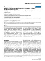
Mesenchymal stem cells used during this studyFigure 1
Mesenchymal stem cells used during this study. Typical morphological aspects of mesenchymal stem cells observed
through culture flask (A). Mesenchymal stem cells expression of CD73 and CD90 antigens was attested by flow cytometry (B).
20 µm
5µm
A
B
Respiratory Research 2005, 6:125 />Page 4 of 13
(page number not for citation purposes)
1 hour, 3 hours, 24 hours and 96 hours thereafter. Planar
whole body images were acquired with Helix Elscint scan-
ner (GE Healthcare) using a medium energy collimator.
Images were acquired on a 256 × 256 matrix using a win-
dow centered at 245 keV. The distance between the chest
of animals and the detector was fixed at 65 mm. In analy-
sis of the scintigraphic images, regions of interest (ROIs)
were placed over lungs, liver and spleen on anterior inci-
dence, and over kidneys on posterior incidence. The
whole body count was determined by the mean counts on
both incidences. Total counts in the ROIs were corrected
with physical decay of
111
In and with body count.
After sacrifice lung, liver, heart, spleen, kidneys and bone
marrow were harvested. Organs were weighted and
assayed for radioactivity using a Muller counter (Ludlum
Measurements), after what they were snap-frozen in liq-
uid nitrogen, whereas cytospins of bone marrow were
realized. Sample sections (15 µm) and bone marrow cyt-
ospins were exposed to a photographic film within 24–96
hours and autoradiographic films were developed.
GFP labeling, in vivo engraftment and viability controls,
and GFP-labeled MSCs intravenous infusions
GFP labeling
MSCs were labeled by green fluorescent protein (GFP)
after stable viral gene transduction with LNCX-GFP vector.
GFP fluorescence from first-passage transduced MSCs was
checked by flow cytometry. Non-specific fluorescence was
determined using MSCs that were not transduced. GFP-
labeling stability was assayed by flow cytometry using
tenth-passage GFP-labeled MSCs.
In-vivo engraftment and viability controls
Animals were lightly anesthetized and GFP-labeled MSCs
were injected, at a dose of 2.10
6
cells, through the rib cage,
into the right lung lower lobe. After recovering, animals
were housed 3 weeks either in normoxic condition, or
hypoxic condition. Animals were sacrificed after the 3
weeks and the lung was harvested, snap-frozen in liquid
nitrogen. The frozen sample sections (15 µm) were ana-
lyzed by tree-dimensional confocal laser microscopy.
GFP-labeled MSCs intravenous infusions
Second-passage GFP-labeled MSCs were sequentially
infused intravenously at the dose of 10
6
MSCs. The first
infusion indicated the first day of the 3 weeks chronic
hypoxia. Both hypoxic and control rats were infused twice
a week during 3 weeks.
After sacrifice lung, liver, heart, spleen, kidneys and bone
marrow were harvested. Organs were weighed and snap-
frozen in liquid nitrogen. The frozen sample sections (15
µm) of the different organs were analyzed by tree-dimen-
sional confocal laser microscopy. Data was collected with
sequential laser excitation to eliminate bleed through and
acquired on a 1024 × 1024 matrix using a 110 µm pinhole
and an optical section thickness of 0.31 µm. The system
was made up of a FV500 confocal microscope (Olympus)
using FluoView500 software and a 488 nm argon laser.
The GFP protein was also researched on frozen sections by
immunohistochemistry. Sections of harvested organs
were incubated with a rabbit polyclonal antibody against
GFP (1/200, Santa Cruz Biotechnology) and were
revealed either by a conjugated goat anti-rabbit alexa-594
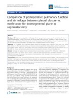
Pulmonary radioactivityFigure 3
Pulmonary radioactivity. Pulmonary repartition was
measured in vivo from lung region of interest counts on scin-
tigraphies at different times after radiolabeled mesenchymal
stem cells infusion. Counts were normalized by whole body
counts. After 24 hours, radioactivity was stabilized without
differences between control and hypoxic groups.
Control rats
Hypoxics rats
0 1 3 24 96
0
10
20
30
40
50
60
70
80
Pulmonary count / Whole body count (%±SEM)
Time post-injection (h)
52.8
±3.4
62.3
±6.3
57.1
±5.4
59.5
±3.5
29.8
±6.3
25.8
±1.2
25.7
±4.8
37.9
±2.3
30.7
±2.1
36.0
±1.0
NS
n=2+2
NS
n=4+4
NS
n=6+6
NS
n=2+2
NS
n=3+3
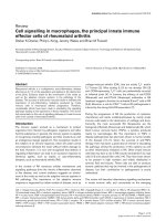
Early dynamic distribution of mesenchymal stem cells in vivoFigure 2
Early dynamic distribution of mesenchymal stem
cells in vivo. Sequential whole body scintigraphies after infu-
sion of indium-111 labeled mesenchymal stem cells were
acquired from injection up to 96 h. After pulmonary reten-
tion, a liver and spleen repartition was observed. A lung
domiciliation was indicated by lungs radioactivity stabilization.
Bone radioactivity was linked with bone marrow homing
after 24 hours.
Since infusion 1h
3h 24h
Infusion site
Lungs
Liver
Spleen
Kidneys
100%
0%
Bone
Respiratory Research 2005, 6:125 />Page 5 of 13
(page number not for citation purposes)
(1/400, Molecular Probes) or by a conjugated goat anti-
rabbit horseradish peroxydase (1/400, Biosource).
Bone marrow homing detection
Cytospins of bone marrow aspirates from control and
hypoxic rats were realized 3 days after a unique GFP-
labeled MSCs infusion and 3 days after the end of GFP-
labeled MSCs infusion during the 3-week hypoxia expo-
sure. The percentage of fluorescent cells was estimated for
each rat in five random fields by microscopy using Opti-
mas software (Imasys). Thin slices (12 µm) of frozen bone
sections were cut in the metaphysis of tibia from five
injected rats. Fluorescence (GFP) was directly observed by
confocal microscopy and adipocytes were detected after
counterstaining with DAPI (4,6-diamidino-2-phenylin-
dole, AbCys) [13].
Detection of GFP transgene and protein by PCR and
western blotting
After sequential infusions, organs were harvested. From
each animal, GFP transgene and protein were assayed by
PCR and Western blotting.
PCR
Total DNA was extracted using QIAamp DNA Mini Kit
(Qiagen, Hilden, Germany) according to the manufac-
turer's instructions. It was analyzed by PCR for GFP trans-
gene presence using a set of primer generating a 249 bp
amplicon: forward, GCGACGTAAACGGCCACAAGTTC
and reverse, CGTCCTTGAAGAAGATGGTGCGC. DNA
was subjected to PCR for 35 cycles of 94°C for 30 seconds,
58°C for 60 seconds, 72°C for 30 seconds, with a final
elongation step of 10 minutes at 72°C.
Western blotting
Organs were crushed by Turrax and homogenized with
lysis buffer [1% sodium deoxycholate, 0.1% SDS, 1% tri-
ton X-100, 10 mM Tris-HCl (pH 8.0), 150 mM NaCl and
an inhibitor protease cocktail (chymotrypsin-, thermo-
lysin-, papain-, pronase-, pancreatic extract- and trypsin-
inhibitor; Roche)] and centrifuged at 20,000 g for 1 h.
After purifying and concentrating small proteins from
each sample (Centriprep Centrigugal Devices YM-30MW,
Millipore) with a nominal molecular weight limit of 30
kDa, proteins were separated on a SDS/12% polyacryla-
mide gel and then transferred to a nitrocellulose mem-
brane (Amersham). Blots were blocks for 2 h at room
temperature with 5% (w/v) non-ft dried milk in Tris-buff-
ered saline [10 mM Tris-HCl (pH 8.0) and 150 mM NaCl]
containing 0.05% Tween 20. The membrane was incu-
bated overnight at 4°C with rabbit polyclonal antibody
against GFP (1/400, Santa Cruz Biotechnology). The blot
was then incubated with the conjugated goat anti-rabbit
horseradish peroxydase (1/1000, Biosource) 2 h at room
temperature. Immunoreactive proteins were detected with
the ECL Western blotting detection system (Amersham).
Pulmonary enzymatic digestion
Lung from 5 non-hypoxic and 5 hypoxic MSCs-injected
rats were cultured after enzymatic digestion. Briefly, rat
lungs were harvested, mechanically dissected and the thin
pieces were digested with collagenase (0.5 mg/ml, 1 hour
at 37°C, Sigma). After wash, the suspension was passed
through a cell strainer to remove undigested block and
wash in PBS with FCS (20%, Hyclone). Then, the suspen-
sion was incubated in trypsin (30 minutes at 37°C, Invit-
rogen), wash twice in PBS-FCS, counted, plated and
incubated at 37°C in a humidified atmosphere of 95% air
5% C0
2
. The first medium change was after 2 days and
twice a week thereafter. The GFP fluorescence was checked
after 1 and 2 weeks.
Statistical analysis
Data are presented as mean +/-SEM with statistical signif-
icance tested using the two tailed paired t-test or the
Mann-Whitney test.
Results
Hypoxia-induced pulmonary arteries remodeling and
pulmonary hypertension
The hypoxia-induced pulmonary artery hypertension was
checked by echocardiography (data not shown). This is
pulmonary artery remodeling model already validated
and previously reported by our team [14].
Mesenchymal stem cells
Cultured bone marrow-derived cells had a typical fibrob-
last-like morphology and were evenly distributed on the
plate after 2 days (fig. 1A). Cells attachment was observed
at about 3–4 h and 80–90% of confluence was typically
Table 1: Harvested organs radioactivity. The radioactivity repartition in different organs, measured ex vivo after animals sacrifice 96 h
after radiolabeled mesenchymal stem cells infusion, was normalized by organ weight and by infused activity. The results were
corrected by time decay and are presented as mean +/-SEM.
Control group rats Hypoxic group rats
Lungs 17.22 % ± 6.92 25.26 % ± 2.78
Liver 41.28 % ± 19,62 29.39 % ± 12.42
Spleen 20.23 % ± 13.59 9.07 % ± 2.83
Kidneys 21.16 % ± 13.01 14.75 % ± 6.93
Respiratory Research 2005, 6:125 />Page 6 of 13
(page number not for citation purposes)
reached by day 6–7. The average cell viability, determined
by exclusion of trypan blue, was approximately 90%.
CD73 and Thy-1/CD90 were expressed in these MSCs
whereas the haematopoietic lineage marker CD45 was not
(fig. 1B). These growth patterns and surface markers
expression were similar to those of normal rat bone mar-
row-derived MSCs previously described [12]. Retroviral
infection of MSCs had not modified their morphology or
viability. The GFP-labeling efficiency was about 98% and
the labeling stability was assayed until tenth passage (data
not shown).
Dynamic distribution of radiolabeled-MSCs after a single
infusion
The distribution of radioactivity after infusion of the radi-
olabeled-MSCs was imaged from the end of infusion up to
96 h after. This imaging provides an immediate indication
of the initial cells distribution. Since radiolabeled-MSCs
intravenous infusion, the radioactivity was first observed
to accumulate into the lungs, and gradually, the radioac-
tivity was observed in the liver. At 3 h after cell infusion,
the radioactivity was observed in the spleen. Kidneys and
bone were widely observed at 24 h (fig. 2).
In order to quantify the distribution of
111
In, the specific
radioactivity of each organ was calculated as a percentage
of the total body counts related to the organs region of
interest (ROIs) counts. The pulmonary radioactivity was
about 50–60% (fig. 3) in both hypoxic and control rats
from infusion and at 1 h. This pulmonary radioactivity
decreased afterwards and stabilized by about 30% in both
groups at 3 h after infusion. No significant difference in
lungs ROIs counts was observed between hypoxic rats and
control rats (tab. 1).
To observe the distribution of the infused-cells in the
lungs, autoradiography of lungs sections were performed
(fig. 4A). These films showed homogenous distribution of
the radioactivity in both groups. Furthermore, radioactiv-
ity was not observed in the lumen of large diameter pul-
monary arteries, proving that the infused cells were not
agglomerated into the pulmonary vessels lumen.
Bone marrow from radiolabeled-MSCs infused-rats was
also harvested and exposed to autoradiographic film. We
therefore showed that infused-MSCs homed in bone mar-
row at 96 h after infusion in both groups (fig. 4B).
In-vivo engraftment and viability controls
In order to have positive controls of GFP signals for con-
focal images interpretation, we first directly injected GFP-
labeled cells into a freshly harvested lung (fig. 5A) and
compared to non-injected freshly harvested lung (fig. 5B).
To check the in-vivo engraftment and viability of the MSCs
into lungs, we have directly injected GFP-labeled MSCs
into the right lower lobe of the lung and housed animals
either in normoxic or hypoxic conditions during 3 weeks.
The tolerance of these injections was good and no animals
died or showed rejection. From confocal microscopy
observation centered on the injection injury (fig. 5C), we
observed GFP signals proving the lung engraftment capac-
ity and the viability of the MSCs after 3 weeks (fig. 5D).
No difference in the appearance of MSCs was observed
between hypoxic and non-hypoxic rats.
Distribution of GFP-labeled MSCs after sequential
infusions
After sequential infusions during the 3-week hypoxia
exposure, we examined the harvested lungs sections from
control and hypoxic rats. Only few GFP-labeled MSCs
were observed per lung sections in both control and
hypoxic rats. Moreover when observed, the GFP-labeled
MSCs were localized in the lung parenchyma and rarely
close to the vascular lumen in both control (fig. 6A) and
hypoxic (fig. 6B, 6C, 6D) rats. To localize these cells, we
then performed the GFP detection in lungs using immu-
nohistochemistry and peroxydase revelations (data not
shown). No signal linked to MSCs localization was
observed into the media of pulmonary arteries. Rarely,
GFP-labeled MSCs were observed close to the adventitial
layer of remodeled vessels. So we confirm the absence of
GFP-labeled cells into the remodeled pulmonary arteries.
GFP cells were also and better observed on liver (fig. 7A)
and spleen sections (fig. 7B) with the same aspect. No dif-
ference in the repartition of GFP-labeled cells was
observed in these organs between normoxic and hypoxic
groups confirming the absence of pulmonary domicilia-
tion enhanced by hypoxia.
AutoradiographiesFigure 4
Autoradiographies. Autoradiographies of organs frozen
sections were realized after animals sacrifice, by 96 h after
radiolabeled mesenchymal stem cells infusion. Lung images
showed a homogenous repartition and the absence of radio-
activity into main arteries that appeared in negative (A,
arrows). Lonely signals on bone marrow cytospins confirmed
the mesenchymal stem cells homing and excluded free
indium bone uptake (B). In all cases no differences in reparti-
tion between control and hypoxic groups were observed
(see tab. 1).
Control rats Hypoxics rats
Lungs:
Bone marrow
cytospins:
A
B
Respiratory Research 2005, 6:125 />Page 7 of 13
(page number not for citation purposes)
The GFP transgene was found in lungs by PCR (fig. 8A)
and the GFP protein was recovered in lungs by western
blotting (fig. 8B) confirming the presence of GFP-cells
into the lungs.
To extract and culture the engrafted GFP-labeled cells
from lungs following the same protocol of three-week cell
injection and hypoxic exposure, we enzymatically
digested lung from 5 control and 5 hypoxic injected rats
and cultured. However this experiment failed to obtain
cultured GFP-labeled cells suggesting that only few num-
bers of GFP-labeled cells localized into the lung both in
normoxic and hypoxic group.
Bone marrow homing and engraftment
The fluorescent cell ratio was evaluated on bone marrow
cytospins by averaging the results of five views fields for
each slide (tab. 2). Compared to a single infusion, we
observed an increase of fluorescent cell ratios with
sequential infusions (tab. 2) while hypoxia appeared to
enhance bone marrow homing. Moreover, on slices of rat
tibial bone after GFP-labeled MSCs infusion, we observed
In-vivo engraftment and viability controlsFigure 5
In-vivo engraftment and viability controls. GFP signals were researched by confocal microscopy on lungs frozen sections.
In a first step, GFP-labeled MSCs were directly injected in ex-vivo excised lungs in order to provide positive control (A, arrow)
for confocal images interpretation whereas a non-injected freshly harvested lung served as negative control (B). Then, MSCs
were directly injected in the right lower lobe of the lung in vivo and rats placed in normoxic or hypoxic conditions for three
weeks. Frozen sections of lungs were observed after three weeks in confocal microscopy to provide in vivo positive engraft-
ment and viability controls. Indeed, the injection site was visualized macroscopically (C, arrows) and GFP signals were seen
centered on the injection injury (D, arrows). Bar = 50 µm, a indicates artery.
a
A
CD
B
Respiratory Research 2005, 6:125 />Page 8 of 13
(page number not for citation purposes)
fluorescent cells localized between adipocytes (fig. 9A) in
contrast to non-infused control rats (fig. 9C). Surprisingly,
their appearance in some part looked like the surrounding
adipocytes counterstained by DAPI (same size and shape)
(fig. 9B). We therefore concluded that MSCs are able to
home into bone with preserved viability.
Discussion
Mesenchymal stem cells
Bone marrow comprises both haematopoietic and non-
haematopoietic cells among these last mesenchymal stem
cells can be found. MSCs in culture can be characterized
by their adhesivity, fusiform shape and presence of spe-
cific membrane surface antigens. In culture after two pas-
sages we showed that more than 90% of collected cells
were MSCs. Transduction by GFP did not alter these prop-
erties. We did not study the effect of gamma irradiation on
the MSCs phenotype. One of the results of our study is
that no engraftment intolerance was observed. In accord-
ance with previous studies our results demonstrated that
infused MSCs could be found several weeks after infusion.
In a precedent study, MSCs were isolated from the recep-
tor organs after in vivo infusion and cultured successfully,
confirming their viability after domiciliation [15]. Moreo-
ver these authors concluded that MSCs could by them-
selves immuno-privileged. In our study, MSCs
morphology and fluorescent labeling were also kept intact
and no inflammatory reactions were observed in the sur-
rounding tissue. We then concluded in the absence of
graft rejection.
In our study, despite the fact that rats have not been irra-
diated, we also observed bone marrow homing of MSCs,
as previously described for haematopoietic cells after
Mesenchymal stem cell localization in lungsFigure 6
Mesenchymal stem cell localization in lungs. GFP-labeled MSCs (arrows) were localized essentially into the pulmonary
parenchyma without difference between the non-hypoxic (A) and the hypoxic group (B, C, D). Bar = 50 µm, a indicates artery,
b indicates bronchiole.
a
b
a
A
CD
B
a
a
Respiratory Research 2005, 6:125 />Page 9 of 13
(page number not for citation purposes)
intravenous infusion in immuno-competent animals
[16]. This homing could be significantly increased after
irradiation [17,18]. In the present study, we observed
bone marrow homing of MSCs that was increased by
sequential infusions. In bone, some GFP-labeled cells
even displayed an adipogenic phenotype, proving in vivo
their viability. Chondrogenic differentiation of MSCs has
already been observed in vivo in bone after intravenous
infusion in neonatal mice [15]. Nevertheless further stud-
ies are required to confirm adipogenic differentiation.
Finally, we showed in our hypoxic rat model that 3 weeks
after the first intravenous infusion, MSCs remain detecta-
ble, viable and functional.
Pulmonary domiciliation
In our model, adhesive bone marrow derived CD45
-
CD73
+
CD90
+
MSCs were localized into the pulmonary
parenchyma. After a first phase of pulmonary arteries
retention, some MSCs reached the systemic circulation
and were distributed mainly in the spleen, and the liver.
These cells are essentially observed into the parenchyma
of these organs and their presence was confirmed by the
detection of the GFP protein in Western Blotting and by
detection of the transgene in PCR analysis from lung sam-
ples.
This observed global cells distribution is in agreement
with previous study [11]. These results leaded some
authors to conclude that the pulmonary retention was not
specific and without any precise localization neither in the
parenchyma nor in the vasculature and to hypothesize
that stem cells infusion induces only passive embolism or
endothelium adhesion. In our study, we also failed to cul-
ture GFP-labeled cells from injected rat lungs suggesting
Mesenchymal stem cell localization in liver and spleenFigure 7
Mesenchymal stem cell localization in liver and spleen. GFP signals were observed in liver (A) and spleen (B) from fro-
zen sections after GFP-labeled mesenchymal stem cells infusions observed in confocal microscopy. Hypoxia did not modify
their repartition. Arrows refer to GFP signals. Bar = 50 µm.
Uninfused liver
Uninfused spleen
A
B
Infused control liver Infused hypoxic liver
Infused control liver spleen Infused hypoxic spleen
Respiratory Research 2005, 6:125 />Page 10 of 13
(page number not for citation purposes)
GFP transgene and protein detectionFigure 8
GFP transgene and protein detection. After sequential infusions, lungs were harvested. PCR (A) and western blot (B)
confirmed presence of GFP transgene and GFP protein in harvested lungs 96 hours after the last infusion in both groups.
209
124
80
49.1
34.8
28.9
20.6
7.1
Hypoxics ratsControl rats
Negative
control
Hypoxics rats Control rats
H
2
O
Positive
control
B
A
Respiratory Research 2005, 6:125 />Page 11 of 13
(page number not for citation purposes)
that only few adhesive bone marrow-derived CD45
-
CD73
+
CD90
+
MSCs were localized into the pulmonary
parenchyma.
Since our autoradiography results showed clearly the
absence of radioactivity in the lumen of large diameter
pulmonary artery vasculature, we could exclude the idea
of a simple intravascular retention of MSCs after infusion.
However, we could not exclude MSC localization into the
small pulmonary artery walls. Unfortunately, our isotopic
labeling did not allow us to analyze the signal observed in
peripheral small vessels. Thus, we performed GFP labeling
and immunohistochemistry with peroxydase, which con-
firmed the absence of GFP-labeled cells into the media of
small pulmonary artery, as well as into their lumen.
Effects of hypoxia
Another conclusion of our study is that exposure to
chronic hypoxia did not modify the global repartition of
MSCs in vivo. We demonstrated that after chronic
hypoxia, cells were essentially observed into the lung
parenchyma. The repartition of MSCs in spleen, liver and
bone was unchanged after hypoxic exposure, which
argues for a non-specific pulmonary domiciliation.
In hypoxic model, remodeling occurs in the media but
also in the adventitial. In a recent study, Hayashida et al
demonstrated after bone marrow transplantation, that
donor's stem cells were present in the remodeled adventi-
tia [10]. However in their studies MSCs do not constitute
the major component of the stem cells that can engraft in
the bone marrow. So, it is unlikely that stem cells
observed by Hayashida et al could be mesenchymal stem
cells. Moreover, Davie et al showed localizations of
infused endothelial progenitor in adventitial vaso-vaso-
rum vessels [19]. In another study using the same chronic
hypoxic rat model, it has been shown that endothelial
progenitor cells could be observed after intravenous infu-
sion in the pulmonary arterioles wall [20]. In this latter
study, GFP signal was observed in the parietal wall. How-
ever the authors did not focus on what it was observed in
control. In our study, fluorescence was observed on the
media layer but without difference between hypoxic and
control groups and we concluded that artifacts are linked
to auto-fluorescence.
Using monocrotaline model of pulmonary hypertension,
Zhao et al [21] founded local endothelial progenitor cell
domiciliation after infusion. However, monocrotaline
induces disruption of endothelial layer, which could have
a positive impact on this domiciliation contrarily to our
hypoxic model without endothelial damaged. We can
speculate that in our model, endothelium constitutes a
barrier stopping the MSCs incorporation. Our study can-
not exclude any participation of mesenchymal stem cells
to adventitial remodeling but, in our particular experi-
mental conditions, we did not observed any significant
and specific recruitment into pulmonary arterial vascula-
ture after hypoxia exposure. This fact is a major limit to
MSCs therapies with intravenous injection.
We showed that MSC bone marrow homing is increased
by hypoxia. This interesting finding needs to be confirmed
by further studies before speculating a specific effect of
hypoxia on MSCs homing, mobilization and migration.
Conclusion
The major conclusion of our study is that, in our model of
hypoxic pulmonary artery remodeling without endothe-
lial disruption, the adhesive bone marrow-derived CD45
-
CD73
+
CD90
+
MSCs are not significantly integrated in the
parietal wall after repeated intravenous infusion whereas
it has been described in systemic vascular remodeling
such graft vasculopathy and arteriosclerosis with endothe-
lial progenitor cells.
List of Abbreviations used
α-MEM modified eagle medium alpha
DAPI 4,6-diamidino-2-phenylindole
DNA deoxyribonucleic acid
ECL enhanced chemiluminescence
FCS fetal calf serum
GFP green fluorescent protein
HSC haematopoietic stem cells
Table 2: Bone marrow homing. The evaluation of bone marrow homing, after unique or sequential infusions by evaluation of
fluorescent cells ratio on bone marrow cytospins, is presented as mean +/-SEM with statistical significance tested using the Mann-
Whitney test.
Control group rats Hypoxic group rats
Unique infusion 2.57 ± 1.48 % 2.76 ± 1.53 % NS
Sequential infusions 13.01 ± 6.41 % 17.27 ± 5.69 % p < 0.05
p < 0.02 p < 0.01
Respiratory Research 2005, 6:125 />Page 12 of 13
(page number not for citation purposes)
MSC mesenchymal stem cells
PCR polymerase chain reaction
ROI regions of interest
SDF-1 stromal cell derived factor-1
SDS sodium dodecyl sulfate
SEM standard error of mean
Authors' contributions
GYR conducted the majority of the research experiments.
PV and JCP helped with the GFP detection. NB carried out
the hypoxic model and helped with GFP detection in
bone. JD and PC provided the cell culture equipment. VE
conceived the experimental study, participated in its
design and coordination and conducted the isotopic
experiments. GYR and VE participated in writing and
preparation of the manuscript. All authors read and
approved the final manuscript.
Acknowledgements
Confocal microscopy analysis was done at the PPF Analyse des systèmes
biologiques, Université François Rabelais, faculté de Médecine, Tours,
France. This work was in part supported by grants from the Conseil
Général d'Indre-et-Loire, France, and from IFR135.
References
1. Sata M, Saiura A, Kunisato A, Tojo A, Okada S, Tokuhisa T, Hirai H,
Makuuchi M, Hirata Y, Nagai R: Hematopoietic stem cells differentiate
into vascular cells that participate in the pathogenesis of atheroscle-
rosis. Nat Med 2002, 8:403-409.
2. Galmiche MC, Koteliansky VE, Briere J, Herve P, Charbord P: Stro-
mal cells from human long-term marrow cultures are mes-
enchymal cells that differentiate following a vascular smooth
muscle differentiation pathway. Blood 1993, 82:66-76.
3. Pereira RF, Halford KW, O'Hara MD, Leeper DB, Sokolov BP, Pollard
MD, Bagasra O, Prockop DJ: Cultured adherent cells from mar-
row can serve as long-lasting precursor cells for bone, carti-
lage, and lung in irradiated mice. Proc Natl Acad Sci U S A 1995,
92:4857-4861.
4. Prockop DJ: Marrow stromal cells as stem cells for nonhemat-
opoietic tissues. Science 1997, 276:71-74.
5. Ferrari G, Cusella-De Angelis G, Coletta M, Paolucci E, Stornaiuolo
A, Cossu G, Mavilio F: Muscle regeneration by bone marrow-
derived myogenic progenitors. Science 1998, 279:1528-1530.
6. Horwitz EM, Prockop DJ, Fitzpatrick LA, Koo WW, Gordon PL, Neel
M, Sussman M, Orchard P, Marx JC, Pyeritz RE, Brenner MK: Trans-
plantability and therapeutic effects of bone marrow-derived
mesenchymal cells in children with osteogenesis imperfecta.
Nat Med 1999, 5:309-313.
7. Jorgensen C, Djouad F, Apparailly F, Noel D: Engineering mesen-
chymal stem cells for immunotherapy. Gene Ther 2003,
10:928-931.
8. Barbash IM, Chouraqui P, Baron J, Feinberg MS, Etzion S, Tessone A,
Miller L, Guetta E, Zipori D, Kedes LH, Kloner RA, Leor J: Systemic
delivery of bone marrow-derived mesenchymal stem cells to
the infarcted myocardium: feasibility, cell migration, and
body distribution. Circulation 2003, 108:863-868.
9. Hyvelin JM, Howell K, Nichol A, Costello CM, Preston RJ, McLoughlin
P: Inhibition of Rho-kinase attenuates hypoxia-induced ang-
iogenesis in the pulmonary circulation. Circ Res 2005,
97:185-191.
10. Hayashida K, Fujita J, Miyake Y, Kawada H, Ando K, Ogawa S, Fukuda
K: Bone marrow-derived cells contribute to pulmonary vas-
cular remodeling in hypoxia-induced pulmonary hyperten-
sion. Chest 2005, 127:1793-1798.
11. Gao J, Dennis JE, Muzic RF, Lundberg M, Caplan AI: The dynamic in
vivo distribution of bone marrow-derived mesenchymal
stem cells after infusion. Cells Tissues Organs 2001, 169:12-20.
12. Lennon DP, Haynesworth SE, Young RG, Dennis JE, Caplan AI: A
chemically defined medium supports in vitro proliferation
and maintains the osteochondral potential of rat marrow-
Bone homingFigure 9
Bone homing. After sacrifice, tibia was harvested and slices were cut out from metaphysis and observed by confocal micros-
copy (A, B). GFP-labeled cells were observed in the same area than adipocytes counterstained by DAPI (white arrows),
whereas no green fluorescent was observed in the area of bone marrow (red arrow). A tibia harvested from a non-injected rat
was used as control (C).
AB
20 µm50 µm 50 µm
C
Publish with BioMed Central and every
scientist can read your work free of charge
"BioMed Central will be the most significant development for
disseminating the results of biomedical research in our lifetime."
Sir Paul Nurse, Cancer Research UK
Your research papers will be:
available free of charge to the entire biomedical community
peer reviewed and published immediately upon acceptance
cited in PubMed and archived on PubMed Central
yours — you keep the copyright
Submit your manuscript here:
/>BioMedcentral
Respiratory Research 2005, 6:125 />Page 13 of 13
(page number not for citation purposes)
derived mesenchymal stem cells. Exp Cell Res 1995,
219:211-222.
13. Caron M, Auclair M, Sterlingot H, Kornprobst M, Capeau J: Some
HIV protease inhibitors alter lamin A/C maturation and sta-
bility, SREBP-1 nuclear localization and adipocyte differenti-
ation. Aids 2003, 17:2437-2444.
14. Bonnet P, Bonnet S, Boissiere J, Le Net JL, Gautier M, Dumas de la
Roque E, Eder V: Chronic hypoxia induces nonreversible right
ventricle dysfunction and dysplasia in rats. Am J Physiol Heart
Circ Physiol 2004, 287:H1023-8.
15. Niyibizi C, Wang S, Mi Z, Robbins PD: The fate of mesenchymal
stem cells transplanted into immunocompetent neonatal
mice: implications for skeletal gene therapy via stem cells.
Mol Ther 2004, 9:955-963.
16. Krause DS, Theise ND, Collector MI, Henegariu O, Hwang S, Gard-
ner R, Neutzel S, Sharkis SJ: Multi-organ, multi-lineage engraft-
ment by a single bone marrow-derived stem cell. Cell 2001,
105:369-377.
17. Jiang Y, Jahagirdar BN, Reinhardt RL, Schwartz RE, Keene CD, Ortiz-
Gonzalez XR, Reyes M, Lenvik T, Lund T, Blackstad M, Du J, Aldrich
S, Lisberg A, Low WC, Largaespada DA, Verfaillie CM: Pluripotency
of mesenchymal stem cells derived from adult marrow.
Nature 2002, 418:41-49.
18. Rombouts WJ, Ploemacher RE: Primary murine MSC show
highly efficient homing to the bone marrow but lose homing
ability following culture. Leukemia 2003, 17:160-170.
19. Davie NJ, Crossno JTJ, Frid MG, Hofmeister SE, Reeves JT, Hyde DM,
Carpenter TC, Brunetti JA, McNiece IK, Stenmark KR: Hypoxia-
induced pulmonary artery adventitial remodeling and neo-
vascularization: contribution of progenitor cells. Am J Physiol
Lung Cell Mol Physiol 2004, 286:L668-78.
20. Nagaya N, Kangawa K, Kanda M, Uematsu M, Horio T, Fukuyama N,
Hino J, Harada-Shiba M, Okumura H, Tabata Y, Mochizuki N, Chiba
Y, Nishioka K, Miyatake K, Asahara T, Hara H, Mori H: Hybrid cell-
gene therapy for pulmonary hypertension based on phagocy-
tosing action of endothelial progenitor cells. Circulation 2003,
108:889-895.
21. Zhao YD, Courtman DW, Deng Y, Kugathasan L, Zhang Q, Stewart
DJ: Rescue of monocrotaline-induced pulmonary arterial
hypertension using bone marrow-derived endothelial-like
progenitor cells: efficacy of combined cell and eNOS gene
therapy in established disease. Circ Res 2005, 96:442-450.