Báo cáo y học: " Private specificities can dominate the humoral response to self-antigens in patients with cryptogenic fibrosing alveolitis " pps
Bạn đang xem bản rút gọn của tài liệu. Xem và tải ngay bản đầy đủ của tài liệu tại đây (375.29 KB, 6 trang )
commentary review reports
primary research
Primary research
Private specificities can dominate the humoral response to
self-antigens in patients with cryptogenic fibrosing alveolitis
Cleo Robinson, Marinella Callow, Sandra Stevenson, Bruce WS Robinson and Richard A Lake
University Department of Medicine, Western Australian Institute for Medical Research, Queen Elizabeth II Medical Centre, Perth, Western Australia
Correspondence: Dr RA Lake, University Department of Medicine, Queen Elizabeth II Medical Centre, 4th Floor, G Block, Nedlands, Perth,
Western Australia 6009. Tel: +618 9346 3127; fax: +618 9346 2816; e-mail;
Introduction
The ILDs are a heterogeneous group of disorders, the most
common of which is CFA. The latter is an inflammatory con-
dition of the lungs that results in scarring, pulmonary failure
and death. The aetiology of the disease is unknown, but the
pathogenesis may involve an immunological reaction to
unidentified antigens in the lung, resulting in tissue damage.
Recent reports suggest a higher prevalence of CFA than
was previously documented (13.2–20.2/100,000 popula-
tion) and a rising mortality rate. The prognosis is universally
Abstract
Background: The pathogenetic mechanisms that underlie the interstitial lung disease cryptogenic
fibrosing alveolitis (CFA) may involve an immunological reaction to unidentified antigens in the lung,
resulting in tissue damage.
Method: In order to identify the range of target autoantigens, we used expression cloning, employing
serum from an index patient as the probe against an expressed cDNA library that was derived from a
tumour cell line. We screened over 5 × 10
5
recombinants and obtained sequence information on three
antigens that had provoked strong responses with immunoglobulin heavy chain class switching,
presumably as a consequence of T-cell recognition.
Results: All of the antigens were identifiable by comparison with sequence data from the US National
Center for Biotechnology Information. Alanyl tRNA synthetase (ATS) was picked on six occasions;
five of these incidences reflected independent recombination events, indicating that the library was
not biased. Antibodies to ATS (anti-PL-12) represent the most common reactivity that defines the
antisynthetase syndrome, which is typically expressed as polymyositis, dermatomyositis and
interstitial lung disease (ILD). The index patient never showed symptoms other than those associated
with alveolitis, even though sera obtained from him over a period of 2 years contained antibodies with
the same specificity. Autoantibodies to ATS were never detected in serial bleeds from 11 other
patients with CFA, and neither did we detect antibodies to the other two antigens identified from the
serum of the index patient.
Conclusion: The humoral response in patients with CFA can be dominated by autoantibodies with
private specificities. This suggests that the antibodies are epiphenomenal and are a secondary feature
of tissue damage induced by some other mechanism.
Keywords: antisynthetase syndrome, cryptogenic fibrosing alveolitis, diagnostic autoantibodies
Received: 24 November 2000
Revisions requested: 12 January 2001
Revisions received: 29 January 2001
Accepted: 31 January 2001
Published: 20 February 2000
Respir Res 2001, 2:119–124
This article may contain supplementary data which can only be found
online at />© 2001 Robinson et al, licensee BioMed Central Ltd
(Print ISSN 1465-9921; Online ISSN 1465-993X)
ATS = alanyl tRNA synthetase; CFA = cryptogenic fibrosing alveolitis; EST = expressed sequence tag; IGBP1 = α
4
immunoglobulin-binding
protein; ILD = interstitial lung disease; SAGE = serial analysis of gene expression; SLE = systemic lupus erythematosus.
Available online />Respiratory Research Vol 2 No 2 Robinson et al
poor, with 50% of patients dying within 5 years. Although
approximately 30% of patients may live for long periods
(>10 years), morbidity is significant and quality of life in
long-term survivors is poor [1].
Diagnosis is traditionally based on an open lung biopsy,
but more recently high-resolution computed tomography
has been used. It has been shown that CFA is associated
with the production of circulating IgG autoantibodies to
antigen(s) that are associated with alveolar lining cells,
and there is evidence for a local humoral immune
response associated with B-lymphocyte aggregates in the
lungs [2–4]. The presence of large numbers of B lympho-
cytes in open lung biopsies may be associated with a poor
prognosis, but it should be noted that B cells are not a
prominent feature of this disease. Although serum levels of
anticytokeratin 8 antibody are increased, it is not clear
whether they are involved in the pathological process or
are epiphenomenal [5]. Antinuclear antibodies have long
been recognized as a feature of CFA [6]. Antitopoiso-
merase II antibodies have also been found in approxi-
mately 37% of sera from patients with CFA [7,8]. In
addition, antibodies to poly (ADP-ribose) polymerase have
been identified in up to 25% of patients [9].
Serological identification of antigens by recombinant
expression cloning can readily be applied to define antigens
at the molecular level. We applied the technique using
serum from a patient with the canonical features of CFA.
Patients and method
Patients
Eleven patients with CFA (six male, five female), eight with
sarcoidosis (five male, three female) and one with sys-
temic lupus erythematosus (SLE; female) were recruited
from the relevant clinics at the Sir Charles Gairdner Hospi-
tal, Perth, Western Australia. The diagnosis of CFA was
based on the following: presence of diffuse mid-, late or
pan-inspiratory crackles, with or without clubbing; the
absence of clinical, radiological or laboratory evidence of
any other ILD; no exposure to agents that are known to be
able to induce ILD (ie dusts, such as asbestos; drugs,
such as nitrofurantoin, amiodarone and bleomycin; and
biological antigens, such as pigeon droppings and inhaled
fungi); diffuse reticular shadowing on chest radiography
without pleural or hilar disease; a high-resolution thoracic
computed tomography scan pattern consistent with CFA;
and lung function tests that revealed the presence of
restriction with reduced gas transfer.
The index case had clubbing and crackles, the radiological
pattern referred to above, negative antinuclear antibodies
and negative rheumatoid factor, a transfer factor that was
56% of the predicted value and an arterial partial oxygen
tension of 63 mmHg. All patients with sarcoidosis had a
clinical pattern that was consistent with sarcoidosis plus
histological evidence of noncaseating granulomata in the
absence of any evidence for other granulomatous disease,
such as tuberculosis or fungal disease. The patient with
SLE had typical subacute cutaneous lupus with breath-
lessness and linear atelactases, but borderline interstitial
disease. She exhibited high levels of circulating anti-SSA,
but was negative for anti-DNA antibodies.
Sera
Blood was obtained by venipuncture from patients attend-
ing the respiratory clinic at the Queen Elizabeth II Medical
Centre in Perth, Western Australia. Patients included 11
with CFA, eight with sarcoidosis, three with ILD and one
with SLE. In addition, sera were obtained from four healthy
laboratory volunteers aged between 29 and 58 years.
Blood was allowed to clot at room temperature, and the
clots were allowed to retract overnight at 4°C. Sera were
clarified by centrifugation and stored in aliquots at –20°C.
These procedures were all approved by the Human Rights
Committee of the University of Western Australia, fulfilled
National Health and Medical Research Council of Aus-
tralia Guidelines on Human Experimentation, and informed
consent was obtained from all patients before enrollment
in the study.
Construction of
λλ
expression library
cDNA was prepared from a tumour cell line, AB1, using
Xho I tagged oligo dT as a primer. cDNA was blunted,
capped with Eco RI adapters and ligated into the λzap
vector (Stratagene, La Jolla, CA, USA). The library, with a
complexity in excess of 1 × 10
6
, was amplified and the
phage stored at 4°C at a concentration of 1 × 10
10
pfu/ml.
The production and characterization of this library has
been described in detail elsewhere [10].
Screening of the library
The index serum was used at a dilution of 1/100. In order
to remove any background reactivity to bacterial proteins,
antibodies from the serum were absorbed onto an
Escherichia coli lysate by admixture followed by centrifuga-
tion. The process was repeated three times. A total of
5×10
5
plaques were screened using the serum, and posi-
tives were identified using an alkaline phosphatase conju-
gated antihuman IgG (Promega, Madison, WI, USA) and
the picoBLUE immunoscreening kit (Stratagene). The posi-
tive clones from the primary screen were picked and
replated over several rounds until they were monoclonal. In
vivo excision of the purified plaques as the pBluescript
phagemid was carried out using the ExAssist helper phage,
as described by the manufacturer (Stratagene). Inserts
were sequenced by standard dideoxy chain termination.
Freckle assay
Clonal phage preparations were titrated and applied to a
bacterial lawn using a plate replicator to achieve a plaque
density of approximately 100/cm
2
. Individual filter lifts
commentary review reports
primary research
containing plaques from each of the clones of interest
were then tested with each of the sera and developed as
indicated above. A reaction was scored on a scale from
negative to +++ within each filter.
Results
The diversity of the autoantibody repertoire in
cryptogenic fibrosing alveolitis
In order to determine the specificity and diversity of
autoantibodies in patients with CFA, we used western
blots. Patients’ sera were applied to blots derived from cell
lines that had originated from the lung. We chose to make
cell extracts from A549, a lung adenocarcinoma cell line,
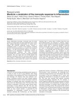
and from Ju77, a mesothelioma cell line. The 10 sera
showed some unique reactivities, but also potential
common reactivities to antigens expressed by A549
(Fig. 1). The pattern of reactivity was identical in the two
cell lines (data not shown). We could identify seven
regions on the blot that might represent serological
responses to a common antigen in these patients (Fig. 1).
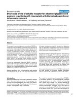
We therefore selected a patient who expressed both
common and unique features for further study. This patient
demonstrated a remarkable stability in the profile of
autoreactivity over a period of 3 years (Fig. 2).
Molecular cloning of antigens associated with
cryptogenic fibrosing alveolitis
A λ expression library with a complexity in excess of
1 million was plated at a density of 3000 plaques/cm
2
. A
total of 5 × 10
5
plaques were tested for reactivity with the
serum of a patient with CFA diluted 1/100. Twenty-two
primaries were picked; of these eight retested positive in
third-round screening, at which stage they were all clonal.
Purified plaques were excised as the pBluescript plasmid
and were subjected to sequence analysis. Seven different
cDNAs were identified from the sequence information
(Table 1). ATS was picked six times from different primary
plaques; five of these six were different recombinants. Two
of these encoded in-frame fusions that incorporated either
39 or 59 amino acid residues from the large open-reading
frame derived from the 5′-untranslated region of the parent
cDNA. The others encoded truncated proteins, missing
the amino-terminal 38, 54 or 447 residues of the parent
968-residue polypeptide, indicating that at least part of
the immune response was directed towards epitopes
expressed by the carboxyl half of the protein. As one might
expect, ATS is well expressed in most cells. Serial analysis
of gene expression (SAGE) libraries [11] indicate that the
gene is highly expressed in normal epithelial cells, fibro-
blasts and a variety of tumour cells.
One recombinant encoded an open-reading frame of 131
amino acids. This cDNA has previously been identified and
sequenced from a size-fractionated library derived from
human brain. The hypothetical protein shows some similar-
ity to the adapter moiety vav2, which functions in some
tyrosine kinase-dependent signalling pathways, but is more
highly related to a gene from Caenorhabditis elegans
named F35A5.8. The function of this gene in the worm is
not clear. Analysis of SAGE libraries shows that the gene is
highly expressed in normal epithelial cells and fibroblasts.
The third recombinant encoded the carboxyl-terminus of
the α
4
immunoglobulin-binding protein (IGBP1) gene,
which has sequence similarity to the yeast protein (TAP42).
Available online />Figure 1
Sera from 10 individuals with a confirmed diagnosis of CFA were used
to probe a western blot prepared by electrophoretic separation of
whole cell (A549) extract. Molecular weight markers (kDa) are shown
to the left, and common reactivities are noted (arrows) to the right.
Figure 2
Western blot (as Fig. 1) using serial blood samples from the index
patient over a period of 2 years (dates marked at the top). Molecular
weight markers (kDa) are shown to the left.
A stretch of consensus motifs in the carboxyl-terminus is
conserved among the related genes of human, mouse,
yeast and rice. IGBP1 is involved in a rapamycin-sensitive
signal transduction pathway in B cells. The gene has previ-
ously been shown to be expressed as a 1.4 kb mRNA tran-
script in most lymphoid tissue, but also in heart, brain,
placenta, skeletal muscle, kidney and pancreas. An antihu-
man IGBP1 antibody detects a 45 kDa protein in human
lymphoid cell lines [12]. Interestingly, SAGE libraries show
that the highest level of expression of this gene is in normal
epithelial cells derived from breast.
Serological recognition of the antigens
We then investigated whether the occurrence of antibod-
ies to each of the antigens was associated with individual
cases of CFA, or with other respiratory diseases of diverse
aetiology.
Purified phage isolates were allowed to form plaques that
were induced with isopropyl-β-thiogalactopyranoside, and
the recombinant proteins were transferred to nitrocellu-
lose membranes. Replicate membranes were probed with
serum (1/50 dilution) and processed as described in
Patients and method. Plaques were scored positive by
visual inspection and in relation to the each of the other
plaques on the membrane. We screened sera from 11
other patients with CFA, nine patients with sarcoidosis,
and one patient with SLE. In addition, we screened the
sera of 15 normal healthy laboratory volunteers. We found
no evidence of antibodies to ATS, IGBP1 or the brain
expressed sequence tag (EST) in any of these sera. Con-
vincingly, the index case retained a strong reaction to each
of these antigens in sera obtained consecutively over the
following 18 months.
Discussion
It is evident that the humoral immune system plays some
role in the pathogenesis of CFA. It is less clear whether
this role is causative or merely epiphenomenal. Although
circulating autoantibodies to lung protein(s) have been
mooted as the pathogenic mechanism, relatively few anti-
gens have actually been characterized and none of these
is accepted as diagnostic for the disease. An autoanti-
body reactive to cytokeratin 8 was defined by western
immunoblotting and by enzyme-linked immunosorbent
assay [5]. Similar data support a role for antibodies that
are reactive with cytokeratin 19 in the process of lung
injury in pulmonary fibrosis [13]. The majority of autoanti-
bodies appear directed against antigens that are not
specifically localized to lung tissue, for example antibodies
to DNA topoisomerase IIα, a nuclear antigen that is consti-
tutively expressed by dividing cells. These antibodies are
prevalent in the sera of some patients, and analysis of the
specific patterns of reactivity with various deletion recom-
binants revealed the existence of multiple epitopes in the
protein [14]. Interestingly, the authors of that study noted
a correlation between the type of epitope and the disease
duration, supporting the hypothesis that the autoantibody
response is an antigen-driven process.
Not many of the antigens associated with CFA actually
show a tissue-specific pattern of expression that is limited
to the lung. This is surprising because the disease is
explicitly manifest as a pulmonary lesion. Lungs may be
targeted simply because their high blood flow means that
they are more likely to trap immune complexes. Alterna-
tively, the majority of antibodies may be epiphenomenal
and associated with tissue damage caused by the primary
lesion. Two of the antigens described here show strong
association with epithelial cells, although they are clearly
expressed in a wider range of tissues. The third antigen,
ATS, is expressed in all tissues at high levels. This high
level of expression is evident from the finding of six inde-
pendent clones encoding ATS in the library that we
probed. The mRNA source of this library originated from a
lung tumour cell line. We chose this library because of its
Respiratory Research Vol 2 No 2 Robinson et al
Table 1
Identity of three CFA antigens and the frequency of serological responses to them
ID Insert size (kb) Full length (kb) Identity Accession Unigene CFA
141/2A 3.6 3.6 ATS D32050 HS75102 1/12
144/1A 3.5 1/12
146/2 3.2 1/12
151/1A 3.2 1/12
147/5A 2.1 1/12
147/5 0.7 1.3 IGBP1 NM_00155 HS136309 1/12
(A4PP) 1
147/4B 0.75 1.2 Brain EST AB007960 HS3631 1/12
complexity and because we knew that it contained large
inserts. In addition, western blots using patients’ sera
strongly suggested that there was no bias in the target
tissue for the dominant antigens. The SAGE analysis,
through its virtual northern programme, showed that each
of the antigens that we cloned are widely expressed.
Each of the reactivities that we defined is associated with T-
cell recognition of the antigen. This follows simply because
we used an isotype-specific second antibody, and class
switching to IgG is a T-cell-dependent event. It seems likely
then that the spectrum of antigen reactivity in the serological
response is dictated by the availability of autoreactive T
cells. How these arise, particularly to highly expressed ubiq-
uitous proteins such as ATS, and why subsets of antinu-
clear antibodies to ubiquitous determinants define particular
syndromes remain unresolved issues.
The antisynthetase syndrome is such a syndrome. It typi-
cally associates polymyositis and dermatomyositis with
complicating ILD, Raynaud’s phenomenon and frequently
polyarthritis. Autoantibodies against aminoacyl-tRNA syn-
thetases (antisynthetases) define the syndrome. Patients
can present with ILD and antisynthetase antibodies, but no
clinically evident myositis. Previous studies have shown
that anti-PL-12 antibodies (anti-ATS) are the most common
in this group, but anti-Jo-1 (antihistidyl-tRNA synthetase),
anti-EJ (antiglycyl-tRNA synthetase) and anti-OJ (anti-
isoleucyl-tRNA synthetase) also occur [15,16]. Patients
usually exhibit reactivity to only one of the synthetases, but
different subsets have occasionally been recorded
together in the same patient [17]. Of interest, autoantibod-
ies to synthetases have been found in mice with graft-
versus-host disease [18], suggesting that they may be a
secondary consequence of tissue damage and cell death.
The serum samples were obtained from 3 months to
4 years after the onset of disease in the patients with CFA,
and 1–5 years after presentation in the sarcoidosis
patients. The SLE patient had been diagnosed with the
disease for 3 months. Immunosuppressive therapy, includ-
ing oral prednisolone, was being taken by five of the CFA
patients and by four of the sarcoidosis patients. The index
patient was on no treatment when the initial sample was
obtained, but he was on immunosuppressive therapy
when the later samples were obtained and was improving
physiologically. The SLE patient was receiving plaquenil
only. Taken together, it seems unlikely that therapy was
associated with the negative results because not all
patients were on steroids and because steroids made no
difference to the results in the index case.
Conclusion
CFA can involve an inappropriate immunological reaction
in the lung interstitia. It is not clear what promotes and
maintains this reactivity. We anticipated that the targets of
the humoral immune response might give us clues to the
originating insult. We demonstrated by western blot that
there were likely to be common as well as unique anti-
genic specificities in the patient population studied.
However, by molecularly cloning and identifying the anti-
gens that were recognized by our index patient, we were
able to demonstrate that they were not recognized by any
other patient. CFA may represent the end stage of a
number of different disease processes, some of which
may be autoantibody mediated. It is also possible that
autoantibodies are epiphenomenal to CFA, representing a
secondary feature of tissue damage induced by some
other mechanism.
References
1. Lake FR: Cryptogenic fibrosing alveolitis: have we made any
progress? Respirology 1996, 1:227–232.
2. Wallace WA, Howie SE, Krajewski AS, Lamb D: The immuno-
logical architecture of B-lymphocyte aggregates in crypto-
genic fibrosing alveolitis. J Pathol 1996, 178:323–329.
3. Wallace WA, Schofield JA, Lamb D, Howie SE: Localisation of a
pulmonary autoantigen in cryptogenic fibrosing alveolitis.
Thorax 1994, 49:1139–1145.
4. Wallace WA, Roberts SN, Caldwell H, Thornton E, Greening AP,
Lamb D, Howie SE: Circulating antibodies to lung protein(s) in
patients with cryptogenic fibrosing alveolitis. Thorax 1994, 49:
218–222.
5. Dobashi N, Fujita J, Ohtsuki Y, Yamadori I, Yoshinouchi T, Kamei
T, Tokuda M, Hojo S, Okada H, Takahara J: Detection of anti-
cytokeratin 8 antibody in the serum of patients with crypto-
genic fibrosing alveolitis and pulmonary fibrosis associated
with collagen vascular disorders. Thorax 1998, 53:969–674.
6. Chapman JR, Charles PJ, Venables PJ, Thompson PJ, Haslam PL,
Maini RN, Turner Warwick ME: Definition and clinical relevance
of antibodies to nuclear ribonucleoprotein and other nuclear
antigens in patients with cryptogenic fibrosing alveolitis. Am
Rev Respir Dis 1984, 130:439–443.
7. Holgate ST, Haslam P, Turner-Warwick M: The significance of
antinuclear and DNA antibodies in cryptogenic fibrosing alve-
olitis. Thorax 1983, 38:67–70.
8. Meliconi R, Bestagno M, Sturani C, Negri C, Galavotti V, Sala C,
Facchini A, Ciarrocchi G, Gasbarrini G, Astaldi Ricotti GC:
Autoantibodies to DNA topoisomerase II in cryptogenic
fibrosing alveolitis and connective tissue disease. Clin Exp
Immunol 1989, 76:184–189.
9. Negri C, Scovassi AI, Cerino A, Negroni M, Borzi RM, Meliconi R,
Facchini A, Montecucco CM, Astaldi Ricotti GC: Autoantibodies
to poly(ADP-ribose)polymerase in autoimmune diseases.
Autoimmunity 1990, 6:203–209.
10. Robinson C, Callow M, Stevenson S, Scott B, Robinson BW,
Lake RA: Serologic responses in patients with malignant
mesothelioma: evidence for both public and private specifici-
ties. Am J Respir Cell Mol Biol 2000, 22:550–556.
11. National Center for Biotechnology Information. Serial analysis of
gene expression. Tag to gene mapping: SAGEmap. http://www.
ncbi.nlm.nih.gov/SAGE/index.cgi
12. Onda M, Inui S, Maeda K, Suzuki M, Takahashi E, Sakaguchi N:
Expression and chromosomal localization of the human
alpha-4/Igbp1 gene, the structure of which is closely related
to the yeast Tap42 protein of the rapamycin-sensitive signal
transduction pathway. Genomics 1997, 46:373–378.
13. Fujita J, Dobashi N, Ohtsuki Y, Yamadori I, Yoshinouchi T, Kamei
T, Tokuda M, Hojo S, Okada H, Takahara J: Elevation of anti-
cytokeratin 19 antibody in sera of the patients with idiopathic
pulmonary fibrosis and pulmonary fibrosis associated with
collagen vascular disorders. Lung 1999, 177:311–319.
14. Grigolo B, Mazzetti I, Borzi RM, Hickson ID, Fabbri M, Fasano L,
Meliconi R, Facchini A: Mapping of topoisomerase II alpha epi-
topes recognized by autoantibodies in idiopathic pulmonary
fibrosis. Clin Exp Immunol 1998, 114:339–346.
Available online />commentary review reports
primary research
15. Targoff IN, Trieu EP, Plotz PH, Miller FW: Antibodies to glycyl-
transfer RNA synthetase in patients with myositis and intersti-
tial lung disease. Arthritis Rheum 1992, 35:821–830.
16. Friedman AW, Targoff IN, Arnett FC: Interstitial lung disease
with autoantibodies against aminoacyl-tRNA synthetases in
the absence of clinically apparent myositis. Semin Arthritis
Rheum 1996, 26:459–467.
17. Gelpi C, Kanterewicz E, Gratacos J, Targoff IN, Rodriguez-
Sanchez JL: Coexistence of two antisynthetases in a patient
with the antisynthetase syndrome. Arthritis Rheum 1996, 39:
692–697.
18. Gelpi C, Martinez MA, Vidal S, Targoff IN, Rodriguez-Sanchez JL:
Autoantibodies to a transfer RNA-associated protein in a
murine model of chronic graft versus host disease. J Immunol
1994, 152:1989–1999.
Respiratory Research Vol 2 No 2 Robinson et al