Báo cáo y học: "Recurrent pneumonia with mild hypogammaglobulinemia diagnosed as X-linked agammaglobulinemia in adults" pot
Bạn đang xem bản rút gọn của tài liệu. Xem và tải ngay bản đầy đủ của tài liệu tại đây (524.53 KB, 5 trang )
Primary research
Recurrent pneumonia with mild hypogammaglobulinemia
diagnosed as X-linked agammaglobulinemia in adults
Kazuhiro Usui*, Yoji Sasahara
†
, Ryushi Tazawa*, Koichi Hagiwara*, Satoshi Tsukada
‡
,
Toshio Miyawaki
§
, Shigeru Tsuchiya
†
and Toshihiro Nukiwa*
*Department of Respiratory Oncology and Molecular Medicine, Institute of Development, Aging, and Cancer, Tohoku University, Sendai, Japan
†
Department of Pediatric Oncology, Institute of Development, Aging, and Cancer, Tohoku University, Sendai, Japan
‡
Department of Medicine III, Osaka University Medical School, Osaka, Japan
§
Department of Pediatrics, Faculty of Medicine, Toyama Medical and Pharmaceutical University, Toyama, Japan
Correspondence: Ryushi Tazawa, MD, Department of Respiratory Oncology and Molecular Medicine, Institute of Development, Aging, and Cancer,
Tohoku University, 4-1 Seiryomachi, Aoba-ku, Sendai 980-8575, Japan. Tel: +81 22 717 8539; fax: +81 22 717 8549;
email:
Introduction
XLA is a prototype of humoral immunodeficiency first
described by Bruton in 1952 [1]. XLA is characterized by
a paucity of circulating B cells and a significant reduction
in the serum immunoglobulin concentrations that predis-
pose the affected patients to frequent and severe bacterial
infections [2]. The BTK gene, which encodes a
cytoplasmic tyrosine kinase, was identified as the gene
responsible for XLA [3,4].
Whereas most XLA patients develop clinical symptoms in
childhood, there might be late-onset XLA cases among
patients with a lower level of serum immunoglobulins who
have often been clinically misdiagnosed as common
Abstract
Background: X-linked agammaglobulinemia (XLA) is a humoral immunodeficiency caused by
disruption of the Bruton’s tyrosine kinase (BTK) gene. Typical XLA patients suffer recurrent and severe
bacterial infections in childhood.
Methods: Flow cytometric analysis of the peripheral monocytes using the anti-BTK antibody was used
to characterize a 27 year old male patient with mild hypogammaglobulinemia (IgG, 635 mg/dl; IgM,
11 mg/dl; IgA, <5 mg/dl). He had suffered from frequent pneumonia since age 25 but had no history
of frequent infections in his childhood or in adolescence. Sequencing of the BTK cDNA obtained from
an Epstein–Barr virus-transformed B lymphoblastoid cell line derived from the bone marrow of the
patient was performed to confirm a genetic defect.
Results: Flow cytometric analysis of cytoplasmic BTK protein in peripheral monocytes indicated that
the patient presents a rare case of adult-onset XLA and that his mother is an XLA carrier. Sequencing
of the BTK gene revealed a deletion of AG in the codon for Glu605 (AGT), resulting in an aberrant
stop codon that truncates the BTK protein in its kinase domain.
Conclusions: This case suggests that some XLA cases may remain undiagnosed because they only
show mild hypogammaglobulinemia and they lack repeated infections in childhood. Flow cytometric
analysis is a powerful method to screen these patients.
Keywords: adult onset, Bruton’s tyrosine kinase, mild hypogammaglobulinemia, recurrent pneumonia, X-linked
agammaglobulinemia
Received: 29 November 2000
Revisions requested: 20 February 2001
Revisions received: 6 March 2001
Accepted: 12 March 2001
Published: 12 April 2001
Respir Res 2001, 2:188–192
This article may contain supplementary data which can only be found
online at />© 2001 Usui et al, licensee BioMed Central Ltd
(Print ISSN 1465-9921; Online ISSN 1465-993X)
BTK = Bruton’s tyrosine kinase; PCR = polymerase chain reaction; XLA = X-linked agammaglobulinemia.
Available online />Available online />commentary review reports
primary research
immunodeficiency, selective IgG or IgA deficiency. Direct
detection of BTK mutations by gene analysis is necessary
for diagnosis of XLA, but it is time consuming, expensive,
and labor intensive to screen these patients.
This article presents a rare case of an adult-onset XLA
patient, the diagnosis of which was indicated by the flow
cytometric analysis of peripheral monocytes using anti-
BTK antibody [5] and was confirmed by the sequencing
analysis of the patient’s BTK gene.
Materials and methods
Flow cytometric analysis of BTK expression in
peripheral monocytes
Flow cytometric analysis of cytoplasmic BTK protein in
peripheral monocytes has been described previously
[5,6]. Briefly, mononuclear cells were surface stained with
phycoerythrin-labeled anti-CD14 antibody, then fixed, per-
mealized, incubated with anti-BTK monoclonal antibody
48-2H [5] or control IgG
1
(Dako, Kyoto, Japan), and then
incubated with fluorescein isothiocyanate-labeled sec-
ondary antibody. The cells were first gated by CD14 to
select monocytes, and then histograms were plotted on
fluorescein isothiocyanate intensity.
Detection of a two base pair deletion in the BTK cDNA
The BTK cDNA of the patient was sequenced as previ-
ously described [7]. Briefly, an Epstein–Barr virus-trans-
formed B lymphoblastoid cell line derived from peripheral
blood of the patient was established and subject to
reverse transcription polymerase chain reaction (PCR) to
amplify the protein coding region of the BTK cDNA, which
was then sequenced.
PCR-based detection of the mutated allele
Based on the sequence information, the normal primer A
(5′-ATGAGAGATTTACTAACAGT-3′), the deletion-spe-
cific primer B (5′-ATGAGAGATTTACTAACTGA-3′), and
the common downstream primer C (5′-AGAGCAAGACT-
GTGTCACCA-3′) were synthesized. Genomic DNA from
the patient, his mother and his brother were extracted from
peripheral blood and amplified by PCR using either primer
A or primer B, together with the common downstream
primer C.
Results
Case report
A 26 year old Japanese crane operator was admitted to
our affiliated hospital with fever, cough and chest pain.
This was followed by admissions to other hospitals with
bacterial pneumonia twice within 18 months. Because the
patient never experienced recurrent infections until
age 25, his B cell numbers or IgG level were not checked
in the routine examination, and he had never been sus-
pected of common variable immunodeficiency or XLA. His
chest X-ray on admission to the hospital in June 1997
showed infiltration in the lower left lobe of the lung with
encapsulated pleural effusion (Fig. 1A). No bronchiectasis
was detected. Because of hypogammaglobulinemia on
laboratory examination (IgG, 635 mg/dl; IgM, 11 mg/dl;
IgA, <5 mg/dl) and the history of repeated pneumonia, the
patient was referred to our hospital for further examination.
The patient had four siblings (Fig. 1E). His sister died
shortly after birth, and his eldest brother, who had a
history of repeated pneumonia, died of drug-induced liver
failure at age 7. The routine hematologic and biochemical
examination of the patient revealed no abnormal findings.
He was negative for both HIV and HTLV-1. Blood type
testing showed that, although his blood type was O, he
had neither anti-A nor anti-B antibodies. Although we did
not directly examine the function of his immunoglobulins,
anti-virus antibodies commonly positive in normal Japan-
ese adults (such as anti-measles, anti-rubella, anti-
cytomegalovirus, and anti-Epstein–Barr virus) were all
negative, indicating that his IgGs were not functional. In
contrast, his cellular immunity was intact because a lym-
phocyte stimulating test by phytohemagglutinin and con-
canavalin A showed normal responses. These findings,
together with his moderate hypoglobulinemia, prompted
us to investigate his B lymphocyte system.
Flow cytometric analysis of BTK expression in
peripheral monocytes
The surface marker examination of the patient’s peripheral
lymphocytes showed marked deficits in the B cell popula-
tions (CD19+, 1%; CD20+, 6%). Measurement of the
BTK protein in CD20+ B cells using anti-BTK monoclonal
antibody 48-2H [5] gave an uninformative result because
only a small number of CD20+ B cells were present (data
not shown). We then measured BTK protein in peripheral
monocytes because they have been reported to express
BTK (Fig. 1B). The patient showed a partial BTK defi-
ciency, and his mother showed a two-peak BTK expres-
sion profile. In females, non-B hematopoietic cells undergo
random inactivation of the X chromosomes [8]. Demon-
stration of a two-peak BTK expression pattern in periph-
eral monocytes is therefore diagnostic of the XLA carrier
state in females [5]. We concluded that the patient has
XLA and that his mother is an XLA carrier. Hypogamma-
globulinemia observed in the patient was considered a
clinical manifestation of his Bruton’s disease.
Detection of a two base pair deletion in the BTK cDNA
To further confirm the diagnosis, the patient’s BTK gene was
sequenced [7] and was found to have a 2 base pair (AG)
deletion in the codon for Glu605 (AGT) in exon 18 that
encodes a part of the kinase domain of the BTK protein. This
deletion places a stop codon just downstream, thus produc-
ing a truncated BTK protein with 604 amino acid residues
instead of the normal BTK protein with 659 residues
(Fig. 1C,D). This mutation has not been reported to date [9].
Respiratory Research Vol 2 No 3 Usui et al
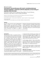
Figure 1
(A) Serial chest radiographs of the patient. The chest X-ray films taken at other hospitals in 1996 reveal infiltration in both the upper and lower
lobes in April, and in the lower lobe of the right lung in November. The chest radiograph on admission to our hospital in June 1997 demonstrates
infiltration in the left lower lobe and the existence of pleural effusion. (B) Flow cytometric analysis of BTK expression in peripheral monocytes. The
solid and the dashed lines indicate cells stained with anti-BTK or control antibody, respectively. FITC, Fluorescein isothiocyanate. (C) The genomic
organization of the human BTK gene and the domain structure of BTK cDNA. Exons 1–19 of the BTK gene, and the BTK cDNA with its functional
domains are shown [14]. Amino acid numbers (1–659) are shown under the cDNA. The arrowhead indicates the position of the mutation identified
in this case. 5UT, 5′-untranslated region; PH, pleckstrin homology domain; TH, Tec homology domain; SH, Src homology domain; 3UT, 3′-
untranslated region. (D) Detection of a 2 base pair deletion in the BTK cDNA. The BTK cDNA of the patient was sequenced as described in
Materials and methods. The chromatograph of the autosequencer shows a 2 base pair deletion. (E) Family pedigree and the PCR-based detection
of the mutated allele. Two generations are depicted. The index case is marked by an arrow. Genomic DNAs from the patient, his mother and his
brother were extracted from peripheral blood and amplified by PCR using either primer A or primer B, together with the common downstream
primer C. Normal genomic DNA gave a band when primer A was used (lane N). The patient’s DNA gave a band when primer B was used (lane D).
The patient’s mother, a carrier of the mutated gene, gave bands when both primers A and B were used (lanes N and D).
PCR-based detection of the mutated allele
Genomic DNA from other family members was then exam-
ined by PCR designed to detect the normal or the
mutated allele separately (Fig. 1E). The patient’s mother
was confirmed to be heterozygous for the mutated BTK
gene. His second-eldest brother, who has no history of
repeated infections, was normal for the BTK gene.
Discussion
Most XLA patients develop clinical symptoms during the
first year of life and, without antibiotics and immunoglobu-
lin replacement, they die in infancy. The case presented
here is the only adult-onset case of the 107 cases in the
XLA registry of the Ministry of Health and Welfare, Japan.
This case clearly illustrates the utility of flow cytometric
analysis for the diagnosis of XLA, and also raises ques-
tions regarding the factors that determine the onset of the
XLA phenotype.
We found that, for the flow cytometric analysis, it is key to
measure BTK expression in peripheral monocytes. Both
XLA patients and XLA carriers are detected as shown
here, and as reported elsewhere [5]. In female XLA carri-
ers, B cells manifest the skewed inactivation of the
mutated X chromosome, reflecting the role of the XLA
gene in early development. Non-B hematopoietic cells in
XLA carriers, on the contrary, undergo random inactivation
of the normal and mutated X chromosomes, and thus the
product of the BTK gene can be detected in B cells and
other hematopoietic cells. This is the reason why demon-
stration of BTK mosaicism in non-B hematopoietic cells
leads to the detection of obligate XLA carriers. The BTK
function in monocytes remains unclear. Monocytes with
deleted BTK are not representative of all that is happening
in the B cells, despite the fact that the detection of the
BTK production in monocytes by flow cytometry is a pow-
erful diagnostic tool for screening XLA patients.
The cause for the delay in the appearance of the pre-
sented patient’s clinical symptoms until age 25 is of much
interest. Mild XLA is clinically likely to occur at any age
incidentally [10,11], and these patients might be misdiag-
nosed as suffering common variable immunodeficiency
[12]. The truncated BTK protein in this patient is possibly
able to function to some extent, although less effectively
than the wild type. This could explain why this patient’s
hypogammaglobulinemia was not severe.
Another explanation for this latency is the contribution of
as yet unidentified factors. Reports have shown that, even
in an XLA family with the identical BTK gene mutation,
some affected males have substantial levels of
immunoglobulins whereas others are nearly agammaglob-
ulinemic [13]. The present patient’s eldest brother had a
history of frequent infections, suggesting that the brother
was also an XLA patient. If this is the case, then as yet
unidentified factors provide the more likely explanation for
the difference in the age of onset for the patient and his
eldest brother.
Although BTK has been identified as a gene responsible
for XLA, the mechanism that links the defect in BTK func-
tion to the development of XLA is not known. This case
provides valuable information, suggesting a direction for
the pursuit of this link, and demonstrates the power of flow
cytometric analysis in diagnosing XLA.
Conclusion
This study presents a rare late-onset XLA case, suggest-
ing that some XLA cases may remain undiagnosed
because they only show mild hypogammaglobulinemia
and they lack repeated infections in childhood. Flow cyto-
metric analysis using the anti-BTK antibody is a powerful
method to screen these patients for BTK deficiency.
References
1. Bruton OC: Agammaglobulinemia. Pediatrics 1952, 9:722–728.
2. Sideras P, Smith CIE: Molecular and cellular aspects of X-
linked agammaglobulinemia. Adv Immunol 1995, 59:135–223.
3. Tsukada S, Saffran DC, Rawlings DJ, Parolini O, Allen RC, Klisak
I, Sparkes RS, Kubagawa H, Mohandas T, Quan S, Belmont JW,
Cooper MD, Conley ME, Witte ON: Deficient expression of a B
cell cytoplasmic tyrosine kinase in human X-linked agamma-
globulinemia. Cell 1993, 72: 279–290.
4. Vetrie D, Vorechovsky I, Sideras P, Holland J, Davies A, Flinter F,
Hammarstroem L, Kinnon C, Levinsky R, Bobrow M, Smith CIE,
Bentley DR: The gene involved in X-linked agammaglobuline-
mia is a member of the src family of protein-tyrosine kinases.
Nature 1993, 361:226–233.
5. Futatani T, Miyawaki T, Tsukada S, Hashimoto S, Kunikata T, Arai
S, Kurimoto M, Niida Y, Matsuoka H, Sakiyama Y, Iwata T,
Tsuchiya S, Tatsuzawa O, Yoshizaki K, Kishimoto T: Deficient
expression of Bruton’s tyrosine kinase is monocytes from X-
linked agammaglobulinemia as evaluated by a flow cytomet-
ric analysis and its clinical application to carrier detection.
Blood 1998, 91:595–602.
6. Hashimoto S, Miyawaki T, Futatani T, Kanegane H, Usui K, Nukiwa
T, Namiuchi S, Matsushita M, Yamadori T, Suemura M, Kishimoto
T, Tsukada S: Atypical X-linked agammaglobulinemia (XLA)
diagnosed in adult. Intern Med 1999, 38:722–725.
7. Ohashi Y, Tsuchiya S, Konno T: A new point mutation involving
a highly conserved leucine in the Btk SH2 domain in a family
with X linked agammaglobulinaemia. J Med Genet 1995, 32:
77–78.
8. Belmont JW: Insights into lymphocyte development from X-
linked immune deficiencies. Trends Genet 1995, 11:112–116.
9. Vihinen M, Brandau O, Branden LJ, Kwan SP, Lappalainen I,
Lester T, Noordzij JG, Ochs HD, Ollila J, Pienaar SM, Riikonen P,
Saha BK, Smith CIE: BTKbase, mutation database for X-linked
agammaglobulinemia (XLA). Nucleic Acids Res 1998, 26:
242–247 [ />10. Ishida F, Kobayashi H, Saito H, Futatani T, Miyawaki T, Kiyosawa
K: The oldest case with X-linked agammaglobulinemia in
Japan lacking Bruton-type tyrosine kinase protein detected by
flow cytometry. Rinsho Ketsueki 1998, 39:44–47.
11. Kornfeld SJ, Haire RN, Strong SJ, Tang H, Sung SS, Fu SM,
Litman GW: A novel mutation (Cys145
→→
Stop) in Bruton’s
tyrosine kinase is associated with newly diagnosed X-linked
agammaglobulinemia in a 51-year-old male. Mol Med 1996, 2:
619–623.
12. Kanegane H, Tsukada S, Iwata T, Futatani T, Nomura K,
Yamamoto J, Yoshida T, Agematsu K, Komiyama A, Miyawaki T:
Detection of Bruton’s tyrosine kinase mutations in hypogam-
maglobulinaemic males registered as common variable
Available online />commentary review reports
primary research
Respiratory Research Vol 2 No 3 Usui et al
immunodeficiency (CVID) in the Japanese Immunodeficiency
Registry. Clin Exp Immunol 2000, 120:512–517.
13. Conley ME, Fitch-Hilgenberg ME, Cleveland JL, Parolini O, Rohrer J:
Screening of genomic DNA to identify mutations in the gene for
Bruton’s tyrosine kinase. Hum Mol Genet 1994, 3:1751–1756.
14. Ohta Y, Haire RN, Litman R, Fu SM, Nelson RP, Kratz J, Kornfeld
SJ, de la Morena M, Good RA, Litman GW: Genomic organiza-
tion and structure of Bruton agammaglobulinemia tyrosine
kinase: localization of mutations associated with varied clinical
presentations and course in X chromosome-linked agamma-
globulinemia. Proc Natl Acad Sci USA 1994, 91: 9062–9066.