Báo cáo y học: "Genomic approaches to research in pulmonary hypertension" pot
Bạn đang xem bản rút gọn của tài liệu. Xem và tải ngay bản đầy đủ của tài liệu tại đây (323.83 KB, 6 trang )
BMPR2 = bone morphogenetic protein type II receptor gene; eNOS = endothelial nitric oxide synthase; FPPH = familial primary pulmonary hyper-
tension; PGI
2
= prostacyclin; PGIS = prostacyclin synthase; PH = pulmonary hypertension; PPAR = peroxisome proliferator-activated receptor;
PPH = primary pulmonary hypertension; Tg+, Tg– = transgenic, nontransgenic littermate; TGF-β = transforming growth factor-β.
Available online />Introduction
Pulmonary hypertension (PH) refers to a spectrum of dis-
eases where the pulmonary artery pressure is elevated. A
new classification of PH has recently been proposed [1].
No cause can be elucidated in primary (or sporadic, idio-
pathic) pulmonary hypertension (PPH). Secondary forms
of PH can occur in association with congenital heart
disease, thromboembolic disease, HIV, anorexigen usage,
and a variety of connective tissue disorders. Familial
primary pulmonary hypertension (FPPH) has been associ-
ated with heterozygous germline mutations in the bone
morphogenetic protein type II receptor gene (BMPR2)
[2,3]. While this recent discovery has generated extreme
interest, the pathobiology of severe PH remains enigmatic.
Recent genomic approaches to investigate PH are
reviewed. Early studies investigated the alterations of
vasoactive and growth factor related genes. Animal
models, using either pharmaceutical approaches, trans-
genics, or targeted disruption of genes, have allowed for
whole animal modeling of specific pathways in the devel-
opment of PH. Progress in medical genetic investigations
has lead to the discovery of a gene (BMPR2) associated
with FPPH. Finally, microarray expression analysis has
been utilized to investigate animal models, and has shown
to be a useful tool providing novel information and better
characterization of the molecular pathobiology of distinct
clinical phenotypes of PH.
Genes involved in the pathobiology of PH
Most investigations of the role of specific genes in the
pathobiology of PH have focused either on the balance of
vasoconstriction and vasodilation or on specific growth
Review
Genomic approaches to research in pulmonary hypertension
Mark W Geraci*, Bifeng Gao*, Yasushi Hoshikawa*, Michael E Yeager*, Rubin M Tuder
†
and Norbert F Voelkel*
*Division of Pulmonary Sciences and Critical Care Medicine, University of Colorado Health Sciences Center, Denver, Colorado, USA
†
Department of Pathology, Johns Hopkins University, School of Medicine, USA
Correspondence: Mark W Geraci, MD, Division of Pulmonary Sciences and Critical Care Medicine, University of Colorado Health Sciences Center,
Campus Box C-272, 4200 East Ninth Avenue, Denver, CO 80262, USA. Tel: +1 303 315 7047; fax: +1 303 315 5632;
e-mail:
Abstract
Genomics, or the study of genes and their function, is a burgeoning field with many new technologies.
In the present review, we explore the application of genomic approaches to the study of pulmonary
hypertension (PH). Candidate genes, important to the pathobiology of the disease, have been
investigated. Rodent models enable the manipulation of selected genes, either by transgenesis or
targeted disruption. Mutational analysis of genes in the transforming growth factor-β family have proven
pivotal in both familial and sporadic forms of primary PH. Finally, microarray gene expression analysis is
a robust molecular tool to aid in delineating the pathobiology of this disease.
Keywords: genetic mutation, knockout mouse, microarray, pulmonary hypertension, transgenic mouse
Received: 16 February 2001
Revisions requested: 13 March 2001
Revisions received: 22 March 2001
Accepted: 3 April 2001
Published: 1 May 2001
Respir Res 2001, 2:210–215
This article may contain supplementary data which can only be found
online at />© 2001 BioMed Central Ltd
(Print ISSN 1465-9921; Online ISSN 1465-993X)
Available online />commentary
review
reports primary research
factors, inflammatory mediators, or ion channels. Another
approach has been to compartmentalize the vasculature,
and focus the investigations on the endothelium, smooth
muscle cells, and the adventitia/extracellular matrix.
Christman et al initially reported an imbalance of prosta-
cyclin (PGI
2
) and thromboxane metabolites in the urine of
patients with both primary and secondary forms of PH,
with more vasoconstrictor thromboxane metabolites in
patients with PH [4]. Giaid et al similarly studied the
expression of endothelin-1 in the lungs of patients with
PH, and showed increased expression by both in situ
hybridization and immunohistochemistry [5]. Overexpres-
sion of 5-lipoxygenase and 5-lipoxygenase activating
protein was shown in endothelial cells of plexiform lesions
and inflammatory cells in patients with PPH, suggesting
that overexpression of enzymes involved in generation of
inflammatory mediators may play a role in the pathogene-
sis of PPH [6]. As there is an imbalance of PGI
2
and
thromboxane, we wondered whether PPH patients had
diminished synthetic enzyme for PGI
2
. We demonstrated,
by in situ hybridization, western analysis and immunohis-
tochemistry, that patients with PPH have decreased lung
tissue prostacyclin synthase (PGIS) [7]. A comprehensive
histochemical analysis of plexiform lesions was performed
by Cool et al [8]. This analysis showed that the endothe-
lial cells of plexiform lesions express, intensely and uni-
formly, the vascular endothelial growth factor receptor
KDR. The analysis by Cool et al also showed that the
cells segregate phenotypically into cyclin-kinase inhibitor
p27/kip1-negative cells in the central core of the plexi-
form lesion and p27/kip1-positive cells in peripheral
areas adjacent to incipient blood vessel formation. Using
immunohistochemistry and three-dimensional reconstruc-
tion techniques, the plexiform lesions were shown to be
dynamic vascular structures characterized by at least two
endothelial cell phenotypes. Despite these powerful
investigations, a unifying pathobiological scheme has
remained elusive.
Animal models of PH
Commonly utilized models of PH in animals are the
chronic hypoxic model and the monocrotaline model. Inter-
estingly, monocrotaline causes PH in the rat, but not the
mouse. Exactly how closely the animal models recapitulate
human disease remains a source of debate. These two
models have, however, been useful for hypothesis testing
and determining the response of genetically altered
animals. Several specific genes have been targeted for
investigation in rodent models.
5-Lipoxygenase
Mice with targeted disruption of 5-lipoxygenase were sub-
jected to chronic hypoxia [9]. These mice developed less
right ventricular hypertrophy than matched controls, sup-
porting the hypothesis that 5-lipoxygenase is involved in
pulmonary vascular tone in rodent hypoxia models.
Nitric oxide synthase
Targeted disruption of the endothelial nitric oxide syn-
thase (eNOS) gene results in mice with increased sus-
ceptibility to hypoxic-induced PH [10]. These studies
conclude that eNOS-derived nitric oxide is an important
modulator of the pulmonary vascular response to
chronic hypoxia, and more than 50% of eNOS expres-
sion is required to maintain normal pulmonary vascular
tone [10].
PGIS and prostacyclin receptor
We hypothesized that selective pulmonary overexpres-
sion of PGIS may prevent the development of PH. Trans-
genic mice were created with selective pulmonary PGIS
overexpression using a construct of the 3.7 kb human
surfactant protein-C promoter and the rat PGIS cDNA.
Transgenic mice (Tg+) and nontransgenic littermates
(Tg–) were subjected to a simulated altitude of 17,000
feet for 5 weeks. After exposure to chronic hypobaric
hypoxia, Tg+ mice have lower right ventricular systolic
pressure than do Tg– mice. Histologic examination of the
lungs revealed nearly normal arteriolar vessels in the Tg+
mice in comparison with vessel wall hypertrophy in the
Tg– mice. The Tg+ mice were thus protected from the
development of PH after exposure to chronic hypobaric
hypoxia. We conclude that PGIS plays a major role in
modifying the pulmonary vascular response to chronic
hypoxia. Additional data investigating the prostacyclin
receptor knockout mice support the important modulat-
ing role of PGI
2
since chronic hypoxic PH is more severe
in these prostacyclin receptor knockout mice when com-
pared with the wild-type animals [11]. This has important
implications for the pathogenesis and treatment of
severe PH [12].
Matrix metalloproteinase and serine elastase
Important changes occur in PH in the vascular adventitia,
with increased production of the extracellular matrix. Matrix
metalloproteinases can stimulate the production of mito-
genic co-factors, such as tenascin. Cowan et al recently
showed that direct inhibition of serine elastases led to
complete regression of pathological changes in experi-
mental PH caused by monocrotaline [13].
Vascular endothelial growth factor
In contrast to the human disease, classical rodent
models of hypoxia and monocrotaline lack the clustered
proliferation of endothelial cells. Taraseviciene-Stewart
et al recently showed that chronic administration of a
vascular endothelial growth factor-2 inhibitor in chroni-
cally hypoxic rats lead, first, to endothelial cell death,
then to obliteration of the vessel lumen by proliferating
endothelial cells and, finally, to PH [14]. A broad spec-
trum caspase inhibitor blocked this proliferation. This
model more accurately depicts the cellular events seen
in the human condition.
Respiratory Research Vol 2 No 4 Geraci et al
Gene transfer
The promise of gene transfer therapy remains the ‘Holy
Grail’ for many genetic diseases as well as diseases that
exhibit a specific enzyme deficiency. PH is no exception.
Adenoviral gene transfer has been used in rats to show
diminished response to acute hypoxia. This has been
accomplished by transfer of eNOS [15] and by gene
therapy with PGIS [16]. Long-term benefit in chronic
hypoxia has not been reported. Repeated adenoviral PGIS
transfection has shown some effectiveness in decreasing
PH in rats using the monocrotaline model [17].
Microarray expression analysis of animal
models
We performed microarray analysis of our PGIS Tg+
animals to determine the global changes in gene expres-
sion caused by PGIS overexpression. Transgene negative
littermates were examined as controls. The mRNA from
five transgenic mouse lungs was pooled and compared
with five nontransgenic, sex-matched littermates. Using
strict criteria (a twofold change in expression), we deter-
mined that a definable number of genes was differentially
expressed between the lungs of transgenic and non-
transgenic animals. Of the 6500 genes surveyed, 32
genes showed an increase in expression and 26 showed
a decrease in expression. Table 1 presents genes that
demonstrate the most significant changes in expression
(at least a 2.2-fold change) when comparing the lung
mRNA from transgenic and nontransgenic mice.
Array analysis importantly demonstrated changes in both
peroxisome proliferator-activated receptor (PPAR) λ and
PPAR δ, and we have followed up these studies with work
demonstrating that prostacyclin activates PPAR δ in colo-
rectal cancer [18]. Histochemical analysis in human colo-
rectal tumors demonstrated colocalization of PPAR δ and
cyclooxygenase-2. An experimental condition was created
in which PGI
2
production could be correlated with PPAR δ
transcriptional activity. Transient transfection assays
established that endogenously synthesized PGI
2
could
serve as a ligand for PPAR δ. A stable PGI
2
analog also
induces transactivation of PPAR δ in human colon cancer
cells, demonstrating that endogenous PPAR δ is transcrip-
tionally responsive to PGI
2
[18].
Human medical genetics
FPPH is an autosomal dominant disorder that is indistin-
guishable from sporadic PPH. The disease has reduced
penetrance, and over 90% of patients have no known
family history of the disease [19]. Linkage analysis in
affected families enabled the locus to be defined within a
3 cM region of chromosome 2q33. Using a positional can-
didate-gene strategy, two groups were subsequently able
to independently confirm that heterozygous germline
mutations in BMPR2 cause FPPH [2,3]. Using a high-
throughput denaturing high-performance liquid chromato-
graphy approach [20] has enabled the rapid identification
of numerous mutations responsible for haploinsufficiency
of BMPR2 [2]. Furthermore, germline mutations of
BMPR2 have also been identified in ~26% of sporadic
cases of PPH [21]. ‘Sporadic’ cases sometimes actually
represented occult familial cases of PPH [21]. The molec-
ular spectrum of BMPR2 mutations is more fully eluci-
dated in an analysis of 47 European families [22]. The
majority of mutations (58%) are predicted to lead to pre-
mature termination codons. However, mutations in
BMPR2 have not been found in 45% of families with PPH
[22]. A number of possible explanations for this fact are
possible, including mutations in intronic and 3′-untrans-
lated regions that are heretofore not examined, rearrange-
ments in the transcribed gene that may occur, or genetic
heterogeneity perhaps playing a role.
BMPR2 encodes a type II receptor member of the trans-
forming growth factor-β (TGF-β) superfamily. Type II
receptors, which have serine/threonine kinase activity, act
as cell-signaling molecules. Following ligand binding, type
II receptors form heteromeric complexes with membrane-
bound type I receptors. This initiates phosphorylation of
the type I receptor and downstream intracellular Smads
[23]. This pathway is diverse and the specificity in cell
growth and differentiation appears to be mediated through
transcriptional control. The importance of the TGF-β
pathway in vascular disorders is evidenced by the fact that
two other components of this pathway, endoglin and the
activin receptor-like kinase-1 gene, are mutated in heredi-
tary hemorrhagic telangectasia [24,25].
Mutational analysis
Lee et al [26] recently demonstrated that the endothelial
cells within plexiform lesions of patients with PPH expand
in a monoclonal fashion, whereas secondary PH lesions
develop via polyclonal expansion of endothelial cells
Table 1
Genes demonstrating the most significant changes in
expression
Genes with significantly Genes with significantly
increased expression decreased expression
PPAR γ PPAR δ
RAS GTPase Cyclooxygenase-2
Focal adhesion kinase Multidrug resistance protein
Keratinocyte growth factor receptor α-Catenin
Epidermal growth factor TGF-β and TGF-β receptor
IL-7 and IL-17 receptors Wilm’s tumor gene
Cathepsins C, D, and E BCR-abl
PPAR, Peroxisome proliferator-activated receptor; TGF, transforming
growth factor.
[26,27]. The finding of monoclonal growth implies that, as
in neoplasia, genetic mutations may occur which provide a
selective growth advantage for a single endothelial cell.
The TGF-β family of signaling molecules inhibits the prolif-
eration of endothelial cells by modulating proteins involved
in cell cycle control and angiogenesis [23]. Mutations in
TGF-β signaling molecules have been implicated in initia-
tion and progression of cancers and atherosclerotic
plaques, because insertions or deletions within a
10-adenine microsatellite region in exon 3 of the TGF-βRII
gene have been demonstrated [28,29]. An 8-guanine
region within exon 3 of Bax, a proapoptotic member of the
Bcl-2 gene family, is similarly prone to instability [30].
To investigate whether cells within plexiform lesions
exhibit microsatellite instability and mutations in TGF-
microsatellite instability signaling genes, Yeager et al per-
formed microdissection of plexiform lesions from patients
with sporadic PPH and those with secondary forms of PH
[31]. The results showed that: first, the endothelial cells
within PPH lesions are genetically unstable, with 50% of
lesions demonstrating microsatellite instability; second,
one-third of the lesions from PPH show mutation of at
least one allele of TGF-βRII, but none of the secondary PH
or normal lungs display mutations; and, finally, 21%
percent of lesions in PPH show Bax mutations, whereas
none of the secondary PH or normals show this mutation.
Furthermore, we have performed mutational analysis of the
microdissected plexiform lesions from five patients with
FPPH. In total, 22 lesions from 5 patients were analyzed
for mutations of TGF-βRII and Bax. We report here that
none of the 22 lesions examined showed mutations of
TGF-βRII or Bax, in contrast to the lesions of patients with
spontaneous PPH. In summary, the monoclonal expansion
of endothelial cells seen in sporadic PPH may result from
mutations in regulatory genes such as TGF-βRII and Bax.
Expression analysis of human PPH
Gene microarray technology [32] now permits the analysis
of the gene expression profile of lung tissue obtained from
patients with primary PH to compare with that found in
normal lung tissue. Because the vascular lesions are
homogeneously distributed throughout the entire lung, a
tissue fragment of the lung is probably representative of
the whole lung. RNA extracted from such fragments is
likely to provide meaningful information regarding the
changes in gene expression pattern in PPH when com-
pared with structurally normal lung tissue. We can model
the range of normality by examining a sufficient number of
lung tissue samples. Methods exist for determining coordi-
nation in expression data using cluster expression profiles.
Cluster analysis can give clues to the pathogenesis by dis-
playing genes whose expression is altered in a coordinate
manner. Finally, an important goal is to discern sets of
genes that differentiate between normal and disease
states — or discrimination analysis. Building discrimination
models has a long history in statistical pattern recognition
and machine learning, and has been applied to cancer
paradigms using gene expression data [33]. For our study,
we used Affymetrix oligonucleotide microarrays (human
FL) to characterize the expression pattern in the lung
tissue obtained from six patients with PPH, including two
patients with FPPH, and from six patients with histologi-
cally normal lungs [34].
Although the number of patient samples was small, gene
dendogram, cluster analysis and concordant expression
differences show that there are categorical and robust
differences in the profile of expressed genes between
structurally normal lungs, lungs from patients with
sporadic PPH, and lungs from patients with FPPH. We
began our study of differential gene expression in PPH
with the assumption that sporadic PPH is a disease with
typical and dramatic histological features, which are suffi-
ciently distinct from the structurally normal lung but essen-
tially indistinguishable from those features found in FPPH
lungs. We found that only 307 genes were significantly
different in their expression when PH tissues were com-
Available online />commentary
review
reports primary research
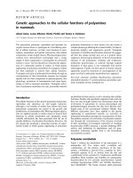
Figure 1
Dendogram showing the relatedness of gene expression profiles
between normal lungs (N), sporadic primary pulmonary hypertension
(PPH) lungs, and familial primary pulmonary hypertension (FPPH)
lungs. Total RNA from the lung was assayed using Affymetrix HU FL
arrays. GeneSpring
®
software was used to generate an experimental
tree by k-tuple means analysis. The relatedness of each sample to one
another is depicted by the dendogram. Blue lines, normal samples;
green lines, FPPH samples; and red lines, sporadic PPH. The degree
of relatedness is proportional to the length of the lines. Yellow lines,
The PPH samples originate from a different phylogeny to the six normal
samples or the three FPPH samples, which originate as depicted from
the black lines. F
PPH refers to a patient whose family history could not
be determined, but whose expression pattern suggests a familial form.
The black box surrounds a group of genes that appear to be
differentially expressed between sporadic PPH and all other samples,
and might represent discriminating genes for this condition.
pared with structurally normal lung tissues. Genes encod-
ing ribosomal, mitochondrial and cytoskeletal proteins and
genes encoding ion channels and enzymes were differen-
tially expressed between PH and normal lungs. Several
transcription factor genes and genes related to cyclin-
dependent kinases were different in their expression, indi-
cating that the PH gene signature reflects a profound
imbalance in the control of genes involved in cell prolifera-
tion and apoptosis. Furthermore, as shown in Figure 1,
whole-tissue total RNA expression profiles demonstrate
striking differences in the expression signatures between
sporadic and familial PPH. Importantly, the differences in
expression profiles are complemented by independent
gene mutation analysis. Only the plexiform lesions in the
lungs from patients with sporadic PPH [31], not those
lesions in FPPH lungs, display mutations of the Bax and
TGF-βRII genes. It is possible that these mutational differ-
ences may lead to gene expression changes. The RNA
expression data and the DNA mutation data taken
together [31] lead to the conclusion that sporadic and
familial PPH are mechanistically distinct. In summary,
microarray gene expression analysis and profiling is a
useful molecular tool that provides a better characteriza-
tion and understanding of the pathobiology of distinct clin-
ical phenotypes of PH.
Conclusions
Genomic approaches to the investigation of PH in animals
or relevant tissues have vastly expanded our knowledge
about the pathobiology of pulmonary hypertensive dis-
eases. Human genetic analysis will undoubtedly expand
and discover further gene mutations involved in the patho-
genesis of PH. Gene expression profiling of different
animal models of PH, and comparison of these profiles
with human PH, will assist in determining the complex
pathways that comprise the response that we term ‘pul-
monary hypertensive tissue remodeling’.
Acknowledgements
This work was supported by the NHLBI Grant HL60913-01 and by a
grant from the Kinner-Wisham Family Foundation. The authors wish to
thank James Campbell for supporting the establishment of the UCHSC
Microarray Facility. The gene expression analysis was performed at the
University of Colorado Comprehensive Cancer Center Gene Expres-
sion Core Facility.
References
1. Rich S: Executive summary from the World Symposium on
primary pulmonary hypertension 1998. [www.who.int/ncd/
cvd/pph.html].
2. Deng Z, Morse JH, Slager SL, Cuervo N, Moore KJ, Venetos G,
Kalachikov S, Cayanis E, Fischer SG, Barst RJ, Hodge SE,
Knowles JA: Familial primary pulmonary hypertension (gene
PPH1) is caused by mutations in the bone morphogenetic
protein receptor-II gene. Am J Hum Genet 2000, 67:737–744.
3. The International PPH Consortium, Lane KB, Machado RD, Pauci-
ulo MW, Thomson JR, Phillips JA III, Loyd JE, Nichols WC, Trem-
bath RC: Heterozygous germline mutations in BMPR2,
encoding a TGF-
ββ
receptor, cause familial primary pulmonary
hypertension. Nat Genet 2000, 26:81–84.
4. Christman BW, McPherson CD, Newman JH, King GA, Bernard
GR, Groves BM, Loyd JE: An imbalance between the excretion
of thromboxane and prostacyclin metabolites in pulmonary
hypertension. N Engl J Med 1992, 327:70–75.
5. Giaid A, Yanagisawa M, Langleben D, Michel RP, Levy R, Shennib
H, Kimura S, Masaki T, Duguid WP, Stewart DJ: Expression of
endothelin-1 in the lungs of patients with pulmonary hyper-
tension. N Engl J Med 1993, 328:1732–1739.
6. Wright L, Tuder RM, Wang J, Cool CD, Lepley RA, Voelkel NF: 5-
Lipoxygenase and 5-lipoxygenase activating protein (FLAP)
immunoreactivity in lungs from patients with primary pul-
monary hypertension. Am J Respir Crit Care Med 1998, 157:
219–229.
7. Tuder RM, Cool CD, Geraci MW, Wang J, Abman SH, Wright L,
Badesch D, Voelkel NF: Prostacyclin synthase expression is
decreased in lungs from patients with severe pulmonary hyper-
tension. Am J Respir Crit Care Med 1999, 159:1925–1932.
8. Cool CD, Stewart JS, Werahera P, Miller GJ, Williams RL, Voelkel
NF, Tuder RM: Three-dimensional reconstruction of pulmonary
arteries in plexiform pulmonary hypertension using cell-spe-
cific markers. Evidence for a dynamic and heterogeneous
process of pulmonary endothelial cell growth. Am J Pathol
1999, 155:411–419.
9. Voelkel NF, Tuder RM, Wade K, Hoper M, Lepley RA, Goulet JL,
Koller BH, Fitzpatrick F: Inhibition of 5-lipoxygenase-activating
protein (FLAP) reduces pulmonary vascular reactivity and pul-
monary hypertension in hypoxic rats. J Clin Invest 1996,
97:2491–2498.
10. Fagan KA, Fouty BW, Tyler RC, Morris KG Jr, Hepler LK, Sato K,
LeCras TD, Abman SH, Weinberger HD, Huang PL, McMurtry IF,
Rodman DM: The pulmonary circulation of homozygous or
heterozygous eNOS-null mice is hyperresponsive to mild
hypoxia. J Clin Invest 1999, 103:291–299.
11. Hoshikawa Y, Voelkel NF, Gesell TL, Moore MD, Morris KG, Alger
LA, Narumiya S, Geraci MW: Prostacyclin receptor-dependent
modulation of pulmonary vascular remodeling. Am J Respir
Crit Care Med 2001, in press.
12. Geraci MW, Gao B, Shepherd DC, Moore MD, Westcott JY,
Fagan KA, Alger LA, Tuder RM, Voelkel NF: Pulmonary prostacy-
clin synthase overexpression in transgenic mice protects
against development of hypoxic pulmonary hypertension. J
Clin Invest 1999, 103:1509–1515.
13. Cowan KN, Heilbut A, Humpl T, Lam C, Ito S, Rabinovitch M:
Complete reversal of fatal pulmonary hypertension in rats by
a serine elastase inhibitor. Nat Med 2000, 6:698–702.
14. Taraseviciene-Stewart L, Kasahara Y, Alger L, Hirth P, McMahon
GG, Waltenberger J, Voelkel NF, Tuder RM: Inhibition of the
VEGF receptor 2 combined with chronic hypoxia causes cell
death-dependent pulmonary endothelial cell proliferation and
severe pulmonary hypertension. FASEB J 2001, 15:427–438.
15. Janssens SP, Bloch KD, Nong Z, Gerard RD, Zoldhelyi P, Collen
D: Adenoviral-mediated transfer of the human endothelial
nitric oxide synthase gene reduces acute hypoxic pulmonary
vasoconstriction in rats. J Clin Invest 1996, 98:317–324.
16. Geraci M, Gao B, Shepherd D, Allard J, Curiel D, Westcott J,
Voelkel N: Pulmonary prostacyclin synthase overexpression by
adenovirus transfection and in transgenic mice [abstract].
Chest 1998, 114:99S.
17. Nagaya N, Yokoyama C, Kyotani S, Shimonishi M, Morishita R,
Uematsu M, Nishikimi T, Nakanishi N, Ogihara T, Yamagishi M,
Miyatake K, Kaneda Y, Tanabe T: Gene transfer of human
prostacyclin synthase ameliorates monocrotaline-induced
pulmonary hypertension in rats. Circulation 2000, 102:2005–
2010.
18. Gupta RA, Tan J, Krause WF, Geraci MW, Willson TM, Dey SK,
DuBois RN: Prostacyclin-mediated activation of peroxisome
proliferator-activated receptor delta in colorectal cancer. Proc
Natl Acad Sci USA 2000, 97:13275–13280.
19. Loyd JE, Butler MG, Foroud TM, Conneally PM, Phillips JA III,
Newman JH: Genetic anticipation and abnormal gender ratio
at birth in familial primary pulmonary hypertension. Am J
Respir Crit Care Med 1995, 152:93–97.
20. O’Donovan MC, Oefner PJ, Roberts SC, Austin J, Hoogendoorn
B, Guy C, Speight G, Upadhyaya M, Sommer SS, McGuffin P:
Blind analysis of denaturing high-performance liquid chro-
matography as a tool for mutation detection. Genomics 1998,
52:44–49.
Respiratory Research Vol 2 No 4 Geraci et al
Available online />commentary
review
reports primary research
21. Thomson JR, Machado RD, Pauciulo MW, Morgan NV, Humbert
M, Elliott GC, Ward K, Yacoub M, Mikhail G, Rogers P, Newman
J, Wheeler L, Higenbottam T, Gibbs JS, Egan J, Crozier A,
Peacock A, Allcock R, Corris P, Loyd JE, Trembath RC, Nichols
WC: Sporadic primary pulmonary hypertension is associated
with germline mutations of the gene encoding BMPR-II, a
receptor member of the TGF-
ββ
family. J Med Genet 2000, 37:
741–745.
22. Machado RD, Pauciulo MW, Thomson JR, Lane KB, Morgan NV,
Wheeler L, Phillips JA 3
rd
, Newman J, Williams D, Galie N, Manes
A, McNeil K, Yacoub M, Mikhail G, Rogers P, Corris P, Humbert
M, Donnai D, Martensson G, Tranebjaerg L, Loyd JE, Trembath
RC, Nichols WC: BMPR2 haploinsufficiency as the inherited
molecular mechanism for primary pulmonary hypertension.
Am J Hum Genet 2001, 68:92–102.
23. Massague J, Blain SW, Lo RS: TGF-
ββ
signaling in growth
control, cancer, and heritable disorders. Cell 2000, 103:
295–309.
24. McAllister KA, Grogg KM, Johnson DW, Gallione CJ, Baldwin MA,
Jackson CE, Helmbold EA, Markel DS, McKinnon WC, Murrell J:
Endoglin, a TGF-
ββ
binding protein of endothelial cells, is the
gene for hereditary haemorrhagic telangiectasia type 1. Nat
Genet 1994, 8:345–351.
25. Johnson DW, Berg JN, Baldwin MA, Gallione CJ, Marondel I,
Yoon SJ, Stenzel TT, Speer M, Pericak-Vance MA, Diamond A,
Guttmacher AE, Jackson CE, Attisano L, Kucherlapati R, Porteous
ME, Marchuk DA: Mutations in the activin receptor-like kinase
1 gene in hereditary haemorrhagic telangiectasia type 2. Nat
Genet 1996, 13:189–195.
26. Lee SD, Shroyer KR, Markham NE, Cool CD, Voelkel NF, Tuder
RM: Monoclonal endothelial cell proliferation is present in
primary but not secondary pulmonary hypertension. J Clin
Invest 1998, 101:927–934.
27. Tuder RM, Radisavljevic Z, Shroyer KR, Polak JM, Voelkel NF:
Monoclonal endothelial cells in appetite suppressant-associ-
ated pulmonary hypertension. Am J Respir Crit Care Med
1998, 158:1999–2001.
28. Markowitz S, Wang J, Myeroff L, Parsons R, Sun L, Lutterbaugh J,
Fan RS, Zborowska E, Kinzler KW, Vogelstein B: Inactivation of
the type II TGF-
ββ
receptor in colon cancer cells with
microsatellite instability. Science 1995, 268:1336–1338.
29. McCaffrey TA, Du B, Consigli S, Szabo P, Bray PJ, Hartner L,
Weksler BB, Sanborn TA, Bergman G, Bush HL: Genomic insta-
bility in the type II TGF-
ββ
1 receptor gene in atherosclerotic
and restenotic vascular cells. J Clin Invest 1997, 100:
2182–2188.
30. Rampino N, Yamamoto H, Ionov Y, Li Y, Sawai H, Reed JC,
Perucho M: Somatic frameshift mutations in the BAX gene in
colon cancers of the microsatellite mutator phenotype.
Science 1997, 275:967–969.
31. Yeager ME, Halley GR, Golpon HA, Voelkel NF, Tuder RM:
Microsatellite instability of endothelial cell growth and apop-
tosis genes within plexiform lesions in primary pulmonary
hypertension. Circ Res 2001, 88:E2–E11.
32. Lockhart DJ, Dong H, Byrne MC, Follettie MT, Gallo MV, Chee
MS, Mittmann M, Wang C, Kobayashi M, Horton H, Brown EL:
Expression monitoring by hybridization to high-density
oligonucleotide arrays. Nat Biotechnol 1996, 14:1675–1680.
33. Golub TR, Slonim DK, Tamayo P, Huard C, Gaasenbeek M,
Mesirov JP, Coller H, Loh ML, Downing JR, Caligiuri MA, Bloom-
field CD, Lander ES: Molecular classification of cancer: class
discovery and class prediction by gene expression monitor-
ing. Science 1999, 286:531–537.
34. Geraci MW, Moore MD, Gesell TL, Yeager ME, Alger L, Golpon
H, Gao B, Loyd JE, Tuder RM, Voelkel NF: Gene expression pat-
terns in the lungs of patients with primary pulmonary hyper-
tension — a gene microarray analysis. Circ Res 2001, 88:
555–562.