Báo cáo y học: " Collaborative interactions between neutrophil elastase and metalloproteinases in extracellular matrix degradation in threedimensional collagen gels" pps
Bạn đang xem bản rút gọn của tài liệu. Xem và tải ngay bản đầy đủ của tài liệu tại đây (2.16 MB, 7 trang )
Research article
Collaborative interactions between neutrophil elastase and
metalloproteinases in extracellular matrix degradation in three-
dimensional collagen gels
Yunkui Zhu*, Xiangde Liu*, C Magnus Sköld
†
, Hangjun Wang
‡
, Tadashi Kohyama*,
Fu-Qiang Wen*, Ronald F Ertl* and Stephen I Rennard*
*University of Nebraska Medical Center, Omaha, Nebraska, USA
†
Karolinska Hospital, Stockholm, Sweden
‡
Mount Sinai Hospital, Pathology and Laboratory Medicine, Toronto, Ontario, Canada
Correspondence: Stephen I Rennard, MD, University of Nebraska Medical Center, 985125 Nebraska Medical Center, Omaha,
Nebraska 68198-5125, USA. Tel: +1 402 559 7313; fax: +1 402 559 4878; e-mail:
Introduction
Three-dimensional (3D) collagen gel culture has been
used as an in vitro model of in vivo tissue contraction, a
common feature of fibrosis, as well as the resolution of
granulation tissue that characterizes repair [1,2]. Short-
term co-cultures of monocytes with fibroblasts result in the
inhibition of collagen gel contraction [3], while co-cultures
of fibroblasts with neutrophils, or with neutrophil elastase
(NE), augment contraction [4].
Results in the linked study [5] demonstrated that 3D colla-
gen gel contraction was augmented in extended co-cultures
Abstract
Background: Extended culture of monocytes and fibroblasts in three-dimensional collagen gels leads
to degradation of the gels (see linked study in this issue, “Fibroblasts and monocytes contract and
degrade three-dimensional collagen gels in extended co-culture”). The current study, therefore, was
designed to evaluate production of matrix-degrading metalloproteinases by these cells in co-culture
and to determine if neutrophil elastase could collaborate in the activation of these enzymes. Since co-
cultures produce prostaglandin E
2
(PGE
2
), the role of PGE
2
in this process was also evaluated.
Methods: Blood monocytes from healthy donors and human fetal lung fibroblasts were cast into type I
collagen gels and maintained in floating cultures for three weeks. Matrix metalloproteinases (MMPs)
were assessed by gelatin zymography (MMPs 2 and 9) and immunoblotting (MMPs 1 and 3). The role
of PGE
2
was explored by direct quantification, and by the addition of exogenous indomethacin and/or
PGE
2
.
Results: Gelatin zymography and immunoblots revealed that MMPs 1, 2, 3 and 9 were induced by co-
cultures of fibroblasts and monocytes. Neutrophil elastase added to the medium resulted in marked
conversion of latent MMPs to lower molecular weight forms consistent with active MMPs, and was
associated with augmentation of both contraction and degradation (P < 0.01). PGE
2
appeared to
decrease both MMP production and activation.
Conclusion: The current study demonstrates that interactions between monocytes and fibroblasts can
mediate tissue remodeling.
Keywords: collagen degradation, lung fibroblasts, metalloproteinases, monocytes, prostaglandin E
2
Received: 5 March 2001
Revisions requested: 14 May 2001
Revisions received: 13 July 2001
Accepted: 8 August 2001
Published: 4 September 2001
Respir Res 2001, 2:300–305
The complete version of this article is online at
/>© 2001 Zhu et al, licensee BioMed Central Ltd
(Print ISSN 1465-9921; Online ISSN 1465-993X)
3D = three-dimensional; MMP = matrix metalloproteinase; NE = neutrophil elastase; PGE
2
= prostaglandin E
2
.
Available online />Available online />commentary review reports
research article
of fibroblasts and monocytes. Since MMPs play a prominent
role in connective tissue degradation [6–8], the current
study, an extension of this linked study, was designed to
explore the potential role of MMPs in this process.
Materials and methods
See supplementary material for further information.
Cells and cultures
See Supplementary material.
Preparation of collagen gels for three-dimensional co-
culture
Collagen gels were prepared as described previously [9].
For long-term co-culture, the medium was changed every
5 days. The areas of floating gels were measured using an
image analyzer.
To investigate the effect of PGE
2
on collagen degradation,
indomethacin (1 µM) or PGE
2
(0.1 µM) was added to the
medium.
Gelatinase activity assay
Gelatin zymography was performed by modification of a
previously published procedure to identify MMPs 1 and -9
[10,11].
Immunoblot analysis of metalloproteinases
To further identify the MMPs produced, immunoblots for
MMPs 1 and 3 were performed.
Results
Effect of co-culture on gelatinase activity
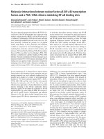
As shown in Figures 1 and 2, fibroblasts alone routinely
released primarily MMP-2 into their surrounding medium, as
identified at the molecular weights of 72 kDa (latent form)
and 66 kDa (active form) (Fig. 1). Over 5 days, elastase
appeared to partially convert some of the latent 72 kDa form
to the 66 kDa form. With increasing incubation time, MMP-2
present in culture medium gradually decreased. Even at day
21, however, there was readily detectable MMP-2, consis-
tent with ongoing release (Fig. 1a). Co-culture of monocytes
and fibroblasts increased both bands of MMP-2 and
resulted in more of the 66 kDa form (Fig. 1b). Co-culture of
fibroblasts with monocytes also induced the release of
MMP-9 (Fig. 1b), which was present as the latent 92 kDa
form. Addition of elastase nearly completely converted the
latent 92 kDa MMP-9 to the active 83 kDa form. With
increasing culture time, the amount of detectable MMP-9 in
co-cultures decreased. In contrast to the co-cultures, mono-
cytes cultured alone released no gelatinolytic activity (data
not shown).
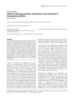
Neutrophil elastase (NE) augmented and PGE
2
inhibited the
conversion of 72 kDa MMP-2 to the 66 kDa form in fibrob-
lasts cultured alone (Fig. 2a). In co-cultures, indomethacin
resulted in a marked increase of conversion of MMP-9 from
the 92 kDa to the 83 kDa form, most readily observed in the
absence of NE, where conversion was minimal (Fig. 2b). The
addition of exogenous PGE
2
decreased the conversion of
MMP-9 to the 83 kDa form MMP-9. Neither indomethacin
nor PGE
2
induced release of gelatinase activity in monocytes
cultured alone (data not shown).
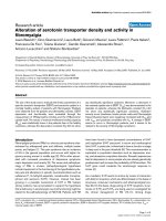
Effect of co-culture on MMP-1
No detectable MMP-1 was observed in cultures of mono-
cytes (Fig. 3). In fibroblasts alone, a trace of MMP-1 was
occasionally detectable. In co-cultures of monocytes and
fibroblasts, however, there was marked induction of
MMP-1, which was present at a size corresponding to the
latent 52 kDa form (Fig. 3). The detectable MMP-1 in co-
cultures was maximal at earlier times, decreasing with
increase cultured time and becoming undetectable by
Figure 1
Matrix metalloproteases 2 and 9 (MMP-2 and MMP-9). Culture media
were harvested after 5, 10, 15 and 21 days under control conditions
and, after 5 days, in the presence of neutrophil elastase (NE) and
subjected to gelatin zymography. (a) Human fetal lung (HFL)
fibroblasts alone. MMP-2 latent (72 kDa) and active (66 kDa) forms are
shown. (b) Blood monocytes (BM) co-cultured with HFL fibroblasts.
MMP-9 latent (92 kDa) and active (83 kDa) forms are shown in
addition to MMP-2 forms. Lane 1: samples were harvested on day 5.
Lane 2: culture medium in the presence of NE, harvested on day 5.
Lanes 3, 4, 5: samples were harvested on days 10, 15 and 21 under
control conditions, respectively.
Figure 2
Prostaglandin E
2
(PGE
2
) and matrix metalloproteases (MMPs) 2 and 9.
Culture media were harvested after four days in the presence of
neutrophil elastase (NE), indomethacin or exogenous PGE
2
. (a)
Fibroblasts alone. MMP-2 latent (72 kDa) and active (66 kDa) forms
are shown. Lane 1: control; lane 2: NE; lane 3: indomethacin; lane 4:
PGE
2
. (b) Monocytes co-cultured with fibroblasts. MMP-9 latent
(92 kDa) and active (83 kDa) forms are shown in addition to MMP-2
forms. Lane 1: control; lane 2: NE; lane 3: indomethacin;
lane 4: NE + indomethacin; lane 5: PGE
2
; lane 6: NE + PGE
2
;
lane 7: NE + indomethacin + PGE
2
.
(a) (b)
(a) (b)
Respiratory Research Vol 2 No 5 Zhu et al
15 days (Fig. 3). The presence of neutrophil elastase for
the first 5 days converted latent MMP-1 to active 42 kDa
and 20 kDa forms. Indomethacin augmented the induction
of MMP-1 in co-culture (Fig. 4).
In contrast, PGE
2
reduced the amount of total MMP-1 and
decreased the conversion of the 52 kDa form to the lower
molecular weight forms in the presence of elastase.
Neither indomethacin nor PGE
2
had an effect on MMP-1
in cultures of monocytes or fibroblasts alone.
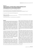
Effect of co-culture on MMP-3
Neither fibroblasts nor monocytes alone released
detectable MMP-3 (Fig. 5). In co-cultures of monocytes
and fibroblasts, however, MMP-3 release was readily
detected in a size corresponding to the latent 57 kDa form
(Fig. 5). MMP-3 release was greatest at the earliest time
points evaluated, and decreased with time becoming
undetectable by 15 days of culture. Addition of NE for the
first 5 days resulted in conversion of the 57 kDa form to
active 47 and 35 kDa forms.
Figure 3
Immunoblot for matrix metalloprotease (MMP)-1. Media were harvested after 5, 10, 15 and 21 days under control conditions and, after five days, in
the presence of neutrophil elastase (NE) and were subjected to immunoblot for MMP-1. (a) Human fetal lung (HFL) fibroblasts alone. (b) Blood
monocytes (BM) co-cultured with HFL fibroblasts. MMP-1 appears in its latent 52 kDa form. NE presence resulted in conversion of latent MMP-1
to active 42 kDa and 20 kDa forms. (c) BM alone. Lane 1: samples harvested on day 5. Lane 2: culture medium in the presence of NE, harvested
at day 5. Lanes 3, 4, 5: samples harvested on days 10, 15 and 21 under control conditions, respectively.
(a) (b) (c)
Figure 4
Effect of prostaglandin E
2
(PGE
2
) on matrix metalloprotease (MMP)-1. After four days, media were harvested for immunoblotting. (a) Human fetal
lung (HFL) fibroblasts alone. Lane 1: control; lane 2: neutrophil elastase (NE); lane 3: indomethacin; Lane 4: PGE
2
. (b) Blood monocytes (BM)
co-cultured with HFL fibroblasts. MMP-1 appears in its latent 52 kDa form. NE presence resulted in conversion of latent MMP-1 to the active
20 kDa form. Lane 1: control; lane 2: NE; lane 3: indomethacin; lane 4: NE + indomethacin; lane 5: PGE
2
; lane 6: NE + PGE
2
;
lane 7: NE + indomethacin + PGE
2
. (c) BM alone. Lane 1: control; lane 2: NE; lane 3: indomethacin; lane 4: PGE
2
.
(a) (b) (c)
Indomethacin augmented the induction of MMP-3 while
PGE
2
reduced the conversion of MMP-3 to lower molecu-
lar weight forms (Fig. 6). Neither indomethacin nor PGE
had an effect on MMP-3 on fibroblasts or monocytes cul-
tured alone.
Discussion
In pulmonary emphysema, various inflammatory mediators
have been suggested to cause tissue destruction and loss
of structure [12–15]. Several lines of evidence support the
concept that neutrophil elastase contributes to the patho-
genesis of emphysema [6,7]. Evidence, including the
marked expansion of macrophage numbers in smokers’
lungs and in studies from genetically altered mice, also
supports a role for macrophage-derived proteases in
emphysema [16,17]. These concepts are not exclusive,
and it is possible that several proteolytic and inflammatory
mechanisms contribute to the development of emphysema.
In the linked study [5], extended co-cultures of fibroblasts
and monocytes augmented collagen gel contraction and
degraded the extracellular matrix. NE added to co-cultures
resulted in a concentration-dependent degradation of colla-
gen. The current study suggests that this increased degra-
Available online />commentary review reports
research article
Figure 5
Immunoblot for matrix metalloprotease (MMP)-3. Media were harvested after 5, 10, 15 and 21 days under control conditions and, after five days, in
the presence of neutrophil elastase (NE) and subjected to immunoblot for MMP-3. (a) Human fetal lung (HFL) fibroblasts alone. (b) Blood
monocytes (BM) co-cultured with HFL fibroblasts. The latent 57 kDa form of MMP-3 is shown; addition of NE for the first five days resulted in
conversion of the 57 kDa form to active 47 kDa and 35 kDa forms. Lane 1: samples were harvested on day 5. Lane 2: culture medium in the
presence of NE, harvested at day 5. Lanes 3, 4, 5: samples harvested on days 10, 15 and 21 under control conditions, respectively. (c) BM alone.
(a) (b) (c)
Figure 6
Effect of prostaglandin E
2
(PGE
2
) on matrix metalloprotease (MMP)-3. Media were harvested for immunoblotting. (a) Human fetal lung (HFL)
fibroblasts alone. Lane 1: control; lane 2: neutrophil elastase (NE); lane 3: indomethacin; lane 4: PGE
2
. (b) Blood monocytes (BM) co-cultured with
HFL fibroblasts. Lane 1: control; lane 2: NE; lane 3: indomethacin; lane 4: NE + indomethacin; lane 5: PGE
2
; lane 6: NE + PGE
2
;
lane 7: NE + indomethacin + PGE
2
. (c) BM alone. Lane 1: control; lane 2: NE; lane 3: indomethacin; lane 4: PGE
2
.
(a) (b) (c)
Respiratory Research Vol 2 No 5 Zhu et al
dation of extracellular collagen may be due to NE activation
of latent MMPs induced in the co-culture conditions.
NE has been demonstrated to result in the augmentation of
contraction [4]. It also has been suggested to play an impor-
tant role in the development of emphysema. In animals,
instillation of NE can result in the development of pulmonary
emphysema [18]. Individuals deficient in α-1 protease
inhibitor, moreover, have an increased susceptibility to the
development of emphysema [19–21]. The current study
suggests the possibility that NE can collaborate with MMPs,
leading to the degradation of extracellular matrix.
The MMPs are a family of proteolytic enzymes [22,23].
Most are released as latent precursors. Proteolytic cleav-
age of the latent forms can result in generation of active
proteases [24,25]. The MMPs differ both in their substrate
specificity and in their mechanisms of activation. Since
some members of the MMP family are capable of activating
other members [26,27], it is likely that proteolytic cascades
may regulate MMP activity. A further degree of regulation of
MMP activity is afforded by the family of inhibitors: tissue
inhibitors of metalloproteinases (TIMPs) [28].
Recent studies in genetically altered mice have suggested
an important role for multiple proteases in the development
of emphysema. Mice deficient in MMP-9, MMP-12, or NE
are resistant to the development of emphysema or skin blis-
ters [8,16]. Mice overexpressing collagenase, however,
develop emphysema [14]. The current study proposes a
possible collaboration among proteases that is responsible
for the tissue degradation associated with the disease.
According to results from this study, several proteases are
induced in co-cultures and activated in the presence of
NE. It is likely that other proteolytic enzymes may also play
a role beyond those evaluated in the current study. In this
context, fibroblasts are known to express cell surface pro-
teases, which may have a major role in regulating the
activity of other mediators in the extracellular milieu
[29–31]. It is of interest that PGE
2
appears to be able to
regulate the protease activity responsible for extracellular
matrix degradation.
It is unlikely that PGE
2
functions directly as a protease
inhibitor. It seems more plausible that PGE
2
regulates pro-
teolytic activity by altering the production of antiproteases,
or by altering the production of components essential in
the proteolytic cascade leading to collagen degradation
[31]. The sequence of proteolytic events, by which NE
leads to collagen degradation, is incompletely defined.
PGE
2
, however, could potentially modulate the proteolytic
cascade, resulting in collagen degradation at a number of
steps. Both PGE
2
and a cascade of proteolytic events that
lead to extracellular matrix degradation have the potential
for serving as paracrine regulators. Such a means of regu-
lation may be particularly important in tissue remodeling. It
seems unlikely that tissue remodeling is accomplished by
individually active fibroblasts. Rather, coordinated activity
within a tissue would seem to be a more appropriate
means to accomplish alteration in tissue structure.
Paracrine regulation would seem to be ideally suited to
accomplish such an effect.
Conclusion
This study demonstrates that monocytes and fibroblasts in
co-culture can release MMPs and degrade extracellular
matrix. Activation of MMPs by NE can augment this
process. PGE
2
can modulate this proteolytic cascade.
These data support a role for collaborative interaction
among inflammatory mediators leading to tissue destruc-
tion in diseases such as emphysema.
References
1. Grinnell F: Fibroblasts, myofibroblasts and wound contraction.
J Cell Biol 1994, 124:401-404.
2. Bell E, Ivarsson B, Merrill C: Production of a tissue-like struc-
ture by contraction of collagen lattices by human fibroblasts
of different proliferative potential in vitro. Proc Natl Acad Sci
USA 1979, 76:1274-1278.
3. Sköld CM, Liu XD, Umino T, Zhu YK, Ertl RF, Romberger DJ,
Rennard SI: Blood monocytes attenuate lung fibroblast con-
traction of three-dimensional collagen gels in coculture. Am J
Physiol Lung Cell Mol Physiol 2000, 279:L667-L674.
4. Skold CM, Liu X, Umino T, Zhu Y, Ohkuni Y, Romberger DJ,
Spurzem JR, Heires AJ, Rennard SI: Human neutrophil elastase
augments fibroblast-mediated contraction of released colla-
gen gels. Am J Respir Crit Care Med 1999, 159:1138-1146.
5. Zhu YK, Skold CM, Liu XD, Wang H, Kohyama T, Wen FQ, Ertl
RF, Rennard SI: Fibroblasts and monocytes contract and
degrade three-dimensional collagen gels in extended culture.
Respir Res 2001, 2:295–299.
6. Thurlbeck WM, Muller NL: Emphysema: definition, imaging, and
quantification. AJR Am J Roentgenol 1994, 163:1017-1025.
7. Senior RM, Tegner H, Kuhn C, Ohlsson K, Starcher BC, Pierce
JA: The induction of pulmonary emphysema with human
leukocyte elastase. Am Rev Resp Dis 1977, 116:469-475.
8. Liu Z, Zhou X, Shapiro SD, Shipley JM, Twining SS, Diaz LA,
Senior RM, Werb Z: The serpin alpha1-proteinase inhibitor is a
critical substrate for gelatinase B/MMP-9 in vivo. Cell 2000,
102:647-655.
9. Mio T, Adachi Y, Romberger DJ, Ertl RF, Rennard SI: Regulation
of fibroblast proliferation in three dimensional collagen gel
matrix. In Vitro Cell Dev Biol 1996, 32:427-433.
10. Zhang Y, McCluskey K, Fujii K, Wahl LM: Differential regulation
of monocyte matrix metalloproteinase and TIMP-1 production
by TNF-alpha, granulocyte-macrophage CSF, and IL-1 beta
through prostaglandin-dependent and -independent mecha-
nisms. J Immunol 1998, 161:3071-3076.
11. Kleiner DE, Stetler-Stevenson WG: Quantitative zymography:
detection of picogram quantities of gelatinases. Anal Biochem
1994, 218:325-329.
12. Dalal S, Imai K, Mercer B, Okada Y, Chada K, D’Armiento JM: A
role for collagenase (matrix metalloproteinase-1) in pul-
monary emphysema. Chest 2000, 117:227S-228S.
13. Finlay GA, Russell KJ, McMahon KJ, D’arcy EM, Masterson JB,
FitzGerald MX, O’Connor CM: Elevated levels of matrix metal-
loproteinases in bronchoalveolar lavage fluid of emphysema-
tous patients. Thorax 1997, 52:502-506.
14. D’Armiento J, Dalal SS, Okada Y, Berg RA, Chada K: Collage-
nase expression in the lungs of transgenic mice causes pul-
monary emphysema. Cell 1992, 71:955-961.
15. Carp H, Janoff A: Possible mechanisms of emphysema in
smokers. In vitro suppression of serum elastase-inhibitory
capacity by fresh cigarette smoke and its prevention by
antioxidants. Am Rev Respir Dis 1978, 118:617-621.
16. Hautamaki RD, Kobayashi DK, Senior RM, Shapiro SD: Require-
ment for macrophage elastase for cigarette smoke-induced
emphysema in mice. Science 1997, 277:2002-2004.
17. Marques LJ, Teschler H, Guzman J, Costabel U: Smoker’s lung
transplanted to a nonsmokker long term detection of smok-
er’s macrophages. Am J Respir Crit Care Med 1997, 156:1700-
1702.
18. Massaro G, Massaro D: Retinoic acid treatment abrogates
elastase-induced pulmonary emphysema in rats. Nat Med
1997, 3:675-677.
19. Laurell CB, Eriksson S: The electrophoretic alpha 1-globulin
pattern of serum in alpha 1-antitrypsin deficiency. Scand J
Clin Lab Invest 1963, 15:132-140.
20. Crystal RG: Beta 1-antitrypsin deficiency, emphysema, and
liver disease. J Clin Invest 1990, 85:1343-1352.
21. Fujita J, Nelson NL, Daughton DM, Dobry CA, Spurzem JR, Irino
S, Rennard SI: Evaluation of elastase and antielastase balance
in patients with bronchitis and pulmonary emphysema. Am
Rev Respir Dis 1990, 142:57-62.
22. Birkedal-Hansen H, Moore WG, Bodden MK, Windsor LJ,
Birkedal-Hansen B, DeCarlo A, Engler JA: Matrix metallopro-
teinases: a review. Crit Rev Oral Biol Med 1993, 4:197-250.
23. Woessner JF: The matrix metalloproteinase family. In Matrix
metalloproteinases. Edited by Parks WC, RP Mecham. San
Diego: Academic Press, 1988:1-13.
24. Nagase H, Suzuki K, Enghild JJ, Salvesen G: Stepwise activation
mechanisms of the precursors of matrix metalloproteinases 1
(tissue collagenase) and 3 (stromelysin). Biomed Biochim
Acta 1991, 50:749-754.
25. Ferry G, Lonchampt M, Pennel L, de Nanteuil G, Canet E, Tucker
GC: Activation of MMP-9 by neutrophil elastase in an in vivo
model of acute lung injury. FEBS Lett 1997, 402:111-115.
26. Swiderski RE, Dencoff JE, Floerchinger CS, Shapiro SD, Hun-
ninghake GW: Differential expression of extracellular matrix
remodeling genes in a murine model of bleomycin-induced
pulmonary fibrosis. Am J Pathol 1998, 152:821-828.
27. Suzuki K, Enghild JJ, Morodomi T, Salvesen G, Nagase H: Mecha-
nisms of activation of tissue procollagenase by matrix metal-
loproteinase 3 (stromelysin). Biochemistry 1990, 29:10261-
10270.
28. Brown PD: Synthetic inhibitors of matrix metalloproteinases.
In Matrix Metalloproteinases. Edited by Parks WC, RP Mecham.
San Diego: Academic Press, 1988:243-256.
29. Li H, Bauzon DE, Xu X, Tschesche H, Cao J, Sang QA: Immuno-
logical characterization of cell-surface and soluble forms of
membrane type 1 matrix metalloproteinase in human breast
cancer cells and in fibroblasts. Mol Carcinog 1998, 22:84-94.
30. Konttinen YT, Ceponis A, Takagi M, Ainola M, Sorsa T, Sutinen M,
Salo T, Ma J, Santavirta S, Seiki M: New collagenolytic
enzymes/cascade identified at the pannus-hard tissue junc-
tion in rheumatoid arthritis: destruction from above. Matrix
Biol 1998, 17:585-601.
31. Li L, Eisen AZ, Sturman E, Seltzer JL: Protein tyrosine phospho-
rylation in signalling pathways leading to the activation of
gelatinase A: activation of gelatinase A by treatment with the
protein tyrosine phosphatase inhibitor sodium orthovanadate.
Biochim Biophys Acta 1998, 1405:110-120.
32. Wahl LM, Katona IM, Wilder RL, Winter CC, Haraoui B, Scher I,
Wahl S: Isolation of human mononuclear cell subsets by
counter flow centrifugal elutriation (CCE). I. Characterization
of B-lymphocyte, T-lymphocytes and monocyte-enriched frac-
tions by flow cytometric analysis. Cell Immunol 1984, 85:373-
383.
Supplementary material
Materials and methods
Cells and cultures
Human fetal lung fibroblasts (cell line HFL-1), obtained
from the American Type Culture Collection (Rockville, MD,
USA), were cultured with Dulbecco’s Modified Eagle
Medium (DMEM) supplemented with 10% fetal-calf
serum, 50 U/ml penicillin, 50 µg/ml streptomycin and
0.25 µg/ml fungizone. The cells were cultured in 100 mm
tissue culture dishes (Falcon, Becton Dickinson Labware,
Lincoln Park, NJ, USA). The fibroblasts were passaged
every week. Subconfluent fibroblasts were trypsinized
(trypsin-EDTA; 0.05% trypsin, 0.53 mM EDTA-4 Na) and
used for collagen gel culture. Fibroblasts used in these
experiments were between cell passages 14 and 16.
Blood monocytes were isolated from blood cells of healthy
blood donors [32]. Cell suspensions were > 96% mono-
cytes by the criteria of cell morphology on Wright stained
cytosmears. Monocytes were stored at 4°C and were
used for co-culture within 4 hours after isolation.
Reagents
Human neutrophil elastase was purchased from ECP
(Owensville, MO, USA). Prostaglandin E
2
(PGE
2
) and
indomethacin were purchased from Sigma (St. Louis, MO,
USA). Tissue culture supplements and media were pur-
chased from GIBCO (Life Technologies, Grand Island,
NY, USA). Fetal calf serum was purchased from Biofluid
(Rockville, MD, USA).
Preparation of collagen gels
Tendons were excised from rat tails, and the tendon
sheath and other connective tissue were removed care-
fully. After repeated washing with Tris-buffered saline
(TBS, 0.9% NaCl, 10 mM Tris, pH 7.5) and serial concen-
trations of ethanol (from 50% to 100%), type I collagen
was extracted in 6 mM hydrochloric acid at 4°C for
24 hours. Protein concentration was determined by weigh-
ing a lyophilized aliquot from each lot of collagen solution.
Sodium dodecyl sulfate polyacrylamide gel electrophore-
sis routinely determined no detectable protein other than
type I collagen.
Gelatinase activity assay
Conditioned media were concentrated fivefold by ethanol
precipitation and re-suspension in distilled H
2
O. The
samples were dissolved in twofold electrophoresis sample
buffer (0.5 M Tris-HCL, pH 6.8, 2% SDS, 20% glycerol,
0.1% bromophenol blue), and heated for 5 min at 95°C.
Thirty microliters of each sample were loaded in each lane,
and electrophoresis was performed with a Mini Elec-
trophoresis Cell (BIO-RID, Hercules, CA, USA) at 200 V.
After electrophoresis, the gels were gently soaked with
2.5% (v/v) Triton-X 100 at 20°C for 30 min, then incu-
bated in the metalloproteinase buffer (0.06 M Tris-HCl, pH
7.5, containing 6 mM CaCl
2
and 1 µM ZnCl) for 18 hours
at 37°C. The gels were stained with 0.4% (w/v)
Coomassie blue and rapidly destained with 30% (v/v)
methanol, 10% acetic acid. The gels were dried directly
between cellophane sheets (Pharmacia Biotech, San
Francisco, CA, USA).
Available online />commentary review reports
research article
Immunoblot analysis of metalloproteinases
Supernatant media from 3D cultures were concentrated
10-fold by precipitation with ethanol, resuspended in dis-
tilled H
2
O and mixed with twofold sample buffer (0.5 M
Tris-HCl, pH 6.8, 2% SDS, 0.1% bromphenol blue, 0.5%
ß-mercaptoethanol, 20% glycerol). After heating for 3 min
at 95°C, 30 µl of each sample was loaded for elec-
trophoresis with a Mini Electrophoresis Cell (BIO-RAD,
Hercules, CA). The proteins were transferred to a PVDF
transfer membrane (BIO-RAD, Hercules, CA, USA) in
electrophoresis buffer (20 mM Tris-HCl, pH 8.0, 150 mM
glycine, 20% methanol) at 20 V for 35 min with a Semi-dry
Electrophoretic Transfer Cell (BIO-RAD, Hercules, CA,
USA). The blots were blocked in 5% fat-free milk in PBS-
Tween at room temperature for 1 hour, then exposed to
primary antibodies (mouse anti-human MMP-1, MMP-2,
MMP-3 or MMP-9 antibodies; Calbiochem, Cambridge,
MA, USA), and subsequently detected using HRP conju-
gated rabbit anti-mouse IgG (ICN Biomedical, Costa
Mesa, CA, USA) in conjunction with an enhanced chemilu-
minescence detection system (ECL, Amersham Pharma-
cia Biotech, Little Chalfont, Buckinghamshire, England).
Respiratory Research Vol 2 No 5 Zhu et al