Báo cáo y học: "Distinct signal transduction processes by IL-4 and IL-13 and influences from the Q551R variant of the human IL-4 receptor alpha chain" docx
Bạn đang xem bản rút gọn của tài liệu. Xem và tải ngay bản đầy đủ của tài liệu tại đây (319.69 KB, 11 trang )
Available online />Page 1 of 11
(page number not for citation purposes)
Respiratory Research
Vol 3 No 1
/>Kruse
et al.
Research article
Distinct signal transduction processes by IL-4 and IL-13 and
influences from the Q551R variant of the human IL-4 receptor
alpha chain
Susanne Kruse, Sandra Braun and Klaus A Deichmann
University Children's Hospital, University of Freiburg, Mathildenstr. 1, 79106 Freiburg, Germany
Correspondence: Klaus A Deichmann -
Abstract
Background: Although IL-4 and IL-13 share the IL-13 receptor, IL-13 exhibits unique functions. To
elicit the cellular basis of these differences, signal transduction processes have been compared.
Additionally, the role of the IL-4 receptor alpha (IL-4Rα) variant Q551R was investigated.
Methods: Peripheral blood mononuclear cells from donors were stimulated with IL-4 and IL-13. The
phosphorylation status of effector substrates was detected by immunostaining. Binding of SHP-2 to IL-
4Rα was investigated by using synthetic peptides.
Results: SHP-2 bound IL-4Rα synthetic peptide; this binding was reduced in the presence of the
R551 variant. Stimulation with IL-4 increased SHP-1 phosphorylation, however, stimulation with IL-13
increased SHP-2 phosphorylation. PI3-kinase phosphorylation was elevated following stimulation with
IL-13 in all individuals and with IL-4 only in R551 individuals. Jak1, Tyk2 and IRS-2 signals were
reduced after IL-13 stimulation in Q551 individuals. STAT3 phosphorylation was markedly increased
in R551 individuals, following stimulation with both IL-4 and IL-13. However, STAT3 was only detected
immediately in nuclear extracts from variant individuals after stimulation with IL-13; in wildtype
individuals STAT3 was only detected after IL-4 treatment.
Conclusion: IL-4 and IL-13 appear to promote distinct signal transduction cascades. SHP-1 seems to
be predominately activated by IL-4 and to influence the PI3-kinase, in contrast, SHP-2 seems to be
predominately activated by IL-13 and to influence Jak1, Tyk2 and IRS-2. Both phosphatases control
STAT3. In the presence of the variant R551, SHP-1/2 activation is reduced and signal transduction is
altered. STAT3 signaling appears be further regulated on the level of nuclear translocation.
Keywords: asthma, IL-4, IL-13, SHP, STAT3
Introduction
Asthma and atopy represent a group of complex diseases
with a broad variety of clinical phenotypes. The individual
risk of developing atopic diseases seems to be influenced
by genetic susceptibility and environmental factors. During
the past decade, a great number of studies have tried to in-
vestigate the genetic basis of atopy. Functional studies of
genetic variants contributing to the susceptibility of asthma
and atopy become immensely important in an effort to un-
derstand the complex immunological processes underlying
the development of these diseases. Two important regula-
tors of the human immune system are the pleiotropic cy-
tokines IL-4 and IL-13. They play major roles in stimulating
B-cell proliferation and in influencing B-cell differentiation
towards IgE production. In addition, IL-4 shifts the Th1/Th2
balance of activated Th cells towards Th2 cells [1–3]. The
effect of IL-13 on human T cells, if any, is still unknown.
Among others roles, these two cytokines play important
roles in the development of inflammatory diseases [4]. B-
cell activation and Th2 type immune responses underlie at-
opic disorders. Animal models have revealed that IL-13 in-
duces the pathophysiological features of asthma
Received: 21 November 2001
Revisions requested: 5 February 2002
Revisions received: 22 May 2002
Accepted: 28 May 2002
Published: 14 August 2002
Respir Res 2002, 3:24
© 2002 Kruse et al., licensee BioMed Central Ltd
(Print ISSN 1465-9921; Online ISSN 1465-993X)
Respiratory Research Vol 3 No 1 Kruse et al.
Page 2 of 11
(page number not for citation purposes)
independently of IL-4, but is strongly dependent on IL-4Rα
[5,6], whereas IL-4 initiates a more general inflammatory re-
sponse. Furthermore, IL-13 null mice fail to clear helminthic
infections, fail to generate goblet cells responsible for mu-
cus overproduction in asthmatics, and fail to recover basic
IgE levels even after stimulation with IL-4 [7]. Thus it seems
likely, that IL-4 and IL-13 play distinct roles at the cellular
level, e.g. in signal transduction.
IL-4 and IL-13 share a functional receptor, named the IL-13
receptor. It is composed of a 140 kDa high affinity binding
chain (IL-4R
α) plus the IL-13Rα1 chain (60–70 kDa) [8].
Both chains are members of the hematopoietin receptor
superfamily [9]. Association studies with common polymor-
phisms in the coding part of the human (hu) IL-4R
α gene
suggest the involvement of this gene in atopy, systemic lu-
pus erythematosus and transplant rejection [10–14]. Func-
tional studies have revealed that amino acid variants of the
huIL-4R
α protein, I50V (in Japanese individuals), S478P
and Q551R = Q576R (where Q551R is the mature pro-
tein) (in Caucasians), strongly influence the structure and
consequently the substrate binding and signaling process-
es of this chain [12,15,16].
IL-4 and IL-13 promote activation of a number of cell sub-
strates such as kinases of the Janus type (e.g Jak1), insulin
receptor-like substrates (IRS-1/2), Phosphatidylinositol 3-
kinase (PI3-kinase) and the transcription factor STAT6, the
latter being a unique substrate for the IL-4R
α pathway [17].
After binding of IL-4 and IL-13, activation of the receptor-
associated kinases (Jak) takes place, followed by the direct
activation of IRS-1/-2 (acting as an interface between sig-
naling proteins with Src homology-2 domains [SH2 pro-
teins]) and STAT6 (an IL-4-specific transcription factor)
and further signal transduction cascades (e.g. IRS-1/2 ini-
tiates PI3-kinase). PI3-kinase is a lipid kinase that phospho-
rylates the inositol ring of phosphatidylinositol and related
compounds at the 3-prime position. The products of these
reactions are thought to serve as second messengers (e.g.
in growth signaling pathways).
In addition, both IL-4 and IL-13 initiate signal transduction
cascades through further effector substrates of the IL-
13R
α1 chain like the kinase Tyk2 and STAT3 [18], which
are activated via phosphorylation. Activated STAT3 mole-
cules dimerize, translocate to the nucleus and finally serve
as transcription factors for various genes (e.g. IRF-1, junB
or glycoprotein 130) [19,20]. Activation of effector sub-
strates and translocation in the case of STAT are negatively
controlled by phosphatases (SHPs, SHIP), which generally
regulate growth and functional responses of hematopoietic
cells through tyrosine phosphorylation of proteins [21].
Regulation is further accomplished by specific inhibitors
such as SSI-1/SOCS-1 or PIAS3 [22,23].
The fact that some of the defects in IL-13 null mice cannot
be overcome even by high IL-4 concentrations [6] points
towards similar yet distinct signal transduction cascades.
The aim of this study was to prove this hypothesis. Addi-
tionally, the role of the functional IL-4R
α variant Q551R has
been studied.
Materials and methods
Typing of IL-4R
α
polymorphisms
Typing of polymorphisms was performed as described pre-
viously [16,24]. Mainly, DNA was extracted from peripheral
blood leukocytes following standard protocols and column
purified (DNA midi kit; Qiagen, Germany). To amplify the
target DNA in the polymorphic regions prior to RFLP anal-
ysis, the following oligonucleotide primer pairs, incorporat-
ing restriction endonuclease sites, were used (the
respective restriction enzyme sites are in brackets): I50V =
5'-GCCTCCGTTGTTCTCAGGTA-3' and 5'-TCTGTC-
CTCGCATCCGTGAT-3' (BstZ17 I); E375A = 5'-
TTAGCCGGGCCACAAAGGCC-3' and 5'-TGGAGAT-
CAGCAAGACAGTC-3' (StuI); S478P = 5'-CTTACCG-
CAGCTTCAGGTAC-3' and 5'-
TTTCTGGCTCAGGTTGGGGC-3' (KpnI); Q551R = 5'-
GGCCCCCACCAGTGGCGATC-3' and 5'-GCAAG-
CAGGCTTGAGAAGGC-3' (PvuI). PCR was carried out in
a volume of 10
µl containing 30 ng DNA, 5 pmol of each
primer, 0.06 U Taq polymerase (Pharmacia, Uppsala, Swe-
den); and a 2 mmol dNTP mix. Annealing temperatures
were 60
°C for I50V, 64°C for E375A, 59°C for S478P and
60
°C for Q551R. Restriction digestion was performed in a
volume of 10
µl containing 5 µl of the PCR product and the
buffer recommended by the supplier for 90 min at 37
°C.
The fragments were resolved on 10% or 12% polyacryla-
mide gels. In each reaction individuals with known geno-
types were included as positive and negative controls. The
genotyping was performed by two investigators unaware of
the phenotypes.
Cell culture and stimulation
Peripheral blood mononuclear cells (PBMCs) derived from
two different groups of probands (homozygous Q551 =
wildtype and homozygous R551 = variant) were cultured
for 48 h at 37
°C with 5% CO
2
in RPMI-1640 medium con-
taining 2 mM L-glutamine, 20 mM HEPES, 100 U/ml peni-
cillin, 50 mg/ml streptomycin and 10% fetal calf serum
(FCS) (PAN Systems GmbH, Aidenbach, Germany). Sam-
ples from at least two different individuals in each group
were investigated in all the following experiments.
Cells were made quiescent for 5–16 h in RPMI-1640/
glutamine/HEPES/penicillin/streptomycin and 1% FCS.
After that all cultured samples were pooled (separately for
each proband) and then divided into equal amounts prior to
stimulation. Cells (1–2
× 10
7
) were stimulated with 100 nM
human IL-4 (PAN Systems GmbH) or IL-13 (Sigma, De-
Available online />Page 3 of 11
(page number not for citation purposes)
isenhofen, Germany) for 5–15 min at 37°C. Cell pellets
were then stored at -80
°C. 293 fibroblasts were cultured
for 48 h in Dulbeccos'MEM/Glutamax-1/sodiumpyruvate/
4,500 mg sodium pyruvate/l-glucose/pyridoxine/penicillin/
streptomycin and 10% FCS. Cells were then stimulated
with IL-4 and IL-13.
Immunoprecipitation and immunoblot
After thawing, cells were suspended in lysis buffer (10 mM
Tris [pH 7.8], 5 mM EDTA, 50 mM NaCl, 30 mM pyrophos-
phate, 50 mM sodium fluoride, 20
µM sodium orthovanad-
ate, 1%Triton X-100, 1 mM phenylmethylsulfonyl fluoride, 5
µg/ml aprotinin, 1 µg/ml pepstatin A and 10 µg/ml leupep-
tin; 10
8
cells/ml buffer) and incubated for 60 min at 4°C. In-
soluble material was removed by centrifugation and equal
amounts of cell lysates (BioRad protein assay, BioRad Lab-
oratories, Munich, Germany) were incubated with 1
µg/ml
of the appropriate antibodies (PI3-kinase and p85
α [BIO-
MOL, Hamburg, Germany]; Tyk2 and IRS-2 [Santa Cruz Bi-
otechnology, Heidelberg, Germany]; STAT3, SHP-1 and
SHP-2 [Transduction Laboratories, Lexington, USA]; and
Jak1 [Upstate Biotechnology, Lake Placid, USA]) for 16 h
at 4
°C. Antibodies were precipitated with protein A sepha-
rose beads (CL-4B; Pharmacia, Germany). After four wash-
es with lysis buffer, proteins were analyzed on SDS-PAGE
and transferred onto polyvinylidene difluoride filters (Milli-
pore, Bedford, UK). The residual binding sites on the filters
were blocked with TPBS (150 mM sodium chloride, 3 mM
potassium chloride, 1 mM potassium dihydrogen phos-
phate, 7 mM disodium hydrogen phosphate, 0.05% Tween
20) and 5% non-fat dried milk overnight. The filters were in-
cubated with anti-phosphotyrosine antibodies (Santa Cruz
Biotechnology), the appropriate horseradish-peroxidase
(HRP)-coupled secondary antibodies (DAKO GmbH, Ger-
many) and developed using a chemiluminescence kit (ECL;
Amersham, Germany). Filters were stripped, washed in
TPBS and immunostained with the respective antibody
used for precipitation to control for protein concentrations.
Preparation of nuclear extracts
Nuclear extracts were prepared as described for electro-
phoretic mobility shift assays (EMSA) [25]. Immunoprecip-
itation and immunoblots were performed as described
above. STAT3 in the extract was detected with anti-tyrosine
(Santa Cruz) or anti-STAT3 (Transduction Laboratories)
antibodies. STAT6 was detected with anti-STAT6 antibody
(Dianova). Experiments were repeated several times using
at least two different individuals in each group. For concen-
tration curves, 0, 5, 10, 50 and 100 nM of IL-4 or IL-13
were used for stimulation. In time-course experiments cells
were stimulated with 100 nM IL-4 or IL-13 for 0, 1, 5, 15 or
20 min. To test for purity of the extracts, immunoprecipita-
tion was performed with anti-Tyk2 antibody. Tyk2 was not
detectable in all cases (data not shown in Fig. 4C, 4D).
Immunofluorescence
PBMCs were grown and stimulated as described above,
and then transferred to culture chamber slides (Falcon;
Becton Dickinson, Franklin Lakes, USA) and spun down at
800
× g (Megafuge 3.0R; Heraeus Instruments, Hanau,
Germany). This procedure was repeated after each incuba-
tion step. Cells were stained as described for the immuno-
blots (see above). A FITC-conjugated goat anti-rabbit
antibody (DAKO) was used as a secondary antibody. The
B cells were examined under a Zeiss Axioplan2 micro-
scope (C Zeiss GmbH, Jena, Germany).
In vitro binding assays
This assay was performed as previously described [12,16].
The following synthetic peptides, corresponding to the ami-
no acids 545–558 of the mature IL-4R
α, were used:
wildtype (Q551) phosphorylated Y550 (NH
2
-
SAPTSG(PY)QEFVHAVE-COOH) and mutant (R551)
phosphorylated Y550 (NH
2
-SAPTSG(PY)REFVHAVE-
COOH) (INTERACTIVA Biotechnology GmbH, Ulm, Ger-
many). Amino acids 726–784 of IL-4R
α, expressed in Es-
cherichia coli, were available as a control peptide (the
corresponding DNA was amplified by PCR at 55
°C using
primers 5'-GGGGGGATCCAGGTCCTCGCCCCCTA-
CAAC-3' and 5'-GGGGGGATCCGGGGGTCTGGCTT-
GAGCTCT-3', cloned into pQE-30 [Qiagen, Hilden,
Germany] and in E. coli BL21pLysS, and affinity purified by
Ni-NTA agarose [Qiagen] according to standard proto-
cols). Further control peptides from the amino acids of the
I4R-motif of the IL4R
α : wildtype unphosphorylated Y497
(NH
2
-LVIAGNPAYRSFSNSLSQSP-COOH), wildtype
phosphorylated Y497 (NH
2
-LVIAGNPA(pY)RSFSN
SLSQSP-COOH) and mutant phosphorylated Y497 (NH
2
-
LVIAGNPA(pY)RSFSN PLSQSP-COOH) were also used.
The peptides were coupled to Affigel 10 beads (BioRad
Laboratories, München, Germany) at a ratio of 3 mg pep-
tide per ml of beads. Afterwards, sufficient binding was
confirmed by testing for proteins in the supernatant (Bio-
Rad protein assay).
To assess the binding of cellular proteins to the peptides,
20
µl of peptide-conjugated beads were incubated with
lysates from IL-13-activated cells (3
× 10
7
cells). The pep-
tide-associated SHP-2 were analyzed by immunoblotting
with specific antibodies (monoclonal anti- SHP-2; Trans-
duction Laboratories) and developed using a chemilumi-
nescence kit (ECL; Amersham, Germany).
Results
First, all blood donors were typed for the common IL-4Rα
variants I50V, E375A, S478P and Q551R [15,16]. Those
individuals bearing the intracellular R551 variant of the IL-
4
α (homozygous R551 = variant) and no other intracellular
variant, and those bearing no intracellular variant at all (ho-
mozygous Q551 = wildtype) were selected for the experi-
Respiratory Research Vol 3 No 1 Kruse et al.
Page 4 of 11
(page number not for citation purposes)
ments. All probands showed the extracellular I50V variant.
Two or three probands in each group were examined and
all experiments were repeated at least twice. PBMCs were
stimulated either with IL-4 or IL-13. Unstimulated cells
served as controls. Effector substrates of the IL-13 recep-
tor were investigated in cytoplasmatic extracts, and in the
case of STAT3 also in nuclear extracts. SHP-2 binding was
assayed using synthetic peptides of IL-4R
α. Examples of
the experiments are shown in all figures. There was no ob-
vious variation between the different groups of probands.
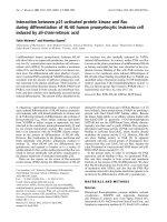
SHP-2 binding to synthetic peptides
In vitro experiments using synthetic peptides revealed
strong binding of SHP-2 to the IL-4R
α in the region of the
amino acids 445–558. However, reduced binding was
seen in the presence of the R551 variant. No binding was
seen with the control peptides (Fig. 1 and data not shown).
SHP-1/2
Investigating cytoplasmatic extracts after IL-4 or IL-13 stim-
ulation, SHP-1 phosphorylation was generally reduced in
individuals bearing the variant R551 compared to wildtype
Q551 individuals. Furthermore, in wildtype individuals
SHP-1 phosphorylation was markedly increased after stim-
ulation with IL-4. IL-13 also induced SHP-1 phosphoryla-
tion to a slightly lesser extent (Fig. 2A).
SHP-2 phosphorylation was generally reduced in individu-
als bearing the variant R551 compared to the wildtype
Q551, where SHP-2 phosphorylation was markedly in-
creased after stimulation with IL-13. IL-4 also induced
SHP-2 phosphorylation in the variant (Fig. 2B).
PI3-Kinase, Jak1, Tyk2 and IRS-2
In cytoplasmic extracts, phosphorylation of the p85α subu-
nit of PI3-kinase was markedly increased after stimulation
with IL-13 in both groups of probands. After IL-4 stimula-
tion, enhanced phosphorylation was seen in the presence
of the R551 variant, but not in wildtype Q551 individuals
and unstimulated controls (Fig. 3A).
In the case of Jak1, elevated phosphorylation was seen in
individuals bearing the variant R551 after stimulating with
IL-13, compared to the wildtype Q551 (Fig. 3B).
Tyk2 phosphorylation appeared to be down-regulated after
stimulation with IL-13 in wildtype cells, whereas no effect
was seen for the R551 variant. Indeed, in variant cells both
IL-4 and IL-13 seemed to activate Tyk 2 to a great extent
(Fig. 3C). The same situation described for Tyk2 – that is,
Figure 1
In vitro binding assay using synthetic IL-4R
α peptides (amino acids
545–558 = Q551/R551; control amino acids = 726–784). To assess
the binding of cellular proteins to the peptides, 20
µl of peptide-conju-
gated Affigel beads were incubated with lysates from IL-13-activated
cells (3
× 10
7
cells). The peptide-associated SHP-2 were analyzed by
immunoblotting with specific antibodies (monoclonal anti SHP-2;
Transduction Laboratories) and developed using a chemiluminescence
kit (ECL; Amersham, Germany). Cells were (1): wildtype Q551; (2): var-
iant R551; (3): control. SHP =SH2 containing phosphatase.
6+3
ZW
4
YDU
5
FRQWU
SHSWLGHV
Figure 2
Phosphorylation of SHP-1 and SHP-2. PBMCs (1–2
× 10
7
) from differ-
ent probands (wildtype Q551 or variant R551) were stimulated with IL-
4 or IL-13 for 10 min. Cells were suspended in lysis buffer [10 mM Tris
(pH 7.8), 5 mM EDTA, 50 mM NaCl, 30 mM pyrophosphate, 50 mM
sodium fluoride, 20
µM sodium orthovanadate, 1%Triton X-100, 1 mM
phenylmethylsulfonyl fluoride, 5
µg/ml aprotinin, 1 µg/ml pepstatin A
and 10
µg/ml leupeptin; 10
8
cells/ml buffer] and incubated for 60 mins
at 4
°C. Insoluble material was removed by centrifugation and equal
amounts of cell lysates (BioRad protein assay, BioRad Laboratories)
were incubated with 1
µg/ml of the appropriate antibodies, anti-SHP1
or anti-SHP-2, followed by western blotting and immunostaining with
either p-Tyr or the respective control antibody. (A). SHP-1, 3= WT/IL-
13; 2= WT/IL-4; 1= WT unstimulated. (stain 2 min); 6= R551/ IL-13;
5= R551/ IL-4; 4= R551 unstimulated. (stain 5 min); (B). SHP-2, 3=
WT/ IL-13; 2= WT/ IL-4; 1= WT unstimulated; 6= R551/ IL-13; 5=
R551/ IL-4; 4= R551 unstimulated. PBMC = Peripheral blood mononu-
clear cell; SHP =SH2 containing phosphatase; WT = wildtype.
$
,/
,/
QR
:LOGW\SH4
,/
,/
QR
9DULDQW5
DQWL6+3
DQWLS7\U
DQWL6+3
%
,/
,/
QR
:LOGW\SH4
,/
,/
QR
9DULDQW5
DQWLS7\U
Available online />Page 5 of 11
(page number not for citation purposes)
down-regulation after IL13 stimulation in case of the
wildtype situation – applies to IRS-2 (Fig. 3D).
STAT3
In cytoplasmic extracts, STAT3 phosphorylation was mark-
edly increased after stimulation with IL-4 as well as IL-13 in
individuals bearing the R551 variant compared to wildtype
individuals and unstimulated controls (Fig. 3E).
STAT in nuclear extracts
In nuclear extracts from R551 positive individuals, STAT3
was predominately found when stimulating with IL-13 for
10 min, while almost no STAT3 was detectable when stim-
ulating with IL-4. Interestingly, the opposite was seen in
Q551 homozygeous individuals. STAT3 was only found af-
ter stimulation with IL-4 and almost no STAT3 proteins
were detectable after stimulation with IL-13 for 10 min (Fig.
4A). Exactly the same results were achieved when staining
Figure 3
Effector substrates. Phosphorylation of PI3-kinase, Jak-1, Tyk2, IRS-2 and-STAT3. PBMCs (1–2
× 10
7
) from different probands (wildtype Q551 or
variant R551) were stimulated with IL-4 or IL-13 for 10 mins. Cells were suspended in lysis buffer [10 mM Tris (pH 7.8), 5 mM EDTA, 50 mM NaCl,
30 mM pyrophosphate, 50 mM sodium fluoride, 20
µM sodium orthovanadate, 1%Triton X-100, 1 mM phenylmethylsulfonyl fluoride, 5 µg/ml apro-
tinin, 1
µg/ml pepstatin A and 10 µg/ml leupeptin; 10
8
cells/ml buffer] and incubated for 60 mins at 4°C. Insoluble material was removed by centrifu-
gation and equal amounts of cell lysates (BioRad protein assay, BioRad Laboratories) were incubated with 1
µg/ml of the appropriate antibodies.
Immunoprecipitation was performed using anti-PI3-kinase (p85
α), anti-Jak-1, anti-Tyk2, anti IRS-2 or anti-STAT3, followed by western blotting and
immunostaining with either p-Tyr or the respective control antibody. (A). PI3-kinase (p85
α), 6= R551/ IL-4; 5= R551/ IL-13; 4= R551 unstimulated.;
3= WT and IL-13; 2= WT and IL-4; 1= WT unstimulated. (B). Jak-1, 6= R551/ IL-4; 5= R551/ IL-13; 4= R551 unstimulated.; 3= WT/ IL-4; 2= WT/
IL-13; 1= WT unstimulated. (C). Tyk2, 6= R551/ IL-4; 5= R551/ IL-13; 4= R551 unstimulated.; 3= WT/ IL-4; 2= WT/ IL-13; 1= WT unstimulated.
(D). IRS-2, 6= R551/ IL-4; 5= R551/ IL-13; 4= R551 unstimulated.; 3= WT/ IL-4; 2= WT/ IL-13; 1= WT unstimulated. (E). STAT3, 6= R551/ IL-4;
5= R551/ IL-13; 4= R551 unstimulated.; 3= WT/ IL-4; 2= WT/ IL-13; 1= WT unstimulated. JAK =Janus kinase; PBMC = Peripheral blood mononu-
clear cell; SHP =SH2 containing phosphatase; STAT =signal transducer and activator of transcription;WT = wildtype.
$ '
DQWLS7\U
DQWL,56
DQWLS7\U
DQWL3,NLQDVH
&
DQWL7\N
DQWLS7\U
% (
DQWLS7\U
DQWL-DN
,/
,/
QR
:LOGW\SH
4
,/
,/
QR
9DULDQW
5
,/
,/
QR
,/
,/
QR
,/
,/
QR
,/
,/
QR
,/
,/
QR
,/
,/
QR
:LOGW\SH
4
:LOGW\SH
4
:LOGW\SH
4
9DULDQW
5
9DULDQW
5
9DULDQW
5
DQWLS7\U
DQWL67$7
,/
,/
QR
,/
,/
QR
:LOGW\SH
4
9DULDQW
5
Respiratory Research Vol 3 No 1 Kruse et al.
Page 6 of 11
(page number not for citation purposes)
with anti-STAT3 or anti-phosphotyrosine antibodies (re-
sults not shown).
The results were further confirmed by a direct immunofluo-
rescence-staining with anti-STAT3 antibodies in stimulated
and unstimulated B cells (data not shown). The 293 fibrob-
lasts (lacking the common
γ chain) showed the same pat-
tern as wildtype individuals (Fig. 4B). Typing these cells for
the IL-4R
α polymorphisms indeed revealed the Q551 situ-
ation.
As a control STAT6 was analyzed in all nuclear extracts,
and no differences could be detected 10 mins after IL-4 or
IL-13 stimulation.
Different concentrations of IL-4 and IL-13 did not have an
detectable effect on the phosphorylation status in the ex-
periments (data not shown). However, looking at different
time points revealed that the response is delayed in the re-
spective case. That means, for example, in the case of IL-4
stimulation, STAT3 appears after 15–20 min in nuclei of
variant, whereas STAT3 appears immediately in nuclei of
wildtype individuals. In the case of IL-13 stimulation, STAT3
appears immediately in nuclei of variants, whereas its ap-
pearance is delayed in nuclei of wildtype individuals (Fig.
4C, 4D).
Discussion
Our knowledge concerning the importance of IL-4 and IL-
13 in the context of IgE regulation, as well as in the devel-
opment of inflammatory and atopic diseases, increases.
However, it is not yet known if these cytokines play distinct
roles in signal transduction processes. Only mouse models
suggest these unique roles of both cytokines, especially in
the context of asthma.
IL-4 and IL-13 share the IL-13 receptor (IL-4R
α and IL-
13R
α1), which is prominent on human B cells. However, in
this study PBMCs were chosen for investigations, as the
role of IL-13 on other cells (T cells, eosinophils etc.) is not
completely understood. By using a mixture of cells, stimula-
tion with IL-4 would, of course, also lead to activation via
the functional IL-4 receptor (IL-4R
α and the common γ
chain). We chose this model system to reflect the in vivo
and complex medically relevant situation as best as possi-
ble. Therefore, we used freshly isolated PBMCs and cul-
tured them for only 2 days. Impairments due to different cell
and receptor numbers were limited by pooling the cultured
samples before stimulation experiments (for further details
see Materials and methods) and by comparing phosphor-
ylation statuses only in samples within each proband.
Previous studies have shown that polymorphisms in the
gene encoding the IL-4R
α chain exhibit strong influences
on the structure and signal transduction through this recep-
tor chain [12,15,16].
Hershey et al. showed impaired binding of the phosphatase
SHP-1 in the presence of the R551 variant of the IL-4R
α
chain. Possible influences on the transcription factor
STAT6 and elevated CD23 expression were discussed
[12]. However, another study could not repeat these re-
sults [26] and a further study revealed even slightly re-
duced phosphorylation of STAT6, following IL-4 stimulation
of PBMCs derived from individuals bearing the R551 vari-
ant [16]. Moreover, the variant was associated with low-
ered total serum IgE levels [16], in direct contrast to
Hershey's findings. Nevertheless, the impaired SHP-1
binding might well influence intracellular substrates other
than STAT6, which could explain the controversy.
Figure 4
STAT3 and STAT6 in nuclear extracts. PBMC (1–2
× 10
7
) from differ-
ent probands (wildtype Q551 or variant R551) were stimulated with IL-
4 or IL-13. Nuclear extracts were obtained according to the EMSA pro-
tocol. Immunoprecipitation was performed using anti-STAT3, followed
by western blotting and immunostaining with either p-Tyr or ant-STAT3.
Staining with Tyk2 served as a control. In (A) and (B) cells were stimu-
lated for 10 min. In (C) and (D) a time course has been performed. (A).
PBMCs 1= WT unstimulated.; 2= WT/ IL-4, 3= WT/ IL-13; 4= R551
unstimulated.; 5= R551/ IL-4; 6= R551/ IL-13; (B). 293 cells 1=
unstimulated.; 2= IL-4; 3= IL-13; (C). Time-course experiment IL-4. 1=
WT 0 min, 2= WT 1 min, 3= WT 5 min, 4= WT 15 min, 5= WT 20 min,
6= R551 0 min, 7= R551 1 min, 8= R551 5 min, 9= R551 15 min,
10= R551 20 min; (D). Time-course experiment IL-13. 1= WT 0 min,
2= WT 1 min, 3= WT 5 min, 4= WT 15 min, 5= WT 20 min, 6= R551
0 min, 7= R551 1 min, 8= R551 5 min, 9= R551 15 min, 10= R551
20 min. PBMC = Peripheral blood mononuclear cell; WT = wildtype.
$
QR
,/
,/
:LOGW\SH4
QR
,/
,/
9DULDQW5
6WLPXODWLRQIRUPLQ
DQWL 67$7
DQWL 67$7
DQWL7\N
%
6WLPXODWLRQIRUPLQ
FHOOV
QR
,/
,/
DQWL 67$7
DQWL 67$7
DQWL7\N
Available online />Page 7 of 11
(page number not for citation purposes)
In order to study possible the influence of the Q551R vari-
ant on signal transduction processes, and here especially
on the IL-13 receptor, two groups of probands were select-
ed: exclusively homozygous wildtype Q551, or variant
R551 in the intracellular part of the IL-4R
α chain. Influences
from the other intracellular variants such as E375A and
S478P on signal transduction were excluded for clarity.
Their effect will, of course, need to be considered in the fu-
ture, especially because S478P and Q551R are in strong
linkage disequilibrium [16]. Due to the high allelic frequen-
cy of the variant V50, all probands in this study bore the ex-
tracellular variant I50V. The frequency of individuals
showing only the R551 variant and no other polymorphism
in IL-4R
α is less than 1% in the German population (unpub-
lished data).
As the transcription factor STAT3 is an effector substrate
of the IL-13 receptor
α1 chain, our initial interest was to test
whether the variant R551 would influence STAT3 signaling
through an impaired SHP binding and activation capacity
(in accordance with the work of Hershey et al.[12]).
SHP-1/2
STAT3 signaling is controlled by the phosphatase SHP-2
[27]. SHP-2 possesses a structure very similar to SHP-1
[21], which has previously been shown to bind to the Y550
region of IL-4R
α [12]. Therefore, this region was also con-
sidered a potential docking site for SHP-2. By using syn-
thetic peptides it was indeed confirmed that SHP-2 binds
to the Y550 region of IL-4R
α. Furthermore, binding to the
variant R551 (Fig. 1) was impaired as expected [12]. Im-
paired SHP-1 binding with R551 could be repeated [data
not shown].
Interestingly, SHP-1 and SHP-2 seem to be activated dif-
ferentially by IL-4 and IL-13. In wildtype individuals SHP-1
phosphorylation is predominately induced by IL-4, while
SHP-2 phosphorylation is predominately induced by IL-13.
As expected, this phosphorylation was in each case mark-
edly reduced in individuals bearing the variant R551 (Fig.
2A, 2B). The phosphorylation of SHP-2 was also slightly in-
creased in the presence of the variant R551 after stimula-
tion, for example, with IL-4, which was comparable to the
wildtype (Fig. 2B). This observation hints at docking sites
for SHP-2 on the IL-4R
α protein other than Y550, as has
been described earlier [28]. We might even have seen a
multiplied effect due to the presence of the conventional IL-
4 receptor as we worked with cell mixtures (PBMCs, see
above).
In conclusion, depending on the stimulating agent (IL-4 or
IL-13) SHP-1 and SHP-2 evidently regulate different effec-
tor substrates and therefore activate distinct signal trans-
duction cascades.
The two phosphatases have a wide variety of intracellular
substrates [29–32]. We went on to test several substrates
of the IL-13 receptor for phosphorylation (activation status)
after IL-4 or IL-13 treatment.
PI3-kinase, Jak1, Tyk-2 and IRS-2
Stimulation with IL-4 reduced phosphorylation of PI3-ki-
nase compared to stimulation with IL-13 (Fig. 3A). Imani et
al. also found decreased PI3-kinase phosphorylation after
IL-4 treatment and proposed that SHP-1 mediates this ef-
fect [30]. IL-13 does not seem to induce SHP-1 and, as ex-
pected, markedly increased phosphorylation of PI3-kinase
was seen in both wildtype and variant cells (Fig. 3A). Pos-
sibly due to the impaired SHP-1 activation, IL-4 stimulation
slightly increased PI3-kinase phosphorylation in the variant
compared to the wildtype situation.
Jak1 phosphorylation was markedly reduced in wildtype in-
dividuals after IL-13 as compared with IL-4 stimulation (Fig.
3B). This would imply that SHP-2 is responsible for this ef-
fect, as it is specifically activated by IL-13 (stated above).
Jak1 has indeed been shown to interact with SHP-2 [33].
Impaired binding of SHP-2 in the case of the variant R551
consequently leads to increased phosphorylation of Jak1
(Fig. 3B). As this increase in phosphorylation is also higher
than after stimulation with IL-4 in both groups of probands,
one can even speculate that Jak1 is specifically induced by
IL-13.
As for Jak1, the phosphorylations of Tyk2 and IRS-2 were
reduced after IL-13 stimulation in wildtype cells (Fig. 3C,
3D). SHP-2 seems to be responsible for these effects as
well. Though it was suggested that IRS-2 does not associ-
ate with SHP-2 after IL-4 treatment [34], which confirms
the results with IL-4 (no effect, see Fig. 1D), it obviously
does so after IL-13 stimulation. Not much is known yet
about Tyk2, but these results suggest that it is regulated by
SHP-2 after IL-13 stimulation as well. In contrast to Jak1,
no increase in phosphorylation is seen after IL-13 treatment
for Tyk2 and IRS-2 in the presence of the variant. So these
two substrates do not seem to be particularly activated by
IL-13.
In most experiments a rather high level of substrate phos-
phorylation has been seen in the controls, i.e. without addi-
tional stimulation with exogeneously administered IL-4 or
IL-13. Two effects might underlie this observation. First,
certain amounts of IL-4 and IL-13 might be produced by the
cell culture itself, although the incubation has been rather
short at 10 min. Second, the signal might represent a lower
dephosphorylation due to missing SHP-1 and SHP-2 acti-
vation in concordance with our hypothesis.
Respiratory Research Vol 3 No 1 Kruse et al.
Page 8 of 11
(page number not for citation purposes)
STAT3
Figure 5
Hypothetical model of nuclear translocation of STAT3. (A) When stimulating cells from wildtypes (Q551) with IL-4, a potential inhibitor does not bind
to STAT3 and nuclear translocation takes place. (B) When stimulating wildtypes (Q551) with IL-13, a potential inhibitor is able to interact with
STAT3 and prevents it from being translocated. (C) When stimulating variants R551 with IL-4, the inhibitor is able to interact and prevent the STAT3
translocation. (D) When stimulating the variants with IL-13. STAT3 is translocated STAT = signal transducer and activator of transcription.
,/5a
,/5D
,/
67$7
ZLOGW\SH4
,/5D
4
67$7
,/5a
,/
$7*
QXFOHXV
$7*
QXFOHXV
,QKLELWRU
,QKLELWRU
$
%
4
YDULDQW5
,/
,/5D
5
67$7
,/5a
$7*
QXFOHXV
,QKLELWRU
,/5a
,/5D
5
67$7
$7*
QXFOHXV
,/
,QKLELWRU
& '
6+3 6+3
6+36+3
Available online />Page 9 of 11
(page number not for citation purposes)
As stated above, STAT3 phosphorylation was believed to
be controlled by SHP-2 [27] until recently, when Tenev et
al. reported a control by SHP-1 [35].
The results from this study suggest that STAT3 is regulated
by both phosphatases, SHP-1/2. We therefore only see a
dramatic increase in phosphorylation of STAT3 in the pres-
ence of the R551 variant, after both IL-4 and IL-13 stimula-
tion, due to the impaired binding of SHP-1 or SHP-2 (Fig.
3E). No effect at all was seen in the wildtype situation
where normal regulation of STAT3 by SHP-1/2 takes place.
Interestingly, the signaling of STAT3 seems to be further
regulated on a different level. As a transcription factor it can
act only when present in a dimeric form in the nucleus. Re-
cently, it was reported that although STAT3 was activated
by IL-4 its nuclear translocation was impaired in several ke-
ratinocytic cell lines [36]. We therefore sought similar phe-
nomena. We immediately found STAT3 only in nuclear
extract of wildtype cells after IL-4 treatment, whereas in
R551 variant nuclear extracts it was present at once pre-
dominately after IL-13 stimulation (Fig. 4A), although its
phosphorylation, that means activation in the cytoplasm,
was markedly increased after treatment with both cytokines
(stated above). These results are very specific for the IL-13
receptor. 293 fibroblasts lack the common
γ chain and be-
have the same way as PBMCs (Fig. 4B).
Performing time-course experiments revealed, however,
that we are not faced with a "black and white" situation. As
expected STAT3 molecules appeared in a delayed fashion
(after 20 min) in the nucleus in the respective cases men-
tioned above (Fig. 4C, 4D). We suggest that specific inhib-
itors of activated STAT3 are responsible for the observed
effects, and that they might interact with STAT3 and con-
sequently prevent or delay it from translocating to the nu-
cleus. A variety of inhibitors of the IL-4/IL-13 pathway have
been reported, such as SOCS-1 (also known as SSI-1)
[21]; however, this inhibitor was suggested to prevent acti-
vation of STAT and, for example, act on Jak proteins [37].
So, SOCS-1 does not seem to be the right candidate in
this case, because STAT3 activation was not found to be
abolished. A better candidate seems to be the inhibitor
PIAS3, which was previously shown to specifically bind to
activated STAT3 proteins [23]. In addition, it is possible to
imagine splice variants of STAT3 acting as anti-substrates;
this has previously been suggested (e.g. for STAT6 [21]).
For conformational reasons the inhibitor might not be able
to bind to STAT3 in case of the wildtype situation and IL-4
stimulation, so that STAT3 can be translocated to the nu-
cleus. This is suggested in a hypothetical model (Fig. 5A).
In the case of IL-13 stimulation, the conformational status
of the receptor might allow the inhibitor to interact directly
with STAT3 and prevent the molecule from being translo-
cated (Fig. 5B). In the case of the R551 variant the confor-
mation of IL-4R
α would be altered, so that the situation is
reversed. The inhibitor interacts with STAT3 in case of IL-4
stimulation (Fig. 5C), however, after IL-13 stimulation this
interaction is abolished and consequently STAT3 is trans-
located to the nucleus (Fig. 5D). It might also be that SHP-
1 and SHP-2 are differentially recruited to the receptor de-
pending on the wildtype or variant situation and IL-4 or IL-
13 stimulation.
On the whole, these results help to understand the complex
picture of signal transduction processes in the IL-4/IL-13
pathway. These findings thus provide the first evidence for
distinct roles of IL-4 and IL-13 while acting through the
same IL-13 receptor. They support the idea of IL-13 being
responsible for developing the asthma phenotype by induc-
ing separate intracellular signaling processes independent-
ly of IL-4. If this assumption is correct, variants in the IL-13
protein itself might also be able to add to these specific ef-
fects. Very recently, the R110Q variant in IL-13 was found
to be highly associated with the asthma phenotype, atopic
dermatitis and elevated total serum IgE levels in three dif-
ferent populations [38–40]. The variant Gln110 is thought
to provide a higher binding affinity to the IL-13 receptor and
might therefore influence the IL-13 signaling processes in
a specific way.
Other factors have, of course, to be considered. The distri-
bution of receptors (IL-4R and IL-13R) varies in mononu-
clear cells and bronchial tissues [38], and there are further
regulation mechanisms through which, for example, the
phosphatase SHIP acts on the products of PI3-kinase [41],
specific Jak inhibitors [JAB; [37]], soluble IL-4R
α [42] and
many others. Also, IL-4 might act differently through the IL-
4 receptor than through the IL-13 receptor. Furthermore,
more than one docking site for SHP proteins on the IL-4R
α
chain (stated above) is responsible for activation, and, very
importantly, other functionally relevant polymorphisms exist
in the IL-4R
α gene and in the genes of members of the IL-
4/IL-13 pathway, apart from the polymorphism encoding
the R551 variant, via linkage disequilibrium [43,44]. Further
studies will be necessary to understand the complexity of
the IL-4/IL-13 signaling pathway and also to deduce its ex-
act implications for the development of asthma and atopy.
Conclusion
Using whole-cell in vivo experiments, we present evidence
that IL-4 and IL-13 act through distinct signaling processes
by predominately inducing either the phosphatase SHP-1
following binding of IL-4, or SHP-2 following binding of IL-
13. Moreover, nuclear translocation of STAT proteins
seems to differ following IL-4 versus IL-13 binding. Al-
though some of the effects seen might be due to only IL-4
acting on part of the cells, and IL-4 plus IL-13 on others,
this would still not explain some of the observations regard-
Respiratory Research Vol 3 No 1 Kruse et al.
Page 10 of 11
(page number not for citation purposes)
ing the unique effects of IL-13 on activated STAT3. The
functions of IL-4 and IL-13 are further influenced by some
of the IL-4R
α variants. These findings may also have impli-
cations for the development of asthma or atopy.
Abbreviation
IL-4 = interleukin 4; IL-4Rα = IL-4 receptor alpha chain; IL-
13 = interleukin 13; IL-13R
α1 = IL-13 receptor alpha
chain; IRS = insulin receptor-like substrate; JAK = Janus ki-
nase; PBMC = Peripheral blood mononuclear cell; SHP =
SH2 containing phosphatase; SH2 = src-homology 2;
STAT = signal transducer and activator of transcription.
Acknowledgement
This project is supported by a grant from the German Science Founda-
tion (DFG-De386/2-3).
References
1. Howard M, Farrar J, Hilfiker M, Johnson B, Takatsu K, Hamaoka T,
Paul WE: Identification of a T cell-derived
β cell growth factor
distinct from interleukin 2. J Exp Med 1982, 155:914-923
2. Coffman RL, Lebman DA, Rothman P: Mechanism and regula-
tion of immunoglobulin isotype switching. Adv Immunol 1993,
54:229-270
3. Hu-Li J, Shevach EM, Mizuguchi J, Ohara J, Mosmann T, Paul WE:
B cell stimulatory factor-1 (interleukin 4) is a potent costimu-
lant for normal resting T lymphocytes. J Exp Med 1987,
165:157-172
4. Brombacher F: The role of interleukin-13 in infectious diseases
and allergy. Bioessays 2000, 22:646-656
5. Wills-Karp M, Luyimbazi J, Xu X, Schofield B, Neben TY, Karp CL,
Donaldson DD: Interleukin-13: central mediator of allergic
asthma. Science 1998, 282:2258-2261
6. Grunig G, Warnock M, Wakil AE, Venkayya R, Brombacher F, Ren-
nick DM, Sheppard D, Mohrs M, Donaldson DD, Locksley RM,
Corry DB: Requirement for IL-13 independently of IL-4 in ex-
perimental asthma. Science 1998, 282:2261-2263
7. McKenzie GJ, Bancroft A, Grencis RK, McKenzie AN: A distinct
role for interleukin-13 in Th2-cell-mediated immune respons-
es. Curr Biol 1998, 8:339-342
8. Russell SM, Keegan AD, Harada N, Nakamura Y, Noguchi M, Le-
land P, Friedmann MC, Miyajima A, Puri RK, Paul WE, Leonard WJ:
The interleukin-2 receptor
γ chain is a functional component of
the interleukin-4 receptor. Science 1993, 262:1880-1883
9. Cosman D: The hematopoietin receptor superfamily. Cytokine
1993, 5:95-106
10. Deichmann KA, Bardutzky J, Forster J, Heinzmann A, Kuehr J:
Common polymorphisms in the coding part of the IL-4-recep-
tor gene. Biochem Biophys Res Comm 1997, 231:696-697
11. Deichmann KA, Heinzmann A, Forster J, Dischinger S, Mehl C,
Brueggenolte E, Hildebrandt F, Moseler M, Kuehr J: Linkage and
allelic association of atopy and markers flanking the IL-4-re-
ceptor gene. Clin Exp All 1998, 28:151-155
12. Hershey GKK, Friedrich MF, Esswein LA, Thomas ML, Chatila TA:
The association of atopy with a gain-of-function mutation in
the
α subunit of the interleukin-4 receptor. N Engl J Med 1997,
337:1720-1725
13. Kanemitsu S, Takabayashi A, Sasaki Y, Kuromaru R, Ihara K, Kaku
Y, Sakai K, Hara T: Association of interleukin-4 receptor and in-
terleukin-4 promoter gene polymorphisms with systemic lu-
pus erythematosus. Arthritis Rheum 1999, 42:1298-1300
14. Hackstein H, Kluter H, Fricke L, Hoyer J, Bein G: The IL-4 receptor
alpha-chain variant Q576R is strongly associated with de-
creased kidney allograft survival. Tissue Antigens 1999,
54:471-477
15. Mitsuyasu H, Izuhara K, Mao XQ, Gao PS, Arinobu Y, Enomoto T,
Kawai M, Sasaki S, Dake Y, Hamasaki N, Sirakawa T, Hopkin JM:
Ile50Val variant of IL-4R alpha upregulates IgE synthesis and
associates with atopic asthma. Nat Genet 1998, 19:119-120
16. Kruse S, Japha T, Tedner M, Hauschildt Sparholt S, Forster J, Kue-
hr J, Deichmann KA: The polymorphisms S503P and Q576R in
the interleukin-4 receptor
α gene are associated with atopy
and influence the signal transduction. Immunology 1999,
96:365-371
17. Gessner A, Roellinghoff M: Biologic functions and signaling of
the interleukin-4 receptor complexes. Immunobiology 2000,
201:285-307
18. Orchansky PL, Kwan R, Lee F, Schrader JW: Characterization of
the cytoplasmic domain of interleukin-13 receptor-alpha. J
Biol Chem 1999, 274:20818-20825
19. Takeda T, Kurachi H, Yamamoto T, Nishio Y, Nakatsuji Y, Morishige
K, Miyake A, Murata Y: Crosstalk between the interleukin-6 (IL-
6)-JAK-STAT and the glucocorticoid-nuclear receptor path-
way: synergistic activation of IL-6 response element by IL-6
and glucocorticoid. J Endocrinol 1998, 159:323-330
20. O'Brien CA, Manolagas SC: Isolation and characterization of
the human gp130 promoter. Regulation by STATs. J Biol Chem
1997, 272:15003-15010
21. Nelms K, Keegan AD, Zamorano J, Ryan JJ, Paul WE: The IL-4 re-
ceptor: signaling mechanisms and biologic functions. Annu
Rev Immunol 1999, 17:701-738
22. Naka T, Narazaki M, Hirata M, Matsumoto T, Minamoto S, Aono A,
Nishimoto N, Kajita T, Taga T, Yoshizaki K, Akira S, Kishimoto T:
Structure and function of a new STAT-induced STAT inhibitor.
Nature 1997, 387:924-929
23. Chung CD, Liao J, Liu B, Rao X, Jay P, Berta P, Shuai K: Specific
inhibition of Stat3 signal transduction by PIAS3. ience 1997,
278:Sc1803-1805
24. Wjst M, Kruse S, Illig T, Deichmann KA: Asthma and IL-4 recep-
tor alpha gene variants. Eur J Immunogenet 2002, 29:263-268
25. Ausubel FM, Brent R, Kingston RE, Moore DD, Seidman JG, Smith
JA, Struhl K: Preparation of nuclear extracts. In Current Proto-
cols in Molecular Biology.
26. Edited by John Wiley & Sons Inc. USA, 1997, 11-1212
27. Wang HY, Shelburne CP, Zamorano J, Kelly AE, Ryan JJ, Keegan
AD: Cutting edge: effects of an allergy-associated mutation in
the human IL-4R alpha (Q576R) on human IL-4-induced signal
transduction. J Immunol 1999, 162:4385-4389
28. Servidei T, Aoki Y, Lewis SE, Symes A, Fink JS, Reeves SA: Coor-
dinate regulation of STAT signaling and c-fos expression by
the tyrosine phosphatase SHP-2. J Biol Chem 1998, 273:6233-
6241
29. Jiang H, Harris MB, Rothman P: IL-4/IL-13 signaling beyond
JAK/STAT. J All Clin Immunol 2000, 105:1063-1070
30. Haque SJ, Harbor P, Tabrizi M, Yi T, Williams BR: Protein-tyrosine
phosphatase SHP-1 is a negative regulator of IL-4- and IL-13-
dependent signal transduction. J Biol Chem 1998, 273:33893-
33896
31. Imani F, Rager KJ, Catipovic B, Marsh DG: Interleukin-4 (IL-4) in-
duces phosphatidylinositol 3-kinase (p85) dephosphorylation.
Implications for the role of SHP-1 in the IL-4-induced signals
in human B cells. J Biol Chem 1997, 272:7927-7931
32. Kuhne MR, Pawson T, Lienhard GE, Feng GS: The insulin recep-
tor substrate1 associates with the SH2-containing phosphoty-
rosine phosphatase Syp. J Biol Chem 1993, 268:11479-11481
33. Gadina M, Stancato LM, Bacon CM, Larner AC, O'Shea JJ: In-
volvement of SHP-2 in multiple aspects of IL-2 signaling: evi-
dence for a positive regulatory role. J Immunol 1998, 160:4657-
4661
34. Yin T, Shen R, Feng GS, Yang YC: Molecular characterization of
specific interactions between SHP-2 phosphatase and JAK ty-
rosine kinases. J Biol Chem 1997, 272:1032-1037
35. Sun XJ, Pons S, Wang LM, Zhang Y, Yenush L, Burks D, Myers
MGJr, Glasheen E, Copeland NG, Jenkins NA, Pierce JH, White
MF: The IRS-2 gene on murine chromosome 8 encodes a
unique signaling adapter for insulin and cytokine action. Mol
Endocrinol 1997, 11:251-262
36. Tenev T, Bohmer SA, Kaufmann R, Frese S, Bittorf T, Beckers T,
Bohmer FD: Perinuclear localization of the protein-tyrosine
phosphatase SHP-1 and inhibition of epidermal growth factor-
stimulated STAT1/3 activation in A431 cells. Eur J Cell Biol
2000, 79:261-271
37. Wery-Zennaro S, Letourneur M, David M, Bertoglio J, Pierre J:
Binding of IL-4 to the IL-13Ralpha(1)/IL-4Ralpha receptor
complex leads to STAT3 phosphorylation but not to its nuclear
translocation. FEBS Lett 1999, 464:91-96
38. Endo TA, Masuhara M, Yokouchi M, Suzuki R, Sakamoto H, Mitsui
K, Matsumoto A, Tanimura S, Ohtsubo M, Misawa H, Miyazaki T,
Available online />Page 11 of 11
(page number not for citation purposes)
Leonor N, Taniguchi T, Fujita T, Kanakura Y, Komiya S, Yoshimura
A: A new protein containing an SH2 domain that inhibits JAK
kinases. Nature 1997, 387:921-924
39. Heinzmann A, Mao XQ, Akaiwa M, Kroemer RT, Gao PS, Ohshima
K, Umeshita R, Abe Y, Braun S, Yamashita T, Roberts MH, Sugim-
oto R, Arima K, Arinobu Y, Yu B, Kruse S, Enomoto T, Dake Y, Ka-
wai M, Shimazu S, Sasaki S, Adra CN, Kitaichi M, Inoue H,
Yamauchi K, Tomichi N, Kurimoto F, Hamasaki N, Hopkin JM, Izu-
hara K, Shirakawa T, Deichmann KA: Genetic variants of IL-13
signaling and human asthma and atopy. Hum Mol Genet 2000,
9:549-959
40. Graves PE, Kabesch M, Halonen M, Holberg CJ, Baldini M,
Fritzsch C, Weiland SK, Erickson RP, von Mutius E, Martinez FD:
A cluster of seven tightly linked polymorphisms in the IL-13
gene is associated with total serum IgE levels in three popu-
lations of white children. J Allergy Clin Immunol 2000, 105:506-
513
41. Liu X, Nickel R, Beyer K, Wahn U, Ehrlich E, Freidhoff LR, Bjorksten
B, Beaty TH, Huang SK: An IL13 coding region variant is asso-
ciated with a high total serum IgE level and atopic dermatitis
in the German multicenter atopy study (MAS-90). J All Clin Im-
munol 2000, 106:167-170
42. Giallourakis C, Kashiwada M, Pan PY, Danial N, Jiang H, Cambier
J, Coggeshall KM, Rothman P: Positive Regulation of IL-4 Medi-
ated Proliferation by the SH2-Containing Inositol 5'-Phos-
phatase (SHIP). J Biol Chem 2000, 275:29275-29282
43. Kruse S, Forster J, Kuehr J, Deichmann KA: Characterization of
the membrane-bound and a soluble form of human IL-4 recep-
tor alpha produced by alternative splicing. Int Immunol 1999,
11:1965-1970
44. Shirakawa T, Deichmann KA, Izuhara K, Mao XQ, Adra CN, Hopkin
JM: Atopy and asthma: genetic variants of IL-4 and IL-13 sign-
aling. Immunol Today 2000, 21:60-64
45. Ober C, Leavitt SA, Tsalenko A, Howard TD, Hoki DM, Daniel R,
Newman DL, Wu X, Parry R, Lester LA, Solway J, Blumenthal M,
King RA, Xu J, Meyers DA, Bleecker ER, Cox NJ: Variation in the
interleukin 4-receptor alpha gene confers susceptibility to
asthma and atopy in ethnically diverse populations. Am J Hum
Genet 2000, 66:517-526