Báo cáo y học: "The molecular basis of lung cancer: molecular abnormalities and therapeutic implications" potx
Bạn đang xem bản rút gọn của tài liệu. Xem và tải ngay bản đầy đủ của tài liệu tại đây (705.43 KB, 15 trang )
BioMed Central
Page 1 of 15
(page number not for citation purposes)
Respiratory Research
Open Access
Review
The molecular basis of lung cancer: molecular abnormalities and
therapeutic implications
Pierre P Massion* and David P Carbone
Address: Vanderbilt-Ingram Comprehensive Cancer Center, Vanderbilt University Medical Center, Nashville Tennessee, U.S.A. 37232-6838
Email: Pierre P Massion* - ; David P Carbone -
* Corresponding author
microarraybiomarkermolecular
Abstract
Lung cancer is the number one cause of cancer-related death in the western world. Its incidence
is highly correlated with cigarette smoking, and about 10% of long-term smokers will eventually be
diagnosed with lung cancer, underscoring the need for strengthened anti-tobacco policies. Among
the 10% of patients who develop lung cancer without a smoking history, the environmental or
inherited causes of lung cancer are usually unclear. There is no validated screening method for lung
cancer even in high-risk populations and the overall five-year survival has not changed significantly
in the last 20 years. However, major progress has been made in the understanding of the disease
and we are beginning to see this knowledge translated into the clinic.
In this review, we will summarize the current state of knowledge regarding the cascade of events
associated with lung cancer development. From subclinical DNA damage to overt invasive disease,
the mechanisms leading to clinically and molecularly heterogeneous tumors are being unraveled.
These lesions allow cells to escape the normal regulation of cell division, apoptosis and invasion.
While all subtypes of non-small cell lung cancer have historically been treated the same, stage-for-
stage, recent technological advances have allowed a better understanding of the molecular
classification of the disease and provide hypotheses for molecular early detection and targeted
therapeutic strategies.
Introduction
The pathogenesis of lung cancer involves the accumula-
tion of multiple molecular abnormalities over a long
period of time [1,2]. Genomic instability is universally
found during accumulation of these hits [3]. The altera-
tions can happen at the level of gene silencing through
methylation, DNA sequence changes, DNA segment
amplification or deletion or whole chromosome gains or
losses. These changes occur early in normal-appearing tis-
sues that do not have the characteristics of cancer cells.
Microdissection of lesions of the bronchial epithelium as
well as of invasive tumors has provided purified tissue for
the analysis of point mutations [4], chromosomal dele-
tions [5], microsatellite instability [6,7] and DNA methyl-
ation patterns [8].
The most common early genetic alterations in non-small
cell lung cancer involve loss of genomic regions of chro-
mosomes 3p and 9p, deletions of chromosomal arm on
5p and mutations of p53 and K-ras [9]. Loss of
Published: 07 October 2003
Respiratory Research 2003, 4:12
Received: 17 July 2003
Accepted: 07 October 2003
This article is available from: />© 2003 Massion and Carbone; licensee BioMed Central Ltd. This is an Open Access article: verbatim copying and redistribution of this article are permit-
ted in all media for any purpose, provided this notice is preserved along with the article's original URL.
Respiratory Research 2003, 4 />Page 2 of 15
(page number not for citation purposes)
chromosomal regions on chromosomes 3p and 9p have
been recognized as early events [10] and identified in pre-
invasive lesions and in the normal appearing epithelium
of smokers [11,12]. In contrast, p53 and K-ras mutations
have been seen primarily in later stages of preneoplasia or
frank invasive lesions [9]. Amplification of large regions
on the q arm of chromosome 3 has been characterized in
invasive carcinomas [13] only recently in preinvasive
lesions [14].
The historical focus of much of this research has been to
identify and study the role of specific genetic abnormali-
ties in tumor cells related to chromosomal abnormalities,
inactivation of specific tumor suppressor genes, the activa-
tion of specific oncogenes, the expression of hormone
receptors and growth factor production associated with
the development of cancer. More recently, the contribu-
tion of stromal interactions, angiogenesis, apoptosis, and
epigenetic phenomena such as posttranslational modifi-
cation of critical genes has been the subject of intense
research. The recent completion of the first draft of the
human genome sequence [15] and the availability of high
throughput technologies (e.g. microarrays) have
prompted investigators to propose studies to discover
common genetic abnormalities in both pre- and invasive
lung cancers and to test these markers for their potential
use in early detection strategies. In this paper we will
review the genetic basis of lung cancer progression using a
stepwise approach from point mutation to invasion and
address its therapeutic implications.
Early events in oncogenesis
Mutations
In the last 20 years somatic mutations have been identi-
fied and associated with the development of cancer. These
mutations, involving tumor suppressor genes or onco-
genes, may or may not be rate-limiting events. Epidemio-
logical data support that groups of cells accumulate
several key mutations [16]. The model of the mutator phe-
notype proposed by Loeb suggests that cells develop a pre-
disposition for mutations early on [3]. This phenotype
may be hereditary, yet the key genes remain to be discov-
ered. In the lung, DNA damage can fail to be repaired,
resulting in misincorporated nucleotides and therefore
mutations. Spontaneous errors of replication attributed to
DNA polymerase occur at a rate of 1/10,000 to 1/100,000
base pairs depending on the polymerase. These intrinsic
mutations may be an important component underlying
genomic instability and eventually tumor growth. We will
illustrate this point by commenting on 3 classical exam-
ples: k-ras, p53 and p16.
K-ras mutations are most commonly seen in 30% of ade-
nocarcinomas of the lung [17] but much less frequently in
other subtypes. K-ras, once mutated (most frequently
codon 12 G-T transversions), can transform airway epithe-
lial cells [18,19] by activating the ERK-MAP kinase path-
way. Because K-ras mutation is found early in alveolar
atypical hyperplasia, a presumed precursor lesion to ade-
nocarcinomas [20], this may be an important step in the
genesis of this subtype of lung cancer. Mutant ras trans-
genic mice develop adenocarcinomas of the lung as well,
supporting this hypothesis.
p53 is a prototype tumor suppressor gene that is the most
common genetic lesion in human cancers [21] and is thus
well suited for analysis of the mutational spectrum in
human cancers. p53 mutations are most commonly seen
in squamous carcinoma and small-cell carcinoma of the
lung. Mutations predominantly represent G to T transver-
sions consistent with causation by bulky DNA adducts
such as the polycyclic hydrocarbons frequently found in
the lungs of smokers [22]. The p53 tumor suppressor gene
is mutated in over two thirds of lung cancers [23]. When
mutated, p53 can function as an oncogene and accumu-
late in the cytoplasm [24]. Mutated p53 exhibits a pro-
longed half-life and can thus be found to be overexpressed
in about 50% of lung cancers by immunohistochemistry
[25]. Although not consistently associated with prognos-
tic significance, there is little doubt that p53 mutations
play a key role in tumor development by dysregulation of
cell-cycle control and apoptosis.
p16, a tumor suppressor gene and critical member of the
Rb pathway, is inactivated in over 40% of NSCLCs. Previ-
ous studies have demonstrated that point mutations, loss
of heterozygosity on 9p21, or hypermethylation of the
gene provide alternate mechanisms of inactivation in 30–
50% of NSCLCs [26]. Tumors arising in smokers are
found to more frequently harbor point mutations or
homozygous deletions as the mechanism of loss of p16
function [27]. The relationship between tobacco and the
loss of p16 points to new mechanisms involving smoking
in the pathogenesis of lung cancer.
Mutagens
Cigarette smoking is a major risk factor for 85% of lung
cancers. Approximately one in ten life-smokers will
develop lung cancer, suggesting individual differences in
susceptibility [28]. The susceptibility to lung cancer is
being approached by molecular epidemiology and identi-
fying links between genes involved in DNA repair, poly-
morphisms in the cytochrome p450 enzymes and the
metabolizing capability of glutathione s-transferase or
acetylation [29,30]
The majority of lung cancers are diagnosed among ex-
smokers [31]. This suggests that the accumulation of
molecular damage during cigarette exposure has set a cas-
cade of events in motion that leads to the diagnosis of
Respiratory Research 2003, 4 />Page 3 of 15
(page number not for citation purposes)
cancer often decades after smoking cessation. Risk factors
for lung cancer from smoking (first publicly recognized in
the 1964 Report of the U.S. Surgeon General), include
total consumption, age at initiation, and years of smok-
ing. Other risk factors include occupational and environ-
mental exposure (asbestos, uranium, radiation), diet
(vitamin A, vitamin E, cholesterol), and host (familial
aggregation) and genetic factors. Some of the components
of cigarette smoke implicated in lung cancer are now rec-
ognized. Cigarette smoking is a complex mixture and
includes substances that are responsible for DNA adduct
formation such as polycyclic aromatic hydrocarbons
(PAH), aromatic amines, and tobacco-specific nitro-
samines (NKK). These form DNA adducts that may escape
normal adduct repair mechanisms and result in heritable
alterations in DNA sequence. The resulting conversion of
G-C base pairs to T-A leads to activation of the K-ras onco-
gene and inactivation of the p53 tumor suppressor gene
[32]. The activated form of benzopyrene (BaP) is BPDE
and can cause DNA adducts, and, in addition to point
mutations, can also lead to single strand chromatid breaks
that are more frequent in lung cancers [33]. One of the
concerning facts in this process is that people who start
smoking at young ages seem to be have greater amounts
of permanent DNA alterations than smokers who start
smoking at an older age [34].
Chromosomal changes
Cancer cells are characterized not only by mutations but
also by a series of chromosomal aberrations including
deletions and amplifications [35]. The chromosomal
regions with frequent losses are found in regions coding
for essential tumor suppressor genes and DNA repair
genes that may be involved in the pathogenesis of several
tumor types [36]. Large areas of deletions (e.g. chromo-
some 3p, 9p) or amplifications (e.g. 1q, 3q) are com-
monly seen across the genome of lung cancer. Higher rates
of chromosomal changes as determined by loss of hetero-
zygosity (LOH) and CGH have been found in SqCa than
in adenocarcinoma of the lung [37,38].
The most common alterations involve loss of regions of
chromosomes 3p21 and 9p21, deletions of chromosomal
arm on 5q21 and mutations of p53 associated with LOH
on 17p and K-ras point mutations [9]. Interestingly, loss
of chromosomal regions on chromosomes 3p and 9p
have been recognized as early events [10] and identified in
preinvasive lesions and in the normal appearing epithe-
lium of smokers [11,12]. In contrast, p53 and K-ras muta-
tions have been seen in a high percentage of later stages
progression and in early invasive lesions [9].
LOH at chromosome 3p14 was evaluated in smokers and
ex-smokers and found to be more frequent in current
smokers (22/25 cases) than in former smokers (5/11
cases), a high frequency that correlated with a high meta-
plasia index [12]. This implies that not only are these
chromosomal changes frequent in normal appearing
bronchial epithelia but that cells with these changes may
regress after smoking cessation and be replaced by cells
without this damage. The dynamics of this process is very
poorly understood at this time and represents an interest-
ing area of future research.
Lung cancer allelotypes have been investigated in detail
and have recently identified new regions of allelic loss
using high throughput technologies [39]. Interestingly,
differences between smokers and non-smokers have
shown LOH on chr. 9 and 17 targets for p16 and p53,
respectively [27]. LOH and chromosomal gain is less prev-
alent at all sites in cancer from non-smokers [27].
Patterns of chromosomal copy number abnormalities in
squamous carcinomas of the lung using CGH analysis
have been published recently [40–42] and show particu-
larly common amplified regions on chromosomal arms
1q, 3q, 5p, 8q, 11q, 12p, 17q and 20q. Among many areas
of genomic abnormality, amplification of chromosomal
region 3q26 was found to be the most prevalent abnor-
mality in squamous carcinoma of the lung followed by a
deletion of chromosome 3p. Limitations of chromosome-
based CGH include its relatively poor genomic resolution
(~10–20 MB) [43,44], lack of sensitivity for detection of
aberrations involving megabase sized regions, inability to
provide quantitative information about the magnitudes
of genome copy number and the insensitivity of CGH to
detect aberrations such as translocations that do not alter
copy number. Most of these limitations can be overcome
by viewing the chromosomes as the framework onto
which information is mapped with high-resolution arrays
of cloned probes.
Accumulation of specific chromosomal abnormalities has
been correlated with clinical and pathological data in
NSCLC. Chromosomal abnormalities have been recently
correlated with clinical outcome for a variety of cancers
[45–47], but often the genes responsible for the observed
biology are unknown or only partly known. As mentioned
above, 3q amplification is a common finding among
many squamous carcinomas of non-lung origin. In partic-
ular, amplification of that region is seen in squamous car-
cinoma of the head and neck [48], esophageal cancers
[49], and cervical cancers including cervical dysplasia
[50]. In our recent study in NSCLC, among many ampli-
fied genes found in chromosome 3q26 (Figure 1), some
are candidate oncogenes (phosphatidylinositol-3 kinase
catalytic subunit, PIK3CA) or are described to be involved
in tumor progression including the somatostatin gene
(SST), p63 (p53 homolog gene), telomerase RNA compo-
nent gene (hTER), and neutral endopeptidase (NEP) [13].
Respiratory Research 2003, 4 />Page 4 of 15
(page number not for citation purposes)
Human cytogenetic methods such as fluorescence in situ
hybridization (FISH) are particularly useful in analyses of
genomic organization, and copy number in individual
cells and are applicable to tissue microarrays. Rather than
look at the individual impact of isolated changes, we have
begun efforts to "cluster" changes into groups of changes
associated with a clinical feature. In an effort to find pat-
terns associated with lung cancer histological subtypes
based on array CGH profiling, we first identified 50 clones
(most of which were on chr. 3) that best correlated with
histological subtype using correlation and permutation
analysis. Hierarchical clustering showed a clear pattern of
gains and losses for squamous carcinoma, while the pat-
tern for adenocarcinoma was less distinct (Figure 2). We
then used an automatic classification method to assign
tumor profiles to histological subtypes using a subset of
20 clones. The K-nearest-neighbor classification method
correctly assigned 32/37 samples (87%) to proper histo-
logical subtype. The best multi-gene model found had a
leave-one-out accuracy of 89.2%. Gene copy numbers as
measured by array CGH are, collectively, an excellent indi-
cator of histological subtype [51]. These data support the
hypothesis that clusters of genes or groups of biomarkers
may be more useful than single markers have been in the
past as diagnostic, prognostic or predictive markers.
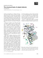
Specific translocation is another chromosomal abnormal-
ity, but it is much less commonly observed in lung cancer
than hematologic or mesodermal tumors [52]. Chromo-
somal translocations modify gene function through the
deregulated expression of cellular proto-oncogenes with-
out altering the structure of the protein product or by
Array comparative genomic hybridization on a squamous carcinoma of the lungFigure 1
Array comparative genomic hybridization on a squamous carcinoma of the lung. A. array CGH profile on a squamous carci-
noma of the lung labeled with Cy3 against normal DNA with Cy5. Each data point in presented mean (n = 4) ± coefficient of
variance (CV=STD/Mean). B. View of Chromosome 3 array CGH profile on the same squamous carcinoma of the lung showing
the size of the amplicon.
Respiratory Research 2003, 4 />Page 5 of 15
(page number not for citation purposes)
generating and expressing a chimeric protein with growth-
promoting activities. Recently, Dang et al. identified a
chromosome 19–15 translocation associated with overex-
pression of Notch 3 [53]. The authors developed a trans-
genic mouse model overexpressing Notch 3 causing
neonatal mortality with a phenotype suggestive of alveo-
lar cell hyperplasia. These data suggest that Notch3 over-
expression prevents epithelial differentiation and this
may play a significant role in promoting oncogenesis in a
subset of lung cancers [54].
Genomic instability
Genome instability is a fundamental characteristic of can-
cer initiation and progression. However, our understand-
ing of the time when instability occurs during
progression, the rate of instability, and the mechanisms
leading to instability is far from complete. Instability can
arise from different pathways. In a small fraction of lung
tumors, mismatch repair deficiency leads to microsatellite
instability at the nucleotide sequence level. In other
tumors, abnormal chromosome number (aneuploidy) is
the dominant feature [55]. The progressive accumulation
of mutations, loss of apoptotic control and regulation of
cell proliferation, and the appearance of aneusomy are
associated with worsening dysplasia phenotypes and may
reflect underlying dysregulation of mechanisms control-
ling genomic fidelity. Less clear than microsatellite insta-
bility is the importance of specific defects in DNA repair
in lung cancer. It is known that polymorphisms in DNA
repair genes XPD (codon 312 Asp/Asp vs Asp/Asn) have
been found to be associated with impaired efficiency of
DNA repair and apoptotic function in lung cancer [56].
New techniques, however, are allowing us to assess these
changes in individual or small numbers of preneoplastic
cells. Copy number changes in single cells can be assessed
by FISH probes. Microdissection of dysplastic epithelium
has provided purified tissue for the analysis of point
mutations [4], chromosomal deletions [5], microsatellite
instability [6,7] and DNA methylation patterns [8]. We
thus may be able to ultimately derive a sequential pattern
of development for genetic abnormalities in preneoplastic
lung epithelium.
Role of viruses in lung tumorigenesis
The understanding of lung cancer molecular approaches
has led to the development of transgenic models using
viral antigens, including SV40 large T antigen and polyo-
mavirus (PyV) large and middle T antigens that result in a
high frequency of tumors. No common respiratory viruses
have been conclusively incriminated in the development
of lung cancer, but several have been implicated. Human
papilloma virus (HPV), for example, has been associated
with lung cancer and in particular lung cancer arising in
women [57]. Simian Virus 40 has been incriminated in
the development of mesothelioma [58]; Epstein Bar Virus
(EBV) has been suspected to be involved in the
Hierarchical clustering analysis of NSCLCs using array comparative genomic hybridizationFigure 2
Hierarchical clustering analysis of NSCLCs using array comparative genomic hybridization. Cluster analysis using the 50 BAC
clones closely correlated with histological subtype allowed accurate discrimination between SqCa and AdCa. K-nearest-neigh-
bor classification was used to formally test the ability to predict subtype from array CGH profile. Cross-validation yielded 24/
27 (89%) correct histological classification. Green squares: increase in copy number of a specific BAC clone, red squares:
decrease in copy number.
Respiratory Research 2003, 4 />Page 6 of 15
(page number not for citation purposes)
development of papillomas, mesotheliomas and lympho-
mas of the lung. Many PCR-based assays, however, have
attempted to correlate bronchogenic carcinomas with res-
piratory viruses without success. Recent advances in pro-
teomics may be useful in studying the role of viral
infection in airway epithelial cell transformation. The pro-
teomic analysis of tumors may allow the identification of
peptide sequences specific to pathogens otherwise
ignored in tumorigenesis.
Viruses have also been used (e.g., adenovirus) to facilitate
gene entry into cells (adenovirus-mediated gene transfer)
or in in vivo gene therapy of human lung cancer using
wild-type p53 delivered by retrovirus [59].
Genomic instability causing lung tumorigenesis
A multistep process for clonal evolution
Genetic changes are seen in the transition from normal to
intraepithelial cancer to invasive disease. The understand-
ing of the timing of instability during progression, the rate
of instability and the mechanisms leading to instability
are far from complete. Chronic exposure to carcinogens
initiates a process characterized by genetic abnormalities,
phenotypic changes and clonal overgrowth throughout
the lungs [60]. Measures of genomic instability follow
rates of loss of heterozygosity [39] and accumulation of
other genomic abnormalities [55]. In the airways, progres-
sively more severe and more frequent abnormalities are
seen in preinvasive lesions [61]. The progressive accumu-
lation of genomic abnormalities associated with clonal
growth among populations of tumor cells are well
described and favor the clonal progression of cancer. Yet
cancer remains a rare event if one considers the total
number of bronchial epithelial cells and the proliferation
rate of patches of clonal abnormalities [62,63].
While lung cancer originates from one or a few airway epi-
thelial cells, it is clear that exposure of the whole airway
mucosa to tobacco smoke could cause the entire bron-
chial tree to be at increased risk of developing lung cancer,
leading to the concept of field cancerization. Field cancer-
ization was first proposed in the fifties [64] and its molec-
ular correlates later confirmed in the airways of human
smokers [65,66]. Field cancerization is also demonstrated
by the elevated Ki-67 labeling index in the airways of
smokers at more than one site [67]. Although the risk of
developing lung cancer increases with the presence of
such preinvasive lesions, no one has identified the molec-
ular determinants of preinvasive lesions that may predict
irreversible progression to lung cancer.
Carcinogenesis in the airways has proved to be multistep
and multifocal and yet clonal in nature. Multiple lines of
evidence support the concept of clonal progression of
tumors. First, at the chromosomal level, abnormalities
found in invasive tumors and their metastases are
extremely highly correlated [68,69]. Similarly, allelic
losses or microsatellite abnormalities found to be in pre-
invasive lesions are found in similar frequencies in inva-
sive lesions [7,63]. The issue remains complex as a small
fraction of tumors appear to be truly independent syn-
chronous primaries and different p53 mutations have
been found in synchronous preinvasive lesions [70]. The
prolonged, multistep nature of lung cancer development
makes this disease process potentially amenable to chem-
opreventive interventions that should be optimally
applied in the earliest preinvasive phases.
Significance of genomic instability in lung tumorigenesis
Some preinvasive lesions are committed to develop into
invasive cancer [71,72]. One critical question that remains
is the identification of that specific subset of the plethora
of genetic changes in a given lesion that predisposes that
lesion to develop into frank cancer. The literature suggests
that the number of molecular abnormalities accumulated
in the epithelium underlies tumor progression independ-
ent of light microscopically observable morphological
abnormalities [4,73]. This observation raises the possibil-
ity that genomic instability itself may be independently
predictive of tumor progression. Consistent with this
hypothesis, the relationship between clonal chromosome
alterations and various clinical parameters was evaluated
in 70 patients with non-small cell lung cancer [47]. An
increased number of marker chromosomes were observed
in patients having a higher number of packs of cigarettes
smoked over years.
Epigenetic alterations of gene expression in lung cancer
Gene function loss can be mediated by deletion of large
chromosomal regions or by inactivation of gene function
from genetic mutation, or due to epigenetic modifications
of DNA such as promoter hypermethylation or histone
deacetylation.
DNA adducts
One marker for significant carcinogen exposure is the
level of DNA adducts in normal DNA. DNA adducts are
covalent modifications of the DNA that result from expo-
sure to specific activated carcinogens. In addition to being
markers of carcinogen exposure, it is possible that these
adducts may directly alter regulation of transcription of
tumor suppressor or oncogenes [74]. The distribution of
benzo[a]pyrene diol epoxide (BPDE) adducts along exons
of the p53 gene in BPDE-treated HeLa cells and bronchial
epithelial cells has been mapped at nucleotide resolution
[22]. Cigarette smokers have higher adduct levels than
non-smokers. Because DNA adduct levels in tumor tissue
and in blood lymphocytes have been associated with lung
cancer [75,76] and because these levels correlate with
daily or lifetime cigarette consumption and do not reverse
Respiratory Research 2003, 4 />Page 7 of 15
(page number not for citation purposes)
after smoking cessation [77], DNA adducts have been pro-
posed as potential biomarkers of risk for lung cancer.
In an attempt to identify risk factors associated with the
level of DNA adduct accumulation, Wiencke et al. studied
DNA adducts in current and former smokers and found
that in current smokers the most important variable was
the number of cigarettes smoked per day. In contrast, they
found that in ex-smokers, the most important variable
was age at initiation [34]. Mechanisms responsible for the
relationship between DNA adduct levels and age of initia-
tion are unknown, and the relative contribution of
decreased adduct removal by DNA repair or cell turnover
or increased adduct formation at younger ages is yet to be
determined. Prospective study is needed to follow current
and ex-smokers over time to determine the value of
adduct levels in risk assessment.
DNA adducts have been associated with smoking status
and shown to be more prevalent among women. In a
matched case-control study nested within the prospective
Physicians' Health Study, there was an increased level of
DNA adducts in active smokers who developed lung can-
cer as compared to controls; a finding that was not found
among former or non smokers [78]. Women smokers may
be at higher risk of developing lung cancer for a given
tobacco exposure and women also seem to accumulate
aromatic/hydrophobic DNA adducts at a faster rate then
men [79]. DNA adduct levels were higher in women even
when corrected for smoking dose packs of cigarettes
smoked either per day or over years.
Methylation
Among epigenetic alterations, gain of methylation in nor-
mally unmethylated CpG islands around gene transcrip-
tion start sites is an increasingly recognized and important
means of altered gene expression in tumors [80]. The
genes affected include over half of the tumor suppressor
genes that cause familial cancers when mutated in the
germline, and the selective advantage for genetic and epi-
genetic dysfunction in these genes is very similar in spo-
radic cases. In contrast to genetic mutations that require
two hits to inhibit both alleles, aberrant methylation is a
dynamic process over multiple division cycles and may
cause increasing degrees of gene function loss by increas-
ing the density of methylation on promoter regions. "CpG
islands," the targets of DNA methyltransferase, are associ-
ated with the transcription start sites in almost half of
human genes [81]. Dense methylation of cytosines within
CpG islands causes heritable gene silencing [82]. Aberrant
methylation can begin very early in tumor progression by
causing loss of cell cycle control (p16) [83], loss of mis-
match repair function (MLH1) [84] and loss of cell-cell
interaction (E-cadherin). The exact mechanism by which
hypermethylation may cause tumor progression is still
unknown. In fact, there is still debate as to whether meth-
ylation is a result rather than a cause of gene function loss
[85]. Promoter region hypermethylation has been pro-
posed as an excellent tumor marker. In lung cancer, com-
mon methylated loci were found in both tumor and
sputum DNA and were detected in the sputum for up to 3
years before the diagnosis of cancer [86].
Acetylation
The dynamics of chromatin formation suggest that the
association of DNA methylation and histone deacetyla-
tion may cause silencing of hypermethylated genes in
tumors. During transcription, chromatin unfolds and
allows ribosomal access to the DNA. Acetylation of his-
tone tails on the nucleosome is associated with chromatin
unfolding and increased regional transcriptional activity.
Histone deacetylases (HDACs) modulate chromatin
structure by regulating acetylation of core histone pro-
teins. Deacetylation of histones is thus associated with
compacting the DNA and transcriptional repression. In
lung cancer cell lines, for example, de-acetylation of his-
tone 3 correlated with retinoic acid refractoriness, a phe-
nomenon related to RARbeta promoter methylation in a
subset of cell lines [87]. Inhibitors of HDACs have already
shown to decrease the level of a series of oncoproteins
[88] suggesting a potential role as antitumor therapeutic
agents.
From genetic abnormalities to biomarkers for lung cancer
Lung cancer is a heterogeneous disease. The specific
genetic abnormalities mentioned above have thus far
proven to be of limited use individually as biomarkers for
lung cancer. However, the completion of the first draft of
the human genome sequence [15] and the availability of
high throughput technologies (e.g. microarray) have
prompted us to look in an unbiased way for complex pat-
terns of genetic abnormalities that may be better associ-
ated with both pre- and invasive lung cancers and
potential markers for use in early detection strategies.
Genomic arrays
DNA amplification and deletion in lung cancers of vari-
ous histological subtypes have been analyzed by genomic
approaches. We recently published the results of such
analysis in a series of 37 NSCLCs [13]. With this tech-
nique, we demonstrated substantial genomic differences
between squamous carcinomas and adenocarcinomas
that are consistent with earlier chromosome based com-
parative genomic hybridization studies [40–42]. The sig-
nificant difference in the total number of abnormalities
between squamous carcinomas and adenocarcinomas
suggests that they may differ in the level of genome insta-
bility and/or in the mechanisms by which they progress.
Chromosome 3q is a common area of chromosomal gain
in a variety of solid tumors. When early lesions are treated,
Respiratory Research 2003, 4 />Page 8 of 15
(page number not for citation purposes)
they are known to prevent progression to invasive cancer.
As discussed above, particularly common were amplified
regions on chromosomal arms 1q, 3q, 5p, 8q, 11q, 12p,
17q and 20q, but gene amplification in chromosomal
region 3q26 was the most prevalent abnormality. Among
many amplified genes found in this region in a variety of
solid tumors, some have been called potential candidate
oncogenes (phosphatidylinositol-3 kinase catalytic subu-
nit, PIK3CA) or genes suspected to be involved in tumor
progression including the somatostatin gene (SST), p63
(p53 homologue gene), telomerase RNA component gene
(TERC) and neutral endopeptidase gene (NEP). These pat-
terns may ultimately be more predictive than analysis of
expression of any single genes.
Expression arrays
RNA expression patterns may be more functionally rele-
vant than DNA copy number changes, as most of these
copy number changes affect cellular behavior via altered
expression of included genes. The microarray technology
developed in the mid 90's offers the hope that a genetic
fingerprint of these tumors can be developed associated
with clinical features. Beyond the need for better classifi-
cation of lung cancers, this technical revolution opens a
window of understanding to the world of tumor behavior
(disease progression, recurrence, response to therapy) as
well as to the mechanisms of tumor development. Tumor
expression profiles are also influenced by the surrounding
non-malignant cells. The combination of tumor and cell
line profiling allows for the study of the regulatory role of
both entities [89].
Efforts in classifying lung cancers based on microarray
analysis revealed subclasses of adenocarcinomas. Selected
genes allow the discrimination between primary lung can-
cer and metastasis of extrapulmonary sites [90]. Studies of
expression profiles of adenocarcinomas of the lung using
different chips commercially available [90] or custom
arrays [91,92] identified different classes of tumors with
some overlap. Four classes of adenocarcinomas were
found to have specific prognosis and molecular signature.
These were characterized respectively 1) by expression of
cell cycle or proliferation genes, 2) by expression of neu-
roendocrine markers, 3) by expression of markers of alve-
olar origin, and 4) by expression of ODC or glutathione S-
transferase [91]. The neuroendocrine subclass was found
to have outcome significantly worse than the others. The
hope is that these subclass differences will point towards
new molecular therapeutic opportunities for these sub-
sets. Interestingly, when applied to neuroendocrine
tumors, cDNA microarrays found poor correlations
between genes expressed in carcinoid and SCLC [93],
tumors that may be morphologically similar but that
behave very different clinically.
Protein profiling
Recent advances in protein profiling have suggested a
poor correlation between gene expression and protein
expression. Perhaps more significantly, it is now well
established that protein activity is often highly regulated
by post-translational modifications such as proteolysis
and phosphorylation. Neither protein expression levels
nor post-translational modification can be assessed by
genomic or cDNA microarray technologies, prompting
interest in evaluation of protein expression, commonly
referred to as "proteomics".
Investigators, including those at our institution, have
attempted to use several proteomic methods of analysis,
including 2Dgel and IHC, to identify biomarkers in
tumors [94–97] in body fluids such as bronchoalveolar
lavage [98] of patients with or without cancer. We recently
acquired experience in this method for profiling of pro-
teins in cancer tissue [99]. We applied MALDI-MS to 79
surgically resected lung cancers and 14 normal tissues.
Software written by Dr. Jason Moore at Vanderbilt allows
assignment of protein peaks in the mass spectral data
across samples into unique "bins" corresponding to
unique peptide species with correction for multiply
charged ions. Hierarchical clustering of the resulting data
has allowed the identification of patterns distinguishing
between tumor and normal as well as histological sub-
groups. For example, to identify proteomic patterns that
distinguish primary NSCLC from metastases to the lung,
we compared protein expression profiles obtained from
34 primary NSCLCs with those from 7 other types of lung
metastatic tumors, including 5 metastases to the lung
from other sites and 2 lung metastases from previously
resected NSCLC in the training cohort. We identified 24
MS signals that could discriminate all of the primary
NSCLC from non-primary NSCLC in the training cohort,
and were able to perfectly classify blinded samples in a
test cohort [100]. Proteomic patterns from primary
tumors with prognostic discriminatory power were identi-
fied as well and are potentially very useful in the clinical
management of lung cancer. Although requiring prospec-
tive validation, these data bring proof of concept to an
approach that may be found to be very powerful at select-
ing surgical candidates and other therapeutic strategies
based on novel biological targets.
Identification of biomarkers
Biomarkers are needed to identify patients at high risk for
lung cancer and to identify surrogate endpoints for
response to chemoprevention strategies.
Despite the societal need for the early diagnosis of lung
cancer, no role for biomarkers has yet been established for
decision-making in intraepithelial neoplasia of the lung.
Technical procedures such as tissue processing, use of
Respiratory Research 2003, 4 />Page 9 of 15
(page number not for citation purposes)
antibody reagents and data interpretation need to be
developed and standardized. A comprehensive and inte-
grated approach linking laboratory findings of IEN of the
lung with clinical features holds the potential to identify
clinically relevant genetic and protein markers of
carcinogenesis.
The number of potential lung cancer-related genes is rap-
idly growing. Once identified, genes and proteins may be
tested in large populations of patients by immunohisto-
chemical or cytogenetic techniques on tissue microarrays
[100]. This high throughput method allows the screening
of hundreds of lung cancer samples on a single glass slide
and will allow retrospective analysis of material stored
with associated clinical outcome. The arrays typically
comprise core biopsies 0.6 mm in diameter of different
tumors and uninvolved lung from the same individuals
retrieved from the pathology archives of various institu-
tions [101] (Figure 3). A firmer understanding of the rela-
tionship of relevant protein and genetic markers to
clinical and pathologic status could lead to more accurate
estimates of the anatomic extent of disease, risk of recur-
rence, and most effective intervention.
From Genetic abnormalities to early detection and new
therapies
The identification of early molecular events such as chro-
mosomal gain or loss that predicts tumor development
Tissue microarrays (TMAs) of lung cancerFigure 3
Tissue microarrays (TMAs) of lung cancer. TMAs are comprised of core biopsies of 0.6 mm in diameter of different tumors and
of uninvolved lung from the same individuals. We retrieved 240 NSCLC tissue blocks from the pathological archives of Vander-
bilt University between 1989 and 2001 and arrayed them in triplicate onto 4 separate TMAs. Tissue microarrays allow high
throughput analysis of molecular markers identified in squamous lung neoplasia.
Tissue Microarray for NSCLC
240 NSCLC - 4 TMA’s
Punches in triplicate
Normal match control
Squamous cell 98
Adenocarcinoma 91
Large cell 13
Carcinoid 10
Neuroendocrine 7
Small cell 5
Sarcoma 4
Adenosquamous 3
Respiratory Research 2003, 4 />Page 10 of 15
(page number not for citation purposes)
suggests that early detection of lung cancer could be
approached by means of molecular analysis. Sputum sam-
ple analysis for DNA methylation or chromosomal abnor-
malities by FISH may represent approaches suitable for
early detection. The analysis of sputum, bronchial biop-
sies of preinvasive lesions using new detection methods
such as fluorescence bronchoscopy [102], as well as
exhaled breath condensate for tumor metabolites may be
shown to be efficient ways of assessing high risk individu-
als. Early detection by low dose computed tomography
scanning is being evaluated prospectively with the
National Lung Cancer Screening Trial in 50,000 smokers.
The addition of molecular studies may significantly
increase the sensitivity and specificity of this new strategy
for early detection.
Several therapeutic approaches to cancer have been devel-
oped to reduce undesirable expression of gene product or
otherwise inhibit its function: (1) gene therapy (e.g. Ade-
novirus-p53) gene-specific ribozymes, which are able to
break down specific RNA sequences, or with antisense oli-
gonucleotides, (2) small molecule inhibition of receptor
tyrosine kinases, (3) inhibition of p21(ras) farnesylation
either by inhibition of farnesyl transferase or synthesis
inhibition of farnesyl moieties, and (4) specific antibody
approaches (e.g. anti-HER2 or anti-VEGF). We will touch
on a couple of these approaches below.
Specific molecular targets
p53
Recently several phase I studies have evaluated the safety,
biological effect and different routes of administration of
adenoviral-mediated p53 gene therapy in various tumor
types. These studies indicate that adenovirus-mediated
p53 gene therapy and introduction of wild-type p53 into
tumor cells represents a potentially valuable tool for the
therapy of many types of human cancers [103] mainly by
causing cell-cycle arrest or apoptosis [104,105]. When
injected intra-tumorally, wt-p53 has shown to be
expressed in patients with p53 mutations and 3/7 patients
showed regression of tumor size [106]. Using the wild-
type p53 recombinant adenovirus, the same group of
investigators showed in phase I trial that 16/25 had stabi-
lization of disease and 2 had partial remissions [107].
One of the major limitations of the intra-tumoral
approach is the inefficient delivery of genes of interest
within the tumor mass. We have shown that intra-alveolar
delivery of the gene in patients with bronchioloalveolar
carcinoma led to objective responses.
GFR antagonists
Several epithelial tumors express EGFR with and without
EGFR amplification [108]. This EGFR overexpression is
associated with increased ligand production and hyperac-
tive receptor function. About a third or more of NSCLC
showed overexpressed EFGR [109]. Overexpression of
EFGR was also associated with poor prognosis of patients
with NSCLC [110]. Low-grade bronchial preinvasive
lesions have also been shown to overexpress EGFR [111].
EGFR expression has been found to be elevated in meta-
plastic biopsies when compared to normal biopsies in
active smokers [112] and that when co-expressed with
p53 may predict squamous cell carcinoma development.
Interruption of this autocrine pathway with receptor anti-
bodies (extracellular domain of the protein) or tyrosine
kinase inhibitors (competition with the kinase ATP bind-
ing site) can cause tumor regression [113,114]. ZD1839,
IRESSA and OSI-774 (Tarceva) are potent and specific
inhibitors of the tyrosine kinase moiety of EGFR.
Response rates in heavily pretreated patients with NSCLC
vary between 10–18% in the IDEAL trials [115,116],
which may seem low but is actually far higher than any
standard chemotherapy and represents a major benefit for
these low-toxicity oral agents. Studies are proposed to
investigate the value of EGFR inhibition in combination
therapy, in earlier stage NSCLC and in lung cancer chem-
oprevention (STOP trials, SPORE Trial of Lung Cancer
Prevention). Such chemoprevention trials with molecular
and morphologic (preinvasive lesions) surrogate end-
points may suggest reversibility of lesions. However, the
rate of spontaneous regression of these preinvasive lesions
is, as yet, poorly characterized.
Kras: Farnesyl transferase inhibitor Zarnestra
K-ras was one of the first oncogenes implicated in human
cancer. Development of retroviral vectors containing ani-
tsense K-ras constructs or inhibitors of ras function may
reduce proliferation or tumorigenicity. Farnesyltrans-
ferase enzyme activity is required to transfer farnesyl iso-
prenoid to the Ras c-terminus to anchor it to the cell
membrane. This step is critical for Ras activation as an
oncogene. The ras protein is known to undergo a series of
post-translational modifications at the c-terminal CAAX
motif, which forms a thioether bond of p21 ras with far-
nesyl and ties it to the plasma membrane [117]. At the cell
surface, ras relays growth regulatory signals from receptor
tyrosine kinases to various pathways of cell signal trans-
duction. Unfortunately the currently available inhibitors
work best with activated H-ras, a rare finding in lung can-
cer rather than the more common K-ras activation. Also
not well explained is the observation that antitumor activ-
ity is very poorly correlated with measurable activation of
any of the ras genes. However, several farnesyltransferase
inhibitors are currently being tested in the clinic.
R115777-Zarnestra is also being proposed in the clinic in
a secondary chemoprevention trial. This trial is essentially
based on the efficacy of FTI-276 on established lung ade-
nomas (considered to be premalignant lesions of the
lung) from A/J mice exposed to 4-(methylnitrosamino)-1-
(3-pyridyl)-1-butanone, a tobacco-related carcinogen
Respiratory Research 2003, 4 />Page 11 of 15
(page number not for citation purposes)
[118]. Analysis of the tumors showed a 60% reduction in
tumor multiplicity and a 42% reduction in tumor inci-
dence as well as a significant reduction in tumor volume
(approximately 58%).
COX-2 inhibition
Cyclooxygenase-2 (COX-2) is an inducible enzyme that
catalyzes the production of prostanoids. COX-2 can acti-
vate carcinogens in tobacco smoke [119], and COX-2
expression may play a role in angiogenesis by correlating
with VEGF levels [120]. In addition, COX-2 activity may
have a role in inhibiting apoptosis and modulating
immune responses [121]. While nonsteroidal anti-
inflammatory drugs have shown to reduce the risk of
colorectal cancer, no such evidence yet exists in lung can-
cer. COX-2 inhibition has proven to reduce lung cancer
cell growth in vitro [122]. In vivo, COX-2 has shown to
cause persistent remission in patients otherwise refractory
to lung cancer. COX-2 overexpression is a marker of poor
prognosis in early stage NSCLC [123,124]. COX-2 inhibi-
tors are being evaluated in combination therapy for che-
moprevention and therapy for lung cancer.
Other targeted strategies
Other targets include antibodies against VEGF ligand,
EGFr or HER2 and inhibition of proteosome activity to
counteract NFκB activation. All of these are currently in
large scale clinical trials. Markers identified as being over-
expressed in lung cancers represent potential immuno-
therapy targets even if no significant function can be
found for the marker protein. An example is the recent
identification of frequent overexpression of the cancer tes-
tis antigens from the microarray studies [125]. These
genes are already being tested as vaccine targets in
melanoma, and are only recently recognized as being
overexpressed in the majority of non-small cell lung
cancers.
Conclusions
A large number of genetic pathways associated with can-
cer development are being discovered at a rapid pace. The
clinical impact of this recent knowledge on disease man-
agement is still relatively small, but real and growing. Lit-
tle progress has been made in lung cancer
chemoprevention, yet preventing, inhibiting and revers-
ing the preneoplastic changes leading to cancer may ulti-
mately prove a much more tractable goal than treating
advanced disease. The slow process of carcinogenesis
makes this period an open window for chemoprevention
so that the intervention occurs when genetic instability is
still controllable.
Abbreviations
CGH: comparative genomic hybridization, LOH: loss of
heterozygosity, FISH: fluorescence in situ hybridization,
TMA: tissue microarray, NSCLC: non small cell lung
cancer
Acknowledgement
This work was supported by the Vanderbilt Ingram Cancer Center SPORE
in Lung Cancer from the National Institutes of Health, the Flight Attendants
Medical Research Institute, the Damon Runyon Foundation, the Office of
Research and Development, and the Department of Veterans Affairs. The
authors thank Tamara Lasakow for editorial assistance with the manuscript.
References
1. Knudson A. G., Jr.: Genetics of human cancer. Genetics 1975, 79
Suppl:305-316.
2. Peto R, Roe FJ, Lee PN, Levy L and Clack J: Cancer and ageing in
mice and men. Br J Cancer 1975, 32:411-426.
3. Loeb LA: Mutator phenotype may be required for multistage
carcinogenesis. Cancer Res 1991, 51:3075-3079.
4. Chung GT, Sundaresan V, Hasleton P, Rudd R, Taylor R and Rabbitts
PH: Sequential molecular genetic changes in lung cancer
development. Oncogene 1995, 11:2591-2598.
5. Sundaresan V, Ganly P, Hasleton P, Rudd R, Sinha G, Bleehen NM and
Rabbitts P: p53 and chromosome 3 abnormalities, character-
istic of malignant lung tumours, are detectable in preinva-
sive lesions of the bronchus. Oncogene 1992, 7:1989-1997.
6. Miozzo M, Sozzi G, Musso K, Pilotti S, Incarbone M, Pastorino U and
Pierotti MA: Microsatellite alterations in bronchial and spu-
tum specimens of lung cancer patients. Cancer Res 1996,
56:2285-2288.
7. Mao L, Lee DJ, Tockman MS, Erozan YS, Askin F and Sidransky D:
Microsatellite alterations as clonal markers for the detection
of human cancer. Proc Natl Acad Sci U S A 1994, 91:9871-9875.
8. Belinsky SA, Nikula KJ, Baylin SB and Issa JP: Increased cytosine
DNA-methyltransferase activity is target-cell- specific and
an early event in lung cancer. Proc Natl Acad Sci U S A 1996,
93:4045-4050.
9. Thiberville L, Payne P, Vielkinds J, LeRiche J, Horsman D, Nouvet G,
Palcic B and Lam S: Evidence of cumulative gene losses with
progression of premalignant epithelial lesions to carcinoma
of the bronchus. Cancer Res 1995, 55:5133-5139.
10. Sundaresan V, Heppell-Parton A, Coleman N, Miozzo M, Sozzi G, Ball
R, Cary N, Hasleton P, Fowler W and Rabbitts P: Somatic genetic
changes in lung cancer and precancerous lesions. Ann Oncol
1995, 6:27-31; discussion 31-2.
11. Wistuba ,II, Lam S, Behrens C, Virmani AK, Fong KM, LeRiche J, Samet
JM, Srivastava S, Minna JD and Gazdar AF: Molecular damage in
the bronchial epithelium of current and former smokers. J
Natl Cancer Inst 1997, 89:1366-1373.
12. Mao L, Lee JS, Kurie JM, Fan YH, Lippman SM, Lee JJ, Ro JY, Broxson
A, Yu R, Morice RC, Kemp BL, Khuri FR, Walsh GL, Hittelman WN
and Hong WK: Clonal genetic alterations in the lungs of cur-
rent and former smokers. J Natl Cancer Inst 1997, 89:857-862.
13. Massion PP, Kuo WL, Stokoe D, Olshen AB, Treseler PA, Chin K,
Chen C, Polikoff D, Jain AN, Pinkel D, Albertson DG, Jablons DM and
Gray JW: Genomic copy number analysis of non-small cell
lung cancer using array comparative genomic hybridization:
implications of the phosphatidylinositol 3-kinase pathway.
Cancer Res 2002, 62:3636-3640.
14. Massion PP, Taflan PM, Rahman SMJ, Yildiz P, Shyr Y, Edgerton ME,
Westfall MD, Roberts JR, Pietenpol JA, Carbone DP and Gonzalez AL:
Significance of p63 amplification and overexpression in lung
cancer development and prognosis. Cancer Res 2003,
63:7113-7121.
15. Venter JC, Adams MD, Myers EW, Li PW, Mural RJ, Sutton GG, Smith
HO, Yandell M, Evans CA, Holt RA, Gocayne JD, Amanatides P,
Ballew RM, Huson DH, Wortman JR, Zhang Q, Kodira CD, Zheng
XH, Chen L, Skupski M, Subramanian G, Thomas PD, Zhang J, Gabor
Miklos GL, Nelson C, Broder S, Clark AG, Nadeau J, McKusick VA,
Zinder N, Levine AJ, Roberts RJ, Simon M, Slayman C, Hunkapiller M,
Bolanos R, Delcher A, Dew I, Fasulo D, Flanigan M, Florea L, Halpern
A, Hannenhalli S, Kravitz S, Levy S, Mobarry C, Reinert K, Remington
K, Abu-Threideh J, Beasley E, Biddick K, Bonazzi V, Brandon R, Cargill
M, Chandramouliswaran I, Charlab R, Chaturvedi K, Deng Z, Di
Francesco V, Dunn P, Eilbeck K, Evangelista C, Gabrielian AE, Gan W,
Respiratory Research 2003, 4 />Page 12 of 15
(page number not for citation purposes)
Ge W, Gong F, Gu Z, Guan P, Heiman TJ, Higgins ME, Ji RR, Ke Z,
Ketchum KA, Lai Z, Lei Y, Li Z, Li J, Liang Y, Lin X, Lu F, Merkulov
GV, Milshina N, Moore HM, Naik AK, Narayan VA, Neelam B, Nussk-
ern D, Rusch DB, Salzberg S, Shao W, Shue B, Sun J, Wang Z, Wang
A, Wang X, Wang J, Wei M, Wides R, Xiao C, Yan C, Yao A, Ye J,
Zhan M, Zhang W, Zhang H, Zhao Q, Zheng L, Zhong F, Zhong W,
Zhu S, Zhao S, Gilbert D, Baumhueter S, Spier G, Carter C, Cravchik
A, Woodage T, Ali F, An H, Awe A, Baldwin D, Baden H, Barnstead
M, Barrow I, Beeson K, Busam D, Carver A, Center A, Cheng ML,
Curry L, Danaher S, Davenport L, Desilets R, Dietz S, Dodson K,
Doup L, Ferriera S, Garg N, Gluecksmann A, Hart B, Haynes J, Haynes
C, Heiner C, Hladun S, Hostin D, Houck J, Howland T, Ibegwam C,
Johnson J, Kalush F, Kline L, Koduru S, Love A, Mann F, May D,
McCawley S, McIntosh T, McMullen I, Moy M, Moy L, Murphy B, Nel-
son K, Pfannkoch C, Pratts E, Puri V, Qureshi H, Reardon M, Rod-
riguez R, Rogers YH, Romblad D, Ruhfel B, Scott R, Sitter C,
Smallwood M, Stewart E, Strong R, Suh E, Thomas R, Tint NN, Tse S,
Vech C, Wang G, Wetter J, Williams S, Williams M, Windsor S, Winn-
Deen E, Wolfe K, Zaveri J, Zaveri K, Abril JF, Guigo R, Campbell MJ,
Sjolander KV, Karlak B, Kejariwal A, Mi H, Lazareva B, Hatton T,
Narechania A, Diemer K, Muruganujan A, Guo N, Sato S, Bafna V,
Istrail S, Lippert R, Schwartz R, Walenz B, Yooseph S, Allen D, Basu
A, Baxendale J, Blick L, Caminha M, Carnes-Stine J, Caulk P, Chiang
YH, Coyne M, Dahlke C, Mays A, Dombroski M, Donnelly M, Ely D,
Esparham S, Fosler C, Gire H, Glanowski S, Glasser K, Glodek A,
Gorokhov M, Graham K, Gropman B, Harris M, Heil J, Henderson S,
Hoover J, Jennings D, Jordan C, Jordan J, Kasha J, Kagan L, Kraft C,
Levitsky A, Lewis M, Liu X, Lopez J, Ma D, Majoros W, McDaniel J,
Murphy S, Newman M, Nguyen T, Nguyen N, Nodell M, Pan S, Peck
J, Peterson M, Rowe W, Sanders R, Scott J, Simpson M, Smith T,
Sprague A, Stockwell T, Turner R, Venter E, Wang M, Wen M, Wu
D, Wu M, Xia A, Zandieh A and Zhu X: The sequence of the
human genome. Science 2001, 291:1304-1351.
16. Steen HB: The origin of oncogenic mutations: where is the pri-
mary damage? Carcinogenesis 2000, 21:1773-1776.
17. Westra WH, Slebos RJ, Offerhaus GJ, Goodman SN, Evers SG, Ken-
sler TW, Askin FB, Rodenhuis S and Hruban RH: K-ras oncogene
activation in lung adenocarcinomas from former smokers.
Evidence that K-ras mutations are an early and irreversible
event in the development of adenocarcinoma of the lung.
Cancer 1993, 72:432-438.
18. Niklinski J, Niklinska W, Laudanski J, Chyczewska E and Chyczewski
L: Prognostic molecular markers in non-small cell lung
cancer. Lung Cancer 2001, 34 Suppl 2:S53-8.
19. Lacal JC, Srivastava SK, Anderson PS and Aaronson SA: Ras p21 pro-
teins with high or low GTPase activity can efficiently trans-
form NIH/3T3 cells. Cell 1986, 44:609-617.
20. Cooper CA, Carby FA, Bubb VJ, Lamb D, Kerr KM and Wyllie AH:
The pattern of K-ras mutation in pulmonary adenocarci-
noma defines a new pathway of tumour development in the
human lung. J Pathol 1997, 181:401-404.
21. Harris CC: 1995 Deichmann Lecture p53 tumor suppressor
gene: at the crossroads of molecular carcinogenesis, molec-
ular epidemiology and cancer risk assessment. Toxicol Lett
1995, 82-83:1-7.
22. Denissenko MF, Pao A, Tang M and Pfeifer GP: Preferential forma-
tion of benzo[a]pyrene adducts at lung cancer mutational
hotspots in P53. Science 1996, 274:430-432.
23. Bennett WP, Colby TV, Travis WD, Borkowski A, Jones RT, Lane DP,
Metcalf RA, Samet JM, Takeshima Y, Gu JR and et al.: p53 protein
accumulates frequently in early bronchial neoplasia. Cancer
Res 1993, 53:4817-4822.
24. Stewart ZA and Pietenpol JA: p53 Signaling and cell cycle
checkpoints. Chem Res Toxicol 2001, 14:243-263.
25. Carbone DP, Mitsudomi T, Chiba I, Piantadosi S, Rusch V, Nowak JA,
McIntire D, Slamon D, Gazdar A and Minna J: p53 immunostaining
positivity is associated with reduced survival and is imper-
fectly correlated with gene mutations in resected non-small
cell lung cancer. A preliminary report of LCSG 871. Chest
1994, 106:377S-381S
26. Belinsky SA: Role of the cytosine DNA-methyltransferase and
p16INK4a genes in the development of mouse lung tumors.
Exp Lung Res 1998, 24:463-479.
27. Sanchez-Cespedes M, Decker PA, Doffek KM, Esteller M, Westra
WH, Alawi EA, Herman JG, Demeure MJ, Sidransky D and Ahrendt
SA: Increased loss of chromosome 9p21 but not p16 inactiva-
tion in primary non-small cell lung cancer from smokers. Can-
cer Res 2001, 61:2092-2096.
28. Peto R, Darby S, Deo H, Silcocks P, Whitley E and Doll R: Smoking,
smoking cessation, and lung cancer in the UK since 1950:
combination of national statistics with two case-control
studies. Bmj 2000, 321:323-329.
29. Wright GS and Gruidl ME: Early detection and prevention of
lung cancer [In Process Citation]. Curr Opin Oncol 2000,
12:143-148.
30. Hecht SS: Cigarette smoking and lung cancer: chemical mech-
anisms and approaches to prevention. Lancet Oncol 2002,
3:461-469.
31. Burns DM: Primary prevention, smoking, and smoking cessa-
tion: implications for future trends in lung cancer
prevention. Cancer 2000, 89:2506-2509.
32. Hecht SS: Tobacco smoke carcinogens and lung cancer. J Natl
Cancer Inst 1999, 91:1194-1210.
33. Wei Q, Gu J, Cheng L, Bondy ML, Jiang H, Hong WK and Spitz MR:
Benzo(a)pyrene diol epoxide-induced chromosomal aberra-
tions and risk of lung cancer. Cancer Res 1996, 56:3975-3979.
34. Wiencke JK, Thurston SW, Kelsey KT, Varkonyi A, Wain JC, Mark EJ
and Christiani DC: Early age at smoking initiation and tobacco
carcinogen DNA damage in the lung. J Natl Cancer Inst 1999,
91:614-619.
35. Mitelman F, Mertens F and Johansson B: A breakpoint map of
recurrent chromosomal rearrangements in human
neoplasia. Nat Genet 1997, 15 Spec No:417-474.
36. Knuutila S, Aalto Y, Autio K, Bjorkqvist AM, El-Rifai W, Hemmer S,
Huhta T, Kettunen E, Kiuru-Kuhlefelt S, Larramendy ML, Lushnikova
T, Monni O, Pere H, Tapper J, Tarkkanen M, Varis A, Wasenius VM,
Wolf M and Zhu Y: DNA copy number losses in human
neoplasms. Am J Pathol 1999, 155:683-694.
37. Wistuba ,II, Berry J, Behrens C, Maitra A, Shivapurkar N, Milchgrub S,
Mackay B, Minna JD and Gazdar AF: Molecular changes in the
bronchial epithelium of patients with small cell lung cancer.
Clin Cancer Res 2000, 6:2604-2610.
38. Sato S, Nakamura Y and Tsuchiya E: Difference of allelotype
between squamous cell carcinoma and adenocarcinoma of
the lung. Cancer Res 1994, 54:5652-5655.
39. Girard L, Zochbauer-Muller S, Virmani AK, Gazdar AF and Minna JD:
Genome-wide allelotyping of lung cancer identifies new
regions of allelic loss, differences between small cell lung
cancer and non-small cell lung cancer, and loci clustering.
Cancer Res 2000, 60:4894-4906.
40. Balsara BR, Sonoda G, du Manoir S, Siegfried JM, Gabrielson E and
Testa JR: Comparative genomic hybridization analysis detects
frequent, often high- level, overrepresentation of DNA
sequences at 3q, 5p, 7p, and 8q in human non-small cell lung
carcinomas. Cancer Res 1997, 57:2116-2120.
41. Bjorkqvist AM, Husgafvel-Pursiainen K, Anttila S, Karjalainen A, Tam-
milehto L, Mattson K, Vainio H and Knuutila S: DNA gains in 3q
occur frequently in squamous cell carcinoma of the lung, but
not in adenocarcinoma. Genes Chromosomes Cancer 1998,
22:79-82.
42. Petersen I, Bujard M, Petersen S, Wolf G, Goeze A, Schwendel A,
Langreck H, Gellert K, Reichel M, Just K, du Manoir S, Cremer T, Die-
tel M and Ried T: Patterns of chromosomal imbalances in ade-
nocarcinoma and squamous cell carcinoma of the lung.
Cancer Res 1997, 57:2331-2335.
43. Kallioniemi A, Kallioniemi OP, Piper J, Tanner M, Stokke T, Chen L,
Smith HS, Pinkel D, Gray JW and Waldman FM: Detection and
mapping of amplified DNA sequences in breast cancer by
comparative genomic hybridization. Proc Natl Acad Sci U S A
1994, 91:2156-2160.
44. Bentz M, Plesch A, Stilgenbauer S, Dohner H and Lichter P: Minimal
sizes of deletions detected by comparative genomic
hybridization. Genes Chromosomes Cancer 1998, 21:172-175.
45. Bockmuhl U, Schluns K, Kuchler I, Petersen S and Petersen I:
Genetic imbalances with impact on survival in head and neck
cancer patients. Am J Pathol 2000, 157:369-375.
46. Gray JW and Collins C: Genome changes and gene expression
in human solid tumors. Carcinogenesis 2000, 21:443-452.
47. Feder M, Siegfried JM, Balshem A, Litwin S, Keller SM, Liu Z and Testa
JR: Clinical relevance of chromosome abnormalities in non-
small cell lung cancer. Cancer Genet Cytogenet 1998, 102:25-31.
Respiratory Research 2003, 4 />Page 13 of 15
(page number not for citation purposes)
48. Yamaguchi K, Wu L, Caballero OL, Hibi K, Trink B, Resto V, Cairns
P, Okami K, Koch WM, Sidransky D and Jen J: Frequent gain of the
p40/p51/p63 gene locus in primary head and neck squamous
cell carcinoma. Int J Cancer 2000, 86:684-689.
49. Pack SD, Karkera JD, Zhuang Z, Pak ED, Balan KV, Hwu P, Park WS,
Pham T, Ault DO, Glaser M, Liotta L, Detera-Wadleigh SD and
Wadleigh RG: Molecular cytogenetic fingerprinting of esopha-
geal squamous cell carcinoma by comparative genomic
hybridization reveals a consistent pattern of chromosomal
alterations. Genes Chromosomes Cancer 1999, 25:160-168.
50. Heselmeyer K, Schrock E, du Manoir S, Blegen H, Shah K, Steinbeck
R, Auer G and Ried T: Gain of chromosome 3q defines the tran-
sition from severe dysplasia to invasive carcinoma of the
uterine cervix. Proc Natl Acad Sci U S A 1996, 93:479-484.
51. Aliferis CF, Hardin D and Massion PP: Machine learning models
for lung cancer classification using array comparative
genomic hybridization. Proc AMIA Symp 2002:7-11.
52. Zhou JY, Taguchi T, Siegfried JM, Jhanwar SC, Resau J and Testa JR:
Characterization of 9q;15q whole-arm translocation deriva-
tives in non- small cell lung carcinomas by fluorescence in
situ hybridization. Cancer Genet Cytogenet 1993, 69:1-6.
53. Dang TP, Gazdar AF, Virmani AK, Sepetavec T, Hande KR, Minna JD,
Roberts JR and Carbone DP: Chromosome 19 translocation,
overexpression of Notch3, and human lung cancer. J Natl Can-
cer Inst 2000, 92:1355-1357.
54. Dang TP, Eichenberger S, Gonzalez A, Olson S and Carbone DP:
Constitutive activation of Notch3 inhibits terminal epithelial
differentiation in lungs of transgenic mice. Oncogene 2003,
22:1988-1997.
55. Lengauer C, Kinzler KW and Vogelstein B: Genetic instability in
colorectal cancers. Nature 1997, 386:623-627.
56. Butkiewicz D, Rusin M, Enewold L, Shields PG, Chorazy M and Harris
CC: Genetic polymorphisms in DNA repair genes and risk of
lung cancer. Carcinogenesis 2001, 22:593-597.
57. Cheng YW, Chiou HL, Sheu GT, Hsieh LL, Chen JT, Chen CY, Su JM
and Lee H: The association of human papillomavirus 16/18
infection with lung cancer among nonsmoking Taiwanese
women. Cancer Res 2001, 61:2799-2803.
58. Testa JR, Carbone M, Hirvonen A, Khalili K, Krynska B, Linnainmaa K,
Pooley FD, Rizzo P, Rusch V and Xiao GH: A multi-institutional
study confirms the presence and expression of simian virus
40 in human malignant mesotheliomas. Cancer Res 1998,
58:4505-4509.
59. Carbone DP and Minna JD: In vivo gene therapy of human lung
cancer using wild-type p53 delivered by retrovirus [editorial;
comment]. J Natl Cancer Inst 1994, 86:1437-1438.
60. Auerbach O, Stout AP, Hammond EC and Grafinkel L: Changes in
bronchial epithelum in reation to cigraette smoking and in
relation to lung cancer. N Engl J Med 1961, 265:253-267.
61. Hirsch FR, Merrick DT and Franklin WA: Role of biomarkers for
early detection of lung cancer and chemoprevention. Eur
Respir J 2002, 19:1151-1158.
62. Park IW, Wistuba ,II, Maitra A, Milchgrub S, Virmani AK, Minna JD and
Gazdar AF: Multiple clonal abnormalities in the bronchial epi-
thelium of patients with lung cancer. J Natl Cancer Inst 1999,
91:1863-1868.
63. Wistuba ,II, Behrens C, Milchgrub S, Bryant D, Hung J, Minna JD and
Gazdar AF: Sequential molecular abnormalities are involved
in the multistage development of squamous cell lung
carcinoma. Oncogene 1999, 18:643-650.
64. Slaughter DP, Southwick HW and Smejkal W: Field cancerization
in oratl stratified squamous epithelium: clinical implications
of multicentric origin. Cancer 1953, 6:963-968.
65. Hittelman WN, Voravud N, Shin DM, Lee JS, Ro JY and Hong WK:
Early genetic changes during upper aerodigestive tract
tumorigenesis. J Cell Biochem Suppl 1993:233-236.
66. Smith AL, Hung J, Walker L, Rogers TE, Vuitch F, Lee E and Gazdar
AF: Extensive areas of aneuploidy are present in the respira-
tory epithelium of lung cancer patients. Br J Cancer 1996,
73:203-209.
67. Lee JJ, Liu D, Lee JS, Kurie JM, Khuri FR, Ibarguen H, Morice RC,
Walsh G, Ro JY, Broxson A, Hong WK and Hittelman WN: Long-
term impact of smoking on lung epithelial proliferation in
current and former smokers. J Natl Cancer Inst 2001,
93:1081-1088.
68. Petersen S, Aninat-Meyer M, Schluns K, Gellert K, Dietel M and
Petersen I: Chromosomal alterations in the clonal evolution
to the metastatic stage of squamous cell carcinomas of the
lung. Br J Cancer 2000, 82:65-73.
69. Levin NA, Brzoska PM, Warnock ML, Gray JW and Christman MF:
Identification of novel regions of altered DNA copy number
in small cell lung tumors. Genes Chromosomes Cancer 1995,
13:175-185.
70. Lavieille JP, Gazzeri S, Riva C, Reyt E, Brambilla C and Brambilla E: p53
mutations and p53, Waf-1, Bax and Bcl-2 expression in field
cancerization of the head and neck. Anticancer Res 1998,
18:4741-4749.
71. Venmans BJ, van Boxem TJ, Smit EF, Postmus PE and Sutedja TG:
Outcome of bronchial carcinoma in situ. Chest 2000,
117:1572-1576.
72. Bota S, Auliac JB, Paris C, Metayer J, Sesboue R, Nouvet G and Thi-
berville L: Follow-up of bronchial precancerous lesions and
carcinoma in situ using fluorescence endoscopy. Am J Respir
Crit Care Med 2001, 164:1688-1693.
73. Hung J, Kishimoto Y, Sugio K, Virmani A, McIntire DD, Minna JD and
Gazdar AF: Allele-specific chromosome 3p deletions occur at
an early stage in the pathogenesis of lung carcinoma. Jama
1995, 273:1908.
74. Wiencke JK: DNA adduct burden and tobacco carcinogenesis.
Oncogene 2002, 21:7376-7391.
75. Cheng YW, Chen CY, Lin P, Huang KH, Lin TS, Wu MH and Lee H:
DNA adduct level in lung tissue may act as a risk biomarker
of lung cancer. Eur J Cancer 2000, 36:1381-1388.
76. Vulimiri SV, Wu X, Baer-Dubowska W, de Andrade M, Detry M, Spitz
MR and DiGiovanni J: Analysis of aromatic DNA adducts and
7,8-dihydro-8-oxo- 2'- deoxyguanosine in lymphocyte DNA
from a case-control study of lung cancer involving minority
populations. Mol Carcinog 2000, 27:34-46.
77. Phillips DH, Hewer A, Martin CN, Garner RC and King MM: Corre-
lation of DNA adduct levels in human lung with cigarette
smoking. Nature 1988, 336:790-792.
78. Tang D, Phillips DH, Stampfer M, Mooney LA, Hsu Y, Cho S, Tsai WY,
Ma J, Cole KJ, She MN and Perera FP: Association between car-
cinogen-DNA adducts in white blood cells and lung cancer
risk in the physicians health study. Cancer Res 2001,
61:6708-6712.
79. Mollerup S, Ryberg D, Hewer A, Phillips DH and Haugen A: Sex dif-
ferences in lung CYP1A1 expression and DNA adduct levels
among lung cancer patients. Cancer Res 1999, 59:3317-3320.
80. Burbee DG, Forgacs E, Zochbauer-Muller S, Shivakumar L, Fong K,
Gao B, Randle D, Kondo M, Virmani A, Bader S, Sekido Y, Latif F,
Milchgrub S, Toyooka S, Gazdar AF, Lerman MI, Zabarovsky E, White
M and Minna JD: Epigenetic inactivation of RASSF1A in lung
and breast cancers and malignant phenotype suppression. J
Natl Cancer Inst 2001, 93:691-699.
81. Antequera F and Bird A: Number of CpG islands and genes in
human and mouse. Proc Natl Acad Sci U S A 1993, 90:11995-11999.
82. Jones PA and Laird PW: Cancer epigenetics comes of age. Nat
Genet 1999, 21:163-167.
83. Belinsky SA, Nikula KJ, Palmisano WA, Michels R, Saccomanno G,
Gabrielson E, Baylin SB and Herman JG: Aberrant methylation of
p16(INK4a) is an early event in lung cancer and a potential
biomarker for early diagnosis. Proc Natl Acad Sci U S A 1998,
95:11891-11896.
84. Baylin SB, Belinsky SA and Herman JG: Aberrant methylation of
gene promoters in cancer concepts, misconcepts, and
promise. J Natl Cancer Inst 2000, 92:1460-1461.
85. Baylin S and Bestor TH: Altered methylation patterns in cancer
cell genomes: cause or consequence? Cancer Cell 2002,
1:299-305.
86. Palmisano WA, Divine KK, Saccomanno G, Gilliland FD, Baylin SB,
Herman JG and Belinsky SA: Predicting lung cancer by detecting
aberrant promoter methylation in sputum. Cancer Res 2000,
60:5954-5958.
87. Suh YA, Lee HY, Virmani A, Wong J, Mann KK, Miller W. H., Jr.,
Gazdar A and Kurie JM: Loss of retinoic acid receptor beta gene
expression is linked to aberrant histone H3 acetylation in
lung cancer cell lines. Cancer Res 2002, 62:3945-3949.
88. Yu X, Guo ZS, Marcu MG, Neckers L, Nguyen DM, Chen GA and
Schrump DS: Modulation of p53, ErbB1, ErbB2, and Raf-1
Respiratory Research 2003, 4 />Page 14 of 15
(page number not for citation purposes)
expression in lung cancer cells by depsipeptide FR901228. J
Natl Cancer Inst 2002, 94:504-513.
89. Virtanen C, Ishikawa Y, Honjoh D, Kimura M, Shimane M, Miyoshi T,
Nomura H and Jones MH: Integrated classification of lung
tumors and cell lines by expression profiling. Proc Natl Acad Sci
U S A 2002, 99:12357-12362.
90. Bhattacharjee A, Richards WG, Staunton J, Li C, Monti S, Vasa P, Ladd
C, Beheshti J, Bueno R, Gillette M, Loda M, Weber G, Mark EJ, Lander
ES, Wong W, Johnson BE, Golub TR, Sugarbaker DJ and Meyerson M:
Classification of human lung carcinomas by mRNA expres-
sion profiling reveals distinct adenocarcinoma subclasses.
Proc Natl Acad Sci U S A 2001, 98:13790-13795.
91. Garber ME, Troyanskaya OG, Schluens K, Petersen S, Thaesler Z,
Pacyna-Gengelbach M, van de Rijn M, Rosen GD, Perou CM, Whyte
RI, Altman RB, Brown PO, Botstein D and Petersen I: Diversity of
gene expression in adenocarcinoma of the lung. Proc Natl Acad
Sci U S A 2001, 98:13784-13789.
92. Beer DG, Kardia SL, Huang CC, Giordano TJ, Levin AM, Misek DE,
Lin L, Chen G, Gharib TG, Thomas DG, Lizyness ML, Kuick R, Haya-
saka S, Taylor JM, Iannettoni MD, Orringer MB and Hanash S: Gene-
expression profiles predict survival of patients with lung
adenocarcinoma. Nat Med 2002, 8:816-824.
93. Anbazhagan R, Tihan T, Bornman DM, Johnston JC, Saltz JH, Weiger-
ing A, Piantadosi S and Gabrielson E: Classification of small cell
lung cancer and pulmonary carcinoid by gene expression
profiles. Cancer Res 1999, 59:5119-5122.
94. Brichory F, Beer D, Le Naour F, Giordano T and Hanash S: Pro-
teomics-based identification of protein gene product 9.5 as a
tumor antigen that induces a humoral immune response in
lung cancer. Cancer Res 2001, 61:7908-7912.
95. Stoeckli M, Chaurand P, Hallahan DE and Caprioli RM: Imaging
mass spectrometry: a new technology for the analysis of pro-
tein expression in mammalian tissues. Nat Med 2001,
7:493-496.
96. Celis JE, Celis P, Ostergaard M, Basse B, Lauridsen JB, Ratz G, Ras-
mussen HH, Orntoft TF, Hein B, Wolf H and Celis A: Proteomics
and immunohistochemistry define some of the steps
involved in the squamous differentiation of the bladder tran-
sitional epithelium: a novel strategy for identifying metaplas-
tic lesions. Cancer Res 1999, 59:3003-3009.
97. Chaurand P, Stoeckli M and Caprioli RM: Direct profiling of pro-
teins in biological tissue sections by MALDI mass
spectrometry. Anal Chem 1999, 71:5263-5270.
98. Noel-Georis I, Bernard A, Falmagne P and Wattiez R: Database of
bronchoalveolar lavage fluid proteins. J Chromatogr B Analyt
Technol Biomed Life Sci 2002, 771:221-236.
99. Yanagisawa K, Shyr Y, Xu BJ, Massion PP, Larsen PH, White BC, Rob-
erts JR, Edgerton M, Gonzalez A, Nadaf S, Moore JH, Caprioli RM and
Carbone DP: Proteomic patterns of tumour subsets in non-
small-cell lung cancer. Lancet 2003, 362:433-439.
100. Kononen J, Bubendorf L, Kallioniemi A, Barlund M, Schraml P,
Leighton S, Torhorst J, Mihatsch MJ, Sauter G and Kallioniemi OP:
Tissue microarrays for high-throughput molecular profiling
of tumor specimens [see comments]. Nat Med 1998, 4:844-847.
101. Massion PP and Gray JW: Molecular Cytogenetic Explorations
of Human Genome. Genomic Technologies: Present and Future Evolu-
tion of Genomic Technologies 2002.
102. Lam S, Kennedy T, Unger M, Miller YE, Gelmont D, Rusch V, Gipe B,
Howard D, LeRiche JC, Coldman A and Gazdar AF: Localization of
bronchial intraepithelial neoplastic lesions by fluorescence
bronchoscopy. Chest 1998, 113:696-702.
103. Swisher SG, Roth JA, Komaki R, Gu J, Lee JJ, Hicks M, Ro JY, Hong
WK, Merritt JA, Ahrar K, Atkinson NE, Correa AM, Dolormente M,
Dreiling L, El-Naggar AK, Fossella F, Francisco R, Glisson B, Grammer
S, Herbst R, Huaringa A, Kemp B, Khuri FR, Kurie JM, Liao Z, McDon-
nell TJ, Morice R, Morello F, Munden R, Papadimitrakopoulou V, Pis-
ters KM, Putnam J. B., Jr., Sarabia AJ, Shelton T, Stevens C, Shin DM,
Smythe WR, Vaporciyan AA, Walsh GL and Yin M: Induction of
p53-regulated genes and tumor regression in lung cancer
patients after intratumoral delivery of adenoviral p53 (INGN
201) and radiation therapy. Clin Cancer Res 2003, 9:93-101.
104. Nguyen DM, Wiehle SA, Koch PE, Branch C, Yen N, Roth JA and
Cristiano RJ: Delivery of the p53 tumor suppressor gene into
lung cancer cells by an adenovirus/DNA complex. Cancer Gene
Ther 1997, 4:191-198.
105. Cai DW, Mukhopadhyay T, Liu Y, Fujiwara T and Roth JA: Stable
expression of the wild-type p53 gene in human lung cancer
cells after retrovirus-mediated gene transfer. Hum Gene Ther
1993, 4:617-624.
106. Roth JA, Nguyen D, Lawrence DD, Kemp BL, Carrasco CH, Ferson
DZ, Hong WK, Komaki R, Lee JJ, Nesbitt JC, Pisters KM, Putnam JB,
Schea R, Shin DM, Walsh GL, Dolormente MM, Han CI, Martin FD,
Yen N, Xu K, Stephens LC, McDonnell TJ, Mukhopadhyay T and Cai
D: Retrovirus-mediated wild-type p53 gene transfer to
tumors of patients with lung cancer. Nat Med 1996, 2:985-991.
107. Swisher SG, Roth JA, Nemunaitis J, Lawrence DD, Kemp BL, Car-
rasco CH, Connors DG, El-Naggar AK, Fossella F, Glisson BS, Hong
WK, Khuri FR, Kurie JM, Lee JJ, Lee JS, Mack M, Merritt JA, Nguyen
DM, Nesbitt JC, Perez-Soler R, Pisters KM, Putnam J. B., Jr., Richli
WR, Savin M, Waugh MK and et al.: Adenovirus-mediated p53
gene transfer in advanced non-small-cell lung cancer. J Natl
Cancer Inst 1999, 91:763-771.
108. Arteaga CL and Johnson DH: Tyrosine kinase inhibitors-ZD1839
(Iressa). Curr Opin Oncol 2001, 13:491-498.
109. Brabender J, Danenberg KD, Metzger R, Schneider PM, Park J, Salonga
D, Holscher AH and Danenberg PV: Epidermal growth factor
receptor and HER2-neu mRNA expression in non- small cell
lung cancer Is correlated with survival. Clin Cancer Res 2001,
7:1850-1855.
110. Salomon DS, Brandt R, Ciardiello F and Normanno N: Epidermal
growth factor-related peptides and their receptors in human
malignancies. Crit Rev Oncol Hematol 1995, 19:183-232.
111. Kurie JM, Shin HJ, Lee JS, Morice RC, Ro JY, Lippman SM, Hittelman
WN, Yu R, Lee JJ and Hong WK: Increased epidermal growth
factor receptor expression in metaplastic bronchial
epithelium. Clin Cancer Res 1996, 2:1787-1793.
112. Rusch V, Klimstra D, Linkov I and Dmitrovsky E: Aberrant expres-
sion of p53 or the epidermal growth factor receptor is fre-
quent in early bronchial neoplasia and coexpression
precedes squamous cell carcinoma development. Cancer Res
1995, 55:1365-1372.
113. Woodburn JR: The epidermal growth factor receptor and its
inhibition in cancer therapy. Pharmacol Ther 1999, 82:241-250.
114. Giaccone G: Clinical impact of novel treatment strategies.
Oncogene 2002, 21:6970-6981.
115. Baselga J: The EGFR as a target for anticancer therapy focus
on cetuximab. Eur J Cancer 2001, 37 Suppl 4:S16-22.
116. Herbst RS: Dose-comparative monotherapy trials of ZD1839
in previously treated non- small cell lung cancer patients.
Semin Oncol 2003, 30:30-38.
117. Adjei AA: Blocking oncogenic Ras signaling for cancer
therapy. J Natl Cancer Inst 2001, 93:1062-1074.
118. Lantry LE, Zhang Z, Yao R, Crist KA, Wang Y, Ohkanda J, Hamilton
AD, Sebti SM, Lubet RA and You M: Effect of farnesyltransferase
inhibitor FTI-276 on established lung adenomas from A/J
mice induced by 4-(methylnitrosamino)-1-(3-pyridyl)-1-
butanone. Carcinogenesis 2000, 21:113-116.
119. Wiese FW, Thompson PA and Kadlubar FF: Carcinogen substrate
specificity of human COX-1 and COX-2. Carcinogenesis 2001,
22:5-10.
120. Gallo O, Franchi A, Magnelli L, Sardi I, Vannacci A, Boddi V, Chiarugi
V and Masini E: Cyclooxygenase-2 pathway correlates with
VEGF expression in head and neck cancer. Implications for
tumor angiogenesis and metastasis. Neoplasia 2001, 3:53-61.
121. Dannenberg AJ, Altorki NK, Boyle JO, Lin DT and Subbaramaiah K:
Inhibition of cyclooxygenase-2: an approach to preventing
cancer of the upper aerodigestive tract. Ann N Y Acad Sci 2001,
952:109-115.
122. Hida T, Leyton J, Makheja AN, Ben-Av P, Hla T, Martinez A, Mulshine
J, Malkani S, Chung P and Moody TW: Non-small cell lung cancer
cycloxygenase activity and proliferation are inhibited by non-
steroidal antiinflammatory drugs. Anticancer Res 1998,
18:775-782.
123. Kennedy AR, McGundy RB and Little JB: Serial sacrifice study of
pathogenesis of 210Po-induced lung tumors in Syrian golden
hamsters. Cancer Res 1978, 38:1127-1135.
124. Achiwa H, Yatabe Y, Hida T, Kuroishi T, Kozaki K, Nakamura S,
Ogawa M, Sugiura T, Mitsudomi T and Takahashi T: Prognostic sig-
nificance of elevated cyclooxygenase 2 expression in pri-
mary, resected lung adenocarcinomas. Clin Cancer Res 1999,
5:1001-1005.
Publish with BioMed Central and every
scientist can read your work free of charge
"BioMed Central will be the most significant development for
disseminating the results of biomedical research in our lifetime."
Sir Paul Nurse, Cancer Research UK
Your research papers will be:
available free of charge to the entire biomedical community
peer reviewed and published immediately upon acceptance
cited in PubMed and archived on PubMed Central
yours — you keep the copyright
Submit your manuscript here:
/>BioMedcentral
Respiratory Research 2003, 4 />Page 15 of 15
(page number not for citation purposes)
125. Sugita M, Geraci M, Gao B, Powell RL, Hirsch FR, Johnson G, Lapadat
R, Gabrielson E, Bremnes R, Bunn PA and Franklin WA: Combined
use of oligonucleotide and tissue microarrays identifies can-
cer/testis antigens as biomarkers in lung carcinoma. Cancer
Res 2002, 62:3971-3979.