Báo cáo y học: "Prolastin, a pharmaceutical preparation of purified human α1-antitrypsin, blocks endotoxin-mediated cytokine release" potx
Bạn đang xem bản rút gọn của tài liệu. Xem và tải ngay bản đầy đủ của tài liệu tại đây (343.17 KB, 11 trang )
BioMed Central
Page 1 of 11
(page number not for citation purposes)
Respiratory Research
Open Access
Research
Prolastin, a pharmaceutical preparation of purified human
α1-antitrypsin, blocks endotoxin-mediated cytokine release
Izabela Nita
1
, Camilla Hollander
2
, Ulla Westin
2
and Sabina-
Marija Janciauskiene*
1
Address:
1
Department of Medicine, Lund University, University Hospital Malmö, 20502 Malmö, Sweden and
2
Department of Otolaryngology and
Head and Neck Surgery, Lund University, University Hospital Malmö, 20502 Malmö, Sweden
Email: Izabela Nita - ; Camilla Hollander - ; Ulla Westin - Ulla.Peterson-
; Sabina-Marija Janciauskiene* -
* Corresponding author
α1- antitrypsinProlastinmonocytesneutrophilsinflammationendotoxin
Abstract
Background: α1-antitrypsin (AAT) serves primarily as an inhibitor of the elastin degrading proteases,
neutrophil elastase and proteinase 3. There is ample clinical evidence that inherited severe AAT deficiency
predisposes to chronic obstructive pulmonary disease. Augmentation therapy for AAT deficiency has been
available for many years, but to date no sufficient data exist to demonstrate its efficacy. There is increasing
evidence that AAT is able to exert effects other than protease inhibition. We investigated whether
Prolastin, a preparation of purified pooled human AAT used for augmentation therapy, exhibits anti-
bacterial effects.
Methods: Human monocytes and neutrophils were isolated from buffy coats or whole peripheral blood
by the Ficoll-Hypaque procedure. Cells were stimulated with lipopolysaccharide (LPS) or zymosan, either
alone or in combination with Prolastin, native AAT or polymerised AAT for 18 h, and analysed to
determine the release of TNFα, IL-1β and IL-8. At 2-week intervals, seven subjects were submitted to a
nasal challenge with sterile saline, LPS (25 µg) and LPS-Prolastin combination. The concentration of IL-8
was analysed in nasal lavages performed before, and 2, 6 and 24 h after the challenge.
Results: In vitro, Prolastin showed a concentration-dependent (0.5 to 16 mg/ml) inhibition of endotoxin-
stimulated TNFα and IL-1β release from monocytes and IL-8 release from neutrophils. At 8 and 16 mg/ml
the inhibitory effects of Prolastin appeared to be maximal for neutrophil IL-8 release (5.3-fold, p < 0.001
compared to zymosan treated cells) and monocyte TNFα and IL-1β release (10.7- and 7.3-fold, p < 0.001,
respectively, compared to LPS treated cells). Furthermore, Prolastin (2.5 mg per nostril) significantly
inhibited nasal IL-8 release in response to pure LPS challenge.
Conclusion: Our data demonstrate for the first time that Prolastin inhibits bacterial endotoxin-induced
pro-inflammatory responses in vitro and in vivo, and provide scientific bases to explore new Prolastin-based
therapies for individuals with inherited AAT deficiency, but also for other clinical conditions.
Published: 31 January 2005
Respiratory Research 2005, 6:12 doi:10.1186/1465-9921-6-12
Received: 05 November 2004
Accepted: 31 January 2005
This article is available from: />© 2005 Nita et al; licensee BioMed Central Ltd.
This is an Open Access article distributed under the terms of the Creative Commons Attribution License ( />),
which permits unrestricted use, distribution, and reproduction in any medium, provided the original work is properly cited.
Respiratory Research 2005, 6:12 />Page 2 of 11
(page number not for citation purposes)
Background
α1-antitrypsin (AAT) is a glycoprotein, which is the major
inhibitor of neutrophil elastase and proteinase 3 [1,2].
AAT is mainly produced in liver cells, but also in extrahe-
patic cells, such as monocytes, macrophages and pulmo-
nary alveolar cells [3,4]. The average concentration of AAT
in plasma in healthy individuals is 1.3 mg/ml, with a half-
life of 3 to 5 days. AAT is an acute phase protein, and its
circulating levels increase rapidly to concentrations
exceeding 2 mg/ml in response to inflammation or infec-
tion [5]. Individuals with plasma AAT values below 0.7
mg/ml are considered to be AAT deficient [6,7]. Over 75
alleles of AAT have been identified to date, of which at
least 20 affect either the amount or the function of the
AAT molecule in vivo [6-8]. A very common deficiency
allele is termed Z, which differs from the normal M in the
substitution of Glu 342 to Lys [7,9,10]. This single amino
acid exchange causes spontaneous polymerization of the
AAT, markedly impeding its release into the circulation
[11]. The retained material is associated with hepatic dis-
eases [12], while diminished circulating levels lead to
antiproteinase deficiency and higher susceptibility to
elastase mediated tissue injury [13,14]. The alleles of AAT
are inherited in an autosomal codominant manner [2].
Therefore, individuals heterozygous for the Z allele (MZ)
have 30–40% whereas individuals homozygous for the Z
allele (ZZ) have only 10–15% of normal plasma AAT lev-
els [15-17]. Tobacco smoke and air pollution have long
been recognised as risk factors for the development of
chronic obstructive pulmonary disease (COPD); the only
proven genetic risk factor, however, is the severe Z defi-
ciency of AAT [18,19]. Cigarette smokers with AAT-defi-
ciency develop COPD much earlier in life than smokers
with the normal AAT genotype [8,10,11].
The pulmonary emphysema that is associated with inher-
ited AAT deficiency is intimately linked with the lack of
proteinase inhibitor within the lungs that is available to
bind to, and inactivate, neutrophil elastase. On the basis
of clinical observations involving patients with inherited
AAT deficiency and various experimental studies, the
elastase-AAT imbalance hypothesis became widely
accepted as the explanation for lung tissue destruction in
emphysema [20,21]. There is now increasing evidence
that an excessive activity of various proteolytic enzymes in
the lung milieu, including members of the serine, cysteine
and metalloprotease families, may damage the elastin net-
work of lungs [14]. Since the severe ZZ and intermediate
MZ AAT deficiency accounts for less than 1–2% and 8–
18% of emphysema cases, it is believed that the protease-
antiprotease hypothesis provides a rational basis for the
explanation of the development and progression of
emphysema in general [22,23].
Based on the protease-antiprotease hypothesis, augmenta-
tion therapy of emphysema with severe AAT deficiency
was introduced during the 1980s [24]. Intravenous
administration of a pasteurized pooled human plasma
AAT product (Prolastin; Bayer Corporation; Clayton,
North Carolina) is used to increase AAT levels in deficient
individuals [25]. The major concept behind augmenta-
tion therapy is that a rise in the levels of blood and tissue
AAT will protect lungs from continuous destruction by
proteases, particularly neutrophil elastase [26]. For exam-
ple, anti-elastase capacity in the lung epithelial lining
fluid has been found to increase to 60–70% of normal in
homozygous Z AAT-deficient individuals subjected to
augmentation therapy [26,27]. Whether this biochemical
normalization of AAT levels influences the pathogenic
processes of lung disease is still under debate. The most
recent results, however, suggest that Prolastin therapy may
have beneficial effects in reducing the frequency of lung
infections and reducing the rate of decline of lung func-
tion [28,29].
There is growing evidence that AAT, in addition to its anti-
proteinase activity, may have other functional activities.
For example, AAT has been demonstrated to stimulate
fibroblast proliferation and procollagen synthesis [30], to
up-regulate human B cell differentiation into IgE-and
IgG4-secreting cells [31], to interact with the proteolytic
cascade of enzymes involved in apoptosis [32,33] and to
express contrasting effects on the post-transcriptional reg-
ulation of iron between erythroid and monocytic cells
[34]. AAT is also known to inhibit neutrophil superoxide
production [35], induce macrophage-derived interleukin-
1 receptor antagonist release [36] and reduce bacterial
endotoxin and TNFα-induced lethality in vivo [37,38]. We
recently demonstrated, in vitro, that both native (inhibi-
tory) and non-inhibitory (polymerised and oxidised)
forms of AAT strongly inhibit lipopolysaccharide-induced
human monocyte activation [39]. AAT appears to act not
just as an anti-proteinase, but as a molecule with broader
anti-inflammatory properties. Data presented in this
study provide clear evidence that Prolastin, a preparation
used for AAT deficiency augmentation therapy, signifi-
cantly inhibits bacterial endotoxin-induced pro-inflam-
matory cell responses in vitro, and suppresses nasal IL-8
release in lipopolysaccharide-challenged individuals, in
vivo.
Materials and Methods
α
1-antitrypsin (AAT) preparations
α1-antitrypsin (Human) Prolastin
®
(Lot 26N3PT2) was a
gift from Bayer (Bayer Corporation, Clayton, North Caro-
lina, USA). This vial of Prolastin contained 1059 mg of
functionally active AAT, as determined by capacity to
inhibit porcine pancreatic elastase. Prolastin was dis-
solved in sterile water for injections provided by
Respiratory Research 2005, 6:12 />Page 3 of 11
(page number not for citation purposes)
manufacture and stored at +4°C. Purified human AAT was
obtained from the Department of Clinical Chemistry,
Malmö University Hospital, Sweden. Native AAT was
diluted in phosphate buffered saline (PBS), pH 7.4. To
ensure the removal of endotoxins, AAT was subjected to
Detoxi-Gel AffinityPak columns according to instructions
from the manufacturer (Pierce, IL, USA). Purified batches
of AAT were then tested for endotoxin contamination
with the Limulus amebocyte lysate endochrome kit
(Charles River Endosafe, SC, USA). Endotoxin levels were
less than 0.2 enzyme units/mg protein in all preparations
used. The concentrations of AAT in the endotoxin-puri-
fied batches were determined according to the Lowry
method [40]. Polymeric AAT was produced by incubation
at 60°C for 10 h. Polymers were confirmed on non-dena-
turing 7.5% PAGE gels.
Monocyte isolation and culture
Monocytes were isolated from buffy coats using Ficoll-
Paque PLUS (Pharmacia, Sweden). Briefly, buffy coats
were diluted 1:2 in PBS with addition of 10 mM EDTA
and layered on Ficoll. After centrifugation at 400 g for 35
min, at room temperature, the cells in the interface were
collected and washed 3 times in PBS-EDTA. The cell purity
and amount were determined in a cell counter Auto-
counter AC900EO (Swelabs Instruments AB, Sweden).
The granulocyte fractions were less than 10%. Cells were
seeded into 12-well cell culture plates (Nunc, Denmark)
at a concentration of 4 × 10
6
cells/ml in RPMI 1640
medium supplemented with penicillin 100 U/ml; strepto-
mycin 100 µg/ml; non-essential amino acids 1×; sodium
pyruvate 2 mM and HEPES 20 mM (Gibco, UK). After 1 h
15 min, non-adherent cells were removed by washing 3
times with PBS supplemented with calcium and magne-
sium. Fresh medium was added and cells were stimulated
with lipopolysaccharide (LPS, 10 ng/ml, J5 Rc mutant;
Sigma, Sweden) in the presence or absence of various con-
centrations of Prolastin (0–16 mg/ml), constant concen-
tration of native or polymerised AAT (0.5 mg/ml) for 18 h
at 37°C, 5% CO
2
.
Neutrophil isolation and culture
Human neutrophils were isolated from the peripheral
blood of healthy volunteers using Polymorphprep TM
(Axis-Shield PoC AS, Oslo, Norway) as recommended by
the manufacture. In brief, 25 ml of anti-coagulated blood
was gently layered over the 12.5 ml of Polymorphprep TM
and centrifuged at 1600 rpm for 35 min. Neutrophils were
harvested as a low band of the sample/medium interface,
washed with PBS, and residual erythrocytes were sub-
jected to hypotonic lysis. Purified neutrophils were
washed in RPMI-1640- Glutamax-1 medium (Gibco-BRL
Life Technologies, Grand Island, NY) supplemented with
0.1% bovine serum albumin (BSA) and resuspended in
the same medium. The neutrophil purity was more than
75% as determined on an AutoCounter AC900EO. Cell
viability was > 95% according to trypan blue staining.
Neutrophils (5 × 10
6
cells/ml) were plated into sterile
ependorf tubes. Zymosan was boiled, washed and soni-
cated. Opsonized zymosan was prepared by incubating
zymosan with serum (1:3) in 37°C water bath for 20 min.
After, zymosan was centrifuged, washed with PBS and re-
suspended at 30 mg/ml. Cells alone or activated with
zymosan (0.3 mg/ml) were exposed to various concentra-
tions of Prolastin (0–8 mg/ml), and native or polymerised
AAT preparations (0.5 mg/ml) for 18 h at 37°C 5% CO
2
.
Cell free supernatants were obtained by centrifugation at
300 g for 10 min, and stored at -80°C until analysis
Cytokine/chemokine analysis
Cell culture supernatants from monocytes and neu-
trophils stimulated with LPS or zymosan alone or in com-
bination with Prolastin, native or polymerised AAT were
analysed to determine TNFα, IL-1β and IL-8 levels by
using DuoSet ELISA sets (R&D Systems, MN, USA; detec-
tion levels 15.6, 3.9, and 31.2 pg/ml, respectively).
Subjects
Seven subjects (four females and three males) of 26–50
(median 38) years of age, non-smokers, non-allergic vol-
unteers participated in the study. All subjects gave written
informed consent before participation in the study. None
of the subjects has a history of respiratory disease and
none took any medication at the study time.
Study Design
At 2-week intervals each subject was submitted to a nasal
challenge with sterile saline, LPS and LPS-Prolastin com-
bination. All experimental sessions were done in the same
room. On each provocation day, the nose was inspected
and cleaned with 8 ml of isotonic NaCl. Between nasal
lavages the subjects stayed in the same building and asked
to keep away from known sources of nasal irritants. The
night was spent in their own homes. All participants com-
pleted a symptom questionnaire. In the first session, the
baseline lavage was taken after instillation to each nostril
of 8 ml of sterile isotonic NaCl. In the next session, the
subjects were challenged with LPS from Escherichia coli
serotype 026:B6, Lot 17H4042 (Sigma-Aldrich, USA). The
provocation solution was prepared prior to use. LPS was
added to 8 ml of sterile 0.9% NaCl to obtain a final con-
centration of 250 µg/ml, and 100 µl of the provocation
solution was sprayed into each nostril, using a needle-less
syringe. In the third session, the subjects were first chal-
lenged with LPS, as described above, and after 30 min
with 2.5 mg of Prolastin into each nostril. Lavage samples
were taken with instillation to each nostril of 8 ml of ster-
ile isotonic NaCl after 2, 6 and 24 h followed by assess-
ment of symptoms by a questionnaire. All subject
Respiratory Research 2005, 6:12 />Page 4 of 11
(page number not for citation purposes)
completed a symptom questionnaire with questions
about nasal and eye irritation, and throat and airway
symptoms. None of the participants reported symptoms
of nasal, eye or throat irritations, and no general symp-
toms such as muscle pain, shivering, were mentioned.
Nasal Lavage
The procedure for nasal lavage was performed according
to a method described by Wihl and co-workers [41]. Each
nasal cavity was lavaged separately with a syringe (60 ml)
to which a plastic nasal olive was connected for close nos-
tril fitting. To prevent lavage spilling into the throat, the
subject was bent forward at an angle of 60° during the
procedure. Equilibrium was maintained between the
mucosal lining and the lavage fluid by injecting the saline
gently into the nasal cavity and drawing it back five times
into the syringe. The lavage was performed in both nos-
trils and samples were collected into a test tube. The sam-
ples were then centrifuged at 1750 rpm, 6°C for 10 min
and immediately frozen at -80°C. The protein concentra-
tion in the lavage fluids was measured by Lowry method
and IL-8 levels were determined by DuoSet ELISA sets
(R&D Systems, MN, USA; detection levels 31.2 pg/ml).
Statistical Analysis
Statistical Package (SPSS for Windows, release 11.5, SPSS
Inc., Chicago) was used for the statistical calculations. The
differences in the means of cell culture experimental
results were analysed for their statistical significance with
the one-way ANOVA combined with a multiple-compari-
sons procedure (Scheffe multiple range test). The equality
of means of experimental results in healthy volunteers
were analysed for statistical significance with independent
two sample t-test and repeated measures of ANOVA using
the SPSS MANOVA procedure />docs/stat38.html. Tests showing p < 0.05 were considered
to be significant.
Results
Concentration-dependent effects of Prolastin on LPS-
induced cytokine release from human monocytes
Various concentrations of Prolastin (0–16 mg/ml) were
added to adherent-isolated human monocytes with or
without LPS (10 ng/ml). Cells stimulated with LPS alone
served as a positive control, while PBS stimulated mono-
cytes served as negative controls. As illustrated in figures
1A and 1B, simultaneous incubation of monocytes with
LPS and Prolastin resulted in a reduction in TNFα and IL-
1β release compared to the cells stimulated with LPS
alone. Inhibition of LPS-induced cytokine release by Pro-
lastin was concentration-dependent and was typically
observed over a concentration range of 0.5–16 mg/ml. At
16 mg/ml the inhibitory effects of Prolastin appeared to
be maximal for both TNFα (10.7-fold, p < 0.001) and IL-
1β (7.3-fold, p < 0.001), compared to LPS alone.
Inhibitory effects at 0.5 mg/ml of AATs on LPS-mediated
IL-1
β
and TNF
α
release
We recently found that simultaneous incubation of
monocytes with LPS and either the inhibitory (native) or
non inhibitory (polymeric) form of AAT resulted in a
reduction in TNFα and IL-1β release compared to the cells
stimulated with LPS alone. At 0.5 mg/ml the effects of
native and polymerised AAT appeared to be maximal
(41). Therefore, we selected a 0.5 mg/ml concentration of
Prolastin, native and polymerised AAT, and compared
their effects on LPS-stimulated cytokine release at 18 h. As
shown in figures 2A and 2B, LPS triggered a significant
release of TNFα and IL-1β (p < 0.001 v medium alone) by
monocytes. At 0.5 mg/ml, native and polymerised AAT
remarkably inhibited LPS-induced TNFα and IL-1β release
(p < 0.001) (Fig. 2). The inhibitory effect of Prolastin (0.5
mg/ml) on LPS-stimulated TNFα release was comparable
in magnitude to that of native or polymeric AAT, whereas
its inhibitory effect on LPS-induced IL-1β release did not
reach significance.
Concentration-dependent effects of Prolastin on
neutrophil IL-8 release
The effects of Prolastin (0–8 mg/ml) on human neu-
trophil IL-8 production are shown in Figure 3A. Neu-
trophils stimulated with opsonized zymosan (0.3 mg/ml)
released a large amount of IL-8 (p < 0.001), compared to
controls. Prolastin inhibited IL-8 release by neutrophils
stimulated with opsonized zymosan (Fig 3A). This inhibi-
tion was concentration-dependant, with maximal sup-
pression of IL-8 release (5.3-fold, p < 0.001 compared to
zymosan treated cells) at 8 mg/ml.
Inhibitory effects at 0.5 mg/ml of native, polymeric AAT
and Prolastin on zymosan-mediated IL-8 release
Neutrophils were stimulated with zymosan (0.3 mg/ml)
or AATs (0.5 mg/ml) either alone or in combination for
18 h and IL-8 protein determined. As illustrated in figure
3B, polymeric and native AAT and Prolastin significantly
inhibited the release of IL-8 protein by activated neu-
trophils. In terms of maximal effect, native AAT >polymer-
ised AAT>Prolastin. It must be noted that native,
polymeric AAT and Prolastin alone showed no effect on
neutrophils, relative to non-treated buffer controls (data
not shown).
Inhibition of the LPS-induced increase in nasal IL-8 release
by Prolastin
To assess the effect of Prolastin on LPS-induced nasal
provocation, IL-8 levels in nasal lavages were measured.
Nasal instillation 25 µg per nostril of LPS alone or in com-
bination with 2.5 mg/ml of Prolastin was performed in
non-smoking and non-allergic volunteers (n = 7, 4
females and 3 males). The IL-8 release in response to LPS
challenge increased over time compared to baseline levels
Respiratory Research 2005, 6:12 />Page 5 of 11
(page number not for citation purposes)
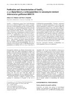
A concentration-response inhibition of lipopolysaccharide-stimulated TNFα (A) and IL-1β (B) release by Prolastin in human blood monocytesFigure 1
A concentration-response inhibition of lipopolysaccharide-stimulated TNFα (A) and IL-1β (B) release by Prolastin in human
blood monocytes. Isolated blood monocytes were treated with LPS (10 ng/ml) alone or together with various concentrations
of Prolastin (0–16 mg/ml) for 18 h. TNFα and IL-1β levels were measured by ELISA. Data are the means of quadruplicate cul-
ture supernatants ± S.E. and are representative of three separate experiments.
A
Prolastin (mg/ml)
0 2 4 6 8 10121416
TNF
D
(pg/ml)
0
2000
4000
6000
8000
10000
12000
Monocytes stimulated
with LPS (10 ng/ml)
B
Prolastin (mg/ml)
0 2 4 6 8 10 12 14 16
IL-1
E
(pg/ml)
0
1000
2000
3000
4000
5000
6000
7000
8000
Monocytes stimulated
with LPS (10 ng/ml)
Respiratory Research 2005, 6:12 />Page 6 of 11
(page number not for citation purposes)
Comparisons of the effects of native (nAAT), polymeric (pAAT) and Prolastin on lipopolysaccharide – stimulated TNFα (A) and IL-β (B) production by human blood monocytes isolated from four healthy donorsFigure 2
Comparisons of the effects of native (nAAT), polymeric (pAAT) and Prolastin on lipopolysaccharide – stimulated TNFα (A)
and IL-β (B) production by human blood monocytes isolated from four healthy donors. Isolated blood monocytes were treated
with LPS (10 ng/ml) alone or together with 0.5 mg/ml nAAT, pAAT or Prolastin for 18 h. TNFα and IL-1β levels were meas-
ured by ELISA. Each bar represent the mean ± S.E. *** p < 0.001.
A
0 LPS nAAT pAAT Prolastin
TNF
D
(pg/ml)
0
2000
4000
6000
8000
10000
12000
14000
Monocytes stimulated with LPS (10 ng/ml)
alone or in combination with AATs (0.5 mg/ml)
B
0 LPS nAAT pAAT Prolastin
IL-1
E
(pg/ml)
0
2000
4000
6000
8000
***
***
***
***
***
Respiratory Research 2005, 6:12 />Page 7 of 11
(page number not for citation purposes)
Effects of AATs on neutrophils activated with zymosanFigure 3
Effects of AATs on neutrophils activated with zymosan. (A) Concentration-dependent effects of Prolastin on IL-8 release from
neutrophils activated with opsonised zymosan. Freshly isolated blood neutrophils were treated with zymosan (0.3 mg/ml) alone
or together with various concentrations of Prolastin (0–8 mg/ml) for 18 h. IL-8 levels were measured by ELISA. Data are the
means of quadruplicate culture supernatants ± S.E. and are representative of three separate experiments. (B) Effects of opson-
ised zymosan alone or together with native (nAAT), polymeric (pAAT) AAT or Prolastin on IL-8 release from neutrophils. The
release of neutrophil IL-8 was measured in cell free supernatants as described in Materials and methods. Neutrophils were
treated for 18 h with a constant amount of zymosan (0.3 mg/ml) alone or together with nAAT, pAAT or Prolastin (0.5 mg/ml)
for 18 h. IL-8 levels were measured by ELISA. Each bar represents the means ± S.E. of three separate experiments carried out
in duplicate repeats. *** p < 0.001
A
Prolastin concentration (mg/ml)
02468
IL-8 (pg/ml)
0
10000
20000
30000
40000
50000
Neutrophils activated
with zymosan (0.3 mg/ml)
B
IL-8 (pg/ml)
0
10000
20000
30000
40000
50000
Neutrophils activated with zymosan (0.3 mg/ml)
alone or in combination with AATs (0.5 mg/ml)
***
***
***
Control
Zymosan
pAAT
nAAT
Prolastin
0
Respiratory Research 2005, 6:12 />Page 8 of 11
(page number not for citation purposes)
(Fig. 4). The levels of IL-8 increased already after 2 h of
LPS challenge (245.7% ± 87) and remained higher after
24 h (310 ± 77.5) compared to baseline (100% ± 19.2).
By contrast, when IL-8 levels were examined in LPS-Pro-
lastin-treated lavage samples, no significant changes in IL-
8 release were observed compared to baseline. In the pres-
ence of Prolastin, the LPS effect on IL-8 release was inhib-
ited (p < 0.05) (Fig. 4).
Disscussion
There is now, however, ample evidence that serine protei-
nase inhibitors (serpins), in addition to their well estab-
lished anti-inflammatory capacity to regulate serine
proteinases activity, may possess broader anti-inflamma-
tory properties. Several studies have shown that the bio-
logical responses of bacterial lipopolysaccharide
(endotoxin) in vivo may be sensitive to serpins. For exam-
ple, the serpin antithrombin, has been shown to protect
animals from LPS-induced septic shock and also to inhibit
IL-6 induction by LPS [42,43]. Our recent study provided
first in vitro evidence that native (inhibitor) and at least
two modified (non-inhibitory i.e. polymeric and oxi-
dised) forms of AAT can block the release of an array of
chemokine and cytokines from LPS-stimulated
IL-8 analysis in nasal lavage of subjects challenged with LPS alone or LPS+Prolastin combinationFigure 4
IL-8 analysis in nasal lavage of subjects challenged with LPS alone or LPS+Prolastin combination. Seven healthy volunteers were
treated with LPS (25 µg/nostril) or with LPS followed 30 min later with Prolastin (2.5 mg/nostril), nasal lavage was collected at
different time points (0, 2, 6 and 24 h) as described in Material and Methods. The concentration of IL-8 (pg/ml) was measured
by ELISA. IL-8 values are expressed as a ratio of IL-8 concentration at selected time point and the basal level. Independent two
sample t-test shows after 6 and 24 h significantly higher levels of IL-8 in subjects treated with LPS compared to LPS+Prolastin.
* p < 0.05
Time (h)
0 2 4 6 8 10 12 14 16 18 20 22 24
IL-8 (% of control)
100
200
300
400
LPS
LPS+Prolastin
*
*
Respiratory Research 2005, 6:12 />Page 9 of 11
(page number not for citation purposes)
monocytes [39]. These studies therefore further support a
central role of serpins in inflammation, not only as the
regulators of proteinase activity, but also as the suppress-
ers of endotoxin induced pro-inflammatory responses. In
line with these findings, we demonstrate here that Prolas-
tin, a preparation of human AAT which is used for aug-
mentation therapy, significantly inhibits endotoxin-
induced pro-inflammatory effects in vitro and in vivo.
Stimulation of human monocytes and neutrophils with
bacterial endotoxin results in the release of a range of
inflammatory mediators including the pro-inflammatory
cytokines (e.g. IL-6, IL-1β and TNFα) and the chemokines
(e.g. MCP-1 and IL-8) [44-46]. Together, these play a cru-
cial role in the recruitment and activation of leukocytes
and the subsequent release of harmful proteases that may
further perpetuate the inflammatory process. We found
that Prolastin significantly inhibits endotoxin-induced IL-
1β and TNFα release by monocytes and IL-8 release by
neutrophils in vitro. The Prolastin exhibited these anti-
inflammatory properties in a concentration-dependent
manner. Its maximal effects were observed with 16 mg/ml
in the monocyte model and with 8 mg/ml in the neu-
trophil model, since doubling these concentrations did
not significantly modify the intensity of the effects.
Indeed, Prolastin markedly prevented endotoxin-induced
cell activation at 0.5–4 mg/ml concentrations, implying
that these lower concentrations of Prolastin might also be
sufficient to inhibit endotoxin effects. It is worth noting
that in order to reduce a potential risk of transmission of
infectious agents the Prolastin preparation is heat-treated
in solution at 60° ± 0.5 for not less than 10 h. Data from
in vitro studies show that heat-treatment results in AAT
polymerization and loss of its inhibitory activity [47,48].
Therefore, in our experimental model we compared anti-
inflammatory effects of Prolastin with those of native and
heat treated (60°C 10 h) AATs. At concentrations used
(0.5 mg/ml), no significant difference was found between
the effects of Prolastin and native or heat-treated (poly-
meric) AAT on endotoxin-induced monocyte TNFα and
neutrophil IL-8 elevation. The median concentrations of
endotoxin-stimulated IL-1β levels also decreased in the
presence of Prolastin but failed to reach statistical signifi-
cance. In general, inhibitory effects on endotoxin-stimu-
lated monocyte IL-1β and neutrophil IL-8 release were
better pronounced by native AAT compared to polymeric
AAT or Prolastin. Similarly, in our previous study we
found that in terms of maximal effect, native AAT >poly-
merised AAT>oxidized AAT were efficient in inhibiting
LPS-stimulated TNFα and IL-1β, and IL-8 release from
monocytes [39]. Further studies will be necessary to better
evaluate how temperature, pH or other physicochemical
challenges may influence anti-inflammatory effectiveness
of AAT preparations.
To explore our hypothesis that AAT functions as a potent
inhibitor of endotoxin-induced effects, we examined
whether Prolastin also inhibits responses to LPS in the
nasal airway, in vivo. In particular, we were interested in
concentrations of the neutrophil chemoattractant, IL-8.
Endotoxin (or LPS) from gram-negative bacteria is a com-
mon air contaminant in a number of occupational condi-
tions, especially those in which exposure to animal waste
or plant matter occurs [44,49-51]. Levels of LPS in such
environments may exceed 20 µg/m
3
air and may be asso-
ciated with respiratory symptoms and nasal inflammation
in exposed persons [52]. For example, nasal inflammation
as evaluated by an increased influx of inflammatory cells
into the nasal airway and increased IL-8 levels, has been
described in persons occupationally exposed to LPS [51].
Moreover, it has been suggested that constitutive levels of
IL-8 might further enhance responses to an inflammatory
stimulus, such as LPS [53]. A number of experimental
studies have shown that a nasal instillation of LPS causes
the cytokine and chemokine reaction [54,55]. In our pilot
study we also showed that instilled defined amounts of
endotoxin (25 µg/per nostril) induce time-dependent
nasal IL-8 release in normal subjects. Two hours after LPS
instillation the IL-8 levels in nasal lavage reached more
than twice the basal level and remained higher during all
the times studied. However, during the next session, when
30 min after challenge with LPS, Prolastin (2.5 mg/ per
nostril) was instilled, no induction of nasal IL-8 release
was found compared to the basal levels. Furthermore, the
protective ability of Prolastin did not disappeared over
study time. We cannot determine from these experiments
whether Prolastin is directly suppressing IL-8 release or
suppressing another inflammatory response that leads to
IL-8 release; nonetheless, our finding suggests that effects
of Prolastin directed against endotoxin-stimulated
inflammatory responses may be beneficial.
Thus, data from both in vitro and in vivo experiments pro-
vide novel evidence that the Prolastin preparation is a
potent inhibitor of endotoxin effects. The major concept
behind augmentation therapy with pooled plasma-
derived AAT has been that a rise in the level of AAT in sub-
jects with severe inherited AAT deficiency would protect
the lung tissue from continued destruction by proteinases
(i.e. primarily leukocyte elastase) [7,56,57]. Recent find-
ings provide evidence that augmentation therapy with
AAT reduces the incidence of lung infections in patients
with AAT-related emphysema [28,58]. Furthermore, Can-
tin and Woods have reported that aerosolized AAT sup-
presses bacterial proliferation in a rat model of chronic
Pseudomonas aeruginosa lung infection [59]. Stockley and
co-workers demonstrated that a short-term therapy of AAT
augmentation not only restores airway concentrations of
AAT to normal, but also reduces levels of leukotriene B4,
a major mediator of neutrophil recruitment and
Respiratory Research 2005, 6:12 />Page 10 of 11
(page number not for citation purposes)
activation. Interestingly, authors have suggested that the
efficacy of AAT augmentation may be most beneficial in
individuals with the most inflammation [29,60]. Data
presented in this study clearly show that Prolastin inhibits
endotoxin-stimulated pro-inflammatory responses, and
thus provides new biochemical evidence supporting the
efficacy of augmentation therapy. The current findings
also suggest that Prolastin may, in fact, be used for
broader clinical applications than merely augmentation
therapy.
Abbreviations
AAT, α1-antitrypsin; COPD, chronic obstructive pulmo-
nary disease; LPS, lipopolysaccharide; ZZ, homozygous
AAT-deficiency variant; MM, wild type AAT variant; PBS,
phosphate buffered saline; EDTA, ethylenediamine-
tetraacetic acid; HEPES, 4-(2-hydroxyethyl)-1-pipera-
zineethanesulfonic acid
Authors' contribution
Izabela Nita, performed cell culture experiments, made
contribution to acquisition of data;
Camilla Hollander, made substantial contribution to
patient study design, material collection and analysis;
Ulla Westin, contributed to study design and data inter-
pretation; Sabina Janciauskiene, contributed to concep-
tion and study design, data interpretation and wrote the
article
Acknowledgments
This work was supported by grants from the Swedish Research Council,
and Department of Medicine, Lund University, Sweden.
References
1. Potempa J, Korzus E, Travis J: The serpin superfamily of protei-
nase inhibitors: structure, function, and regulation. J Biol Chem
1994, 269:15957-15960.
2. Brantly ML: Alpha-1-antitrypsin genotypes and phenotypes. In
Alpha-1-antitrypsin Edited by: Crystal RG. New York, Marcel Dekker;
1996:45-59.
3. Kalsheker N, Morley S, Morgan K: Gene regulation of the serine
proteinase inhibitors alpha1-antitrypsin and alpha1-antichy-
motrypsin. Biochem Soc Trans 2002, 30:93-98.
4. Olsen GN, Harris JO, Castle JR, Waldman RH, Karmgard HJ: Alpha-
1-antitrypsin content in the serum, alveolar macrophages,
and alveolar lavage fluid of smoking and nonsmoking normal
subjects. J Clin Invest 1975, 55:427-430.
5. Travis J, Shieh BH, Potempa J: The functional role of acute phase
plasma proteinase inhibitors. Tokai J Exp Clin Med 1988,
13:313-320.
6. Crystal RG: The alpha 1-antitrypsin gene and its deficiency
states. Trends Genet 1989, 5:411-417.
7. Hutchison DC: Natural history of alpha-1-protease inhibitor
deficiency. Am J Med 1988, 84:3-12.
8. Needham M, Stockley RA: Alpha 1-antitrypsin deficiency. 3:
Clinical manifestations and natural history. Thorax 2004,
59:441-445.
9. Lomas DA, Mahadeva R: Alpha1-antitrypsin polymerization and
the serpinopathies: pathobiology and prospects for therapy.
J Clin Invest 2002, 110:1585-1590.
10. Luisetti M, Seersholm N: Alpha1-antitrypsin deficiency. 1: epi-
demiology of alpha1-antitrypsin deficiency. Thorax 2004,
59:164-169.
11. Carrell RW, Lomas DA: Alpha1-antitrypsin deficiency a model
for conformational diseases. N Engl J Med 2002, 346:45-53.
12. Eriksson S: Alpha 1-antitrypsin deficiency. J Hepatol 1999, 30
Suppl 1:34-39.
13. Wiedemann HP, Stoller JK: Lung disease due to alpha 1-antit-
rypsin deficiency. Curr Opin Pulm Med 1996, 2:155-160.
14. Stockley RA: Alpha-1-antitrypsin deficiency: what next? Thorax
2000, 55:614-618.
15. Sandford AJ, Weir TD, Spinelli JJ, Pare PD: Z and S mutations of
the alpha1-antitrypsin gene and the risk of chronic obstruc-
tive pulmonary disease. Am J Respir Cell Mol Biol 1999, 20:287-291.
16. Talamo RC, Langley CE, Levine BW, Kazemi H: Genetic vs. quan-
titative analysis of serum alpha 1 -antitrypsin. N Engl J Med
1972, 287:1067-1069.
17. Guenter CA, Welch MH, Ferguson S, Henderson L, Hammarsten JF:
Alpha-1-antitrypsin deficiency: heterozygosity, intermediate
levels, and pulmonary disease. Chest 1971, 59:Suppl:16S+.
18. Sandford AJ, Silverman EK: Chronic obstructive pulmonary dis-
ease. 1: Susceptibility factors for COPD the genotype-envi-
ronment interaction. Thorax 2002, 57:736-741.
19. Chow CK: Cigarette smoking and oxidative damage in the
lung. Ann N Y Acad Sci 1993, 686:289-298.
20. Laurell CB, Eriksson S: The electrophoretic alpha-1-globulin
pattern of serum in alpha-1-antitrypsin dificiency. Scand J Clin
Lab Invest 1963, 15:132-140.
21. Gross P, deTreville RT, Babyak MA, Kaschak M, Tolker EB: Experi-
mental emphysema: effect of chronic nitrogen dioxide expo-
sure and of papain on normal and pneumoconiotic lungs.
Aspen Emphysema Conf 1967, 10:357-378.
22. Lieberman J, Winter B, Sastre A: Alpha 1-antitrypsin Pi-types in
965 COPD patients. Chest 1986, 89:370-373.
23. Lieberman J: Intermediate antitrypsin deficiency. Am Rev Respir
Dis 1990, 141:1078.
24. Gadek JE, Klein HG, Holland PV, Crystal RG: Replacement ther-
apy of alpha 1-antitrypsin deficiency. Reversal of protease-
antiprotease imbalance within the alveolar structures of PiZ
subjects. J Clin Invest 1981, 68:1158-1165.
25. Wewers MD, Casolaro MA, Sellers SE, Swayze SC, McPhaul KM,
Wittes JT, Crystal RG: Replacement therapy for alpha 1-antit-
rypsin deficiency associated with emphysema. N Engl J Med
1987, 316:1055-1062.
26. Hubbard RC, Brantly ML, Sellers SE, Mitchell ME, Crystal RG: Anti-
neutrophil-elastase defenses of the lower respiratory tract in
alpha 1-antitrypsin deficiency directly augmented with an
aerosol of alpha 1-antitrypsin. Ann Intern Med 1989, 111:206-212.
27. Stoller JK, Aboussouan LS: alpha1-Antitrypsin deficiency . 5:
intravenous augmentation therapy: current understanding.
Thorax 2004, 59:708-712.
28. Lieberman J: Augmentation therapy reduces frequency of lung
infections in antitrypsin deficiency: a new hypothesis with
supporting data. Chest 2000, 118:1480-1485.
29. Stockley RA, Hill AT, Hill SL, Campbell EJ: Bronchial inflamma-
tion: its relationship to colonizing microbial load and
alpha(1)-antitrypsin deficiency. Chest 2000, 117:291S-3S.
30. Dabbagh K, Laurent GJ, Shock A, Leoni P, Papakrivopoulou J, Cham-
bers RC: Alpha-1-antitrypsin stimulates fibroblast prolifera-
tion and procollagen production and activates classical MAP
kinase signalling pathways. J Cell Physiol 2001, 186:73-81.
31. Jeannin P, Lecoanet-Henchoz S, Delneste Y, Gauchat JF, Bonnefoy JY:
Alpha-1 antitrypsin up-regulates human B cell differentia-
tion selectively into IgE- and IgG4- secreting cells. Eur J
Immunol 1998, 28:1815-1822.
32. Ikari Y, Mulvihill E, Schwartz SM: alpha 1-Proteinase inhibitor,
alpha 1-antichymotrypsin, and alpha 2-macroglobulin are
the antiapoptotic factors of vascular smooth muscle cells. J
Biol Chem 2001, 276:11798-11803.
33. Daemen MA, Heemskerk VH, van't Veer C, Denecker G, Wolfs TG,
Vandenabeele P, Buurman WA: Functional protection by acute
phase proteins alpha(1)-acid glycoprotein and alpha(1)-anti-
trypsin against ischemia/reperfusion injury by preventing
apoptosis and inflammation. Circulation 2000, 102:1420-1426.
34. Graziadei I, Gaggl S, Kaserbacher R, Braunsteiner H, Vogel W: The
acute-phase protein alpha 1-antitrypsin inhibits growth and
proliferation of human early erythroid progenitor cells
(burst-forming units-erythroid) and of human erythroleuke-
Publish with BioMed Central and every
scientist can read your work free of charge
"BioMed Central will be the most significant development for
disseminating the results of biomedical research in our lifetime."
Sir Paul Nurse, Cancer Research UK
Your research papers will be:
available free of charge to the entire biomedical community
peer reviewed and published immediately upon acceptance
cited in PubMed and archived on PubMed Central
yours — you keep the copyright
Submit your manuscript here:
/>BioMedcentral
Respiratory Research 2005, 6:12 />Page 11 of 11
(page number not for citation purposes)
mic cells (K562) in vitro by interfering with transferrin iron
uptake. Blood 1994, 83:260-268.
35. Bucurenci N, Blake DR, Chidwick K, Winyard PG: Inhibition of
neutrophil superoxide production by human plasma alpha 1-
antitrypsin. FEBS Lett 1992, 300:21-24.
36. Churg A, Dai J, Zay K, Karsan A, Hendricks R, Yee C, Martin R, Mac-
Kenzie R, Xie C, Zhang L, Shapiro S, Wright JL: Alpha-1-antitrypsin
and a broad spectrum metalloprotease inhibitor, RS113456,
have similar acute anti-inflammatory effects. Lab Invest 2001,
81:1119-1131.
37. Jie Z, Cai Y, Yang W, Jin M, Zhu W, Zhu C: Protective effects of
alpha 1-antitrypsin on acute lung injury in rabbits induced by
endotoxin. Chin Med J (Engl) 2003, 116:1678-1682.
38. Libert C, Van Molle W, Brouckaert P, Fiers W: alpha1-Antitrypsin
inhibits the lethal response to TNF in mice. J Immunol 1996,
157:5126-5129.
39. Janciauskiene S, Larsson S, Larsson P, Virtala R, Jansson L, Stevens T:
Inhibition of lipopolysaccharide-mediated human monocyte
activation, in vitro, by alpha1-antitrypsin. Biochem Biophys Res
Commun 2004, 321:592-600.
40. Lowry OH, Rosebrough NJ, Farr AL, Randall RJ: Protein measure-
ment with the Folin phenol reagent. J Biol Chem 1951,
193:265-275.
41. Wihl JA, Baumgarten CR, Petersson G: Contralateral differences
among biomarkers determined by a modified nasal lavage
technique after unilateral antigen challenge. Allergy 1995,
50:308-315.
42. Dickneite G, Leithauser B: Influence of antithrombin III on coag-
ulation and inflammation in porcine septic shock. Arterioscler
Thromb Vasc Biol 1999, 19:1566-1572.
43. Souter PJ, Thomas S, Hubbard AR, Poole S, Romisch J, Gray E: Anti-
thrombin inhibits lipopolysaccharide-induced tissue factor
and interleukin-6 production by mononuclear cells, human
umbilical vein endothelial cells, and whole blood. Crit Care Med
2001, 29:134-139.
44. Heumann D, Roger T: Initial responses to endotoxins and
Gram-negative bacteria. Clin Chim Acta 2002, 323:59-72.
45. Harada A, Sekido N, Akahoshi T, Wada T, Mukaida N, Matsushima K:
Essential involvement of interleukin-8 (IL-8) in acute
inflammation. J Leukoc Biol 1994, 56:559-564.
46. Anderson P, Phillips K, Stoecklin G, Kedersha N: Post-transcrip-
tional regulation of proinflammatory proteins. J Leukoc Biol
2004, 76:42-47.
47. Chow MK, Devlin GL, Bottomley SP: Osmolytes as modulators of
conformational changes in serpins. Biol Chem 2001,
382:1593-1599.
48. Huntington JA, Pannu NS, Hazes B, Read RJ, Lomas DA, Carrell RW:
A 2.6 A structure of a serpin polymer and implications for
conformational disease. J Mol Biol 1999, 293:449-455.
49. Schwartz DA, Thorne PS, Yagla SJ, Burmeister LF, Olenchock SA,
Watt JL, Quinn TJ: The role of endotoxin in grain dust-induced
lung disease. Am J Respir Crit Care Med 1995, 152:603-608.
50. Schwartz DA, Thorne PS, Jagielo PJ, White GE, Bleuer SA, Frees KL:
Endotoxin responsiveness and grain dust-induced inflamma-
tion in the lower respiratory tract. Am J Physiol 1994,
267:L609-17.
51. Peden DB, Tucker K, Murphy P, Newlin-Clapp L, Boehlecke B,
Hazucha M, Bromberg P, Reed W: Eosinophil influx to the nasal
airway after local, low-level LPS challenge in humans. J Allergy
Clin Immunol 1999, 104:388-394.
52. Blaski CA, Watt JL, Quinn TJ, Thorne PS, Schwartz DA: Nasal lav-
age cellularity, grain dust, and airflow obstruction. Chest 1996,
109:1086-1092.
53. Sigsgaard T, Bonefeld-Jorgensen EC, Kjaergaard SK, Mamas S, Peder-
sen OF: Cytokine release from the nasal mucosa and whole
blood after experimental exposures to organic dusts. Eur
Respir J 2000, 16:140-145.
54. Danuser B, Rebsamen H, Weber C, Krueger H: Lipopolysaccha-
ride-induced nasal cytokine response: a dose-response
evaluation. Eur Arch Otorhinolaryngol 2000, 257:527-532.
55. Besancon-Watelet C, Bene MC, Montagne P, Faure GC, Jankowski R:
Eosinophilia and cell activation mediators in nasal
secretions. Laryngoscope 2002, 112:43-46.
56. Stoller JK: Alpha-1 antitrypsin deficiency: an under-recog-
nized but important issue for respiratory therapists. Respir
Care 2003, 48:1022-1024.
57. Hill AT, Bayley DL, Campbell EJ, Hill SL, Stockley RA: Airways
inflammation in chronic bronchitis: the effects of smoking
and alpha1-antitrypsin deficiency. Eur Respir J 2000, 15:886-890.
58. Dirksen A, Dijkman JH, Madsen F, Stoel B, Hutchison DC, Ulrik CS,
Skovgaard LT, Kok-Jensen A, Rudolphus A, Seersholm N, Vrooman
HA, Reiber JH, Hansen NC, Heckscher T, Viskum K, Stolk J: A ran-
domized clinical trial of alpha(1)-antitrypsin augmentation
therapy. Am J Respir Crit Care Med 1999, 160:1468-1472.
59. Cantin AM, Woods DE: Aerosolized prolastin suppresses bac-
terial proliferation in a model of chronic Pseudomonas aer-
uginosa lung infection. Am J Respir Crit Care Med 1999,
160:1130-1135.
60. Stockley RA, Bayley DL, Unsal I, Dowson LJ: The effect of augmen-
tation therapy on bronchial inflammation in alpha1-antit-
rypsin deficiency. Am J Respir Crit Care Med 2002, 165:1494-1498.