Báo cáo y học: " Association between reduced bronchodilatory effect of deep inspiration and loss of alveolar attachments" pps
Bạn đang xem bản rút gọn của tài liệu. Xem và tải ngay bản đầy đủ của tài liệu tại đây (467.7 KB, 7 trang )
BioMed Central
Page 1 of 7
(page number not for citation purposes)
Respiratory Research
Open Access
Research
Association between reduced bronchodilatory effect of deep
inspiration and loss of alveolar attachments
Nicola Scichilone*
1
, Andreina Bruno
2
, Roberto Marchese
1
,
Antonio Maurizio Vignola
1,2
, Alkis Togias
3
and Vincenzo Bellia
1
Address:
1
Istituto di Medicina Generale e Pneumologia, Cattedra di Malattie dell'Apparato Respiratorio, Università di Palermo, via Trabucco 180,
90146 Palermo, Italy,
2
Istituto di Biomedicina ed Immunologia Molecolare, Consiglio Nazionale delle Ricerche, Via Ugo La Malfa 153, 90146
Palermo, Italy and
3
Division of Allergy and Clinical Immunology, and Division of Respiratory and Critical Care Medicine, Department of
Medicine, Johns Hopkins University, School of Medicine, 5501 Hopkins Bayview Circle 21224, Baltimore, Maryland, USA
Email: Nicola Scichilone* - ; Andreina Bruno - ; Roberto Marchese - ;
Antonio Maurizio Vignola - ; Alkis Togias - ; Vincenzo Bellia -
* Corresponding author
Abstract
Background: We have previously shown that the bronchodilatory effect of deep inspiration is
attenuated in individuals with COPD. This study was designed to investigate whether the
impairment in this effect is associated with loss of alveolar attachments.
Methods: We measured deep inspiration (DI)-induced bronchodilation in 15 individuals with and
without COPD (67 ± 2.2 yrs of age, mean ± SEM) undergoing lobar resection for peripheral
pulmonary nodule. Prior to surgery, we measured TLCO and determined the bronchodilatory
effect of deep inspiration after constricting the airways with methacholine. The number of
destroyed alveolar attachments, as well as airway wall area and airway smooth muscle area, were
determined in tumor-free, peripheral lung tissue.
Results: The bronchodilatory effect of deep inspiration correlated inversely with the % destroyed
attachments (r = -0.51, p = 0.05) and directly with the airway smooth muscle area (r = 0.59, p =
0.03), but not with the total wall area (r = 0.39, p = 0.15).
Conclusion: We postulate that attenuation of airway stretch due to loss of alveolar attachments
contributes to the loss of the bronchodilatory effect of lung inflation in COPD.
Background
We have recently demonstrated that the ability of deep
inspirations to dilate constricted airways is impaired in
subjects with COPD [1]. We have suggested that the lack
of deep inspiration-induced bronchodilation may be one
of the major factors that contribute to persistent airway
narrowing in chronic obstructive pulmonary diseases.
However, the mechanism accounting for the reduction in
the bronchodilatory effect of deep inspiration in COPD
has not yet been elucidated.
When a deep inspiration takes place, radial traction is
applied to the outer airway walls by virtue of the forces of
interdependence between the airways and the surround-
ing parenchyma [2], which are sustained by the connec-
tive tissue of the lungs. As a consequence, lung inflation
Published: 08 June 2005
Respiratory Research 2005, 6:55 doi:10.1186/1465-9921-6-55
Received: 07 March 2005
Accepted: 08 June 2005
This article is available from: />© 2005 Scichilone et al; licensee BioMed Central Ltd.
This is an Open Access article distributed under the terms of the Creative Commons Attribution License ( />),
which permits unrestricted use, distribution, and reproduction in any medium, provided the original work is properly cited.
Respiratory Research 2005, 6:55 />Page 2 of 7
(page number not for citation purposes)
produces transient airway distension. If the airways are
constricted, the stretch imposed by airway distension may
produce bronchodilation [3,4].
The loss of the bronchodilatory effect of lung inflation in
COPD may result from factors that unlink the paren-
chyma from the airways. COPD is accompanied by
destructive changes of alveolar walls and consequent
reduction in the number of alveolar attachments on the
airways [5-7]. We postulated that, because of the alveolar
wall destruction, mechanical decoupling between airways
and parenchyma results in diminished airway wall stretch,
thus impairing the postulated primary step in the mecha-
nism of bronchodilation by deep inspiration. The current
study showed that the reduction in alveolar attachments
on the airways correlates with the reduction in the bron-
chodilatory ability of deep inspiration.
Methods
In order to obtain lung tissue for morphometric and cor-
relative analyses, we enrolled subjects undergoing lobar
resection for peripheral pulmonary nodule, with the
assumption that a significant proportion would have been
smokers and would presumably show evidence of
reduced integrity of alveolar attachments [6]. Subjects
were recruited from the Unit of Thoracic Surgery, "V. Cer-
vello" Palermo Hospital, Italy. After giving their informed
consent, subjects were referred to the Istituto di Medicina
Generale e Pneumologia, Palermo University, for poten-
tial participation in this protocol. Exclusion criteria for
participation in the study were a large involvement of the
lung and/or mediastinal organs by the tumor, a history of
myocardial infarction, congestive heart failure, arrhyth-
mia, and any unstable clinical condition, such as bron-
chial exacerbations. The diagnosis of COPD was made
according to the diagnostic criteria of the GOLD (Global
Initiative for Chronic Obstructive Lung Disease) guide-
lines [8]. Several subjects with COPD were using bron-
chodilators and three subjects were using inhaled
corticosteroids. The study was approved by the local Ethic
Committee.
Study design
Clinical and functional assessment
Prior to surgical lung resection, each subject underwent a
clinical evaluation and a complete respiratory functional
assessment that included spirometry, and determinations
of lung volume and of the single-breath CO diffusing
capacity.
The clinical evaluation included a questionnaire that
derives from the IUALTD (International Union Against
Lung and Tuberculosis Disease) bronchial symptom ques-
tionnaire [9] and a physical examination. Total lung
capacity (TLC) was determined by bodyplethysmography
(Sensor Medics Corporation V6200 AutoBox, Yorba
Linda, CA). TLC was expressed as percent predicted based
on the prediction equation of Goldman and Becklake
[10]. Single-breath diffusing capacity for CO was deter-
mined using a fully-computerized water-sealed Stead-
Wells spirometer (Baires System; Biomedin, Padua, Italy)
and the transfer factor of the lung for CO (TLCO) was
measured. At least two determinations of TLCO that were
within 5% of each other were obtained, and the highest
value was retained for analysis.
In a series of subsequent visits, the bronchodilatory effect
of deep inspirations was determined, as previously
described [1,11]. Inhaled short-acting β-agonists and/or
anticholinergic agents were withheld for at least 8 h and
long-acting β-agonists for at least 24 h before each visit
that involved either lung function or methacholine bron-
choprovocation. A series of single dose methacholine
bronchoprovocations (a single dose per challenge) were
performed, in the absence of deep inspirations, using
stepwise increasing doses of methacholine. All subjects
started with 0.025 mg/ml and increased the dose by a fac-
tor of 2 at each visit until an at least 15% reduction in
inspiratory vital capacity (IVC) from baseline, under deep
breath prohibition (Figure 1). Methacholine was deliv-
ered through an ampul-dosimeter (Mefar Elettromedicali;
Bovezzo, Italy), which was activated by an inspiratory
effort for 0.5 seconds at a time.
To measure IVC, the subject forcefully expired from end-
tidal volume to residual volume (partial expiratory
maneuver) and immediately inhaled to total lung capacity
(IVC = TLC – RV). The maneuver continued with a forced
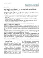
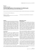
Schematic of the single dose methacholine bronchoprovoca-tion protocol designed to induce at least a 15% reduction in inspiratory vital capacity (IVC), in the absence of deep breaths, and to calculate the bronchodilatory effect of deep inspirationsFigure 1
Schematic of the single dose methacholine bronchoprovoca-
tion protocol designed to induce at least a 15% reduction in
inspiratory vital capacity (IVC), in the absence of deep
breaths, and to calculate the bronchodilatory effect of deep
inspirations. The combination spirometric maneuvers (partial
forced expiration followed by full forced expiration) used to
determine IVC are also depicted.
Respiratory Research 2005, 6:55 />Page 3 of 7
(page number not for citation purposes)
expiration to RV. This forced expiration allowed us to also
calculate FEV
1
and FVC. At baseline, 3 acceptable com-
bined partial/maximal forced expiratory maneuvers were
performed, and the best was retained for analysis. Subjects
were then instructed to abstain from taking deep breaths
for a period of 20 minutes. Thereafter, the single dose of
methacholine was administered as five tidal breaths fol-
lowed, 3 minutes later, by a single partial/maximal com-
bined spirometric measurement, as described above. If the
targeted reduction in IVC (at least 15%) was not attained,
another single dose challenge was performed. This was
done during the same session (at one hour) if the reduc-
tion in IVC was within 5% of baseline, or postponed to
the next day. Single dose provocations with increasing
doses were conducted in this manner until the expected
level of reduction in IVC was reached. The provocation in
which the targeted reduction in IVC from baseline was
attained was extended with 4 deep inspirations taken
immediately after the post-methacholine IVC spirometry.
Following these deep inspirations, another IVC maneuver
was performed. The difference between the IVC obtained
after the 4 deep inspirations and the post-methacholine
IVC that preceded the 4 deep inspirations was used to cal-
culate the bronchodilatory effect of deep inspirations.
This was expressed as percent change from the post-meth-
acholine IVC (% bronchodilation).
We have used IVC in studying the effects of deep inspira-
tion because, assuming that TLC does not change [12], the
primary determinant of a change in IVC is the change in
residual volume. Although IVC is sensitive to the effects of
deep inspirations, its actual measurement, in contrast to
that of FVC or FEV
1
, is not influenced by a lung inflation
maneuver because RV is reached through a partial forced
expiration. FEV
1
and FVC data were utilized in secondary
analyses.
Morphometric assessment
For tissue morphometry, we applied the methodology
previously described by Saetta and colleagues [6]. Two to
seven randomly selected tissue blocks were taken from the
subpleural parenchyma of the resected lobe that was
tumor-free. Specimens were fixed in 10% neutral buffered
formalin (pH 7.2) for at least 24 h and embedded in par-
affin wax. Four-µm sections were attached to microscope
slides pretreated with polylysine solution (Sigma
Aldrich). After dewaxing and rehydratation, all slides were
stained with haematoxylin and eosin. Tissue samples were
coded and evaluated blindly by two independent investi-
gators using a light microscope (Leica, Wetzlar, Ger-
many). The images were analyzed by a computerized
system (Quantimet 500 MC software, Leica, Wetzlar,
Germany).
All airways with internal diameter ≤ 2 mm were retained
for analysis. Non-respiratory bronchioles with incomplete
walls at the edges of the sections or with a short/long
diameter ratio < 1/3 were excluded. After this selection,
each patient had at least four non-respiratory bronchioles
suitable for morphometry. In each airway, the external
perimeter (P
e
), the internal perimeter along the subepi-
thelial basement membrane (P
bm
), the lumenal diameter
(Dl), the external area (Ae), the internal area (Ai), and the
muscle area (WAm) were evaluated. The thickness of the
nonrespiratory bronchioles (wall area, WAtot) was
obtained by the difference between the external area and
the internal areas (WA = Ae – Ai). Values of Dl, WAm and
WAtot were normalized by dividing for P
bm
[13].
According to the method of Saetta and colleagues [6],
alveolar attachments (AAi) were identified as the alveolar
septa that extend from the outer wall of the nonrespira-
tory bronchioles. Those attachments showing rupture or
discontinuity were defined as destroyed alveolar
attachments (AAd). The number of destroyed alveolar
attachments, expressed as a percentage over the total
number of alveolar attachments, represented the primary
outcome of the study. The data used for analysis were
averages of those obtained independently from each the
two study pathologists.
Data analysis
Linear regression analysis was performed to correlate the
bronchodilatory effect of deep inspirations with the mor-
phometric variables obtained from the lung tissue. Sec-
ondary analysis using the same approach was employed
to assess the relationship between the magnitude of bron-
choconstriction that was induced in the absence of deep
breaths (in terms of IVC and FEV1) and the morphometric
variables. Unpaired t-tests were used to assess differences
between groups. In all analyses, two-tailed values of p =
0.05 were considered statistically significant.
Results
Descriptive findings
A total of fifteen subjects took part in the study (age: 67 ±
2.2 yrs, mean ± SEM). Seven of them had a diagnosis of
COPD, confirmed by our clinical and functional evalua-
tion. No subject received a diagnosis of asthma. Eleven
out of the 15 subjects were smokers (69 ± 27 pack-years,
mean ± SD). None of the non-smokers received the diag-
nosis of COPD. Baseline lung function and lung tissue
morphometric characteristics for each individual are pre-
sented in Table 1.
The median single methacholine dose required to induce
the targeted reduction in IVC, in the absence of deep
inspiration, was 25 mg/ml (range: 0.025–75 mg/ml). The
% reduction in IVC in the protocol devoid of deep
Respiratory Research 2005, 6:55 />Page 4 of 7
(page number not for citation purposes)
inspiration was 20 ± 1.8% (mean ± SEM). The % bron-
chodilation by deep inspiration was 4.3 ± 2.1% with a
range of -13% to 18%.
Correlative findings
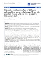
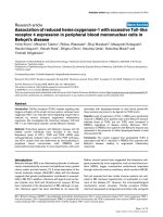
We found a significant inverse correlation between the
bronchodilatory effect of deep inspiration and the per-
centage of destroyed alveolar attachments (r = -0.51, p =
0.05, Figure 2). In addition, the bronchodilatory effect of
deep inspiration correlated directly with the airway
smooth muscle area (r = 0.59, p = 0.03). In contrast, no
correlation with the magnitude of the total wall area (r =
0.39, p = 0.15) was observed. The multiple regression
analysis, in which the bronchodilatory effect of deep
inspiration is the dependent variable, and the percent of
destroyed alveolar attachments and the airway smooth
muscle area serve as independent variables, yielded a p
value of 0.02; however, neither the alveolar attachment (p
= 0.09) nor the airway smooth muscle (p = 0.06) entered
the model. The bronchodilatory effect of deep inspiration
did not differ between subjects with COPD and the 4 sub-
jects without COPD, but with a history of smoking (2.6 ±
4.2% vs. 5.2 ± 3.6%, respectively; p = 0.68). Similarly, no
differences were found between COPD subjects and the
non-COPD smokers with respect to the percentage of
destroyed alveolar attachments (40 ± 7.2% vs. 35 ± 3.9%,
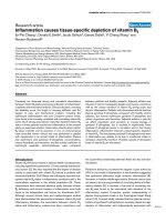
respectively; p = 0.64). When the entire group was
considered, the % destroyed attachments showed a strong
inverse correlation with TLCO% predicted (r= -0.75, p =
0.003) (Figure 3).
Table 1: Functional and morphometric characteristics of study participants.
Mean ± SEM Range
FEV1, % predicted 82 ± 6.2 43–118
FEV1/FVC 0.65 ± 0.03 0.44–0.77
TLC, % predicted 117 ± 14.3 82–164
TLCO, % predicted 74 ± 3.9 55–92
WAtot/Pbm, µm 80 ± 7.9 52–154
WAm/Pbm, µm 11 ± 1.6 6–22
AAd, % 33 ± 4.0 12–61
Relationship between the bronchodilatory effect of deep inspiration and the percentage of destroyed alveolar attachmentsFigure 2
Relationship between the bronchodilatory effect of deep inspiration and the percentage of destroyed alveolar attachments.
Respiratory Research 2005, 6:55 />Page 5 of 7
(page number not for citation purposes)
Discussion
We have recently documented the lack of deep inspira-
tion-induced bronchodilation in COPD [1]. The results of
this study confirm and extend our previous report.
Herein, we provide a possible explanation for this phe-
nomenon, by showing that the impairment of the bron-
chodilatory effect of deep inspiration is associated with
reduction in the alveolar attachments to the airway walls.
A significant correlation is not a proof for a causative rela-
tionship, but it is a pre-requisite for it. Moreover, there is
good theoretical reason to propose that the reduction in
the number of alveolar attachments is the most important
factor responsible for the impairment in deep inspiration-
induced bronchodilation, in smokers and individuals
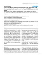
with COPD. A body of evidence has suggested that the
effect of deep inspiration on the airways is a function of
the interdependence between the airways and the paren-
chyma [14,15], provided by the alveolar attachments that
act by distending the airways when lung volume increases
(Figure 4), and by the relative magnitudes of airway and
parenchymal hystereses [16]. According to this theory,
equal degrees of hysteresis result in no effect of a deep
inspiration on airway caliber. If parenchymal hysteresis
prevails, such as in COPD [17], a deep inspiratory maneu-
ver fails to dilate airways, and may even result in
bronchoconstriction. Therefore, the impairment in the
bronchodilatory effect of deep inspiration in subjects with
COPD could be explained by the increased ratio of paren-
chymal over airway hysteresis.
We reasoned that structural alterations of the lung paren-
chyma, specifically the destruction of alveolar attach-
ments, would reduce the effectiveness of the distending
forces in a manner that a deep inspiration would not be
capable of stretching narrowed airways and/or reopen
closed airways. However, other explanations need to be
considered: first, increased airway smooth muscle mass
could render the muscle too stiff to stretch, or generate
higher forces that could counteract bronchodilation.
However, we found that larger smooth muscle area was
related to stronger bronchodilation by deep inspiration.
Second, COPD could be associated with enhancement of
a bronchoconstriction reflex that is activated by lung infla-
tion, or with the failure to release bronchodilatory agents.
Third, under a condition of reduced stretch, the airway
smooth muscle could develop peculiar rearrangement of
the contractile elements that would induce a state of
increased resistance to the effect of deep inspiration.
Finally, in a condition of lung hyperinflation, which is
often recorded in subjects with emphysema, the ampli-
tude of a deep inspiration could be severely reduced.
Corsico et al. [18] recently reported findings that have
similarities to ours. The authors showed that the loss of
alveolar attachments is associated with a
bronchoconstrictor effect of deep inspiration. In our pre-
Relationship between TLCO and the percentage of destroyed alveolar attachmentsFigure 3
Relationship between TLCO and the percentage of destroyed alveolar attachments.
Respiratory Research 2005, 6:55 />Page 6 of 7
(page number not for citation purposes)
vious study, which was conducted on subjects with COPD
[1], we observed that, in those subjects with the lowest
TLCO, deep inspirations led to bronchoconstriction,
instead of bronchodilation. We have the same observa-
tion in this study (Figure 2), in individuals who are
among those with the highest percentage of destroyed
alveolar attachments (>40%). In the study of Corsico and
coworkers, the percentage of destroyed alveolar attach-
ments is higher than that of the current study (46% vs.
33%). It is plausible that mild parenchymal alterations,
such as those observed in smokers [6,19], would attenuate
bronchodilation by deep inspirations, whereas more
advanced abnormalities of the lung would convert the
beneficial effect of deep inspiration into a detrimental
one. Whereas the morphometric approach was identical
in the two studies, the functional assessment was differ-
ent, in that, Corsico and colleagues employed the baseline
ratio of maximal over partial expiratory flows (M/P),
which may be a measure of deep inspiration-induced dis-
tensibility, rather than deep inspiration-induced
bronchodilation. In other words, our protocol assesses the
consequences of the deep inspiratory maneuver after the
maneuver is completed, whereas the M/P ratio describes
the difference in flow between a partial and a maximal
expiration without necessarily predicting what the state of
airway will be at the end of the maneuver.
The correlation between the loss of the bronchodilatory
ability of deep inspiration and the loss of alveolar attach-
ments becomes even stronger if it is viewed in the context
of the fact that the range of deep inspiration-induced
bronchodilation that we observed in our subjects was
quite narrow (18 to -13%). Overall, bronchodilation was
substantially reduced in this group (4.3 ± 2.1%), com-
pared to an average value of around 20% that we would
have expected in healthy individuals of the same age,
based on our previously published data [11]. It is also
important to note that the average bronchodilation by
deep inspiration we report in this group of subjects is the
same as in a group of individuals, all diagnosed with
COPD, that we have reported earlier [1]. Although the
number of subjects is too small for meaningful conclu-
sions to be drawn, it is interesting that the deep inspira-
tion effect appeared to be reduced even in smokers
without the diagnosis of COPD. In our previous study on
subjects with COPD, the bronchodilatory effect of deep
inspiration correlated with TLCO, but not with spiromet-
ric outcomes such as FEV
1
or FEV
1
/FVC [1]. The significant
inverse correlation between TLCO and the percentage of
destroyed attachments we found in this study offers an
explanation for the above-cited relationship.
The lack of correlation between the thickness of airway
wall and the attenuation of the bronchodilatory effect of
deep inspiration is also in agreement with the report by
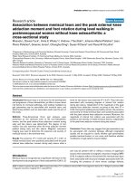
Pathology picture showing the intact (a) and the destroyed (b) alveolar attachmentsFigure 4
Pathology picture showing the intact (a) and the destroyed (b) alveolar attachments.
Respiratory Research 2005, 6:55 />Page 7 of 7
(page number not for citation purposes)
Corsico and colleagues [18], and indicates that, when
parenchymal destruction is present, airway wall factors
play a secondary role in determining the magnitude of the
beneficial effects of deep inspiration. This may be differ-
ent in asthma, where parenchymal involvement appears
to be minimal [20,21]. The presence of a direct correlation
between the bronchodilatory ability of deep inspiration
and the airway smooth muscle area is difficult to explain.
One possibility is that increased smooth muscle mass
leads to more bronchoconstriction and this may, up to a
point, increase the bronchodilatory effects of deep inspi-
ration by increasing radial traction [15,22]. Indeed, we
have previously shown that the bronchodilatory effect of
deep inspiration cannot be measured when the induced
bronchoconstriction is relatively small [3].
Conclusion
The results of our study support the hypothesis that the
attenuation of airway stretch due to loss of alveolar attach-
ments represents an important cause for the impairment
in the bronchodilatory effect of lung inflation in COPD.
Whether the progressive impairment of the beneficial
effect by deep inspiration has clinical and prognostic
implications in these subjects needs to be addressed in
future studies.
Competing interests
Dr. Togias' participation in this work was supported by
NIH grant RO1 HL61277.
The authors declare that they have no competing interest.
Authors' contributions
NS conceived and designed the study, performed the clin-
ical and functional assessments, analyzed and interpreted
the data, drafted the manuscript; AB carried the morpho-
metric assessment and participated to the interpretation
of the findings; RM carried the clinical and functional
assessments and participated to the interpretation of the
data; AMV participated to the design and the coordination
of the study and the interpretation of the results; AT con-
ceived and participated to the design of the study, the
analysis of the data and the interpretation of the results,
and contributed significantly to draft the manuscript; VB
helped in the design and the organization of the study, as
well as the interpretation of the results.
Acknowledgements
The authors wish to thank the colleagues of the Unit of Thoracic Surgery,
"V. Cervello" Hospital, Palermo (Dr. Balistreri, Dr. Regio, Dr. Agneta, Dr.
Caronia, Dr. Mazzotta) who provided the lung tissue, and the personnel of
the Unit of Pathology, "V. Cervello" Hospital, Palermo, who provided the
tissue blocks. The authors are also indebted to the national Council of
Research of Palermo for providing the equipment for morphometric anal-
ysis. All authors read and approved the final version of the manuscript.
References
1. Scichilone N, Marchese R, Catalano F, Vignola AM, Togias A, Bellia V:
Bronchodilatory effect of deep inspiration is absent in sub-
jects with mild COPD. Chest 2004, 125(6):2029-35.
2. Fairshter RD: Effect of a deep inspiration on expiratory flow in
normals and patients with chronic obstructive pulmonary
disease. Bull Eur Physiopathol Respir 1986, 22(2):119-25.
3. Scichilone N, Kapsali T, Permutt S, Togias A: Deep inspiration-
induced bronchoprotection is stronger than
bronchodilation. Am J Respir Crit Care Med 2000, 162:910-916.
4. Scichilone N, Permutt S, Togias A: The lack of the bronchopro-
tective and not the bronchodilatory ability of deep inspira-
tion is associated with airway hyperresponsiveness. Am J
Respir Crit Care Med 2001, 163(2):413-9.
5. Boushy SF, Aboumrad MH, North LB, Helgason AH: Lung recoil
pressure, airway resistance, and forced flows related to mor-
phologic emphysema. Am Rev Respir Dis 1971, 104:551-61.
6. Saetta M, Ghezzo H, Kim WD, King M, Angus G, Wang N, Cosio G:
Loss of alveolar attachments in smokers. Am Rev Respir Dis
1985, 132:894-900.
7. Snider GL, Kleinerman J, Thurlbeck WM, Bengali ZH: The definition
of emphysema. Report of a National Hearth, Lung and Blood
Institute, Division of Lung Disease Workshop. Am Rev Respir
Dis 1985, 132:182-5.
8. Pauwels RA, Buist AS, Calverley PM, Jenkins CR, Hurd SS: Global
strategy for the diagnosis, management, and prevention of
chronic obstructive pulmonary diseases. Am J Resp Crit Care
Med 2001, 163:1256-1276.
9. Abramson MJ, Hensley MJ, Saunders NA, Wlodarczyk JJ: Evaluation
of a new asthma questionnaire. J Asthma 1991, 28:129-139.
10. Goldman H, Becklake M: Respiratory function tests. Normal
values at median altitudes and the prediction of normal
results. Am Rev Respir Dis 1959, 79:457-467.
11. Scichilone N, Marchese R, Catalano F, Togias A, Vignola AM, Bellia V:
The bronchodilatory effect of deep inspiration diminishes
with aging. Respir Med 2004, 98(9):838-43.
12. Kirby J, Juniper E, Hargreave F, Zamel N: Total lung capacity does
not change during methacholine-stimulated airway
narrowing. J Appl Physiol 1986, 61:2144-2147.
13. Turato G, Zuin R, Miniati M, Baraldo S, Rea F, Beghe B, Monti S, For-
michi B, Boschetto P, Harari S, Papi A, Maestrelli P, Fabbri LM, Saetta
M: Airway inflammation in severe chronic obstructive pul-
monary disease: relationship with lung function and radio-
logic emphysema. Am J Respir Crit Care Med 2002, 166:105-10.
14. Mead J, Takishima T, Leith D: Stress distribution in lungs: a
model of pulmonary elasticity. J Appl Physiol 1970, 28:596-608.
15. Moreno RH, Hogg JC, Pare PD: Mechanics of airway narrowing.
Am Rev Respir Dis 1986, 133(6):1171-80.
16. Froeb HF, Mead J: Relative hysteresis of the dead space and
lung in vivo. J Appl Physiol 1968, 25(3):244-8.
17. Fairshter R: Airway hysteresis in normal subjects and individ-
uals with chronic airflow obstruction. J Appl Physiol 1985,
58:1505-1510.
18. Corsico A, Milanese M, Baraldo S, Casoni GL, Papi A, Riccio AM, Cer-
veri I, Saetta M, Brusasco V: Small airway morphology and lung
function in the transition from normality to chronic airway
obstruction. J Appl Physiol 2003, 95(1):441-7.
19. Finkelstein R, Ma H, Ghezzo H, Whittaker K, Fraser R, Cosio M:
Morphometry of small airways in smokers and its relation-
ship to emphysema type and hyperresponsiveness. Am J Respir
Crit Care Med 1995, 152:267-276.
20. O'Byrne PM, Postma DS: The many faces of airway inflamma-
tion. Asthma and Chronic Obstructive Diseases. Am J Resp Crit
Care Med 1999, 159:541-566.
21. Bousquet J, Jeffery P, Busse W, Johnson M, Vignola A: From bron-
choconstriction to airways inflammation and remodeling
(State of the Art). Am J Respir Crit Care Med 2000, 161:1720-1745.
22. Macklem PT: A theoretical analysis of the effect of airway
smooth muscle load on airway narrowing. Am J Respir Crit Care
Med 1996, 153(1):83-9.