Báo cáo y học: " Cigarette smoke induces IL-8, but inhibits eotaxin and RANTES release from airway smooth muscle" pot
Bạn đang xem bản rút gọn của tài liệu. Xem và tải ngay bản đầy đủ của tài liệu tại đây (331.09 KB, 10 trang )
BioMed Central
Page 1 of 10
(page number not for citation purposes)
Respiratory Research
Open Access
Research
Cigarette smoke induces IL-8, but inhibits eotaxin and RANTES
release from airway smooth muscle
Ute Oltmanns
1
, Kian F Chung
1
, Matthew Walters
2
, Matthias John
3
and
Jane A Mitchell*
2
Address:
1
Experimental studies National Heart & Lung Institute, Imperial College, London SW36LY, UK,
2
Cardiothoracic Pharmacology, National
Heart & Lung Institute, Imperial College, London SW36LY, UK and
3
Department of Pneumology, University Hospital Charite, Berlin, Germany
Email: Ute Oltmanns - ; Kian F Chung - ; Matthew Walters - ;
Matthias John - ; Jane A Mitchell* -
* Corresponding author
Abstract
Background: Cigarette smoke is the leading risk factor for the development of chronic
obstructive pulmonary disease (COPD) an inflammatory condition characterised by neutrophilic
inflammation and release of proinflammatory mediators such as interleukin-8 (IL-8). Human airway
smooth muscle cells (HASMC) are a source of proinflammatory cytokines and chemokines. We
investigated whether cigarette smoke could directly induce the release of chemokines from
HASMC.
Methods: HASMC in primary culture were exposed to cigarette smoke extract (CSE) with or
without TNFα. Chemokines were measured by enzyme-linked immunosorbent assay (ELISA) and
gene expression by real time polymerase chain reaction (PCR). Data were analysed using one-way
analysis of variance (ANOVA) followed by Bonferroni's t test
Results: CSE (5, 10 and 15%) induced IL-8 release and expression without effect on eotaxin or
RANTES release. At 20%, there was less IL-8 release. TNFα enhanced CSE-induced IL-8 release
and expression. However, CSE (5–30%) inhibited TNFα-induced eotaxin and RANTES production.
The effects of CSE on IL-8 release were inhibited by glutathione (GSH) and associated with the
induction of the oxidant sensing protein, heme oxygenase-1.
Conclusion: Cigarette smoke may directly cause the release of IL-8 from HASMC, an effect
enhanced by TNF-α which is overexpressed in COPD. Inhibition of eotaxin and RANTES by
cigarette smoke is consistent with the predominant neutrophilic but not eosinophilic inflammation
found in COPD.
Background
Chronic obstructive pulmonary disease (COPD) is a
major public health problem that is currently ranking as
the fourth leading cause of death in the world [1]. It is
characterised by progressive and largely irreversible air-
flow limitation associated with symptoms such as cough,
sputum production, and dyspnea. A chronic inflamma-
tory response of the lung to noxious particles, most nota-
bly tobacco smoke, but also occupational dusts and air
pollution, is currently considered as the underlying
Published: 19 July 2005
Respiratory Research 2005, 6:74 doi:10.1186/1465-9921-6-74
Received: 14 April 2005
Accepted: 19 July 2005
This article is available from: />© 2005 Oltmanns et al; licensee BioMed Central Ltd.
This is an Open Access article distributed under the terms of the Creative Commons Attribution License ( />),
which permits unrestricted use, distribution, and reproduction in any medium, provided the original work is properly cited.
Respiratory Research 2005, 6:74 />Page 2 of 10
(page number not for citation purposes)
pathological mechanism leading to this clinical condition
[1]. However, the link between inhalation of harmful sub-
stances, such as cigarette smoke, bronchial inflammation
and the development of airflow limitation is not com-
pletely understood.
Currently, cessation of smoking is the only intervention
that slows down disease progression in COPD [2].
Although only a minority of smokers develop symptoms
of COPD, there is evidence that even in the lungs of
asymptomatic smokers the numbers of inflammatory
cells are increased [3,4]. COPD is associated with the
release and overexpression of many pro-inflammatory
cytokines and chemokines including TNF-a and IL-8 [5].
Several studies have shown that cigarette smoke is capable
of activating lung macrophages as well as resident lung
cells such as epithelial cells and fibroblasts to release var-
ious inflammatory mediators including TNFα and the
neutrophil chemokine, IL-8 [6-8]. These mediators
together with proteases produced by activated neutrophils
and macrophages are capable of sustaining inflammation
and damaging lung structures. Specifically, the accumula-
tion of neutrophils in the lung has been associated with
more severe disease [9]. The precise mechanisms leading
to neutrophil influx into the lungs of smokers remain
unknown, but this may involve the release of neutrophil-
specific chemokines such as IL-8.
By contrast, the airways of patients with allergic asthma
are chracterised by a different profile of activated leuko-
cytes. Unlike COPD, where neutrophils predominate, in
asthma the eosinophil is present in large numbers, likely
to be the result of eosinophil chemoattractants such as
eotaxin or RANTES [10].
Human airway smooth muscle cells (HASMC) represent
an important structural component of the airway wall. In
addition to their traditionally accepted role as contractile
cells, HASMC produce neutrophil and eosinophil chemo-
tactic factors such as IL-8, eotaxin and RANTES [11-13].
The production of chemokines by these cells is of particu-
lar relevance considering the anatomical localization with
proximity to the vasculature. Many substances capable of
activating the airway smooth muscle synthetic capacity
have been identified, mainly cytokines such as IL-1β,
TNFα and TGFβ [11,14,15]. However, the effects of ciga-
rette smoke on chemokine production from HASMC are
not known.
Therefore, in this study we have exposed HASMC to ciga-
rette smoke and assessed effects on the induction and
release of the chemokines IL-8, eotaxin and RANTES.
Methods
Materials
Tissue culture reagents were obtained from Sigma (Poole,
UK). Cell culture plasticware was purchased from Falcon
Labware (Becton Dickinson, Oxford, UK). Recombinant
human TNFα and matched antibody pairs for IL-8,
eotaxin and RANTES enzyme-linked immunosorbent
assays (ELISA) were purchased from R&D Systems (Duo-
Set, Abingdon, UK). Antibodies were purchased from Cal-
biochem (heme oxygenase-1) and Biogenesis, Poole, UK
(GAPDH). Protease inhibitor cocktail was obtained from
Roche Diagnostic (Lewes, UK). All other chemical rea-
gents were obtained from Sigma (Poole, UK).
Isolation and culture of human airway smooth muscle cells
Human airway smooth muscle was obtained from lobar
or main bronchus from patients undergoing lung resec-
tion for carcinoma of the bronchus. The smooth muscle
was dissected out under sterile conditions and placed in
culture as previously described [16]. Cells were main-
tained in Dulbecco's modified Eagle's medium (DMEM)
containing 10% fetal calf serum supplemented with
sodium pyruvate (1 mM), L-glutamine (2 mM), non-
essential amino acids (1:100), penicillin (100 U/ml)/
streptomycin (100 µg/ml) and amphotericin B (1.5 µg/
ml) in a humidified atmosphere at 37°C in air/CO
2
(95:5
% vol/vol). At confluence, HASMC cultures exhibited a
typical hill-and-valley appearance. Immunofluorescence
techniques for calponin, smooth muscle α-actin and
myosin heavy chain revealed that more than 95% of the
cells displayed the characteristics of smooth muscle cells
in culture. HASMC at passages 3–7 from 9 different
donors were used in the studies described below.
Cigarette Smoke Extract
Cigarette smoke extract (CSE) was prepared by combust-
ing four full strength Marlboro cigarettes (filters removed)
through a modified 60 ml syringe apparatus and passing
the smoke through 100 mls of DMEM. Each cigarette
yielded 5 draws of the syringe (to 60 ml mark), with each
individual draw taking approximately 10 seconds to com-
plete. This solution represents '100%' strength. Smoked
medium was then passed through a 0.25 µM filter in order
to sterilise the solution. Smoked medium was diluted to
the required strength in DMEM and placed upon the cells
immediately afterwards.
Cell treatment
Prior to the experiments, confluent cells were growth-
arrested by FCS deprivation for 24 h in DMEM supple-
mented with sodium pyruvate (1 mM), L-glutamine (2
mM), non-essential amino acids (1:100), penicillin (100
U/ml)/ streptomycin (100 µg/ml), amphotericin B (1,5
µg/ml), insulin (1 µM), transferrin (5 µg/ml), ascorbic
acid (100 µM) and bovine serum albumin (0,1 %). Cells
Respiratory Research 2005, 6:74 />Page 3 of 10
(page number not for citation purposes)
were then exposed to smoke (0–30%) in the presence and
absence of TNFα (1 ng/ml). In additional experiments
cells were pretreated with 100 µM glutathione (GSH) for
30 min before exposure to CSE.
Cell viability
HASMC viability was assessed by the mitochondrial-
dependent reduction of 3-(4,5-dimethylthiazol-2-yl)-2,5-
diphenyltetrazolium bromide (MTT) to formazan. Cells
grown in 96-well plates were treated as indicated above,
washed with PBS and 100 µl MTT solution (1 mg/ml) was
added to each well. After 1 hour of incubation at 37°C,
the MTT solution was removed and the converted dye was
solubilized with 100 µl DMSO. The OD was measured
using a spectrophotometer set to 550 nm. None of the
conditions studied cause visual morphology markers of
apoptosis over the time course studied (not shown).
Cytokine assay
Cell supernatants were harvested 24 hours after stimula-
tion and stored at -70°C until assayed for RANTES,
eotaxin and IL-8. Cytokine levels were determined by
using specific sandwich enzyme-linked immunosorbent
assays (ELISA) according to the manufacturers'
instructions.
RT-PCR and Real-time PCR
Total RNA was isolated from HASMC after 6 hours using
the RNeasy Mini Kit (Qiagen, Crawley, UK) according to
the manufacturer's instructions. cDNA was generated by
reverse transcription (RT) using random hexamers. The
cDNA (42 ng/reaction) was used as a template in the sub-
sequent polymerase chain reaction (PCR) analyses. Tran-
script levels were determined by real-time PCR (Rotor
Gene 3000, Corbett Research, Australia) using the Sybre
Green PCR Master Mix Reagent Kit (Promega, San Luis
Obispo, USA). The sequence for IL-8 PCR primer were
sense 5'-GCCAACACAGAAATTATTGTAAAGCTT and anti-
sense 5'-CCTCTGCACCCA GTTTTCCTT'. Primers for
GAPDH were sense 5'-ATTCCATGGCACCGT CAAGGCT
and antisense 5'-TCAGGTCCACCACTGACACGT. Primers
were used at a concentration of 0.5 µM for real-time PCR
in each reaction. Cycling conditions for real-time PCR
were as follows: step 1, 15 min at 95°C; step 2, 15 sec at
94°C; step3, IL-8: 25 sec at 60°C, GAPDH: 25 sec at 64°C;
step 4, 22 sec at 72°C, with repeat from step 2 to step 4 for
40 times. Data from the reaction were collected and ana-
lysed by the complementary computer software (Corbett
Research, Australia). Relative quantitations of gene
expression were calculated using standard curves and nor-
malized to GAPDH.
Western immunoblot analysis for heme oxygenase-1
Confluent HASMC were exposed to CSE (0–20 %). After
24 hours of incubation, cells were rinsed with ice-cold
wash buffer (PBS containing 2 mM PMSF) and scraped off
the culture dish. HASMC were pelleted by centrifugation
at 1000 RPM at 4°C for 5 min and lysed in radioimmuno-
precipitation assay (RIPA) buffer (PBS containing 0.5%
sodium deoxycholate, 0.1% sodium dodecyl sulphate
(SDS), 1% Igepal and 1 tablet protease inhibitor cocktail
10 ml
-1
buffer). Samples were solubilized by sonication
followed by centrifugation (10,000 × g, 4°C, 4 min). Pro-
tein concentrations were determined using the BCA pro-
tein assay kit (Pierce, Rockford, USA). Lysates were boiled
for 10 min and total protein extracts (40 µg/lane) were
separated by SDS-polyacrylamide gel electrophoresis
(SDS-Page) on a 4–12 % acrylamide precast gel (Novex,
Invitrogen, Paisley, UK). The separated proteins were
transferred electrophoretically to a nitrocellulose mem-
brane in transfer buffer (Novex) and the membrane was
then blocked with 5% nonfat dry milk in TBS containing
0.1% Tween 20 (TBST) for at least 1 hour at room temper-
ature. Blots were then incubated overnight at 4°C with an
anti-HO-1 antibody in TBST containing 5% dried nonfat
milk at a 1:1000 dilution. The next day, the membrane
was washed 3 times with TBST and then incubated for 1
hour with a 1:2000 dilution of goat anti-mouse HRP-con-
jugated secondary antibody in TBST containing 5% non-
fat dry milk. The membrane was then washed as before
and visualized by enhanced chemiluminescent (ECL)
solution (Amersham, Buckinghamshire, UK). Membranes
were reprobed with a mouse anti-GAPDH monoclonal
antibody (1:5000, Biogenesis, Poole, UK) in order to
show the amount of protein loaded. Signals were quanti-
fied by scanning densitometry using software from Ultra-
Violet Products (UVP) (Cambridge, UK). Densitometry
data were normalized for GAPDH values.
Statistics
Data are presented as mean ± SEM. Data were compared
using one-way analysis of variance (ANOVA) followed by
Bonferroni's t test post hoc to determine statistical differ-
ences. A p value < 0.05 was considered significant. Sigma-
Stat software (Jandel Scientific, Germany) was used for
statistical analysis.
Results
Effects of cigarette smoke extract on IL-8 expression and
protein release
Under control culture conditions, IL-8 release from
HASMC was below the detection limit of the ELISA over
the 24-hour experimental period. Increasing concentra-
tions of smoke induced a 'bell-shaped' response curve for
IL-8 release by HASMC. Maximum induction of IL-8
release was seen at a concentration of 15 % CSE (baseline
0 pg/ml; 15% CSE 70.3 ± 8.6 pg/ml, p < 0.001; figure 1).
However, at concentration of 20% and 30%, the release of
IL-8 was lower. In order to assess whether CSE-induced
upregulation of IL-8 production from HASMC at up to
Respiratory Research 2005, 6:74 />Page 4 of 10
(page number not for citation purposes)
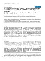
Effect of increasing concentrations of CSE on IL-8 production from HASMCFigure 1
Effect of increasing concentrations of CSE on IL-8 production from HASMC. (A) Cells were stimulated with CSE concentra-
tions from 5–30% for 24 hours. Cell free supernatants were assessed for IL-8 by ELISA. n = 3 from 1 donor. Similar results
were obtained from 2 other donors. *** p < 0.001; * p < 0.05 compared to untreated cells. (B) Effect of CSE (10%) on IL-8
mRNA expression in HASMC. Cells from 4 different donors were used for the experiments. Data were normalized to
GAPDH expression and are expressed as mean ± SEM. (C) HASMC viability in the presence of CSE (0–30%) was assessed by
using the MTT test. Results are expressed as percentage of untreated control cells (mean ± SEM, n = 3).
u
n
tre
a
t
e
d
CSE 10%
0.0
0.1
0.2
0.3
0.4
Ratio IL-8/GAPDH
A
B
0 5 7.510152030
0
25
50
75
100
CSE %
IL-8 (pg/ml)
*
*
***
0 5.07.510152030
0
50
100
150
CSE (%)
MTT (% of control)
C
Respiratory Research 2005, 6:74 />Page 5 of 10
(page number not for citation purposes)
15% concentration was the result of increased IL-8 gene
transcription, we measured IL-8 mRNA expression by real-
time PCR. Stimulation of HASMC with CSE (10%) for 6
hours led to increased IL-8 mRNA expression (Ratio IL-8/
GAPDH: baseline 0.075 ± 0.03, 10% CSE 0.21 ± 0.09, fig-
ure 1). Viability of cells exposed to CSE remained
unchanged up to concentrations of 15 % (104.8 ± 3.2 %
of control) but declined at concentrations of 30% ciga-
rette smoke (50.1 ± 9.7 % of control, figure 1).
Role of oxidative stress in cigarette smoke-induced IL-8
release
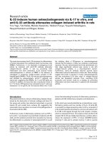
The stimulatory effects of CSE were greatly inhibited by
pre-treatment of cells with GSH (100 µM; 10 % CSE 270.8
± 72.5 pg/ml, 10 % CSE + GSH 70.9 ± 10.8 pg/ml, figure
2), which quenches extracellular oxidative stress [17].
Heme oxygenase-1 is expressed in most cell types and is
highly inducible by oxidative stress. In order to investigate
whether CSE exposure causes an intracellular oxidative
stress response in HASMC, we measured the expression of
heme-oxygenase-1 levels before and after exposure to
smoke by western blot analysis. HASMC expressed detect-
able levels of heme-oxygenase-1 when cultured under
control conditions. However, heme-oxygenase-1 levels
were increased when cells were treated with CSE (5–20 %;
figure 2).
(A) Effect of glutathione (GSH) on cigarette smoke-induced IL-8 release from HASMCFigure 2
(A) Effect of glutathione (GSH) on cigarette smoke-induced IL-8 release from HASMC. Cells were pretreated with 100 µM
GSH for 30 min before adding CSE (10%). Data are expressed as mean ± standard error of the mean (SEM). (B) CSE induced
heme oxygenase-1 (HO-1) expression in HASMC. Cells were exposed to CSE (0–20%) for 24 hours. HO-1 expression was
detected by western blotting. The blot shown in the upper panel was stripped and reprobed using a GAPDH antibody to show
equal protein loading. A representative example of three identical experiments is shown. In the lower panel densitometric anal-
ysis of HO-1 expression, normalized by GAPDH expression, is shown.
u
ntr
eated
G
S
H
CSE 10%
CSE
1
0% + G
SH
0
100
200
300
400
500
IL-8 ( pg/ml)
A
B
5010
20
%CSE
HO-1
GAPDH
p32
p37
0 5 10 20
0
1
2
3
CSE (%)
ratio HO-1/GAPDH
Respiratory Research 2005, 6:74 />Page 6 of 10
(page number not for citation purposes)
Effect of cigarette smoke extract and TNF
α
on IL-8 release
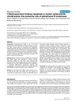
TNFα induced a concentration dependent release of IL-8
from HASMC at 0.1 to 10 ng/ml (not shown), with 1 ng/
ml representing an approximate EC
50
concentration. Fur-
thermore, TNFα (1 ng/ml) acted in synergy with CSE on
the release of IL-8 from HASMC (figure 3). This synergy
was observed across the concentration range of 5–15%
CSE with maximum effect seen at 10% smoke (TNFα
232.1 ± 18.4 pg/ml, 10% CSE + TNFα 628.5 ± 64.2 pg/ml,
p < 0.001, figure 3). This synergy was lost with CSE 20%
and in fact at 30% there was inhibition of IL-8 release. In
line with protein release, TNFα synergised with CSE
(10%) in induction of IL-8 mRNA (figure 3). Cell viability
remained unchanged in cells treated with either TNFα
alone or in combination with CSE at concentrations of up
to 10%. AT CSE 15%, cell viability declined mildly with
further reduction seen at CSE 20–30% (Figure 3).
Effect of cigarette smoke extract on eotaxin and RANTES
release
Similar to observations made with IL-8 release, levels of
either eotaxin or RANTES were below the level of detec-
tion in medium from cells cultured under basal condi-
tions. Similar to IL-8, incubation of cells with TNFα (1 ng/
ml) for 24 hours induced increased levels of both eotaxin
and RANTES released by the cells (Figure 4; RANTES
637.5 ± 84.5 pg/ml; eotaxin 177.1 ± 25.9 pg/ml). How-
ever, by contrast to IL-8, CSE at all concentrations (5%–
30%) failed to induce release of either eotaxin or RANTES
from HASMC (figure 4). Furthermore, CSE did not syner-
gise with TNFα in the release of either eotaxin or RANTES.
In fact, CSE inhibited the release of these chemokines
when induced by TNFα (figure 4) This effect was not
reversed by pre-treatment of HASMC with GSH (100 µM)
before adding cigarette smoke solution (data not shown).
Discussion
Cigarette smoke extracts up to 20% activated HASMC to
release IL-8, an important mediator of neutrophilic
inflammation in COPD, and enhanced the release of IL-8
induced by TNFα. The effect of CSE on IL-8 production
was associated with enhanced IL-8 gene transcription and
increased expression of HO-1, an indicator of intracellular
oxidative stress. By contrast, CSE had no effect on release
of the eosinophil chemotactic chemokines eotaxin and
RANTES at baseline levels and potently inhibited TNFα-
induced release of these chemokines. These results indi-
cate that HASMC may release IL-8 directly on contact with
cigarette smoke extracts and contribute to airway neu-
trophilic inflammation in COPD; the overexpression of
TNFα in COPD may augment this response. In addition,
the effect of cigarette smoke on HASMC may also explain
why there is little eosinophilic response in COPD since
the release of RANTES and eotaxin which have eosi-
nophilic chemotactic effects is inhibited. Our hypothesis
of selective induction of neutrophil accumulation in the
lungs by smoke is supported by a recent study where [18]
cigarette smoke increased neutrophil but reduced eosi-
nophil numbers in the lavage fluid of ovalbumin-sensi-
tized mice.
Neutrophils are considered as important inflammatory
cells in the pathogenesis of COPD because of their ability
to release various substances with harmful effects on lung
structures, such as oxidants, cytokines and especially pro-
teases [19,20]. Various mechanisms may account for the
accumulation of neutrophils in the lungs of cigarette
smokers such as delayed transit time, reduced apoptosis
or enhanced migration from the vasculature due to
increased expression of adhesion molecules [21-24]. In
addition, cigarette smoke induces the release of IL-8, a
potent chemotactic factor and activator for neutrophils
[25]. Elevated levels of IL-8 were found in the BAL-fluid of
smokers compared to nonsmokers and correlated posi-
tively with neutrophil counts in BAL fluid of smokers [8].
Cigarette smoke induces IL-8 production from various
pulmonary residential cells such as monocytes, macro-
phages, epithelial cells and fibroblasts [6-8,26]. In line
with this notion, we show here that cigarette smoke is a
potent stimulus for IL-8 production from HASMC. The
increased levels of IL-8 release were directly associated
with increases in IL-8 gene expression, as measured by real
time PRC. It should be noted, however, that the effects on
mRNA levels could be due to either increased gene expres-
sion or increased message stability. The in-vivo relevance
of this observation is demonstrated by a recent study
which showed infiltration of the airway smooth muscle
layer with neutrophils in patients with COPD [27].
While the release of IL-8 induced by cigarette smoke
extract up to 20% alone was modest, this effect was poten-
tiated by TNFα. Smoke alone, or smoke together with
TNFα, also induced gene transcription of IL-8. The ability
of cigarette smoke to release IL-8 in the presence of TNFα
may explain the persistence of neutrophilic inflammation
in cigarette smokers, where inflammatory cytokines,
including TNFα, are usually present in the lung [28]. In
mice, TNFα appears to be a central mediator for smoke-
induced inflammation and connective tissue breakdown
[29].
By contrast, CSE did not induce the release of eotaxin or
RANTES. In fact, it inhibited the release of these chemok-
ines in TNFα-stimulated cells, an effect that could not be
explained by reduced cell viability and was not reversed
by addition of the antioxidant GSH. Interestingly, we also
observed smaller levels of IL-8 release with higher concen-
trations of CSE above 20%. This was not entirely due to
cell death. In addition the potentiation of TNFα release of
IL-8 was lost, and indeed at 30% concentration there was
Respiratory Research 2005, 6:74 />Page 7 of 10
(page number not for citation purposes)
CSE synergises with TNFα (1 ng/ml) in inducing IL-8 release and expression in HASMCFigure 3
CSE synergises with TNFα (1 ng/ml) in inducing IL-8 release and expression in HASMC. (A) Cells were stimulated with CSE
concentrations from 5–30% for 24 hours in the absence and presence of TNFα (1 ng/ml). IL-8 in the cell-free supernatant was
measured by ELISA. The results shown are those from 3 replicate measurements from cells obtained from one donor. Similar
results were obtained from 2 other donors. *** p < 0.001; ** p < 0.01 compared to cells treated with TNFα only. (B) IL-8
mRNA expression in HASMC exposed to CSE (10%) in the presence and absence of TNFα (1 ng/ml). Cells from 4 different
donors were used for the experiments. Data were normalized to GAPDH expression and are expressed as mean ± SEM. * p <
0.05 compared to untreated cells; § p < 0.05 compared to cells treated with TNFα only. (C) HASMC viability in the presence
of TNFα (1 ng/ml) alone or in combination with CSE (0–30%) was assessed by using the MTT test. Results are expressed as
percentage of cells treated with TNFα only (mean ± SEM, n = 3).
A
B
**
***
***
*
§
057.510152030
0
250
500
750
Control
+TNFα
(1 ng/m l)
CSE %
IL-8 (pg/ml)
u
n
t
r
ea
t
e
d
TNFα
CSE
TNFα +CS
E
0
1
2
Ratio IL8/GAPDH
0 5.07.510152030
0
50
100
150
CSE (%) + TNFα
αα
α (1ng/ml)
MTT (% of TNFα
α
α
α
alone)
C
Respiratory Research 2005, 6:74 />Page 8 of 10
(page number not for citation purposes)
Effect of CSE or TNFα on (A) RANTES and (B) eotaxin release from HASMCFigure 4
Effect of CSE or TNFα on (A) RANTES and (B) eotaxin release from HASMC. Cells were incubated in the presence and
absence of CSE (10 %) or TNFα (1 ng/ml) for 24 hours. Effect of CSE on TNFα-induced (C) RANTES and (D) eotaxin release
from HASMC. Cells were stimulated with CSE concentrations from 5–30% for 24 hours in presence of TNFα (1 ng/ml). Cell
free supernatant was assessed for RANTES and eotaxin by ELISA. Cells from 3 different donors were used for the experi-
ments. Data are expressed as mean ± SEM.
u
n
tre
a
te
d
CSE
10
%
TNFα
0
250
500
750
RANTES (pg/ml)
untreated
CS
E
(
10
%)
TNF
α
0
100
200
eota xin (pg /ml)
AB
CD
0 5 7.510152030
0
100
200
300
CSE (%)
+TNFα (1ng/ml)
eotaxin (pg/ml)
0 5 7.510152030
0
250
500
750
CSE (%)
+TNFα (1n g/m l)
RANTES (pg/ml)
**
§
§§§§§
** **
§
§§§
§§
Respiratory Research 2005, 6:74 />Page 9 of 10
(page number not for citation purposes)
almost complete inhibition of IL-8 release. Because ciga-
rette smoke is a complex insult consisting of more than
4000 different components [30], it is likely that at high
concentrations of CSE, some components achieve concen-
trations that have inhibitory effects on IL-8 release over-
riding the stimulatory effects of other components of
cigarette smoke. Inhibition of eotaxin and RANTES release
at CSE concentrations which stimulated HASMC to
release IL-8 indicates that there is differential sensitivity to
the effects of smoke among the various subsets of chem-
okines. High concentrations of CSE are less likely to be
relevant in vivo. Although it is not known what concentra-
tion of CSE airway smooth muscle cells are exposed to in
vivo, it is likely to be a diluted concentration.
Although the mechanisms involved in the development
of smoke-induced lung diseases are not fully understood,
it is widely accepted that oxidative stress is a key factor
responsible for lung destruction seen in smokers. For
example, oxidants inactivate α
1
-antitrypsin, the major
protease inhibitor in the lung [31] and reactive oxygen
species induce infiltration of neutrophils into the lung
[32], which are an important source of oxidants them-
selves. Our results support the theory that oxidative stress
plays an important role in smoke-related lung diseases.
HASMC exposed to cigarette smoke showed increased
expression of heme-oxygenase-1, an intracellular indica-
tor of oxidative stress. In addition, GSH, an important
intra- and extracellular antioxidant in the lung, inhibited
cigarette smoke-induced IL-8 release in HASMC. Interest-
ingly, the inhibitory effect of cigarette smoke on inhibi-
tion of RANTES and eotaxin was not dependent on
oxidative stress. The mechanisms involved in CSE medi-
ated inhibition of chemokine production remain to be
identified. However, our observations are in line with oth-
ers showing that the release of eotaxin [33,34] and IL-8
can be differencial regulated by inflammatory or anti-
inflamamtory stimuli [35].
Conclusion
It is important to understand how smoking mediates air-
way inflammation in order to identify possible targets for
treatment of patients with chronic obstructive pulmonary
disease. The present study demonstrates that cigarette
smoke stimulates the release of the neutrophil chemotac-
tic cytokine IL-8 but inhibits the production of the eosi-
nophil chemotactic factors eotaxin and RANTES in
HASMC. Considering the anatomical location of the air-
way smooth muscle with proximity to the vasculature, the
data from our study suggest that HASMC play an impor-
tant role in promoting neutrophil migration from the vas-
culature to the interstitium in lung diseases associated
with cigarette smoke and may help to explain why COPD
unlike asthma, is predominantly associated with neu-
trophil recruitment.
Competing interests
The author(s) declare that they have no competing
interests.
Authors' contributions
UO prepared primary cultures of HASMC, carried out
cytokine assays, RT-PCR and real-time PCR, western blot-
ting for heme-oxygenase 1, performed statistical analysis
and drafted the manuscript.
FC participated in the design, coordination of the study
and drafting of the manuscript
MW prepared cigarette smoke extract, participated in cell
treatment and carried out cell viability assays.
MJ participated in drafting the manuscript.
JAM is the chief investigator who conceived the study.
All authors read and approved the final manuscript.
Acknowledgements
This work was supported by a Welcome Trust (U.K.) grant (no: 59857) and
by the Medical Research Council (U.K.).
References
1. Pauwels RA, Buist AS, Calverley MA, Jenkins CR, Hurd SS: Global
Strategy for the Diagnosis, Management, and Prevention of
Chronic Obstructive Pulmonary Disease. NHLBI/WHO Glo-
bal Initiative for Chronic Obstructive Lung Disease (GOLD)
Workshop Summary. Am J Respir Crit Care Med 2001,
163:1256-1276.
2. Culpitt SV, Rogers DF: Evaluation of current pharmacotherapy
of chronic obstructive pulmonary disease. Expert Opin
Pharmacother 2000, 1:1007-1020.
3. Niewoehner DE, Kleinerman J, Rice DB: Pathologic changes in
the peripheral airways of young cigarette smokers. N Engl J
Med 1974, 291:755-758.
4. Mullen JB, Wright JL, Wiggs RB, Pare PD, Hogg JC: Reassessment
of inflammation of airways in chronic bronchitis. Br Med J (Clin
Res Ed) 1985, 291:1235-1239.
5. Chung KF: Cytokines in chronic obstructive pulmonary
disease. Eur Respir J Suppl 2001, 34:50s-59s.
6. Walters MJ, Mitchell JA: Cigarette smoke extract (CSE) stimu-
lates human THP-1 monocytes directly and synergises with
IL-1beta to release IL-8. Br J Pharmacol 2003, 138:44P.
7. Numanami H, Koyama S, Nelson DK, Hoyt JC, Freels JL, Habib MP,
Amano J, Haniuda M., Sato E, Robbins RA: Serine protease inhibi-
tors modulate smoke-induced chemokine release from
human lung fibroblasts. Am J Respir Cell Mol Biol 2003, 29:613-619.
8. Mio T, Romberger DJ, Thompson AB, Robbins RA, Heires A, Rennard
SI: Cigarette smoke induces interleukin-8 release from
human bronchial epithelial cells. Am J Respir Crit Care Med 1997,
155:1770-1776.
9. Stanescu D, Sanna A, Veriter C, Kostianev S, Calcagni PG, Fabbri LM,
Maestrelli P: Airways obstruction, chronic expectoration, and
rapid decline of FEV1 in smokers are associated with
increased levels of sputum neutrophils. Thorax 1996,
51:267-271.
10. Chung KF, Barnes PJ: Cytokines in asthma. Thorax 1999,
54:825-857.
11. John M, Au BT, Jose PJ, Lim S, Saunders M, Barnes PJ, Mitchell JA, Bel-
visi MG, Chung KF: Expression and release of interleukin-8 by
human airway smooth muscle cells: inhibition by Th-2
cytokines and corticosteroids. Am J Respir Cell Mol Biol 1998,
18:84-90.
Publish with BioMed Central and every
scientist can read your work free of charge
"BioMed Central will be the most significant development for
disseminating the results of biomedical research in our lifetime."
Sir Paul Nurse, Cancer Research UK
Your research papers will be:
available free of charge to the entire biomedical community
peer reviewed and published immediately upon acceptance
cited in PubMed and archived on PubMed Central
yours — you keep the copyright
Submit your manuscript here:
/>BioMedcentral
Respiratory Research 2005, 6:74 />Page 10 of 10
(page number not for citation purposes)
12. John M, Hirst SJ, Jose PJ, Robichaud A, Berkman N, Witt C, Twort
CH, Barnes PJ, Chung FK: Human airway smooth muscle cells
express and release RANTES in response to T helper 1
cytokines: regulation by T helper 2 cytokines and
corticosteroids. J Immunol 1997, 158:1841-1847.
13. Chung KF, Patel HJ, Fadlon EJ, Rousell J, Haddad EB, Jose PJ, Mitchell
JA, Belvisi MG: Induction of eotaxin expression and release
from human airway smooth muscle cells by IL-1beta and
TNFalpha: effects of IL-10 and corticosteroids. Br J Pharmacol
1999, 127:1145-1150.
14. Fong CY, Pang L, Holland E, Knox AJ: TGF-beta1 stimulates IL-8
release, COX-2 expression, and PGE(2) release in human
airway smooth muscle cells. Am J Physiol Lung Cell Mol Physiol
2000, 279:L201-207.
15. Chung KF: Airway smooth muscle cells: contributing to and
regulating airway mucosal inflammation? Eur Respir J 2000,
15:961-968.
16. Belvisi MG, Saunders MA, Haddad EL, Hirst SJ, Yacoub MH, Barnes PJ,
Mitchell JA: Induction of cyclo-oxygenase-2 by cytokines in
human cultured airway smooth muscle cells: novel inflam-
matory role of this cell type. Br J Pharmacol 1997, 120:910-916.
17. Rahman I, MacNee W: Lung glutathione and oxidative stress:
implications in cigarette smoke-induced airway disease. Am
J Physiol 1999, 277:L1067-1088.
18. Melgert BN, Postma DS, Geerlings M, Luinge MA, Klok PA, van der
Strate BW, Kerstjens HA, Timens W, Hylkema MN: Short-term
smoke exposure attenuates ovalbumin-induced airway
inflammation in allergic mice. Am J Respir Cell Mol Biol 2004,
30:880-885.
19. Sutherland ER, Martin RJ: Airway inflammation in chronic
obstructive pulmonary disease: comparisons with asthma. J
Allergy Clin Immunol 2003, 112:819-827. quiz 828
20. Dhami R, Gilks B, Xie C, Zay K, Wright JL, Churg A: Acute ciga-
rette smoke-induced connective tissue breakdown is medi-
ated by neutrophils and prevented by alpha1-antitrypsin. Am
J Respir Cell Mol Biol 2000, 22:244-252.
21. MacNee W, Wiggs B, Belzberg AS, Hogg JC: The effect of ciga-
rette smoking on neutrophil kinetics in human lungs. N Engl
J Med 1989, 321:924-928.
22. Finkelstein EI, Nardini M, van der Vliet A: Inhibition of neutrophil
apoptosis by acrolein: a mechanism of tobacco-related lung
disease? Am J Physiol Lung Cell Mol Physiol 2001, 281:L732-739.
23. Di Stefano A, Maestrelli P, Roggeri A, Turato G, Calabro S, Potena P,
Mapp CE, Ciaccia A, Covacev L, Fabbri LM: Upregulation of adhe-
sion molecules in the bronchial mucosa of subjects with
chronic obstructive bronchitis. Am J Respir Crit Care Med 1994,
149:803-810.
24. Riise GC, Larsson S, Lofdahl CG, Andersson BA: Circulating cell
adhesion molecules in bronchial lavage and serum in COPD
patients with chronic bronchitis. Eur Respir J 1994, 7:1673-1677.
25. Adams DH, Lloyd AR: Chemokines: leucocyte recruitment and
activation cytokines. Lancet 1997, 349:490-495.
26. Culpitt SV, Rogers DF, Shah P, De Matos C, Russell RE, Donnelly LE,
Barnes PJ: Impaired inhibition by dexamethasone of cytokine
release by alveolar macrophages from patients with chronic
obstructive pulmonary disease. Am J Respir Crit Care Med 2003,
167:24-31.
27. Baraldo S, Turato G, Badin C, Bazzan E, Beghe B, Zuin R, Calabrese
F, Casoni G, Maestrelli P, Papi A, Fabbri LM, Saetta M: Neutrophilic
infiltration within the airway smooth muscle in patients with
COPD. Thorax 2004, 59:308-312.
28. Kuschner WG, D'Alessandro A, Wong H, Blanc PD: Dose-depend-
ent cigarette smoking-related inflammatory responses in
healthy adults. Eur Respir J 1996, 9:1989-1994.
29. Churg A, Dai J, Tai H, Xie C, Wright JL: Tumor necrosis factor-
alpha is central to acute cigarette smoke-induced inflamma-
tion and connective tissue breakdown. Am J Respir Crit Care Med
2002, 166:849-854.
30. Burns DM: Cigarettes and cigarette smoking. Clin Chest Med
1991, 12:631-642.
31. Evans MD, Pryor WA: Cigarette smoking, emphysema, and
damage to alpha 1-proteinase inhibitor. Am J Physiol 1994,
266:L593-611.
32. Nishikawa M, Kakemizu N, Ito T, Kudo M, Kaneko T, Suzuki M, Udaka
N, Ikeda H, Okubo T: Superoxide mediates cigarette smoke-
induced infiltration of neutrophils into the airways through
nuclear factor-kappaB activation and IL-8 mRNA expression
in guinea pigs in vivo. Am J Respir Cell Mol Biol 1999, 20:189-198.
33. Nie M, Knox AJ, Pang L: {beta}2-Adrenoceptor Agonists, Like
Glucocorticoids, Repress Eotaxin Gene Transcription by
Selective Inhibition of Histone H4 Acetylation. J Immuno
175(1):478-86.
34. Pang L, Knox AJ: Regulation of TNF-alpha-induced eotaxin
release from cultured human airway smooth muscle cells by
beta2-agonists and corticosteroids. FASEB J 2001,
15(1):261-269.
35. Nie M, Corbett L, Knox AJ, Pang L: ifferential regulation of chem-
okine expression by peroxisome proliferator-activated
receptor gamma agonists: interactions with glucocorticoids
and beta2-agonists. J Biol Chem 2005, 280(4):2550-61.