Báo cáo khoa học: "Radiographic closure time of appendicular growth plates in the Icelandic horse" ppsx
Bạn đang xem bản rút gọn của tài liệu. Xem và tải ngay bản đầy đủ của tài liệu tại đây (418.81 KB, 7 trang )
BioMed Central
Page 1 of 7
(page number not for citation purposes)
Acta Veterinaria Scandinavica
Open Access
Research
Radiographic closure time of appendicular growth plates in the
Icelandic horse
Eric Strand*
1
, Linn Camilla Braathen
1
, Mia C Hellsten
1
, Lisel Huse-Olsen
1
and Sigridur Bjornsdottir
2
Address:
1
Equine Teaching Hospital, Norwegian School of Veterinary Science, P.O.Box 8146 Dep. N-0033 Oslo, Norway and
2
Agricultural
Authority of Iceland, Austurvegur 64, 800 Selfoss, Iceland
Email: Eric Strand* - ; Linn Camilla Braathen - ; Mia C Hellsten - ;
Lisel Huse-Olsen - ; Sigridur Bjornsdottir -
* Corresponding author
Abstract
Background: The Icelandic horse is a pristine breed of horse which has a pure gene pool established
more than a thousand years ago, and is approximately the same size as living and extinct wild breeds of
horses. This study was performed to compare the length of the skeletal growth period of the "primitive"
Icelandic horse relative to that reported for large horse breeds developed over the recent centuries. This
information would provide practical guidance to owners and veterinarians as to when the skeleton is
mature enough to commence training, and would be potentially interesting to those scientists investigating
the pathogenesis of osteochondrosis. Interestingly, osteochondrosis has not been documented in the
Icelandic horse.
Methods: The radiographic closure time of the appendicular growth plates was studied in 64 young
Icelandic horses. The results were compared with previously published closure times reported for other,
larger horse breeds. The radiographs were also examined for any signs of developmental orthopaedic
diseases. In order to describe further the growth pattern of the Icelandic horse, the total serum alkaline
phosphatase (ALP) activity was determined and the height at the withers was measured.
Results: Most of the examined growth plates were fully closed at the age of approximately three years.
The horses reached adult height at this age; however ALP activity was still mildly increased over baseline
values. The growth plates in the digits were the first to close at 8.1 to 8.5 months of age, and those in the
regions of the distal radius (27.4 to 32.0 months), tuber olecrani (31.5 to 32.2 months), and the stifle (27.0
to 40.1 months) were the last to close. No horse was found to have osteochondrosis type lesions in the
neighbouring joints of the evaluated growth plates.
Conclusion: The Icelandic horse appears to have similar radiographic closure times for most of the
growth plates of its limbs as reported for large new breeds of horses developed during the past few
centuries. It thus appears that different breeding goals and the intensity of breeding have not altered the
length of the growth period in horses. Instead, it can be assumed that the pristine and relatively small
Icelandic horse has a slower rate of growth. The appendicular skeleton of Icelandic horses has completed
its bone growth in length at approximately 3 years of age, and therefore may be able to enter training at
this time.
Published: 17 July 2007
Acta Veterinaria Scandinavica 2007, 49:19 doi:10.1186/1751-0147-49-19
Received: 11 October 2006
Accepted: 17 July 2007
This article is available from: />© 2007 Strand et al; licensee BioMed Central Ltd.
This is an Open Access article distributed under the terms of the Creative Commons Attribution License ( />),
which permits unrestricted use, distribution, and reproduction in any medium, provided the original work is properly cited.
Acta Veterinaria Scandinavica 2007, 49:19 />Page 2 of 7
(page number not for citation purposes)
Background
The growth plates at the distal radius and the tuber cal-
caneus have been used as indicators of skeletal maturity in
Thoroughbred and Standardbred racing horses [1-3].
These breeds typically enter light training at 1.5 years of
age, and formal race training at 2 years of age. It is widely
thought among horsemen and veterinarians that Icelandic
horses have open growth plates and grow in height until
they are 4 to 5 years of age. As a result of this Icelandic
horses do not receive demanding ridden training until
they have reached that age. It is also thought that the slow
growth rate over an extended period of time protects this
breed from developing osteochondrosis, and other devel-
opmental orthopaedic disorders. To our knowledge, no
study has been made regarding the closure time of the
growth plates in the Icelandic horse, nor has anyone doc-
umented the existence of osteochondrosis in this breed.
The Icelandic horse has developed as an isolated breed
since the settlement of the country in the 8th and 9th cen-
tury. It originates from the medieval horse population of
Norway and probably other countries in Scandinavia and
the British Isles [4]. There is no evidence of introduction
of new blood to the horse population since the end of the
colonization period late in the 10th century [5]. The his-
tory of intense artificial selection of Icelandic horses is rel-
atively short. Organized horse breeding based on different
traits of conformation and performance under saddle has
only been practised for one century [6-8]. For the last two
decades, the breeding values have been obtained by a
multiple-trait animal model (Best Linear Unbiased Pre-
diction, BLUP) [9], accelerating the genetic improvement
of the breed. The Icelandic horse is characterized by its
ability to perform 4 or 5 gaits, and by its good health, and
durability [10]. It is used for pleasure riding, long distance
trekking and special gait competitions and has a wide dis-
tribution in Europe and North America [11].
The Icelandic horse is relatively small. Growth and devel-
opment of the Icelandic horse was studied in the period
1970 – 1980 where the average height at the withers,
measured by rod, was found to be 133 cm for five-year-old
horses [12]. Measurements of horses presented for breed-
ing evaluation in 2001 indicate an increase in the height
of the breed in the last decades, as the average height of
the mares was found to be 136.9 cm (128.0 – 146.0, SD
2.8) and for stallions 138.6 cm (130.0 – 151.0, SD: 3.0)
[13].
The growth plate consists of a plate of hyaline cartilage,
the physeal cartilage, and is seen on radiographs as a radi-
olucent line surrounded by diffuse relatively increased
bone opacity. Endochondral ossification of the growth
plates accounts for most of the linear growth of the long
bones of the horse [14-16]. Cessation of this growth coin-
cides with radiographic closure of the growth plate
[15,16]. Radiographic closure has occurred when there is
no radiolucent line visible in the physeal area. The closure
time of selected growth plates of the limbs has been deter-
mined for some horse breeds [1-3,14,16-20].
Another method of evaluating the maturity of the skele-
ton is the measurement of biochemical parameters that
are associated with growth and remodeling of bone tissue.
Alkaline phosphatase (ALP) concentration in serum can
be used to indicate the level of metabolic activity in the
bone tissue of horses [21-23]. It reflects the active bone
formation which accompanies skeletal modeling in the
growing animal, and it decreases with age as the growth
rate of the skeleton slows down [22-24].
The aims of this study were to determine the approximate
radiographic closure time of the growth plates of the fore-
and hind limbs of the pristine Icelandic horse, and to
compare these closure times with those previously pub-
lished for more recently developed large breeds of horses.
The radiographs were also examined in order to docu-
ment evidence of developmental orthopaedic disease
such as osteochondrosis and osteochondral bone cysts in
the neighbouring joints. In order to further describe the
growth pattern of the Icelandic horse, the total serum
alkaline phosphatase (ALP) activity was determined, and
the height at the withers was measured. This information
would provide practical guidance to owners and veterinar-
ians as to when the skeleton is mature enough to com-
mence formal ridden training, and would be potentially
interesting to those scientists investigating the pathogene-
sis of osteochondrosis.
Methods
Horses
The material consisted of 64 Icelandic horses, including
38 mares, 15 stallions and 11 geldings. Thirty-eight of the
horses were examined in Iceland in late September 2004
and 26 in Norway in January and April 2005. The age of
the horses ranged from 47 days to 52 months at the time
of examination. All the horses were born during the spring
and summer months, the majority in May and June of
each year. Each horse was examined one time. All the
horses were found to be in good nutritional condition.
Further information about the management was collected
from the owners: In Iceland, most of the young horses
that were not in training were kept out-of-doors on large
pastures all year round. The foals were weaned at approx-
imately 6 – 9 months of age, and were either stabled for
the winter months or kept at pasture with access to open
shelters. The feeding consisted of grazing from June to
October/December, and haylage/silage ad lib out-of-
doors during the winter. Mineral supplements were usu-
ally provided by salt licks. In this material, the three-and-
Acta Veterinaria Scandinavica 2007, 49:19 />Page 3 of 7
(page number not for citation purposes)
a-half-year-old horses (42 – 46 months) were being sad-
dle broken, and the four and-a-half-year-olds (48 – 52
months) were in light training at the time of examination.
The management regimes in Norway were similar, except
for supplementary feeding of grain to all horses from
weaning, and that most of the horses in Norway were sta-
bled during the winter months. The horses were privately
owned, and intended for pleasure riding and gait compe-
titions. They had no history of illness or injury.
Radiographic examination
The horses were sedated with a combination of detomi-
dine (Domosedan
®
, Orion Corporation, Turku, Finland)
10–40 µg/kg bwt and butorfanol (Torbugesic
®
, Fort
Dodge Animal Health, Overland Park, Kansas, USA) 20–
30 µg/kg bwt intravenously. Radiographs were taken
using a 80 kV, 15 mA, 1.99 sec portable X-ray machine
(Gierth HF 80/15 plus ULTRA LEICHT, Gierth X-Ray
International GmbH, Riesa, Germany). The focal-film dis-
tance (FFD) was 100 cm, and regular speed screens were
used.
The anatomical regions included in this study were: the
phalanges for all horses 46 days to 24 months of age; and
the carpus, elbow, hock, and stifle for all horses 8 months
to 40 months of age. Two views, in the frontal (cranial to
caudal) and sagittal (lateral to medial) plane, were taken
for each region of the left thoracic and left pelvic limb. Six-
teen different growth plates of the appendicular skeleton
were examined. The radiographs were all interpreted by a
panel consisting of at least three of the five authors and
the classification of each physis was agreed upon by con-
sensus.
For the purpose of this study, the growth plates were clas-
sified as fully open, closing and fully closed, in order of
advancing fusion of the growth plate [2,18,25,26]. A
growth plate was classified as fully open when a distinctly
radiolucent line could be observed spanning the whole
extent of the growth plate region (Fig. 1). A growth plate
was classified as closing when a radiolucent line was
present in the growth plate area, but only intermittently
and surrounded by diffuse relatively increased bone opac-
ity (Fig. 2). A growth plate was classified as fully closed
with total absence of the radiolucent line in the region of
the previous growth plate in the two radiographic projec-
tions (Fig. 3). When there was a difference in appearance
of the growth plates on separate views of the same area,
the growth plate was classified according to the view that
showed the lowest degree of fusion. Other subjective fea-
tures of the growth plates such as width were noted. Time
of closure for each growth plate was defined as the age
range from the youngest horse observed with a fully
closed growth plate, to the age after which all further
horses examined had a fully closed growth plate.
Alkaline phosphatase and height at the withers
Prior to sedation, blood was drawn from the external jug-
ular vein into two 10 ml vials without supplement. The
whole blood was centrifuged later the same day and the
serum frozen to -18°C for later analysis or was analyzed
directly. Total serum alkaline phosphatase (U/L) was
measured with the Modified IFCC method [27]. The
height at the highest point of the withers was measured
with the horse standing square on a level surface. Most of
the horses were measured under mild sedation because
they were not used to extensive handling. In order to have
reference values of total ALP of adult Icelandic horses,
intravenous blood samples were collected from a control
group which consisted of 11 reportedly healthy Icelandic
horses at the age of 7 to 16 years.
Results
Radiographic examination
The growth plates in the first and second distal phalanges
and the proximal third phalanx as well as the proximal
Mt3 and Mc3 were all fully closed in the youngest horses
in this study. The time of closure of the sixteen other
growth plates examined are listed in Table 1, and in Table
2 these same results are listed together with published
data from other horse breeds. The growth plates of the Ice-
landic horses were subjectively characterized as narrow in
most of the regions studied, relative to those present in
large horse breeds, although the width was not objectively
measured.
Alkaline phosphatase and height at the withers
The results of the measurements of total ALP as well as the
height at the withers are plotted against the age of the
horses in Figures 4 and 5 respectively. A geometric trend
line was added to the graphs in both figures. For compar-
ison, the mean value of 242.5 U/L of total ALP of the con-
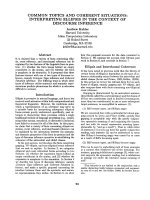
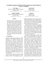
Examples of fully open growth platesFigure 1
Examples of fully open growth plates. A) The proximal
tibia, tuberositas tibia and distal femur of a 46-day-old foal. B)
The tuber olecrani, proximal radius and distal humerus of a
4-month-old foal
Acta Veterinaria Scandinavica 2007, 49:19 />Page 4 of 7
(page number not for citation purposes)
trol group of 11 adult horses was added to Figure 4 as a
dotted horizontal line.
No signs of osteochondrosis or other developmental
orthopaedic disease was found in the neighbouring joints
of the evaluated growth plates.
Discussion
A complete overview of the closure times of the appendic-
ular growth plates requires either following a group of
growing horses for several years, or studying a representa-
tive cross section of individuals at critical points of the
development. Here, a cross-sectional design was chosen,
as groups of individuals of the appropriate age could be
captured on a few occasions during a calendar year. The
material was haphazardly selected from farms in different
locations in Iceland and Norway and is considered to be
representative for the breed without known biases.
The closure times of the appendicular growth plates in the
Icelandic horse listed in Table 1 were found to be similar
to existing data for other horse breeds (see Table 2). The
only exception was a tendency for the growth plates of the
distal radius to close later in Icelandic horses, compared
with Thoroughbred/Quarter horse crosses [14], Brazilian
Thoroughbreds [26], Brazilian Mangalarga [18] and a lim-
ited material of Arabian horses [17]. However, few com-
plete studies are presently available for comparison of our
results. Many of the studies listed in Table 2 are based on
a very limited number of young horses, and/or a limited
number of growth plate regions. No previously published
data could be found for many of the regions now investi-
gated in the Icelandic horse. In general, the differences in
the closure times of the growth plates appear to be mini-
mal between breeds despite of the great variation in adult
sizes. This suggests a slower growth rate in smaller breeds,
such as the Icelandic horse. The consistent "subjec-
tive"observation of relatively narrow growth plates in this
study, compared to the much wider growth plates
observed in adolescent horses of large breeds, also sup-
ports this suggestion. Measuring the actual growth rate of
the Icelandic horse would, however, have required meas-
urements of the size at birth and was beyond the scope of
this study.
The radiographic determination of growth plate closure is
a result of subjective evaluation, and correct interpretation
depends on many factors. To reveal the radiolucent carti-
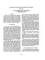
Examples of fully closed growth platesFigure 3
Examples of fully closed growth plates. A) Horse aged
22.8 months. Fully closed growth plates at the proximal sec-
ond phalanx, proximal first phalanx and distal metacarpus. B)
Horse aged 50.8 months. Fully closed growth plates at the
proximal radius and distal humerus. Note the absence of any
radiolucency and diffuse opacity in the region of the previous
growth plate.
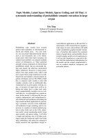
Example of a growth plate classified as closingFigure 2
Example of a growth plate classified as closing. The
carpus of a horse aged 26.7 months. The growth plate at the
distal radius is classified as closing. Note the intermittently
present radiolucent line surrounded by diffuse opacity
(arrow).
Acta Veterinaria Scandinavica 2007, 49:19 />Page 5 of 7
(page number not for citation purposes)
lage at the growth plate, the x-ray beam must be aimed
directly perpendicularly to the growth plate; otherwise
overlapping bone tissue can be misinterpreted as evidence
of fusion of the growth plate. Since the growth plates in
many sites are not flat discs, but undulate to a variable
degree, often in two or more directions, the problem of
overlapping is often present also in good-quality radio-
graphs [28]. In addition, the physeal cartilage becomes
narrower with increasing age [29], which makes it more
difficult to discern between fully open and partially fused
growth plates. Therefore, to minimize interpretation diffi-
culties, two views (cranio – caudal and lateral – medial) of
each region were used. In some cases, it was still difficult
to distinguish between "late" closing and fully closed.
This is a possible explanation to the outliers in the present
study. Other authors have also found what seem to be sin-
gle outliers in their material [14,25].
Total serum alkaline phosphatase (total ALP) consists of
fractions of several tissue-specific isoenzymes. In healthy
Table 2: Previously published reports of closure times (months) of the appendicular growth plates in different horse breeds together
with the results for Icelandic horses in this study.
Growth plate
Breed nProximal
second
phalanx
Proximal
first phalanx
Distal third
metacarpal
Distal
third
metatarsal
Distal
radius
Proximal
radius
Tuber
olecrani
Distal
humerus
Tuber
calcanei
Brazilian Thorougbred [27] 20 20.9–27.6
Thoroughbred [19] 800 8.0–14.0 8.0–14.0
Thoroughbred [20] 53
16.0–24.0
American Standardbred [16] 113 26.0–35.0
Standardbred [13] 14 24.2–31.9
American and Italian
Standardbred [15]
140 26.0–29.0
23.0–27.0
Arabian [14] 2 7.5–7.9 7.5–8.8 7.0–7.5 7.0–7.5 23.2–23.7 13.6–14.0 26.6–29.7 13.6–14.9
Quarter Horse [12] 6 Ca. 18
Thoroughbred-Quarter Horse
Cross [11]
9 6.0–10.0 (tl)*
8.0–11.0 (pl)*
7.0–9.5
(18)**
9.0–12.5 24.0–25.5
Brazilian Manga-larga [17] 7 24.6
Finnhorse [18] 15 24.0–30.0
Icelandic horse, current study 35–56** 8.1 8.1–8.5 8.1–8.5
(16.4)***
8.1–14.9
(16.4)***
27.4–32.0
(39.1)***
14.9 31.5–32.2 8.8–11.0
19.0–26.7
*tl = thoracic limb, pl = pelvic limb, **Depending on region, *** The closure times with single outliers are included in parentheses
Table 1: Radiographic closure time (age range in months) of appendicular growth plates in 64 Icelandic horses
Growth plate n Growth plate fully open Growth plate fully closed = Closure time
Proximal second phalanx (TL) 35 1.5 – 6.1 8.1
Proximal first phalanx (TL) 37 1.5 – 4.3 8.1 – 8.5
Proximal second phalanx (PL) 35 1.5 – 4.3 8.1
Proximal first phalanx (PL) 37 1.5 – 6.1 8.1 – 8.5
Distal third metacarpal 37 1.5 – 4.3 8.1 – 8.5 (16.4)
Distal third metatarsal 37 1.5 – 6.1 8.1 – 14.9 (16.4)
Distal radius 55 1.5 – 22.9 27.4 – 32.0 (39.1)
Proximal radius 56 1.5 – 11.0 14.9
Tuber olecrani 55 1.5 – 26.7 31.5 – 32.2
Medial epicondyle of humerus 56 1.5 – 14.9 15.2
Distal humerus 56 1.5 – 4.3 8.8 – 11.0
Tuber calcanei 56 1.5 – 11.0 19.0 – 26.7
Distal tibia 56 1.5 – 11.0 15.3 – 19.0
Tuberositas tibiae 52 1.5 – 38.6 38.6 – 40.1
Proximal tibia 56 1.5 – 22.8 23.0 – 32.2 (38.6)
Distal femur 53 1.5 – 16.4 19.0 – 27.0
Fully open = distinct radiolucent line spanning the entire extent of the physis; fully closed = no radiolucency in the region of the physis. The
Radiographic closure time was defined as the age range from the youngest horse observed with a fully closed physis, to the age after which all
further horses had a fully closed physis. Single outliers which were in the "closing stage" are in parenthesis.
Acta Veterinaria Scandinavica 2007, 49:19 />Page 6 of 7
(page number not for citation purposes)
young horses only two different isoenzyme fractions
appear in the serum: liver and bone ALP [21]. The level of
total ALP decreases with age, particularly during the first
year of life, mainly due to the decrease of the bone frac-
tion as the skeleton growth rate slows down with age
[22,23]. In horses younger than one year, the bone frac-
tion is 60% of the total ALP, while in horses over five years
of age it has decreased to 20 % [22]. The same pattern in
the changes of total ALP with age was observed in the Ice-
landic horse (see Fig 4). The plots followed a fitted geo-
metric curve that was steepest in the first year of life, and
had almost reached a horizontal line at 38 to 40 months.
At this age the growth plates studied were all closed and
the height at the withers seemed to have reached the adult
level. However, the total ALP in the three- and four-year-
old horses had still not decreased to the baseline value of
the control group of 11 adult horses, and was also higher
than in adult Icelandic horses in a previous study [30].
The mean total ALP in the four-year-old horses was 675.7
U/L, which was actually higher than in the three-year-olds
that had a mean ALP of 497.4 U/L. Although all the
growth plates in the current study were closed at the age of
four years, it has been described that more proximal
growth plates, for example in the pelvis and the vertebral
column, can still be open at this age [31]. It is also known
that considerable remodeling occurs at the physeal sites
for a long time after radiological closure [16]. The four-
year-old horses were in light training which has been
reported to cause an increase in both liver and bone ALP
in Swedish Standardbred trotters up to the age of three
years [21].
Radiographic signs of developmental orthopaedic disease
were not identified in this material nor in an earlier report
based on radiographic examination of the tarsi of 614 Ice-
landic horses in the age of 6 – 12 years [32]. Thus no radi-
ographic survey of Icelandic horses to date has
demonstrated the existence of osteochondrosis type frag-
ments.
Conclusion
This study provides practical information for trainers and
veterinarians working with the Icelandic horse. Tradition-
ally, demanding ridden training of Icelandic horses com-
mences at the age of 4 years at the earliest. According to
the current study, the appendicular skeleton should be
ready for increased load at 3 years of age, as most appen-
dicular growth plates are closed by then. The results also
suggest that the Icelandic horse, with its gene pool estab-
lished over 1000 years ago, has approximately the same
growth period as breeds of horses which have been espe-
cially selected for size during the past few centuries. In our
study the Icelandic horse was also subjectively evaluated
to have relatively narrow growth plates, relative to large
horse breeds, in all age groups suggesting a slower growth
rate. The growth rate of the Icelandic horse needs to be
investigated further, as well as the association between
growth rate and developmental orthopaedic abnormali-
ties.
Competing interests
The author(s) declare that they have no competing inter-
ests.
Authors' contributions
The authors contributed equally to this work. All authors
read and approved the final manuscript.
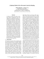
The relationship between height at the withers and age of 63 Icelandic horsesFigure 5
The relationship between height at the withers and age of 63
Icelandic horses. The fitted curve is a geometric model.
80
90
100
110
120
130
140
150
160
0 5 10 15 20 25 30 35 40 45 50 55 60
Age (months)
Height at withers (cm)
The relationship between alkaline phosphatase (ALP, in U/L) and age (in months) of 64 Icelandic horses with a fitted geo-metric curveFigure 4
The relationship between alkaline phosphatase (ALP, in U/L)
and age (in months) of 64 Icelandic horses with a fitted geo-
metric curve. The dotted line at the ALP-level of 242.5 U/L
represents the mean ALP value of the control group of 11
horses, which were 7 to 16 years of age.
0
250
500
750
1000
1250
1500
1750
2000
2250
0 5 10 15 20 25 30 35 40 45 50 55 60
Age (months)
ALP (U/L)
Publish with Bio Med Central and every
scientist can read your work free of charge
"BioMed Central will be the most significant development for
disseminating the results of biomedical research in our lifetime."
Sir Paul Nurse, Cancer Research UK
Your research papers will be:
available free of charge to the entire biomedical community
peer reviewed and published immediately upon acceptance
cited in PubMed and archived on PubMed Central
yours — you keep the copyright
Submit your manuscript here:
/>BioMedcentral
Acta Veterinaria Scandinavica 2007, 49:19 />Page 7 of 7
(page number not for citation purposes)
Acknowledgements
This study was supported by funds from Torsted's Trust Fund for Animal
Welfare and the Norwegian School of Veterinary Science. The authors
acknowledge the breeders providing horses and facilities for the study.
References
1. Pezzoli G, Del Bue M: Valutazione radiografica del grado di svi-
luppo scheletrico nel cavallo trottatore in rapporto all'attiv-
ita' atletica. (Evaluation of bone development in trotting
horses and athletic activity). Folia Vet Lat 1975, 5:399-411.
2. Gabel AA, Spencer CP, Pipers FS: A study of correlation of clo-
sure of the distal radial physis with performance and injury
in the Standardbred. J Am Vet Med Assoc 1977, 170:188-194.
3. Yoshida K, Ueda Y, Masumitsu H: Radiological studies on the
ossification of the Thoroughbreds 2. Closure process in the
distal epiphyseal lines of the radius and the 3rd metacarpal
bone and the proximal epiphyseal line of the proximal pha-
lanx and an assessment system of bone maturity. Bull Equine
Res Inst 1982, 19:18-29.
4. Aðalssteinsson S: Origin and conservation of farm animal pop-
ulations in Iceland. Z Tierzuecht Zuechtsbiol 1981, 98:258-264.
5. Pálsson PA: Er íslenski hesturinn hreinræktaður í 1000 ár? (Is
the Icelandic horse pure bred for a thousand years?). Eiðfaxi
1996, 2:18-19.
6. International Federation of Icelandic Horse Associations:
Breeding [ />]
7. Árnason Th: Genetic studies on conformation and perform-
ance of Icelandic toelter horses. Acta Agric Scand 1984,
34:409-462.
8. Hugason K: Breeding of Icelandic toelter horses: an overview.
Livest Prod Sci 1994, 40:21-29.
9. Árnason Th, van Vleck LD: Genetic Improvement of the horse.
In Genetics of the horse Edited by: Bowling AT, Ruvinsky A. New York:
GABI Publishing; 2000:473-497.
10. Björnsdóttir S, Árnason Th, Lord P: Culling rate of Icelandic
horses due to bone spavin. Acta Vet Scand 2003, 44:161-169.
11. International Federation of Icelandic Horse Associations:
National Breeding Statistics [ />brnews103e.pdf]
12. Árnason Th, Bjarnason Th: Growth and size of Icelandic toelter
horses. Livest Prod Sci 1994, 40:79.
13. Sigurðsson Á, Fróðadóttir H, Jóhannsdóttir LB: Skýrsluhaldið í
hrossarækt 2001. (Annual report on horse breeding 2001).
Freyr 2002, 1:47-50.
14. Fretz PB, Cymbaluk NF, Pharr JW: Quantitative analysis of long-
bone growth in the horse. Am J Vet Res 1984, 45:1602-1609.
15. Smith BL, Auer JA, Taylor TS, Hulse DS, Longnecker MT: Use of
orthopedic markers for quantitative determination of prox-
imal radial and ulnar growth in foals. Am J Vet Res 1991,
52:1456-1460.
16. Uhlhorn H, Eksell P, Carlsten J: Scintigraphic characterization of
distal radial physeal closure in young standardbred race-
horses. Vet Radiol Ultrasound 2000, 41:81-186.
17. Myers VS, Emmerson MA: The age and manner of epiphyseal
closure in the forelegs of two Arabian foals. Vet Radiol Ultra-
sound 1966, 7:39-47.
18. Mamprim MJ, Vulcano LC, Muniz LMR: Estudo radiográfico do
fechamento da epífise distal da rádio em potras de raça
Manga-Larga. (Radiographic study of distal radius epiphyseal
closure in Manga-Larga fillies.). Vet E Zoot 1992, 4:59-62.
19. Koskinen E, Katila T: Effect of 19-Norandrostenololylaurate on
serum testosterone concentration, libido, and closure of dis-
tal radial growth plate in colts. Acta Vet Scand 1997, 38:59-67.
20. Banks WC, Kemler AG, Guttridge H, Kirkham W: Radiography of
the Tuber calcis and its use in Thoroughbred training. Proc
Am Ass Equine Practnrs 1969, 15:273-293.
21. Thorén-Tolling K: Serum Alkaline Phosphatase isonezymes in
the horse – variation with age, training and in different path-
ological conditions. J Vet Med A 1988, 35:13-23.
22. Price JS, Jackson B, Eastell R, Goodship AE, Blumsohn A, Wright I,
Stoneham S, Lanyon LE, Russell RGG: Age related changes in bio-
chemical markers of bone metabolism in horses. Equine Vet J
1995, 27:201-207.
23. Price JS, Jackson BF, Gray JA, Harris PA, Wright IM, Pfeiffer DU, Rob-
ins SP, Eastell R, Ricketts SW: Biochemical markers of bone
metabolism in growing thoroughbreds: a longitudinal study.
Res Vet Sci 2001, 71:37-44.
24. Mäenpää PH, Pirskanen A, Koskinen E: Biochemical indicators of
bone formation in foals after transfer from pasture to stables
for the winter months. Am J Vet Res 1988, 49:1990-1992.
25. Mason TA, Bourke JM: Closure of the distal radial epiphysis and
its relationship to unsoundness in two year old thorough-
breds. Aust Vet J 1973, 49:221-228.
26. Vulcano LC, Mamprim MJ, Muniz LMR, Moreira AF, Luna SPL: Radi-
ographic study of distal radial physeal closure in Thorough-
bred horses. Vet Radiol Ultrasound 1997, 38:352-354.
27. Tietz NW: Clinical Guide to Laboratory Tests Philadelphia, Pennsylvania:
WB Saunders Company; 1995.
28. MacCallum FJ, Brown MP, Goyal HO: An assessment of ossifica-
tion and radiological interpretation in limbs of growing
horses. Br Vet J 1978, 134:366-374.
29. Firth EC, Greydanus Y: Cartilage thickness measurements in
foals. Res Vet Sci 1987, 42:35-46.
30. Seiser M, Strasser A, Hofbauer B: Der Einfluss von Alter und
Geschlecht auf diagnostisch wesentlichen Blutparameter bei
Islandpferden. (The influence of age and gender on diagnos-
tically significant blood parameters in Icelandic horses.). Tier-
arztl Prax 2001, 29(G):324-321.
31. Butler JA, Colles CM, Dyson SJ, Kold SE, Poulos PW: Clinical Radiology
of the Horse Oxford: Blackwell Science Ltd; 2000.
32. Björnsdóttir S, Axelsson M, Eksell P, Sigurdsson H, Carlsten J: Radi-
ographic and clinical survey of degenerative joint disease in
the distal tarsal joints in Icelandic horses. Equine Vet J 2000,
32:268-272.