Báo cáo y học: "The pathophysiological function of peroxisome proliferator-activated receptor-γ in lung-related diseases" pptx
Bạn đang xem bản rút gọn của tài liệu. Xem và tải ngay bản đầy đủ của tài liệu tại đây (392.8 KB, 9 trang )
BioMed Central
Page 1 of 9
(page number not for citation purposes)
Respiratory Research
Open Access
Review
The pathophysiological function of peroxisome
proliferator-activated receptor-γ in lung-related diseases
Tom Hsun-Wei Huang
†
, Valentina Razmovski-Naumovski
†
,
Bhavani Prasad Kota, Diana Shu-Hsuan Lin and Basil D Roufogalis*
Address: Faculty of Pharmacy, A15, University of Sydney, New South Wales, 2006, Australia
Email: Tom Hsun-Wei Huang - ; Valentina Razmovski-Naumovski - ;
Bhavani Prasad Kota - ; Diana Shu-Hsuan Lin - ;
Basil D Roufogalis* -
* Corresponding author †Equal contributors
Peroxisome proliferator-activated receptor-gammarespiratory diseasesasthmachronic obstructive pulmonary diseaselung cancer.
Abstract
Research into respiratory diseases has reached a critical stage and the introduction of novel
therapies is essential in combating these debilitating conditions. With the discovery of the
peroxisome proliferator-activated receptor and its involvement in inflammatory responses of
cardiovascular disease and diabetes, attention has turned to lung diseases and whether knowledge
of this receptor can be applied to therapy of the human airways. In this article, we explore the
prospect of peroxisome proliferator-activated receptor-γ as a marker and treatment focal point of
lung diseases such as asthma, chronic obstructive pulmonary disorder, lung cancer and cystic
fibrosis. It is anticipated that peroxisome proliferator-activated receptor-γ ligands will provide not
only useful mechanistic pathway information but also a possible new wave of therapies for sufferers
of chronic respiratory diseases.
Introduction
It would be fair to say that airway diseases place a signifi-
cant burden on the population in terms of health, social
and economic costs. Leading the way are the chronic pul-
monary disorders such as asthma and lung cancer, riddled
with significant obstacles associated with their various
drug treatments, including limited effectiveness, immu-
nity and side effects. Recent studies delve into the role of
inflammation in the airways and its associated army of
diverse cell types including leukocytes, lymphocytes, neu-
trophils and eosinophils [1]. Modern treatments have
focused on receptor-mediated responses in an attempt to
effectively counteract a specific disease state. Recently, per-
oxisome proliferator-activated receptors (PPAR), in partic-
ular, PPAR-γ, have surfaced as novel immunomodulators
due to their anti-inflammatory actions, most notably in
cardiovascular and diabetes-related diseases [2,3]. This
regulation of inflammatory responses by PPAR-γ has been
extended to processes within the lung, through actions on
both immune and non-immune cells [5]. Widespread
clinical use of PPAR-γ agonists has provided a possible
new direction in the treatment of airway inflammatory
diseases through control of PPAR-γ regulated pathways
[4]. This has uncovered the potential of inhaled PPAR-γ
agonists in the treatment of airway inflammation via the
many cellular targets in the lung such as T lymphocytes,
Published: 09 September 2005
Respiratory Research 2005, 6:102 doi:10.1186/1465-9921-6-102
Received: 17 January 2005
Accepted: 09 September 2005
This article is available from: />© 2005 Huang et al; licensee BioMed Central Ltd.
This is an Open Access article distributed under the terms of the Creative Commons Attribution License ( />),
which permits unrestricted use, distribution, and reproduction in any medium, provided the original work is properly cited.
Respiratory Research 2005, 6:102 />Page 2 of 9
(page number not for citation purposes)
epithelial cells and smooth muscle cells with the possibil-
ity of delivering them locally, with minimal side effects,
compared to the currently available corticosteroids [5].
Current studies have allowed greater insight into the role
of the receptor on the modulation of airway respiratory
diseases by interaction with its agonists, 15-deoxy-∆
12,14
-
prostaglandin J2 (15D-PGJ
2
) and thiazolidinediones
(TZD). This review will summarise the connections
between PPAR-γ interactions with agonists and the mech-
anisms involved in lung cellular processes in chronic dis-
eases such as asthma, lung cancer, cystic fibrosis and
chronic obstructive pulmonary disease (COPD).
PPARs: Background
Since the turn of the decade, the science of receptor-medi-
ated responses has progressed rapidly, uncovering many
unknown pathways of pharmaceutical drug action and,
lately, targeting many diseases where conventional medi-
cine has had limited success. The literature on the PPAR
physiology is extensive. Briefly, the PPARs are a family of
transcription factors belonging to the nuclear hormone
receptor superfamily [6,7]. Three PPAR isoforms, desig-
nated PPAR-α (NR1C1), PPAR-β (also called PPAR-δ,
FAAR, NuC1 or NR1C2) and PPAR-γ (NR1C3) have been
cloned and are differentially expressed in several tissues
including liver, kidney, heart and muscle. PPAR-α prima-
rily regulates cellular lipid metabolism and modulates
inflammation. PPAR-β participates in embryonic develop-
ment, implantation and bone formation. PPAR-γ, which
is the focus of this review, is a key factor in adipogenesis
and is primarily advocated in insulin sensitivity, cell cycle
regulation and cell differentiation [6]. A large proportion
of PPARs actions are mediated through binding to PPAR-
response elements (PPRE) on DNA. PPRE are constituents
of direct repeat (DR) hexameric sequences (AGGTCA),
which are separated by one or two nucleotides (DR-1 and
DR-2 element). Distinct areas such as the DNA binding
and the ligand-independent transactivation domains have
been identified and these influence the transduction of
the PPAR-induced response [8]. PPARs heterodimerise
with the 9-cis-retinoic acid receptors (RXR) and the result-
ant heterodimer subsequently binds to PPRE with the
recruitment of cofactors. PPARs regulate numerous genes
through ligand-dependent transcriptional activation and
repression. This conformational interaction has a pro-
found affect on numerous cellular processes, including
lipid metabolism, glucose homeostasis, cell cycle progres-
sion, cell differentiation, inflammation and extracellular
matrix remodelling [9]. The localisation of a ligand to the
ligand-binding domain results in a conformational
change of the receptor, thereby allowing transactivation of
the appropriate genes [6]. The natural prostaglandin D2
metabolite, 15D-PGJ
2
and synthetic anti-diabetic TZDs
are principal ligands of PPAR-γ and will be the focus of the
review.
Expression and physiological role of PPAR-
γ
in lung
Expression
Historically, the discovery of PPAR-α led to the subse-
quent identification of other isoforms such as PPAR β/δ
and PPAR-γ [10]. The PPAR-γ gene contains three promot-
ers that yield three sub-isoforms, namely, PPAR-γ
1
, PPAR-
γ
2
[11] and PPAR-γ
3
[12]. A comparison of the tissue-dis-
tribution of PPAR-γ transcripts among different species
illustrates the presence of PPAR-γ
1
in a broad spectrum of
tissues such as heart, skeletal muscle, small and large
intestine, kidney, pancreas and spleen, whereas PPAR-γ
2
is
restricted to adipose tissue [6]. Structurally, PPAR-γ
2
con-
tains an additional 30 amino acids at the N-terminal end
relative to PPAR-γ
1
. PPAR-γ
3
is abundant in macrophages,
the large intestine and white adipose tissue [12]. Specific
to the distribution of PPAR-γ in lung, the expression of
PPAR-γ
1
was exhibited at relatively high levels in bovine
lung compared to PPAR-γ
2
. The cellular expression profile
of PPAR-γ in pulmonary tissue has not been well charac-
terised, but studies have uncovered abundant expression
of PPAR-γ in airway epithelium [13], in bronchial submu-
cosa [14], in mononuclear phagocytes such as human
alveolar macrophages (AM) [3], human T lymphocytes
[2], in two different human bronchial epithelial cells,
NL20 and BEAS [15] and human airway smooth muscle
(HASM) cells [2,16]. In HASM cells, PPAR-α but not
PPAR-β was expressed [47]. Primary normal human bron-
chial epithelial cells and human lung epithelial cell lines
BEAS 2B, A549 and NCI-H292 all express PPAR-γ and
PPAR-β, but not PPAR-α [28]. Both PPAR-α and PPAR-γ
are expressed by eosinophils [29]. Mice, rat and human
lung models have been pivotal to the greater understand-
ing of the mechanistic pathways related to PPAR-γ and the
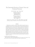
various lung diseases (Figure 1).
Physiology
Although established for glucose metabolism, target cells
for PPAR-γ agonists and the mechanisms by which they
hinder inflammation within the airways are not well
defined [5]. Culminating evidence suggests that PPAR-γ
may act by exerting its influence as a negative immu-
nomodulator regulating inflammatory respiratory
responses (Figure 2). Pro-inflammatory cytokines seem to
be the first point of call. For example, in adipose tissue,
the adipogenic action of the TZD PPAR-γ ligands are
opposed by several pro-inflammatory cytokines, includ-
ing tumour necrosis factor (TNF)-α and interferon (IFN)-
γ (Figure 2). In vitro, the TZDs blocked the effects of TNF-
α on both adipogenesis and insulin sensitivity and, simi-
larly, 15D-PGJ
2
was found to prevent IFN-γ-induced
murine macrophage activation [17].
In murine macrophages and human lung epithelial cell
line A549, expression of PPAR-γ was upregulated by inter-
leukin-4 (IL-4), a cytokine critical for certain subsets of
Respiratory Research 2005, 6:102 />Page 3 of 9
(page number not for citation purposes)
airway inflammation [17,18]. Similarly, IL-4 induced 12/
15-lipoxygenase (12/15-LO), an enzyme capable of gener-
ating PPAR-γ agonists in vivo. 12/15-LO was also highly
expressed in surface airway epithelial cells under basal
conditions [17]. Nitric oxide synthases (NOS) are respon-
sible for the in vivo synthesis of NO, a short-lived molecule
that is an effective bactericidal agent and may also regulate
expression of various pro-inflammatory genes, such as IL-
8, a potent chemoattractant and activator of neutrophils.
Both NOS and IL-8 play an important role in airway host
defence and elevated levels of IL-8 are found in broncho-
alveolar lavage fluid from intrinsic asthmatic patients
[19]. The two PPAR-γ agonists, 15D-PGJ
2
and ciglitazone
dose-dependently blocked the cytokine-induced expres-
sion of the inducible form of NOS. Ciglitazone alone only
slightly affected cytokine-induced IL-8 secretion, however,
the agonist significantly reduced IL-8 secretion from cells
pre-treated with IL-4 [17]. Therefore, PPAR-γ is expressed
and upregulated by IL-4 in airway epithelial cells and
through the activation of airway epithelial, PPAR-γ down-
regulates expression of inflammatory mediators. In
essence, PPAR-γ may act as an anti-inflammatory agent via
12/15-LO-dependent pathways [17].
Certain lung proteins may also be involved. The associa-
tion of PPAR-γ with the recruitment and activation of
peripheral blood monocytes, such as the potent chemok-
ine monocyte chemoattractant protein (MCP)-1, has also
been studied [20]. MCP-1 is produced by lung epithelial
cells during the course of inflammatory lung diseases.
Studies by Momoi's group [20] have demonstrated TZD's
ability to inhibit MCP-1 protein and mRNA expression in
cytokine-treated A549 lung epithelial cells.
The expression and physiological role of PPAR-γ in pul-
monary nonciliated bronchiolar epithelial cells (Clara
cells) and alveolar type II (AT II) epithelial cells has also
been investigated [21]. These cells are highly lipogenic
and are responsible for maintaining pulmonary surfactant
homeostasis [22]. Among the surfactant proteins, SP-B is
a 79-amino acid amphipathic peptide that is synthesised
and produced in Clara cells and AT II epithelial cells. The
SP-B facilitates lamellar body formation in AT II epithelial
cells and phospholipid spreading during the respiratory
cycles. The inhibitory effect of PPAR-γ ligands on SP-B
gene expression reveals a novel mechanism in the
regulation of pulmonary surfactant homeostasis [21]. In
the presence of 15D-PGJ
2
, the transcriptional level of SP-
B was down-regulated in respiratory epithelial cell line
and whole lung explant systems. Similarly, 15D-PGJ
2
sup-
pressed hSP-B gene activity at the -218 to -41 promoter
region in human pulmonary adenocarcinoma H441 cell
line transfected with various hSP-B luciferase reporter
gene constructs.
The intricate multifactorial coordination of PPAR-γ and
CCAAT/enhancer-binding proteins (C/EBP) for lung
development during the perinatal period has also been
displayed [23,24]. C/EBPs is a family of basic leucine-zip-
per transcription factors controlling a wide array of genes
and have been postulated to serve a central role in normal
tissue development and regulation of cell proliferation or
differentiation [25]. C/EBPβ and δ are known to act syner-
gistically with PPAR-γ to promote adipocyte differentia-
tion [23]. C/EBPα gene-deficient mice die shortly after
birth due to abnormal lung histology, including intersti-
tial thickening and hyperproliferation of AT II cells [26].
In developing foetal rat lungs, the C/EBPα, β, δ, and
PPAR-γ
1
mRNA expression was increased by 3- to 5-fold
from Day 18 of gestation, peaking at 1 to 2 days before
birth. However, there was a transient decline of expression
during the first postnatal day and a return to prenatal lev-
els on postnatal Day 5. In the AT II cell line, C/EBPα
mRNA was not detected throughout the developmental
stage; C/EBPβ and δ mRNAs expression was similar to that
of whole lung, with a prenatal rise profile, whereas PPAR-
γ did not display any developmental increase. The expres-
sion of PPAR-γ
2
was not detected in whole lung or in AT II
cell line [24].
Expression of PPAR-γ in various tissues and its role in lung and other organsFigure 1
Expression of PPAR-γ in various tissues and its role in
lung and other organs. PPAR-γ ligands implicated in the
treatment of chronic inflammatory disorders in lung. Activa-
tion of PPAR-γ in heart, intestine, kidney, skeletal muscle,
pancreas, macrophages and adipose tissue results in energy
homeostasis and this effect also found to be crucial in the
pathophysiology of different disorders. Please refer text for
more information.
PPAR-γ
γγ
γ
expression
Heart
Skeletal muscle
Small and large
intestine
Kidney
Pancreas
Spleen
Adipose tissue
Macrophage
• Stimulation of Adipocyte
differentiation
• Insulin sensitisation
• Regulation of
Inflammation and
atherosclerosis
LUNG
Airway epithelium
Bronchial submucosa
Alevolar macrophages
HASMs
T lymphocytes
Vascular smooth muscle
Endothelial cells
Eosinophils
Dendritic cells
Pathophysiology
of chronic and
acute lung
disorders
Respiratory Research 2005, 6:102 />Page 4 of 9
(page number not for citation purposes)
Changes in the metabolism of fatty acids such as arachi-
donic acid may also have detrimental effects on chronic
respiratory diseases including asthma, chronic bronchitis,
cystic fibrosis and bronchiectasis, as well as lung injury
and sepsis [27] The 85-kDa cytosolic phospholipase A
2
(cPLA
2
) plays an essential role in the control of arachi-
donic acid metabolism. It has been shown that cPLA
2
overexpression significantly increased the PPAR-γ-medi-
ated reporter activity and this activation by cPLA
2
may rep-
resent a novel mechanism for the control of airway
inflammation [28].
Asthma and PPAR-
γ
Asthma is a widespread chronic disease, with an increas-
ing incidence among children under 18 years of age [1].
Latest news reports headline the disease and its appear-
ance in the elderly at an alarming rate. Sufferers are
plagued with many undesirable pro-inflammatory events
in the airway, including narrowing and increased produc-
tion of mucous, thickening of the wall and thus reduction
of the airflow through the lungs. This response is accom-
panied by the activation of cell types such as T cells and
eosinophils and histopathological cellular airway restruc-
turing within the airways [4,5,14]. Airway inflammation
and alterations in cellular turnover are histopathologic
features of asthma [4] and recently, research has disclosed
the involvement of PPARs such as PPAR-γ and PPAR-α in
many facets of the disease such as decreasing antigen-
induced airway hyperresponsiveness, lung inflammation,
eosinophilia, cytokine production and serum levels of
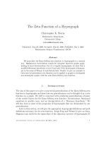
Activation of PPAR-γ by endogenous (15D-PGJ2) and exogenous (TZDs) ligands results in transcription of wide array of genes that can control pathogenesis of acute and chronic disorders in various tissues of lungsFigure 2
Activation of PPAR-γ by endogenous (15D-PGJ2) and exogenous (TZDs) ligands results in transcription of wide
array of genes that can control pathogenesis of acute and chronic disorders in various tissues of lungs. Please
refer text for more information. Abbreviations: 15D-PGJ2: 15-deoxy-∆
12,14
-prostaglandin J2, Cpla2: cytosolic phospholipase A
2
,
TZDs: Thiozolidinediones NSAIDs: Non-steroidal anti-inflammatory drugs MCP:1monocyte chemoattractant protein, G-CSF:
granulocyte-colony-stimulating factor, GM-CSF:granulocyte-macrophage-colony-stimulating factor, KC: keratinocyte-derived
chemokine, NOS: Nitric oxide synthases, SP-B: surfactant proteins-B, MMP-9: matrix metalloproteinase 9, TGF-β: Transform-
ing growth factor-β, IgE and IgG1: Immunoglubulin E and Immuno globulin G1, NF-κB: Nuclear factor-κB, EP2: Prostaglandin E2
receptor, PGE2: Prostaglandin E2, aP2: Adipocyte fatty acid binding protein, UCP 1&3: Uncoupling proteins 1 & 3, Acrp30: Adi-
pocyte complement related factor 30, FATP-1: Fatty acid transport protein-1.
Cytokines
NOS
SP-B MMP 9
MCP-1
TGF-
GATA-3
NF-B
IgE and IgG1
G-CSF,
GM-CSF
and KC
TNF-
EP2 PGE2
RXR
PPAR-γ
γγ
γ
PPRE
Co-activatior
TNF-
aP2
LpL
FATP-1
UCP-1
UCP-3
ACRP-30
Lungs
White adipose tissue
Respiratory Research 2005, 6:102 />Page 5 of 9
(page number not for citation purposes)
antigen-specific IgE [29]. Airway remodelling is character-
ised by the increase in subepithelial membrane (SBM)
and collagen deposition. A recent study displayed a posi-
tive correlation between PPAR-γ expression and SBM
thickening and collagen deposition in the epithelium [4].
In the submucosa, PPAR-γ expression was related to both
SBM thickening and to the number of proliferating cells.
Negative correlation was found between the intensity of
PPAR-γ expression in the bronchial submucosa, the air-
way epithelium and the smooth muscle to the forced
expiratory volume (FEV
1
) values. Inhaled steroids (either
administered alone or in combination with oral steroids)
restrained PPAR-γ expression in all the compartments, cell
proliferation, SBM thickness and collagen deposition,
enhancing apoptotic death in the epithelium and the sub-
mucosa. In this study, T lymphocytes in the bronchial
mucosa failed to express PPAR-γ. Therefore, PPAR-γ may
be an indicator of airway inflammation and remodelling
in asthma (Table 1).
In ovalbumin (OVA)-sensitised BALB/c mice (a murine
model of human asthma), PPAR-γ activation by ciglita-
zone treatment inhibited antigen-induced airway hyperre-
sponsiveness (AHR), basement membrane thickness,
collagen deposition and transforming growth factor
(TGF)-β synthesis, lung inflammation, eosinophilia,
cytokine production (IL-4, IL-5, IL-6 and IL-13), GATA-3
expression and serum levels of antigen-specific IgE and
IgG1. In vitro chemotaxis and antibody-dependent cellu-
lar cytotoxicity in human or rat eosinophils were also pre-
vented. The PPAR-γ antagonist GW9662 reversed the
above effects [5,29,30]. Similarly, PPAR-γ selective agonist
GI 262570 administered intranasally in OVA-induced
BALB/c reduced the elevated allergen-induced bronchoal-
veolar lavage eosinophil and lymphocyte but not neu-
trophil influx. In OVA-pulsed dendritic cells (DC),
rosiglitazone, a PPAR-γ agonist, averted the migration of
antigen-loaded DCs in the mediastinal lymph nodes
(MLN) and reduced the T-cell response in the MLNs [30].
Therefore, PPAR-γ stimulation of DCs may have a poten-
tial therapeutic role in reducing sensitisation to inhaled
allergens.
In similar experiments, PPAR-γ agonist GI 262570, PPAR-
α agonist GW 9578 and dual PPAR-α/γ agonist GW 2331
selectively inhibited allergen-induced bronchoalveolar
lavage eosinophil and lymphocyte influx in OVA-sensi-
tised BALB/c mice. However, PPAR-δ agonist GW 501516
had no effect. There was no inhibition of LPS-induced
bronchoalveolar lavage neutrophil influx or TNF-α and
keratinocyte-derived chemokine (KC) production by all
agonists administered intranasally before the challenge.
In A549 cells, the PPAR agonists did not inhibit intracel-
lular adhesion molecule-1 expression. Thus, in vitro data
suggests that PPAR effects on bronchoalveolar lavage eosi-
Table 1: This table shows PPAR-γ activators, inflammatory mediators affected by PPAR-γ expression and different disorders which can
be controlled by up-regulation of PPAR-γ. Abbreviations: TZDs: Thiozolidinediones, NSAIDs: Non-steroidal anti-inflammatory drugs,
15D-PGJ2: 15-deoxy-∆
12,14
-prostaglandin J2, Cpla2: cytosolic phospholipase A
2
, IL-4: Interleukin-4, MCP:1monocyte chemoattractant
protein, G-CSF: granulocyte-colony-stimulating factor, GM-CSF:granulocyte-macrophage-colony-stimulating factor, KC: keratinocyte-
derived chemokine, NOS: Nitric oxide synthases, SP-B: surfactant proteins-B, MMP-9: matrix metalloproteinase 9, TGF-β:
Transforming growth factor-β, IgE and IgG1: Immunoglubulin E and Immuno globulin G1, NF-κB: Nuclear factor-κB, EP2:
Prostaglandin E2 receptor, PGE2: Prostaglandin E2.
LIGANDS DOWN REGULATION IMPLICATION UP REGULATION IMPLICATION
TZDs (Exogenous) Cytokines (IL-8, IL-4, IL-5, IL-6
and IL-13)
NOS
MCP-1
Asthma and other pulmonary
inflammatory diseases
aP2
UCP1
UCP3
Acrp30
Insulin resistance
Obesity
Hyperlipidaemia
NSAIDs (Exogenous) SP-B
AHR
15D-PGJ2 (Endogenous) TGF-
β
GATA-3
IgE and lgG1
IL-4 (Endogenous) T-cell response
MMP-9
G-CSF and KC
azelaoyl-phosphocholine
(Endogenous)
GM-CSF COPD FATP-1
LPL (Adipose tissue)
Atherosclerosis
Eicosenoids (Endogenous) Cyclin D1
NF-κB
PGE2
EP2
Lung cancer (NSCLC, LCC)
Respiratory Research 2005, 6:102 />Page 6 of 9
(page number not for citation purposes)
nophil and lymphocyte influx may not be mediated by
the antagonism of the NF-κB pathway [31].
Interleukin-5 (IL-5) is the principal regulatory cytokine
mediating eosinophil airway inflammation and extending
the cell's survival. Eosinophils liberate cytotoxic products
at the site of inflammation, thus triggering AHR. IL-5-
stimulated (but not spontaneous) eosinophil survival and
eotaxin-directed chemotaxis was dose-dependently
reduced by the PPAR-γ agonist troglitazone. The results
indicated that upregulation of PPAR-γ in asthma may pre-
vent further activation of pro-inflammatory cells of the
airway [14].
Enzymes may also play a part in the PPAR-γ puzzle. Matrix
metalloproteinase (MMP)-9 (gelatinase B) is a matrix-
degrading enzyme found in human normal bronchial epi-
thelial cells and is involved in airway wall remodelling
generated by inflammatory processes. Activation of PPAR-
γ by rosiglitazone or pioglitazone in human bronchial
epithelial NL20 and BEAS cell lines dose-dependently
limited the expression of MMP-9 gelatinolytic activity
induced by TNF-α and phorbol myristate acetate. In con-
trast, the expression of the local inhibitor of MMP-9, tis-
sue inhibitor type 1, was retained. In this study, however,
transient transfection and electromobility shift assays
affirmed inhibition of nuclear factor (NF)-κB activation
by PPAR-γ agonists, resulting in decreased MMP-9 mRNA
expression [15]. In untreated atopic asthmatic patients,
there was an enhanced expression of PPAR-γ, which sug-
gested signs of airway transformation, including increased
density of the SBM and collagen deposition in the epithe-
lium, with no relation to proliferation or apoptosis. In
contrast, PPAR-γ-expressing cells in the submucosa were
related to both SBM thickening and to the number of
Ki67-, but not caspase-3-expressing-, cells. It was pro-
posed that PPAR-γ might not be involved in epithelial cell
turnover, but rather may manipulate extracellular matrix
accumulation and submucosal cell proliferation [4]
(Table 1).
PPAR-γ activation also influences lung survival factors and
apotosis. In male BALB/c mice, the initial levels of the
cytokines were not affected by the PPAR agonists, rosigli-
tazone or SB 219994. Aerosolised lipopolysaccharide
(LPS) exposure caused a significant increase in neutrophil
numbers in both lung lavage and tissue, however,
lymphomononuclear (LMN) cell numbers in BAL fluid
and lung tissue did not change. On pre-treatment with the
PPAR ligands, the increase in pro-inflammatory cytokines
granulocyte-colony-stimulating factor (G-CSF) and KC
levels was reduced in the lung tissue but not in the lung
lavage fluid. At the trial doses, the PPAR-γ agonists did not
affect LMN cells numbers in the BAL nor lavage or lung tis-
sue homogenate MMP-9 content. Rosiglitazone, when
administered after the LPS insult, reduced the lung tissue
G-CSF and neutrophilia levels and had no effect on KC or
granulocyte-macrophage (GM-CSF) levels. The results
suggested therapeutic similarities between rosiglitazone
and the steroid, dexamethasone [2] (Table 1).
AMs are phagocytes involved in the ingestion and degra-
dation of inhaled particles. This activates a variety of
inflammatory processes involving enhancement of their
cytotoxic capabilities. LPS-induced human AMs treated
with 15D-PGJ
2
and troglitazone showed a significant
reduction of the TNF-α cytokine production. This was
coupled with an increase in the expression of the scaven-
ger receptor CD36 (which contains a functional PPAR-γ
responsive element) and subsequent augmented apop-
totic neutrophil phagocytosis in the ligand-treated AMs
[3]. Therefore, administration of PPAR-γ synthetic ago-
nists such as TZDs may contribute as adjunct therapeutic
agents for airway diseases of the lung, such as asthma
[7,14] (Table 1).
Lung Cancer and PPAR-
γ
Lung cancer is the leading cause of cancer-related death in
developed countries and currently eludes the available
therapies. Consequently, the prognosis of patients with
lung cancer is generally poor, with a 10–15% 5 year sur-
vival rate [32]. High PPAR-γ expression has been sug-
gested as a potential marker for lung cancer and the degree
of PPAR-γ protein appears to correlate with the matura-
tional stage, differentiated phenotype, as well as the
tumour histological type and grade in lung adenocarci-
noma [33,34]. Studies have indicated that upon addition
of PPAR-γ selective agonists, growth of lung cancer cells
was prevented through the induction of differentiation
and apoptosis [35-38]. Additionally, decreased PPAR-γ
expression has been correlated with poor prognosis in
patients with lung cancer, suggesting that the gene expres-
sion may be further diminished as lung cancer progresses
[33]. PPAR-γ-selective agonists such as ciglitazone and
15D-PGJ
2
have diminished the growth of non-small cell
lung cancer (NSCLC) cells through the induction of apop-
tosis, promotion of differentiation and the down-regula-
tion of cell cycle proteins such as Cyclin D1 [35,37].
Treatment with troglitazone and pioglitazone signifi-
cantly reduced the number of lung metastases and
restricted NSCLC tumour progression in vivo [34]. Simi-
larly, combination of ciglitizone with trichostatin (an
inhibitor of histone deacetylase) demonstrated potent
growth-inhibitory and differentiation-inducing activity in
NSCLC, prompting the possibility of combinational dif-
ferentiation therapy for the treatment of lung adenocarci-
nomas [37]. Likewise, untreated large cell carcinoma
(LCC) cells displayed increased NF-κB activity, a pro-sur-
vival mechanism for this cancer in preventing apoptosis.
Upon treatment with thalidomide, the elevated level of
Respiratory Research 2005, 6:102 />Page 7 of 9
(page number not for citation purposes)
NF-κB activity was constrained in the presence of thalido-
mide and the PPAR-γ protein expression in LCC was dose-
dependently increased [32]. Therefore, as activation of
PPAR-γ impedes lung tumour progression, it is feasible
that TZDs may serve as potential therapeutic agents for
both NSCLC and LCC (Table 1).
Another aspect of carcinogenesis is the role of the induci-
ble enzyme, cyclooxygenase (COX)-2. COX-derived pros-
taglandins (PG) exhibit modulation of cell proliferation,
apoptosis, angiogenesis and immunity [39]. Prostaglan-
din E
2
(PGE
2
) is a major COX-2 metabolite and plays an
important role in tumour biology and its function is
mediated through G protein-coupled PGE receptor (EP)
[40]. The NSCLC cell expressing EP2 receptors, a key mod-
ulator of tumor development, has its mRNA and protein
expression significantly attenuated in the presence of
PPAR-γ ligands, GW1929, 15D-PGJ
2
, ciglitazone, troglita-
zone and rosiglitazone [41]. The effects of non-steroidal
anti-inflammatory drugs (NSAIDs) on decreased lung
cancer cell growth have also been examined [42,43].
Sulindac sulfide, a COX inhibitor, activated PPAR-γ at
higher concentration (50 µM). Together with ciglitazone,
sulindac sulfide potently suppressed NSCLC cell growth
[42]. Another COX-2 inhibitor, nimesulide (which is
known to induce PPAR-γ expression), has also had some
success in curbing tumour growth in female nu/nu mice
xenografted with subcutaneous A549 lung tumour cell
line and significantly reduced intratumour PGE
2
levels
[43]. Therefore, the potential therapeutic application of
NSAIDs and TZDs in the treatment and/or prevention of
lung cancer are promising, however more research is still
needed in order to evaluate the long-term safety and effi-
cacy of combined NSAIDs and TZDs in lung cancer [44]
(Table 1).
On the contrary, PPAR-α was not expressed in human
lung cancer cell lines and, thus, respective agonists such as
bezafibrate and prostanoids (PGE
2
and PGF
2α
) did not
inhibit growth of the cancer cell lines by inducing apopto-
sis [35].
Other Respiratory Disorders and PPAR-
γ
Cystic fibrosis is a genetic disorder characterised by func-
tional deficiencies of the reproductive, digestive and respi-
ratory systems. With the help of genetic mapping and
improved, more consistent treatment, patients are
enjoying longer and fulfilled lives. Adding to the
improved outlook, it is believed that respiratory PPAR-γ
expression is altered in tissues deficient in the normal
cystic fibrosis transmembrane regulator protein (CFTR). It
was found that PPAR-γ expression was decreased signifi-
cantly in (CFTR)-regulated tissues (colon, ileum and
lung) from exon 10 CFTR (cftr
_/_
) mice compared to wild-
type mice. In contrast, no differences were found in fat
and liver. In the lung tissue of both mice types, there was
a mixed labelling of both nuclei and cytoplasm localised
to larger bronchi and a diffuse lighter staining of the
remaining tissue [45].
The deficiency of GM-CSF is strongly implicated in the
pathogenesis of pulmonary alveolar proteinosis (PAP), a
rare interstitial lung disease manifested by surfactant accu-
mulation in alveolar airspaces. In PAP individuals, both
PPAR-γ mRNA and the PPAR-γ-regulated lipid scavenger
receptor, CD36 were reduced in AMs when compared to
healthy subjects. PPAR-γ and CD36 deficiency in PAP was
cell type-specific in the lung (i.e. found in AM and not in
bronchial epithelial cells). In vitro and in vivo GM-CSF
treatment of PAP patients fully restored PPAR-γ to healthy
control levels [46].
As for asthma patients, cell-proliferating lesions obstruct
the vessel lumen and promote pulmonary arterial pres-
sure and reduced blood flow in COPD patients [16,47]
(Table 1). In asthma, the eosinophil survival indicator,
GM-CSF, is prominent in bronchoalveolar lavage fluid,
serum and lung tissue. On the contrary, COPD is charac-
terised by neutrophilia [48]. It has been confirmed that
both GM-CSF and the related survival factor, G-CSF are
involved in the survival of the neutrophils. Consequently,
these factors may aggravate and extend the inflammatory
response in neutrophil-related inflammatory lung dis-
eases such as COPD [49,50].
Activation of PPAR-γ by 15D-PGJ
2
and ciglitazone
induced apoptosis and impeded serum-induced cell
growth more effectively than the steroid dexamethasone
in HASM. Moreover, PPAR-γ ligands and dexamethasone
hampered the IL-1β-induced release of GM-CSF. How-
ever, PPAR-γ ligands, but not dexamethasone, similarly
deterred G-CSF release. The above actions of 15D-PGJ
2
were not dependent on the activation of a traditional cell
surface prostanoid receptor. Agents that obstruct prolifer-
ation of HASM cells, as well as CSF release, would repre-
sent potential new therapies to treat COPD and steroid-
insensitive asthma [16] (Table 1).
Conclusion
It appears that chronic lung disorders are not confined to
a particular race, sex or age. Studies delving into respira-
tory diseases have reached a crucial point and the increas-
ing incidence and potential fatality of these debilitating
diseases has emphasised the urgent quest for novel thera-
peutic avenues vital to the control and ultimate elimina-
tion of such disease. The role of PPAR-γ in regulating
adipocyte differentiation and glucose homeostasis has
been established and, consequently, further research has
uncovered its involvement in inflammatory events of car-
diac and, more recently, airway diseases. Antagonism of
Respiratory Research 2005, 6:102 />Page 8 of 9
(page number not for citation purposes)
the pro-inflammatory pathways in respiratory diseases is
the likely mechanism of action of the PPARs and their
respective agonists. Research on the physiological role of
PPAR-γ in the lung is still in its infancy, however, contin-
ued advancement in this field will unravel the co-exist-
ence and interactions of the PPAR-γ gene and related
ligands such as 15D-PGJ
2
and TZDs in the prevention or
treatment of inflammatory respiratory diseases. It is
unlikely that the current PPAR-γ agonists will be used as a
monotherapy in airway diseases such as asthma and lung
cancer. However, with improved comprehension of the
full biological and physiological role of PPAR-γ in these
diseases, novel and more potent agonists could be
designed to include effective administration of anti-
inflammatory therapies with minimal side effects. This
could also extend to tackling more elusive or less common
lung disorders such as cystic fibrosis, PAP and COPD.
It is unanimously agreed that the PPAR-γ anti-inflamma-
tory pathways must be correctly identified for the particu-
lar disease state, as this will have important implications
for the type of treatment and its effective administration.
This would be determined by factors such as the receptor's
presence in the particular sections of the lung (lung tissue
compartment versus airway lumen), its expression in spe-
cific lung cell types and its influence on pro-inflammatory
cytokines, enzymes, proteins, fatty acid metabolism and
subsequent pathways. Therefore, it is anticipated that
PPAR-γ expression will become a potential indicator of
many airway inflammatory diseases leading to a possible
prevention or treatment therapeutic application.
Abbreviations
Peroxisome proliferator-activated receptors (PPAR); 15-
deoxy-∆
12,14
-prostaglandin J2 (15D-PGJ
2
); thiazolidinedi-
ones (TZD); chronic obstructive pulmonary disease
(COPD); PPAR-response element (PPRE); direct repeat
(DR); 9-cis-retinoic acid receptors (RXR); alveolar macro-
phages (AM); human airway smooth muscle (HASM);
tumour necrosis factor (TNF); interferon (IFN); inter-
leukin-4 (IL-4); 12/15-lipoxygenase (12/15-LO); nitric
oxide synthases (NOS); monocyte chemoattractant pro-
tein (MCP); alveolar type II (AT II); surfactant protein,
(SP); CCAAT/enhancer-binding proteins (C/EBP);
cytosolic phospholipase A
2
(cPLA
2
); subepithelial mem-
brane (SBM); forced expiratory volume (FEV
1
); ovalbu-
min (OVA); antigen-induced airway hyperresponsiveness
(AHR); transforming growth factor (TGF); Immunoglubu-
lin E and Immunoglobulin G1 (IgE and IgG1); dendritic
cells (DC); mediastinal lymph nodes (MLN); interleukin-
5 (IL-5); matrix metalloproteinase (MMP); nuclear factor
(NF); lipopolysaccharide (LPS); lymphomononuclear
(LMN); granulocyte-colony-stimulating factor (G-CSF);
keratinocyte-derived chemokine (KC); non-small cell
lung cancer (NSCLC); large cell carcinoma (LCC);
cyclooxygenase (COX); prostaglandin E
2
(PGE
2
); G pro-
tein-coupled PGE receptor (EP); non-steroidal anti-
inflammatory drugs (NSAIDs); ystic fibrosis transmem-
brane regulator protein (CFTR); pulmonary alveolar pro-
teinosis (PAP)
References
1. Serhan CN, Devchand PR: Novel antiinflammatory targets for
asthma. A role for PPARgamma? Am J Respir Cell Mol Biol 2001,
24:658-661.
2. Birrell BA, Patel HJ, McCluskie K, Wong S, Leonard T, Yacoub MH,
Belvisi MG: PPAR-gamma agonists as therapy for diseases
involving airway neutrophilia. Eur Respir J 2004, 24:18-23.
3. Asada K, Sasaki S, Suda T, Chida K, Nakamura H: Antiinflamma-
tory roles of peroxisome proliferator-activated receptor
gamma in human alveolar macrophages. Am J Respir Crit Care
Med 2004, 169:195-200.
4. Benayoun L, Letuve S, Druilhe A, Boczkowski J, Dombret MC,
Mechighel P, Megret J, Leseche G, Aubier M, Pretolani M: Regulation
of peroxisome proliferator-activated receptor gamma
expression in human asthmatic airways: relationship with
proliferation, apoptosis, and airway remodeling. Am J Respir
Crit Care Med 2001, 164:1487-1494.
5. Honda K, Marquillies P, Capron M, Dombrowicz D: Peroxisome
proliferator-activated receptor gamma is expressed in air-
ways and inhibits features of airway remodeling in a mouse
asthma model. J Allergy Clin Immunol 2004, 113:882-888.
6. Kota BP, Huang TH, Roufogalis BD: An overview on biological
mechanisms of PPARs. Pharmacol Res 2004 in press.
7. Mueller C, Weaver V, Vanden Heuvel JP, August A, Cantorna MT:
Peroxisome proliferator-activated receptor gamma ligands
attenuate immunological symptoms of experimental aller-
gic asthma. Arch Biochem Biophys 2003, 418:186-196.
8. Buchan KW, Hassall DG: PPAR Agonists as Direct Modulators
of the Vessel Wall in Cardiovascular Disease. Med Res Rev
2000, 20:350-366.
9. Ferre P: The biology of peroxisome proliferator-activated
receptors: relationship with lipid metabolism and insulin
sensitivity. Diabetes 2004, 53(Suppl 1):S43-S50.
10. Issemann I, Green S: Activation of a member of the steroid hor-
mone receptor superfamily by peroxisome proliferators.
Nature 1990, 347:645-650.
11. Fajas L, Auboeuf D, Raspe E, Schoonjans K, Lefebvre AM, Saladin R,
Najib J, Laville M, Fruchart JC, Deeb S, Vidal-Puig A, Flier J, Briggs MR,
Staels B, Vidal H, Auwerx J: The organization, promoter analy-
sis, and expression of the human PPARgamma gene. J Biol
Chem 1997, 272:18779-18789.
12. Fajas L, Fruchart JC, Auwerx J: PPARgamma3 mRNA: a distinct
PPARgamma mRNA subtype transcribed from an independ-
ent promoter. FEBS Lett 1998, 438:55-60.
13. Sundvold H, Brzozowska A, Lien S: Characterisation of bovine
peroxisome proliferator-activated receptors gamma 1 and
gamma 2: genetic mapping and differential expression of the
two isoforms. Biochem Biophys Res Commun 1997, 239:857-861.
14. Ueki S, Matsuwaki Y, Kayaba H, Oyamada H, Kanda A, Usami A, Saito
N, Chihara J: Peroxisome proliferator-activated receptor
gamma regulates eosinophil functions: a new therapeutic
target for allergic airway inflammation. Int Arch Allergy Immunol
2004, 134:30-36.
15. Hetzel M, Walcher D, Grub M, Bach H, Hombach V, Marx N: Inhibi-
tion of MMP-9 expression by PPARgamma activators in
human bronchial epithelial cells. Thorax 2003, 58:778-783.
16. Patel HJ, Belvisi MG, Bishop-Bailey D, Yacoub MH, Mitchell JA: Acti-
vation of peroxisome proliferator-activated receptors in
human airway smooth muscle cells has a superior anti-
inflammatory profile to corticosteroids: relevance for
chronic obstructive pulmonary disease therapy. J Immunol
2003, 170:2663-2669.
17. Wang AC, Dai X, Luu B, Conrad DJ: Peroxisome proliferator-
activated receptor-gamma regulates airway epithelial cell
activation. Am J Respir Cell Mol Biol 2001, 24:688-693.
18. Huang JT, Welch JS, Ricote M, Binder CJ, Willson TM, Kelly C, Witz-
tum JL, Funk CD, Conrad D, Glass CK: Interleukin-4-dependent
Respiratory Research 2005, 6:102 />Page 9 of 9
(page number not for citation purposes)
production of PPAR-gamma ligands in macrophages by 12/
15-lipoxygenase. Nature 1999, 400:378-382.
19. Folkard SG, Westwick J, Millar AB: Production of interleukin-8,
RANTES and MCP-1 in intrinsic and extrinsic asthmatics.
Eur Respir J 1997, 10:2097-2104.
20. Momoi A, Murao K, Imachi H, Ishida T, Cao WM, Sato M, Takahara J:
Inhibition of monocyte chemoattractant protein-1 expres-
sion in cytokine-treated human lung epithelial cells by
thiazolidinedione. Chest 2001, 120:1293-1300.
21. Yang L, Yan D, Yan C, Du H: Peroxisome proliferator-activated
receptor gamma and ligands inhibit surfactant protein B
gene expression in the lung. J Biol Chem 2003, 278:36841-36847.
22. Du H, Witte DP, Grabowski GA: Tissue and cellular specific
expression of murine lysosomal acid lipase mRNA and
protein. J Lipid Res 1996, 37:937-949.
23. Rosen ED, Spiegelman BM: Molecular regulation of
adipogenesis. Annu Rev Cell Dev Biol 2000, 16:145-171.
24. Barlier-Mur AM, Chailley-Heu B, Pinteur C, Henrion-Caude A, Dela-
court C, Bourbon JR: Maturational factors modulate transcrip-
tion factors CCAAT/enhancer-binding proteins alpha, beta,
delta, and peroxisome proliferator-activated receptor-
gamma in fetal rat lung epithelial cells. Am J Respir Cell Mol Biol
2003, 29:620-626.
25. Lekstrom-Himes J, Xanthopoulos KG: Biological role of the
CCAAT/enhancer-binding protein family of transcription
factors. J Biol Chem 1998, 273:28545-28548.
26. Sugahara K, Iyama KI, Kimura T, Sano K, Darlington GJ, Akiba T,
Takiguchi M: Mice lacking CCAAt/enhancer-binding protein-
alpha show hyperproliferation of alveolar type II cells and
increased surfactant protein mRNAs. Cell Tissue Res 2001,
306:57-63.
27. Nagase T, Uozumi N, Ishii S, Kume K, Izumi T, Ouchi Y, Shimizu T:
Acute lung injury by sepsis and acid aspiration: a key role for
cytosolic phospholipase A2. Nat Immunol 2000, 1:42-46.
28. Pawliczak R, Han C, Huang XL, Demetris AJ, Shelhamer JH, Wu T:
85-kDa cytosolic phospholipase A2 mediates peroxisome
proliferator-activated receptor gamma activation in human
lung epithelial cells. J Biol Chem 2002, 277:33153-33163.
29. Woerly G, Honda K, Loyens M, Papin JP, Auwerx J, Staels B, Capron
M, Dombrowicz D: Peroxisome proliferator-activated recep-
tors alpha and gamma down-regulate allergic inflammation
and eosinophil activation. J Exp Med 2003, 198:411-421.
30. Hammad H, de Heer HJ, Soullie T, Angeli V, Trottein F, Hoogsteden
HC, Lambrecht BN: Activation of peroxisome proliferator-
activated receptor-gamma in dendritic cells inhibits the
development of eosinophilic airway inflammation in a mouse
model of asthma. Am J Pathol 2004, 164:263-271.
31. Trifilieff A, Bench A, Hanley M, Bayley D, Campbell E, Whittaker P:
PPAR-alpha and -gamma but not -delta agonists inhibit air-
way inflammation in a murine model of asthma: in vitro evi-
dence for an NF-kappaB-independent effect. Br J Pharmacol
2003, 139:163-71.
32. DeCicco KL, Tanaka T, Andreola F, De Luca LM: The effect of tha-
lidomide on non-small cell lung cancer (NSCLC) cell lines:
possible involvement in the PPAR{gamma} pathway. Carcino-
genesis 2004, 25:1805-1812.
33. Sasaki H, Tanahashi M, Yukiue H, Moiriyama S, Kobayashi Y,
Nakashima Y, Kaji M, Kiriyama M, Fukai I, Yamakawa Y, Fujii Y:
Decreased perioxisome proliferator-activated receptor
gamma gene expression was correlated with poor prognosis
in patients with lung cancer. Lung Cancer 2002, 36:71-76.
34. Keshamouni VG, Reddy RC, Arenberg DA, Joel B, Thannickal VJ,
Kalemkerian GP, Standiford TJ: Peroxisome proliferator-acti-
vated receptor-gamma activation inhibits tumor progres-
sion in non-small-cell lung cancer. Oncogene 2004, 23:100-108.
35. Tsubouchi Y, Sano H, Kawahito Y, Mukai S, Yamada R, Kohno M,
Inoue K, Hla T, Kondo M: Inhibition of human lung cancer cell
growth by the peroxisome proliferator-activated receptor-
gamma agonists through induction of apoptosis. Biochem Bio-
phys Res Commun 2000, 270:400-405.
36. Inoue K, Kawahito Y, Tsubouchi Y, Yamada R, Kohno M, Hosokawa
Y, Katoh D, Bishop-Bailey D, Hla T, Sano H: Expression of perox-
isome proliferator-activated receptor (PPAR)-gamma in
human lung cancer. Anticancer Res 2001, 21:2471-2476.
37. Chang TH, Szabo E: Enhanced growth inhibition by combina-
tion differentiation therapy with ligands of peroxisome pro-
liferator-activated receptor-gamma and inhibitors of histone
deacetylase in adenocarcinoma of the lung. Clin Cancer Res
2002, 8:1206-1212.
38. Zhang M, Zou P, Bai M, Jin Y, Tao X: Peroxisome proliferator-
activated receptor-gamma activated by ligands can inhibit
human lung cancer cell growth through induction of
apoptosis. J Huazhong Univ Sci Technolog Med Sci 2003, 23:138-140.
39. Stolina M, Sharma S, Lin Y, Dohadwala M, Gardner B, Luo J, Zhu L,
Kronenberg M, Miller PW, Portanova J, Lee JC, Dubinett SM: Spe-
cific inhibition of cyclooxygenase 2 restores antitumor reac-
tivity by altering the balance of IL-10 and IL-12 synthesis. J
Immunol 2000, 164:361-370.
40. Han S, Sidell N, Roser-Page S, Roman J: Fibronectin stimulates
human lung carcinoma cell growth by inducing cyclooxygen-
ase-2 (COX-2) expression. Int J Cancer 2004, 111:322-331.
41. Han S, Roman J: Suppression of prostaglandin E2 receptor sub-
type EP2 by PPARgamma ligands inhibits human lung carci-
noma cell growth. Biochem Biophys Res Commun 2004,
314:1093-1099.
42. Wick M, Hurteau G, Dessev C, Chan D, Geraci MW, Winn RA, Hea-
sley LE, Nemenoff RA: Peroxisome proliferator-activated
receptor-gamma is a target of nonsteroidal anti-inflamma-
tory drugs mediating cyclooxygenase-independent inhibition
of lung cancer cell growth. Mol Pharmacol 2002, 62:1207-1214.
43. Shaik MS, Chatterjee A, Singh M: Effect of a selective cyclooxyge-
nase-2 inhibitor, nimesulide, on the growth of lung tumors
and their expression of cyclooxygenase-2 and peroxisome
proliferator-activated receptor-gamma. Clin Cancer Res 2004,
10:1521-1529.
44. Pang L, Nie M, Corbett L, Knox AJ: Cyclooxygenase-2 expression
by nonsteroidal anti-inflammatory drugs in human airway
smooth muscle cells: role of peroxisome proliferator-acti-
vated receptors. J Immunol 2003, 170:1043-1051.
45. Ollero M, Junaidi O, Zaman MM, Tzameli I, Ferrando AA, Andersson
C, Blanco PG, Bialecki E, Freedman SD: Decreased expression of
peroxisome proliferator activated receptor gamma in cftr-/-
mice. J Cell Physiol 2004, 200:235-244.
46. Bonfield TL, Farver CF, Barna BP, Malur A, Abraham S, Raychaudhuri
B, Kavuru MS, Thomassen MJ: Peroxisome proliferator-acti-
vated receptor-gamma is deficient in alveolar macrophages
from patients with alveolar proteinosis. Am J Respir Cell Mol Biol
2003, 29(6):677-82.
47. Ameshima S, Golpon H, Cool CD, Chan D, Vandivier RW, Gardai SJ,
Wick M, Nemenoff RA, Geraci MW, Voelkel NF: Peroxisome pro-
liferator-activated receptor gamma (PPARgamma) expres-
sion is decreased in pulmonary hypertension and affects
endothelial cell growth. Circ Res 2003, 92:1162-1169.
48. Sullivan S, Broide DH: Compartmentalization of eosinophil
granulocyte-macrophage colony-stimulating factor expres-
sion in patients with asthma. J Allergy Clin Immunol 1996,
97:966-976.
49. Aggarwal A, Baker CS, Evans TW, Haslam PL: G-CSF and IL-8 but
not GM-CSF correlate with severity of pulmonary neu-
trophilia in acute respiratory distress syndrome. Eur Respir J
2000, 15:895-901.
50. Stanford SJ, Pepper JR, Burke-Gaffney A, Mitchell JA: Cytokine-acti-
vated human vascular smooth muscle delays apoptosis of
neutrophils: relevance of interactions between cyclo-oxyge-
nase-2 and colony-stimulating factors. FASEB J 2001,
15:1813-1815.