Báo cáo khoa học: " Evaluation of three commercial bovine ELISA kits for detection of antibodies against Alphaherpesviruses in reindeer (Rangifer tarandus tarandus)" pdf
Bạn đang xem bản rút gọn của tài liệu. Xem và tải ngay bản đầy đủ của tài liệu tại đây (847.33 KB, 10 trang )
BioMed Central
Page 1 of 10
(page number not for citation purposes)
Acta Veterinaria Scandinavica
Open Access
Research
Evaluation of three commercial bovine ELISA kits for detection of
antibodies against Alphaherpesviruses in reindeer (Rangifer
tarandus tarandus)
Carlos G Das Neves*
1
, Matthieu Roger
1,4
, Nigel G Yoccoz
2
, Espen Rimstad
3
and Morten Tryland
1
Address:
1
The Norwegian School of Veterinary Science, Department of Food Safety and Infection Biology, Section of Arctic Veterinary Medicine,
Stakkevollveien 23, NO-9010 Tromsø, Norway,
2
University of Tromsø, Institute of Biology, NO-9037 Tromsø, Norway,
3
The Norwegian School
of Veterinary Science, Department of Food Safety and Infection Biology, Section of Microbiology, Immunology and Parasitology, PO Box 8146,
NO-0033 Oslo, Norway and
4
YROI: Cyclotron et Recherche Biomédicale Technopole – 2 Rue Maxime Rivière – BP 80005 – 97491 Sainte Clotilde
Cedex, Island of Réunion
Email: Carlos G Das Neves* - ; Matthieu Roger - ; Nigel G Yoccoz - ;
Espen Rimstad - ; Morten Tryland -
* Corresponding author
Abstract
Background: The genus Varicellovirus (family Herpesviridae subfamily Alphaherpesvirinae) includes a group of viruses genetically
and antigenically related to bovine herpesvirus 1 (BoHV-1) among which cervid herpesvirus 2 (CvHV-2) can be of importance
in reindeer. These viruses are known to be responsible for different diseases in both wild and domestic animals. Reindeer are a
keystone in the indigenous Saami culture and previous studies have reported the presence of antibodies against
alphaherpesviruses in semi-domesticated reindeer in northern Norway. Mortality rates, especially in calves, can be very high in
some herds and the abortion potential of alphaherpesvirus in reindeer, unlike in bovines, remains unknown.
ELISA kits are the most used screening method in domestic ruminants and given the close genetic relationship between viruses
within this genus, it might be possible to use such kits to screen cervids for different alphaherpesviruses. We have compared
three different commercial ELISA kits in order to validate its use for reindeer and CvHV-2.
Methods: Three commercial bovine ELISA kits (A, B and C), using either indirect (A) or blocking (B and C) ELISA techniques
to detect antibodies against BoHV-1 were tested with sera from 154 reindeer in order to detect antibodies against CvHV-2. A
Spearman's rank-based coefficient of correlation (ρ) was calculated. A dilution trial was performed for all kits. A virus
neutralization test using both BoHV-1 and CvHV-2 was carried out.
Results: Seroprevalence was almost the same with all kits (40–41%). Despite a similar qualitative score, quantitatively kits
classified samples differently and a strong correlation was only identified between Kits B and C. Blocking kits performed better
in both repeatability and in the dilution trial. The virus neutralization results confirmed the ELISA results to a very high degree.
Neutralizing titres ranged from 1:2 to 1:256 and from 0 to 1:16 against CvHV-2 and BoHV-1 respectively.
Conclusion: Results show that the genetic and antigenic similarity between BoHV-1 and CvHV-2 enables the use of a bovine
gB blocking ELISA kit to screen reindeer. The use of an ELISA kit is both cheaper and time saving, allowing screening of large
populations. This study revealed a high number of positive animals against CvHV-2 and its impact and distribution in the general
population should be further evaluated.
Published: 9 March 2009
Acta Veterinaria Scandinavica 2009, 51:9 doi:10.1186/1751-0147-51-9
Received: 10 July 2008
Accepted: 9 March 2009
This article is available from: />© 2009 Das Neves et al; licensee BioMed Central Ltd.
This is an Open Access article distributed under the terms of the Creative Commons Attribution License ( />),
which permits unrestricted use, distribution, and reproduction in any medium, provided the original work is properly cited.
Acta Veterinaria Scandinavica 2009, 51:9 />Page 2 of 10
(page number not for citation purposes)
Background
Viruses in the genus Varicellovirus (family Herpesviridae
subfamily Alphaherpesvirinae) are known to infect and
cause disease in several ruminant species. Of the
alphaherpesviruses infecting ruminants bovine herpesvi-
rus type 1 (BoHV-1), causing the diseases Infectious
Bovine Rhinotracheitis (IBR) and Infectious Pustular Vul-
vovaginitis (IPV), is well-described [1,2]. Other viruses of
this genus related to BoHV-1 are known to cross-react
serologically and have been isolated from semi-domesti-
cated and wildlife ruminant species such as cervid herpes-
virus 2 (CvHV-2, also known as Rangiferine Herpesvirus,
RanHV) from semi-domesticated reindeer (Rangifer taran-
dus tarandus) in Finland and Sweden [3,4]. Serological evi-
dence of alphaherpesvirus infection in reindeer has
further been reported in Greenland [5] and Alaska [6] as
well as in both wild [7] and semi-domesticated reindeer
[8-10] in Norway, although it is unknown which
alphaherpesvirus is circulating in these populations.
Finnmark County in northern Norway (55 047 km
2
) is
the largest reindeer herding area in Norway with an esti-
mate of 168 779 animals in 2005/2006 [11]. In this area
the reindeer are kept in a semi-nomadic way being herded
between summer and winter pastures, and being usually
free-ranging within the borders of their specific herding
districts. Mortality rates in reindeer in Finnmark vary sig-
nificantly between years and reached 47% for calves in
2005–2006 [11]. The impact of CvHV-2 in reindeer mor-
tality or abortion, events commonly associated with other
alphaherpesvirus infections in ruminants [12], remains
unknown.
In Norway the last BoHV-1 infection in cattle was reported
in 1993 [13], and the country has officially eradicated
IBR/IPV although a surveillance program is still ongoing.
According to previous serosurveys [9,10], alphaherpesvi-
rus infections are suspected in semi-domesticated rein-
deer in Finnmark, which is of great epidemiological
importance since cross-species infections between
bovines and reindeer have been shown for BoHV-1 and
CvHV-2 [12].
Many countries use sero-epidemiological surveys of
bovine populations to maintain an active surveillance or
to eradicate IBR/IPV. Different methods for screening for
antibodies against BoHV-1 in cattle have been developed
in several countries. In a study comparing serological
BoHV-1 tests, a blocking Enzyme Linked Immunosorbent
Assay (ELISA) based on glycoprotein B (gB) antigen was
found to be the best option with a sensitivity of 96% and
a specificity of 99% [14]. This was a better score than other
blocking ELISAs based on other glycoprotein antigens
(glycoprotein E), indirect ELISAs or virus neutralization
tests (VNT) [14].
Glycoprotein B plays a decisive role in the interaction
between the virus and host cells during the attachment,
penetration and replication processes of the virus [12].
The nucleotide sequence encoding gB is highly conserved
between BoHV-1 and CvHV-2 [15,16].
Serological cross-reactions have been shown to exist
between different viruses within the Varicellovirus genus
and several studies have calculated coefficients of anti-
genic similarity (R) proving the serological cross-reactivity
between CvHV-2 and BoHV-1 [17-20].
Given the serological cross-reactions within this genus,
serological tests for BoHV-1 based on highly conserved
antigen, such as gB, could be used to detect the presence
of antibodies against alphaherpesviruses in non-bovine
ruminant host species. Since these viruses generally estab-
lish latency and life-long infections in their natural hosts,
the presence of antibodies most likely indicates that the
animals are persistently infected.
There are no standardized methods to conduct serological
testing of reindeer populations and different serological
techniques have been used in smaller sero-surveys carried
out in Alaska [6,21], Norway [7-10] and Greenland [5].
Simultaneously, IBR/IPV eradication campaigns have
many times neglected the status of wild animals as possi-
ble reservoir species for alphaherpesviruses.
To assess the present alphaherpesvirus infection status of
reindeer from different reindeer husbandry districts in
Finnmark, a reliable and feasible serological test was
needed. Three commercial ELISA kits for detecting anti-
bodies against BoHV-1 in cattle were evaluated regarding
their ability to detect antibodies against alphaherpesvi-
ruses in reindeer: one indirect ELISA with BoHV-1 as anti-
gen, and two blocking ELISA kits with BoHV-1 gB as
antigen.
Methods
Origin of samples
A total of 154 serum or plasma samples from four geo-
graphically separated herds from Finnmark County, repre-
senting adults and calves as well as both genders, were
collected in 2004–2005.
Serological testing
The samples were analyzed in duplicate in all the three
commercial kits. The main characteristics for these kits (A,
B and C) are presented in Table 1. The manufacturer's
instructions and kit components were used in Kits B and
C, while for Kit A adaptations were necessary.
Kit A, Infectious Bovine Rhinotracheitis (IBR-Ab) SVANO-
VIR™ (Svanova Biotech AB Sweden), is an indirect ELISA.
Acta Veterinaria Scandinavica 2009, 51:9 />Page 3 of 10
(page number not for citation purposes)
The test wells are coated with a mixture of viral and cellu-
lar proteins from virus-infected cells whereas control wells
are coated with material from non-infected cells of identi-
cal type. The test serum samples were diluted 1:25 and
added to test and control wells. Kit A is based on an indi-
rect method, and because of this the secondary antibodies
provided with the kit (horseradish peroxidase conjugated
anti-bovine IgG monoclonal antibodies) could not be
used, as they would not recognize reindeer antibodies.
They were therefore replaced by a biotin labeled rabbit-
anti-reindeer antibody in a 1:200 dilution and incubated
for 1 h at 37°C [22]. Revelation was achieved using
Streptavidin-β peroxidase (POD-conjugate) diluted
1:10000 (Roche
®
Mannheim, Germany) and incubated
for 1 h at 37°C, followed by orthophenyldiamine (OPD)
as substrate (DakoCytomation
®
Glostrup, Denmark) incu-
bated for 10 min in the dark at 20°C. The enzyme reaction
was stopped by adding 100μL of 1 M H
2
SO
4
per well.
Because positive and negative controls of the ELISA kit
were from cattle, they could not be used in an indirect
ELISA method where the secondary antibody was
replaced. The validation criteria proposed by the manu-
facturer could hence not be used, and samples were there-
fore considered positive when the mean OD of the
antigen well minus the mean OD of the control well was
above zero, which indicates a higher reaction in the anti-
gen well compared to the control well.
Kit B, SERELISA™ IBR/IPV gB Ab Mono Blocking (SYNBI-
OTICS EUROPE SAS, France) is a blocking ELISA in which
two peroxidase conjugated monoclonal antibodies
against the gB protein of BoHV-1 compete with the serum
sample antibodies in binding to gB antigens in the well.
The negative and positive control sera from cattle supplied
with the kit were used. The test serum samples were
diluted 1:2. A competition percentage was calculated
based on the relation between the OD mean of the dupli-
cates and of the controls. Samples with a competition per-
centage above 60% were considered seropositive, between
45–60% doubtful and below 45% seronegative, as recom-
mended when testing cattle serum samples.
Kit C, gB BLOCKING LSI™ (LSI, France – Laboratoire Serv-
ice International), is based on the same blocking design as
Kit B, but with one monoclonal antibody against the gB
protein of BoHV-1 labeled with horseradish peroxidase
(HRP). The negative and positive control sera from cattle
supplied with the kit were used and test serum samples
were diluted 1:2. A competition percentage was calculated
as for Kit B. Samples with a competition percentage above
50% were considered seropositive, between 45–50%
doubtful and below 45% seronegative, as recommended
for cattle.
Sample dilution curves
In order to verify the analytical sensitivity of these kits, a
serial dilution of a panel of four selected serum samples
was performed in parallel for each kit. The starting point
was the initial serum dilution used for each kit (1:25 in Kit
A and 1:2 in Kits B and C). A twofold dilution was con-
ducted, in Kit A from 1:25 to 1:3200, and in Kits B and C
from 1:2 to 1:256. The four samples chosen were all from
herd IV: serum sample 24 was strongly positive in all kits;
Table 1: Major characteristics and modifications of the three commercial bovine ELISA kits used to test reindeer for alphaherpesvirus
antibodies in this study.
ELISA type Well antigen 2nd antibody Revelation
system
Absorbance Validation rules
SVANOVA – A Indirect BoHV-1 unknown antigen
in one well and cells on
another.
Rabbit anti-reindeer
antibody
Streptavidin-POD +
OPD
450 nm OD
S
= (OD
IBR
-
OD
CONTROL
)Sample is positive
if OD
SAMPLE
>0
SYNBIOTICS – B Blocking BoHV-1 gB antigen 2 monoclonal antibodies
(Mabs) anti-gB/
peroxidase
Peroxidase system 450 nm + 620 nm
(for correction)
Validation rules: %P =
[(OD
N
- OD
P
)/OD
N
] ×100
> 80% and OD
N
>0,500
Sample is positive if: %S =
[(OD
N
- OD
S
)/(OD
N
-
OD
P
)] ×100 > 60
Sample is doubtful if:
45<%S<60
LSI – C Blocking BoHV-1 gB antigen 1 monoclonal antibody
anti-gB/HRP labelled
Horseradish
Peroxidase
450 nm + 620 nm
(for correction)
Validation rules: %P =
[(OD
N
- OD
P
)/OD
N
] ×100
> 70% and OD
N
>0,700
Sample is positive if: %S =
[(OD
N
- OD
S
)/(OD
N
-
OD
P
)] ×100 > 50
Sample is doubtful if:
45<%S<50
BoHV-1: Bovine herpesvirus type 1
gB: Glycoprotein B
OPD: orthophenyldiamine
OD
N
: Mean optical density of the negative control sera
OD
P
: Mean optical density of the positive control sera
OD
S
: Mean optical density of the sample sera
Changes of the protocol to adapt the kit to reindeer are depicted in italic.
Acta Veterinaria Scandinavica 2009, 51:9 />Page 4 of 10
(page number not for citation purposes)
serum sample FA16 was moderately positive in all kits;
serum sample FA15 was classified as doubtful in Kit B and
seronegative in Kits A and C, and serum sample FB15 was
negative in all kits.
For Kits B and C the respective positive and negative con-
trol sera from cattle were also tested. For Kit A an addi-
tional sample of water was added as a negative control
and diluted as the other samples using the kit's dilution
buffers.
All dilutions were tested in duplicate and mean optical
density (OD) values were obtained according to the kit's
specifications and used for calculations.
Virus neutralization test (VNT)
Given the serological cross-reaction between BoHV-1 and
CvHV-2 and considering that the ELISA kits were designed
for cattle, VNT was performed on all the reindeer serum
samples to further validate the use of these kits in reindeer
and to confirm their ability to detect antibodies against
CvHV-2.
Reindeer sera were two fold diluted and each dilution
(from 1:2 to 1:256) was incubated with 100 TCID
50
of
CvHV-2 or BoHV-1 at 37°C for 1 h.
A mixture of serum and virus (50 μl) was added to wells
in 96 well plates. To each well, 100 μl of Madin-Darby
bovine kidney cells (MDBK), with calculated area cover-
age of 100%, was added. The medium used was Earles
MEM with addition of 2% foetal calf serum (FCS) and 2%
penicillin-streptomycin (PS 10 000 Units/mL penicillin
and 10 mg/mL streptomycin, SIGMA-ALDRICH, Oslo
Norway). The plates were incubated for 2 days and then
stained according to manufacturer's protocol (Diff-Quik
Staining Protocol, Hamilton Thorne Research). Reading
was performed and titres expressed as the reciprocal of the
highest serum dilution that completely prevented a cyto-
pathic effect (CPE). A reindeer serum sample, obtained
from an animal experimentally infected with CvHV-2,
and a bovine serum sample, obtained from a bovine
infected with BoHV-1, were added as positive controls
and used to calculate the coefficient of antigenic similarity
(R) as previously described by Lyaku et al. [18].
Statistical analysis
As all samples were tested in duplicates, repeatability was
assessed using the absolute difference between the OD
values (variability) calculated for each sample in each kit.
As the distribution of absolute difference was highly
skewed, the 5–95% quantiles (i.e. an interval including
90% of observations with 5% on either side) were used
instead of standard deviation to describe distribution of
individual values.
Because using ranks resulted in more robust statistics [23],
we used Spearman correlation (ρ) to assess the relation-
ships between kits. Calculations were done for two sub-
samples: observations below and above the cut off lines to
assess the relationships between the different kits for the
populations of negative versus positive samples in general
and around the cut-off values. The squared value ρ
2
can be
interpreted in terms of predictive power (explained varia-
bility) of one kit's ranks by the other kit's ranks. P-value
was considered significant if below 0.05. All calculations
were done in R (R Development Core Team 2008).
Results
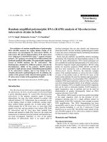
Serological testing
The three ELISA kits produced very similar seropreva-
lences results. Kit A classified 62 reindeer as having anti-
bodies against alphaherpesvirus (40.3%); Kit B 64
seropositive reindeer (41.6%) and three classified as
doubtful and Kit C 63 seropositive reindeer (41.0%) with
one animal classified as doubtful. Results were arranged
in ascending order according to OD difference mean val-
ues for Kit A and to competition percentages for Kits B and
C (Figure 1). The curves confirm that the individual
results were distributed following a sigmoid curve for all
three kits, although a more flattened curve was produced
by Kit A. For Kits B and C most individuals were clustered
in two distinct groups, one up to 20% of competition, rep-
resenting the negative samples, and the other from 85%
upwards, representing the individuals classified as serop-
ositive.
Serology resultsFigure 1
Serology results. Serology results for 154 samples of semi-
domesticated reindeer from Finnmark County, Norway dis-
played in ascending OD for Kit A and in ascending competi-
tion percentage for Kits B and C.
Acta Veterinaria Scandinavica 2009, 51:9 />Page 5 of 10
(page number not for citation purposes)
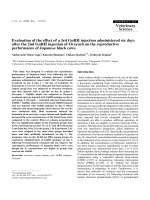
Comparison of three serological kits for detecting antibodies against alphaherpesvirus in reindeer by the ranks of the resultsFigure 2
Comparison of three serological kits for detecting antibodies against alphaherpesvirus in reindeer by the ranks
of the results. The comparison of the kits two by two was done by plotting ranks after sorting the results (OD values for Kit
A and competition percentage for Kit B and C) in ascending order. Results were given a rank position: 1
st
rank being the most
negative and 154
th
rank the most positive. The graphs display the rank obtained per animal in each kit. Lines pass through the
rank closest to the cut-off values for each kit (Kit A cut off value 0; Kits B and C lower cut off value 45%; Kit B higher cut off
value 60%; Kit C higher cut off value 50%). For Kit A, a line (—) passes through the 92
nd
rank (-0.031). For Kit B, a line (···)
passes through the 88
th
rank (47.28%) and another (— —) passes through the 90
th
rank (59.60%). For Kit C, a line (— – —)
passes through the 91
st
rank (45.54%) and represents both cut-off values (higher and lower) as no samples were ranked in
between. 2A: scatter plot displays Kit A and Kit B correlation. 2B: scatter plot displays Kit A and Kit C correlation. 2C: scatter
plot displays Kit B and Kit C correlation.
Acta Veterinaria Scandinavica 2009, 51:9 />Page 6 of 10
(page number not for citation purposes)
Both positive and negative controls for Kit B and C scored
well above the manufacturer's required thresholds.
To reveal if different kits were presenting similar qualita-
tive results (positive, negative or doubtful) a scatter plot,
displaying the results for each animal in each kit com-
pared two by two, was constructed (not displayed). Com-
paring Kits B and C three animals were classified as
doubtful in Kit B and seronegative in Kit C, and one ani-
mal was seropositive in Kit B and classified as doubtful in
Kit C. Comparing Kit A and B two animals were classified
seronegative in Kit A and seropositive in Kit B, whereas
three animals were classified negative in Kit A and doubt-
ful in Kit B. Comparing Kit A and C one animal was clas-
sified seronegative in Kit A and doubtful in Kit C.
Spearman coefficients (variability of one kit's ranks
explained by the other kit's ranks) showed that, despite an
almost absolute agreement of qualitative results (samples
being classified as positive, negative or doubtful) between
the kits, the quantitative results were not as concurrent
(Figure 2 and Table 2). In fact, only between Kits B and C
(Figure 2C) was there evidence for a strong correlation in
ranks both for negative as well as positive samples (P <
0.001). A restricted analysis of samples in the slope of the
curve, shown in Figure 1 (approximately ranks between
68
th
and 111
th
in Figure 2), confirmed the general obser-
vations. A correlation was evident between Kits B and C
for which two clear sub-populations, negative and posi-
tive, outflanking the cut-off value were identified and con-
firmed by VNT. No evidence for a correlation was found
within seropositive or seronegative animals by other kit
comparisons (Figure 2A and 2B) apart from a weak posi-
tive association within positive results for Kit A and Kit B
(Figure 2A), (P = 0.049; all other P-values > 0.09, Table 2).
Repeatability analysis
Kit A had the highest variability between OD duplicates
with a maximum difference in optical density of 2.35 and
a mean difference of 0.37 (5–95% quantiles: [0.018;
1.188]). Kit B had a maximum difference of 0.30 and a
mean of 0.03 (5–95% quantiles [0.001; 0.160]) and Kit C
a maximum difference of 0.27 and mean of 0.06 (5–95%
quantiles [0.001; 0.123]).
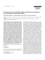
Serial dilution results
Serial dilution curves are displayed in Figure 3. The curves
for Kit A (Figure 3A) displayed some inconstant results for
the first dilutions. The curves for Kits B and C (Figure 3B
and 3C) were comparable to each other. Serum sample 24
(positive in all kits) was classified as positive in all dilu-
tions while the serum sample FA16 (also classified posi-
tive for all kits) became negative at dilution 1:32, 1:64 and
1:200 for Kits C, B and A, respectively. Positive control
samples (Kits B and C) became negative at a dilution of
1:128 in Kit B and 1:8 in Kit C.
Virus neutralization results
The VNT confirmed the ELISA results. All samples that
were classified negative by all the three ELISA kits failed to
neutralize any of the viruses. All samples classified posi-
tive in all kits neutralized CvHV-2, and some of them also
neutralized BoHV-1 though at a lower titre. Neutralizing
titres ranged for CvHV-2 from 1:2 to 1:256 and for BoHV-
1 from 0 to 1:16. No reindeer serum sample neutralized
BoHV-1 to a higher titre than CvHV-2 and the biggest dif-
ference observed between a sample neutralization of
CvHV-2 versus BoHV-1 was of 5 dilutions steps. Samples
that were classified as doubtful in the ELISA kits were sub-
sequently retested and classified as negative, and when
tested in the VNT they also failed to neutralize any of the
viruses. Only one weak positive sample in Kit B, which
was doubtful in Kit C and negative in A failed to neutralize
CvHV-2, while one sample classified as negative in Kit A
but as positive in the other two kits had a low titre for
CvHV-2 (1:2). The reindeer positive control neutralized
CvHV-2 up to 1:128 and BoHV-1 up to 1:16 while the
bovine positive control neutralized BoHV-1 up to 1:32
and CvHV-2 only at 1:2. The coefficient of antigenic simi-
larity between CvHV-2 and BoHV-1 was of R = 8.8. Results
are summarized in Table 3.
Discussion
Serological results obtained with the three different kits
showed that the blocking design kits performed better
than the indirect ones as had been concluded for the use
of similar kits for BoHV-1 [14], and identified that an
alphaherpesvirus serologically related to BoHV-1 is
present in semi-domesticated reindeer in Finnmark. The
blocking kits were found to work efficiently without any
changes to the manufacturers' protocols or pre-defined
cut-off values unlike Kit A which could not be used with-
out adaptations.
Data obtained in the virus neutralization strongly indi-
cates that CvHV-2 is most likely the virus present in this
reindeer population.
The percentage of seropositive reindeer ranged from 40–
42% between kits. The low variation between the kits ver-
ified the consistency of the results. In this study, reindeer
samples were tested in serological kits designed for bovine
sera and it was therefore necessary to verify if the pre-
established cut off values could be used for reindeer sera.
From the data obtained in the kits with blocking design (B
and C) even considerable changes in the cut off values
(10% up or downwards) would not significantly change
the results.
The Spearman coefficient is based on the ranks, reducing
sensitivity to outliers that could affect the Pearson correla-
tion coefficient. The value of Spearman's ρ calculated for
each sub-populations of positive or negative results,
Acta Veterinaria Scandinavica 2009, 51:9 />Page 7 of 10
(page number not for citation purposes)
showed that there was an association between ranks when
the two blocking kits were compared as could have been
expected given they were based on the same blocking
ELISA design. Samples tended to score similar percentages
of competition for Kits B and C even when we analyzed
only those samples flanking the cut-off lines. The cluster-
ing in two populations above and below the cut-off line
with similar quantitative and qualitative results was
shown to be concurrent with the VNT results with the
exception of two samples.
Despite using a different method, Kit A showed qualita-
tive results (animal classified as positive or negative) very
similar to the other two kits. Some association within pos-
itive results between Kits A and B further showed that the
tested ELISA kits correctly classified samples even when
using different methods.
Regarding the samples that scored negative in Kit A while
positive or doubtful in Kits B and C, one could also con-
sider that the difference may be due to a non specific
inhibitory character in the sera or a possible difference in
available epitopes for reaction between the two ELISA
methods.
The analysis of variability serves as an important tool to
study repeatability, and the differences between samples
tested in duplicate in the same plate is a good evaluator. A
mean variability in OD of 0.06 (5–95% [0.001; 0.123])
for Kit C and of 0.03 (5–95% [0.001; 0.160]) for Kit B are
good evidences that gB blocking kits had a better repeata-
bility compared to the indirect ELISA (Kit A), which had a
mean variability in OD of 0.37 (5–95% [0.018; 1.188]). It
is however important to remember that a direct compari-
son is difficult since the protocol of Kit A had to be
adapted to test reindeer sera. In Kits B and C variability
was obtained from the absolute difference between the
observed OD for a given sample (|OD
S1
- OD
S2
|), where
S1 and S2 represent the duplicates of a given test sample.
In Kit A however, there was an intermediate step for the
calculation of the same value (|(OD
IBR1
- OD
CONTROL1
) –
(OD
IBR2
- OD
CONTROL2
)|), where
CONTROL
represents the
control wells,
IBR
the well containing the antigen and 1 and
2 the duplicates. This additional step in Kit A might also
have contributed to the higher variability in Kit A versus
Kits B and C.
If we consider that analytical sensitivity is the largest dilu-
tion of a high-level positive serum in which antibody is no
longer detected, we observed a similar pattern for all kits,
in which sample 24 remained positive at 1:256 for Kits B
and C and at 1:3200 for Kit A. Sample FA16, which was
another strong positive (though not as strong as number
24), became negative at 1:200, 1:64, 1:32 for Kits A, B and
C respectively.
The abnormal curve observed in Kit A (Figure 3A) was
repeated and confirmed and could possibly be explained
by unspecific factors in the sera which interfered with the
binding of the antibodies.
Given the reduced number of samples tested it is difficult
to present a final conclusion for sensitivity, but we might
conclude for Kits B and C that they have a good sensitivity
as positive samples are still detectable 3 to 4 dilution steps
below their testing dilution. Further, it is possible to con-
clude from the three serial dilution curves, that the block-
ing design kits presented a more stable curve with a
moderate decrease in competition percentage when com-
pared to the indirect ELISA kit where OD values changed
abruptly and oscillated even though sensitivity also
seemed to be high considering how the positive samples
scored.
Table 2: Spearman correlation analysis within positive and negative results for the three commercial bovine ELISA kits tested,
compared two by two.
Kits compared Population analysed ρ P
1
Kit A and B Negative
(<88
th
rank for Kit B and <92
nd
rank for Kit A)
0.032 0.767
Positive
(>90
st
rank for Kit B and >92
nd
rank for Kit A)
0.247 0.049
Kit A and C Negative
(<91
st
rank for Kit C and <92
nd
rank for Kit A)
-0.003 0.974
Positive
(>91
st
rank for Kit C and >92
nd
rank for Kit A)
0.213 0.093
Kit B and C Negative
(<88
th
rank for Kit B <91
st
rank for Kit C)
0.481 <0.001
Positive
(>90
th
rank for Kit B and >91
st
rank for Kit C)
0.593 <0.001
1
P-value is considered significant if below 0.05
Acta Veterinaria Scandinavica 2009, 51:9 />Page 8 of 10
(page number not for citation purposes)
When comparing the ELISA designs used in this study, it
is demonstrated from the serology but also from the vari-
ance and serial dilution analysis that the gB blocking
design kits should be preferred to the indirect ELISA kit.
This was also the situation when testing cattle, where the
BoHV-1 gB kits was found more suitable as compared to
kits with an indirect ELISA design [14,24-26]. The lower
performance of Kit A in this trial may have derived from
the adaptations introduced and the conclusions drawn are
therefore only valid regarding its adaptation to test rein-
deer sera as required by the aim of this study.
When comparing the two blocking ELISA kits little differ-
ences can be found, though Kit C gave less doubtful
results and a slightly better repeatability. The positive con-
trol serum of Kit C performed however worse in the dilu-
tion analysis compared to the positive control of Kit B,
becoming negative at dilutions of 1:8 and 1:128 respec-
tively.
Regarding the VNT, Kramps et al. [14] clarified that VNT
did not present sufficient advantages to be the method of
choice for cattle. They showed that the ELISA kits had a
higher sensitivity and specificity and that they were time
and cost saving when large numbers of samples were to be
tested.
Even though the ELISA kits compared in this study were
designed for cattle, the genetic similarity between BoHV-1
and CvHV-2 was sufficient for all kits to detect reindeer
antibodies against CvHV-2. The VNT confirmed this by
showing an unequivocal higher neutralization against
Serial dilution curves of a panel of reindeer serum samples tested in three commercial bovine ELISA kits for detection of alphaherpesvirus antibodiesFigure 3
Serial dilution curves of a panel of reindeer serum samples tested in three commercial bovine ELISA kits for
detection of alphaherpesvirus antibodies. Four samples were selected to illustrate different situations: Serum samples 24
and FA16 were seropositive in all kits; serum sample FA15 was found to be doubtful in Kit B and seronegative in Kits A and C;
serum sample FB15 was classified negative in all kits. The positive and negative cattle sera controls from Kits B and C were also
titrated. In Kit A there were no controls, but water was tested as a negative control. 3A, 3B and 3C displays Kits A, B and C
serial dilutions respectively. In Figure 3A a continuous bold line (—) indicates the cut-off value for Kit A (0.00). In Figures 3B
and 3C a continuous bold line (—) indicates the upper cut-off value for the kits (60% for Kit B and 50% for Kit C) while a dot-
ted bold line (···) indicates the lower cut-off values (45% for both kits).
Acta Veterinaria Scandinavica 2009, 51:9 />Page 9 of 10
(page number not for citation purposes)
CvHV-2 with an average difference of three dilution steps
to BoHV-1. Neutralization against other alphaherpesvi-
ruses was not performed in this study given their unlikely
presence in Norway.
The VNT further showed that the cut-off values of the
ELISA kits were placed at a correct percentage of competi-
tion for Kits B and C and correct OD value for Kit A. Sam-
ples classified as doubtful (Kits B and C) where negative
in the VNT and only one low positive in Kit B (doubtful
in Kit C) and one negative sample in Kit A might have
been misclassified by the ELISA kits, if one wishes to con-
sider the VNT as a potential gold standard test for this type
of wildlife screening.
The present coefficient of antigenic similarity of 8.8 is in
line with previous calculations by Lyaku et al. and Rim-
stad et al. who calculated it to be 9 and 8.8 respectively,
even though the titres against CvHV-2 were lower in this
study (1:256 maximum) than in previous ones, where
reindeer sera neutralized CvHV-2 up to 1: 1024 and 1:512
respectively [18,20].
It is important to clarify that the VNT was used mostly to
confirm the presence of another alphaherpesvirus than
BoHV-1, as would be expected given the BoHV-1 free sta-
tus in cattle in Norway, and not specifically to compare
the performance of ELISA versus VNT despite the agree-
ment found between the two types of tests.
Kits B and C used as antigen the gB glycoprotein which is
strongly immunogenic and induces a humoral response
that appears in an early stage of infection [27]. This
response persists two to three years after infection in cattle
[28]. Because of the persistence of anti-gB antibodies, as
well as the fact that the gB antigen is genetically conserved
between alphaherpesviruses of ruminants, gB can be
regarded as an ideal antigen for serology in wild animals
for which the time of infection is unknown and no vali-
dated serological tests are commercially available.
Conclusion
The blocking ELISA kits using gB as antigen were found to
be preferable to use in serosurveys for alphaherpesvirus in
reindeer. Furthermore the choice of a blocking ELISA ena-
bles all ELISA components to be used and thus gives both
economical and time saving advantages.
With 40% of tested animals presenting antibodies against
alphaherpesviruses, our results indicate that an alphaher-
pesvirus infection is present in reindeer in Finnmark
County.
The virus neutralization results, associated to the inexist-
ence of BoHV-1 in Norway, strengthened and confirmed
the hypothesis that the virus present in this population is
indeed CvHV-2 and that a blocking ELISA commercial kit
can efficiently be used to screen reindeer for the presence
of antibodies against this virus.
These results, in combination with the knowledge of the
biological and economical importance of the closely
related BoHV-1 infection in cattle, should encourage fur-
ther studies of the distribution and impacts of CvHV-2
infection in reindeer in Scandinavia.
Competing interests
The authors declare that they have no competing interests.
Authors' contributions
CDN and MT designed the experiment and analyzed the
data. CDN, MT and MR performed the experiment. CDN
and NGY performed the statistical analysis. ER and CDN
performed the virus neutralization assay. CDN, ER and
Table 3: Virus neutralization test (VNT) on reindeer sera tested in this study.
ELISA Kits results No. of samples Neutralizing antibody titre
[min and max titre] average
Difference between CvHV-2 and BoHV-1
in dilution steps
A B C CvHV-2 BoHV-1 [min – max] average
+ + + 62 [1:2–1:256] 1:45 [0–16] 1:3 [1-5] 3.2
-±- 3 [0] [0] 0
-+± 1 [0] [0] 0
- + + 1 [1:2] [0] 1
87 [0] [0] 0
+ + + Bovine + control [1:2] [1:32] 4
+ + + Reindeer + control [1:128] [1:16] 3
Neutralizing titres are expressed as the reciprocal of the highest serum dilution that completely prevented a cytopathic effect (CPE). For each virus
neutralization, the maximum and minimum titres obtained in each group of samples are presented. Samples were grouped according to the results
they obtained in the different ELISA kits (+ for positive; ± for doubtful; – for negative). [0] represents the absence of neutralization. The difference
between the neutralizing titres against CvHV-2 and BoHV-1 is presented in dilution steps differences.
Publish with Bio Med Central and every
scientist can read your work free of charge
"BioMed Central will be the most significant development for
disseminating the results of biomedical research in our lifetime."
Sir Paul Nurse, Cancer Research UK
Your research papers will be:
available free of charge to the entire biomedical community
peer reviewed and published immediately upon acceptance
cited in PubMed and archived on PubMed Central
yours — you keep the copyright
Submit your manuscript here:
/>BioMedcentral
Acta Veterinaria Scandinavica 2009, 51:9 />Page 10 of 10
(page number not for citation purposes)
MT, drafted the manuscript. MR and NGY further helped
to draft the manuscript. All authors read and approved the
final manuscript.
Acknowledgements
We would like to acknowledge the irreplaceable help in the laboratory
from Eva Marie Breines and Ellinor Hareide, and in the field from the vet-
erinary students, Ingebjørg Nymo, Veronique Poulain, Anett Larsen and
Trine Marhaug. We would also like to thank the staff at Karasjok and
Kautokeino slaughterhouses for their help and hospitality. Finally we would
like to thank The Norwegian Institute for Nature Research for their help
during the sampling of live animals in Finnmark.
This project was supported by the Norwegian Reindeer Development Fund
(RUF).
References
1. Engels M, Ackermann M: Pathogenesis of ruminant herpesvirus
infections. Vet Microbiol 1996, 53:3-15.
2. Pastoret PP, Thiry E, Brochier B, Derboven G: Bovid herpesvirus 1
infection of cattle: pathogenesis, latency, consequences of
latency. Ann Rech Vet 1982, 13:221-235.
3. Ek-Kommonen C, Pelkonen S, Nettleton PF: Isolation of a herpes-
virus serologically related to bovine herpesvirus 1 from a
reindeer (Rangifer tarandus). Acta Vet Scand 1986, 27:299-301.
4. Rockborn G, Rehbinder C, Klingeborn B, Lefler M, Klintevall K, Nik-
kilä T, Landèn A, Nordkvist M: The demonstration of a herpes-
virus, related to bovine herpesvirus 1, in reindeer with
ulcerative and necrotizing lesions of the upper alimentary
tract and nose. Rangifer 1990:373-384.
5. Report on the animal health situation in Greenland 1999
[ />port.pdf].
6. Dieterich RA: Respiratory viruses. In Alaskan wildlife diseases Fair-
banks: University of Alaska; 1981:28-29.
7. Lillehaug A, Vikoren T, Larsen IL, Akerstedt J, Tharaldsen J, Hande-
land K: Antibodies to ruminant alpha-herpesviruses and pes-
tiviruses in Norwegian cervids. J Wildl Dis 2003, 39:779-786.
8. Hyllseth B, Stuen S, Rimstad E, Dahle HK, Tyler NJC: Serologiske
undersøkelser over herpesvirus infekjoner hos rein I
Finnmark og Svalbard. Nor Vet Tidsskr 1993, 105:733-736.
9. Stuen S, Krogsrud J, Hyllseth B, Tyler NJC: Serosurvey of three
virus infections in reindeer in northern Norway and Sval-
bard. Rangifer 1993, 13:215-219.
10. Tryland M, Mørk T, Ryeng KA, Sørensen KK: Evidence of parapox-
, alphaherpes- and pestivirus infections in carcasses of semi-
domesticated reindeer (Rangifer tarandus tarandus) from
Finnmark, Norway. Rangifer 2005, 25:75-83.
11. Anonymous: Ressursregnskap for Reindriftsnæringen fra Rein-
driftsforvaltningen. Alta: Reindriftsforvaltningen; 2007.
12. Thiry J, Keuser V, Muylkens B, Meurens F, Gogev S, Vanderplasschen
A, Thiry E: Ruminant alphaherpesviruses related to bovine
herpesvirus 1. Vet Res 2006, 37:169-190.
13. Nyberg O, Tharaldsen J, Heier BT: The surveillance and control
programme for infectious bovine rhinotracheitis (IBR) and
infectious pustular vulvovaginitis (IPV) in Norway. Annual
Report 2004. National Veterinary Institute, Norway 2004:54-58 [http:/
/www.vetinst.no/nor/content/download/418/3433/file/
vetinst_nokrapp05_6.pdf]. 07 February 2006
14. Kramps JA, Banks M, Beer M, Kerkhofs P, Perrin M, Wellenberg GJ,
Van Oirschot JT: Evaluation of tests for antibodies against
bovine herpesvirus 1 performed in national reference labo-
ratories in Europe. Vet Microbiol 2004, 102:169-181.
15. Griffin AM: The nucleotide sequence of the glycoprotein gB
gene of infectious laryngotracheitis virus: analysis and evolu-
tionary relationship to the homologous gene from other her-
pesviruses. J Gen Virol 1991, 72:393-398.
16. Ros C, Belak S: Studies of genetic relationships between
bovine, caprine, cervine, and rangiferine alphaherpesviruses
and improved molecular methods for virus detection and
identification. J Clin Microbiol 1999, 37:1247-1253.
17. Deregt D, Gilbert SA, Campbell I, Burton KM, Reid HW, Littel-van
den Hurk SVD, Penniket C, Baxi MK: Phylogeny and antigenic
relationships of three cervid herpesviruses. Virus Res 2005,
114:140-148.
18. Lyaku JRS, Nettleton PF, Marsden H: A Comparison of Serologi-
cal Relationships among 5 Ruminant Alphaherpesviruses by
Elisa. Arch Virol 1992, 124:333-341.
19. Martin WB, Castrucci G, Frigeri F, Ferrari M: A serological com-
parison of some animal herpesviruses. Comp Immunol Microbiol
Infect Dis 1990, 13:75-84.
20. Rimstad E, Krona R, Hyllseth B: Comparison of Herpesviruses
Isolated from Reindeer, Goats, and Cattle by Restriction
Endonuclease Analysis. Arch Virol 1992, 123:389-397.
21. Dau J: Caribou management report Units 21D, 22A, 22B, 23,
24 and 26A. In Caribou management report of survey-inventory activities
1 July 1998 – 30 June 2000 Edited by: Healy C. Juneau: Alaska Depart-
ment of Fish and Game; 2001:195-218.
22. Åsbakk K, Gall D, Stuen S: A screening ELISA for brucellosis in
reindeer. Zentralbl Veterinarmed B 1999, 46(9):649-657.
23. Conover WJ, Iman RL: Rank transformations as a bridge
between parametric and nonparametric statistics. American
Statistician 1981, 35:124-129.
24. Perrin B, Bitsch V, Cordioli P, Edwards S, Eloit M, Guerin B, Lenihan
P, Perrin M, Ronsholt L, Van Oirschot JT, et al.: A European com-
parative study of serological methods for the diagnosis of
infectious bovine rhinotracheitis. Rev Sci Tech 1993, 12:969-984.
25. Kramps JA, Perrin B, Edwards S, van Oirschot JT: A European
inter-laboratory trial to evaluate the reliability of serological
diagnosis of bovine herpesvirus 1 infections. Vet Microbiol 1996,
53:153-161.
26. Kramps JA, Magdalena J, Quak J, Weerdmeester K, Kaashoek MJ,
Marisveldhuis MA, Rijsewijk FAM, Keil G, Vanoirschot JT: A Simple,
Specific, and Highly Sensitive Blocking Enzyme-Linked-
Immunosorbent-Assay for Detection of Antibodies to
Bovine Herpesvirus-1. J Clin Microbiol 1994, 32:2175-2181.
27. Schwyzer M, Ackermann M: Molecular virology of ruminant her-
pesviruses. Vet Microbiol 1996, 53:17-29.
28. Kaashoek MJ, Rijsewijk FAM, VanOirschot JT: Persistence of anti-
bodies against bovine herpesvirus 1 and virus reactivation
two to three years after infection. Vet Microbiol 1996,
53:103-110.