Báo cáo thú y: "Survey radiography and computerized tomography imaging of the thorax in female dogs with mammary tumors" pot
Bạn đang xem bản rút gọn của tài liệu. Xem và tải ngay bản đầy đủ của tài liệu tại đây (3.5 MB, 10 trang )
RESEARC H Open Access
Survey radiography and computerized
tomography imaging of the thorax in female
dogs with mammary tumors
Carolina C Otoni
1*
, Sheila C Rahal
1
, Luiz C Vulcano
2
, Sérgio M Ribeiro
3
, Khadije Hette
1
, Tatiana Giordano
1
,
Danuta P Doiche
2
, Renée L Amorim
4
Abstract
Background: Accurate early diagnosis of lung metastases is important for establishing therapeutic measures.
Therefore, the present study aimed to compare survey thoracic radiographs and computerized tomography (CT)
scans to specifically identify lung metastases in female dogs with mammary tumors.
Methods: Twenty-one female dogs, weighing 3 to 34 kg and aged from 5 years to 14 years and 10 months, with
mammary tumors were studied. In all dogs before the imaging examinations, fine-needle aspiration cytology of the
mammary tumors was performed to confirm the diagnosis. Three-view thoracic radiographs were accomplished:
right lateral, left lateral and ventrodorsal views. Sequential transverse images of the thorax were acquired on a
spiral Scanner, before and after intravenous bolus injection of nonionic iodine contrast. Soft-tissue and lung
windows were applied. All the mammary tumors were surgically removed and examined histologically.
Results: The correlati on between the cytological and histological results regarding presence of malignancy was
observed in only 17 cases. In radiographic examinations, no dog displayed signs of lung metastases or thorax chest
lesions. CT detected lung metastasis in two cases, while small areas of lung atelectasis located peripherally were
found in 28.57% of the dogs.
Conclusion: In this study population, spiral CT showed higher sensitivity than chest radiographies to detect lung
metastasis; this indicates that CT should be performed on all female dogs with malignant mammary tumors.
Background
Mammary tumors constitute the most frequent neoplas-
tic disease in female dogs [1,2] . The disease etiology has
not yet been totally elucidated, but there are indications
of a hormonal dependence because the incidence of
tumors is reduced by using early ovariohysterectomy,
with better results when the procedure is performed
before the first estrus [1-3]. In addition, dogs with mam-
mary gland carcinoma spayed less than 2 years before
tumor surgery live longer than dogs spayed earlier in
relation to such surgery [4].
Approximately 35% to 50% of al l mammary tumors in
female dogs are considered malignant by histological
examinations [2,3]. The clinical stage, tumor size and
ovariohysterectomy status are prognostic factors for dog
survival after surgery to treat malignant mammary
tumors [5]. Furthermor e, these tumors may disseminate
through the ly mphatic and blood vessel routes to the
regional lymph nodes and lungs [1,3]. Therefore, the
accurate and early diagnosis of lung metastases is of
considerable importance in the establishment of
therapeutic measures, and approximately 25% and 50%
ofthefemaledogswithmalignantmammarytumors
already present them at the moment of the physical
examination [3].
Among the imaging methods reported as frequentl y
used for lung metastases identification are radiographic
examinations, magnetic resonance and computerized
tomography (CT) [3,6-9]. Radiographically, lung metas-
tasis can be characterized bywell-definednodules,
* Correspondence:
1
São Paulo State University (Unesp), Department of Veterinary Surgery and
Anesthesiology, School of Veterinary Medicine and Animal Science, Botucatu,
SP, Brazil
Otoni et al. Acta Veterinaria Scandinavica 2010, 52:20
/>© 2010 Otoni et al; licensee BioMed Central Ltd. This is an Open Access articl e distributed under the terms of the Creative Commons
Attribution License ( which permits unrestricted use, distribution, and reproduction in
any medium, provided the original work is properly cited.
poorly demarcated nodules or as pleural effusion with-
out any evidence of lung lesions [2]. Although CT is
considered a more sensitive method than radiography
for detecting lung metastases, false-positive or false-
negative results may occur [8,10,11]. Therefore, various
types of CT instruments are constantly being developed
to obtain higher accuracy and p otency. Furthermore, a
shortexposuretimeisveryimportanttominimizethe
effects of cardiovascular and respiratory motion [12].
The present study aimed to compare survey thoracic
radiographs and computerized tomography (CT) scans
in relation to their ability to specifically identify lung
metastases in female dogs with mammary tumors.
Materials and methods
Twenty-one f emale dogs, weighing 3 to 34 kg and aged
from 5 years to 14 years and 10 months (average of 9
years old and 5 months), with mammary tumors were
utilized (Table 1). The time between tumor identifica-
tion and surgical remo val varied from 2 months to 1
year. Two dogs (Cases 6 and 19) had been submitted 3
months previously for regional mastectomy based on
the lymphatic drainage in the other mammary chain.
Four female dogs (Cases 1, 3, 6 and 21) had already
been spayed. Before the imaging examinations in all
female dogs, fine-needle aspiration cytology of the mam-
mary tumors was performed to confirm the diagnosis.
Only those with malignant mammary tumors were
included in the study. The size of the primary tumor
was classified according to maximum diameter as fol-
lows:T1<3cm,T23-5cmandT3>5cm(Table1).
Laboratory tests, including a complete blood cell, urina-
lysis and chemistry panel, were carried out to detect any
metabolic alterations that could contraindicate the
procedures.
Preoperative Radiographic and CT studies
Thoracic radiographs were taken from three views: right
lateral, left lateral and ventrodorsal . Green-sensitive film
(Kodak) and cassettes with screens were used. A focus-
film distance of 90 cm was used with an exposure of
50-70 kV and 3.2-5.0 m As for the lateral v iew and 45-
65 kV and 3.2-5.0 mAs for the ventro dorsal view,
according to the size of each dog. The radiographs were
done at peak inspiration. An X-ray unit type TUR
D800-4, 125 kVp/500 mA capacity, equipped with Pot-
ter-Bucky grid was used. The film was processed with
an automatic processor (Macrotec MX2). To maintain
the quality cont rol of the radiographs, in both ventro-
dorsal and lateral views the thorax should not be
rotated. In addition, the sternum and vertebra should be
superimposed on the ventrodorsal view, and little or no
contact betwe en the diaphragm and the heart should be
seen on the lateral view. The technical quality of the
film was considered satisfactory if there was a clear con-
trast among the structures - pulmonary vessels, heart
and air-filled lungs. The thoracic radiographs were eval-
uated starting from cardiac silhouette, trachea, mediasti-
num and pleural space, and ending at the lungs.
Radiographs were classified as either positi ve or negative
for pulmonary metastases, and other pulmonary dis-
eases. In addition, the heart aspect, the presence of
mediastinal masses, and thoracic w all alteration w ere
evaluated.
To perform CT examinations the female dogs were
premedicated with acepromazine 0.03 mg/kg, IM, and
morphine 0.5 mg/kg, IM. After approximately 10 min-
utes, dissociative anesthesia was induced and maintained
with ketamine 3 mg/kg and diazepam 0.5 mg/kg, admi-
nistered intravenously, with the dogs positioned in ster-
nal recumbency. The lungs were not mechanically
inflated for imaging. The female dogs were placed in
dorsal recumbency with the forelimbs pulled cranially
and the hind limbs caudally. Sequential transverse
images from the first thoracic vertebra to lumbar dia-
phragmatic recess were acquired on a spiral Scanner
(Shimadzu SCT-7800CT). After plain CT study, a con-
trast study was done using 2 ml/kg, IV, bolus injectio n
of nonionic iodine contrast agent (iohexol or meglumine
diatrizoate). The scanning parameters were 120 kVp,
150-180 mA, 2.0-3.0 mm collimation, 2:0 pitch, and 1-
second scan time. The field of view was 168-409 mm
according to the dog’s size, while the matrix was 512 ×
512 mm. The software used to read the CT-images was
2.1.2 eFilm (TM) Lite (TM) (MERGE Healthcare). The
study was carried out using soft-tissue and lung win-
dows, and it was started from the mediastinum to chest
wall in order to detect lesions. The plain CT images
were compared with the contrast images. CT images
were classified as either positive or negative for pulmon-
ary metastases. When nodules were present, their dia-
meters were found by means of a measuring tool from
an image analysis software package. Three experienced
radiologists performed blind evaluation of the radio-
graphs and CT.
Surgical procedures, and Histology
After ovariohysterectomy, ei ther a unilateral, b ilateral or
regional mastectomy was performed, based on an assess-
ment of lymphatic drainage. Lumpectomy was used spe-
cifically in cases when other b enign masses were
present.
The mammary tumors removed at surgery were
immediately fixed in 10% buffered formalin. Semi-serial
sections, 4-μm-thick, were obtained and stained with
hematoxylin and eosin. The histological results were
compared with those obtained previously by fine-needle
aspiration cytology.
Otoni et al. Acta Veterinaria Scandinavica 2010, 52:20
/>Page 2 of 10
Table 1 Description of female dogs with mammary tumors; mammary glands with tumor and tumor size; fine-needle aspiration cytology and histological
diagnosis of the mammary tumors.
Dog description Mammary glands with tumor and
tumor size
Fine-needle aspiration
cytology
Time between tumor
identification and surgical
removal
Histological analysis
Case 1
34 kg-13.8-year-old German Shepherd
(10 y spayed)
Left CrT and CT (T2 - ulcerated) mammary carcinoma 2 mo mammary carcinoma
Case 2
8 kg-11.10-year-old crossbred
(intact)
Left CrA (T1) Left CrA - adenoma
mammary
9 mo Left CrA - mammary carcinoma
Case 3
13.4 kg-10.7-year-old crossbred
(7 y spayed)
Right I (T2 - ulcerated)
Left CA (T1)
Right I - mammary
carcinoma;
Left CA inflammatory
process
3 mo Right I - mammary carcinoma and regional
lymph node metastases; Left CA
-trichoepithelioma
Case 4
11.4 kg-9-year Poodle
(intact)
Left CrA, CA and I (T2);
Right CA (T1)
Left CrA, CA and I -
secretory mammary
carcinoma
Right CA - absent
neoplasia
9 mo Left CrA, CA and I - mammary adenoma;
Right CA - absent neoplasia
Case 5
9.1 kg-9.4-year-old crossbred
(intact)
Right CA (T1) mammary carcinoma 3 mo mammary carcinoma
Case 6
3 kg-5.8-year-old Yorkshire
(3 mo spayed)
Right CrT (T1) mammary carcinoma 9 mo mammary carcinoma
Case 7
3.5 kg-7-year-old crossbred
(intact)
Left CT and CrA (T1); Right CrA (T1) Right CrA and Left CrA -
malignant mixed tumor;
Left CT - epidermal cyst
7 mo Right CrA and Left CrA - malignant mixed
tumor; Left CT - epidermal cyst
Case 8
13 kg-9-year-old Poodle
(intact)
All Left mammary glands (T2);
Right CrA and I (T2)
Left mammary glands -
mammary carcinoma;
Right CrA and I -
malignant mixed tumor
7 mo Left mammary glands - mammary carcinoma;
Right CrA and I -adenocarcinoma
Case 9
7 kg-8.6-year-old
crossbred
(intact)
Left I (T1) adenocarcinoma 1 y benign mixed tumor
Case 10
6.5 kg-8-year-old crossbred
(intact)
Right CA and I (T1) Right CA and I -
malignant mixed tumor
3 mo Right CA and I -malignant mixed tumor
Case 11
22.6 kg-6.8-year-old Cocker Spaniel (intact)
Right I (T1) mammary carcinoma 1 y mammary carcinoma
Otoni et al. Acta Veterinaria Scandinavica 2010, 52:20
/>Page 3 of 10
Table 1: Description of female dogs with mammary t umors; mammary glands with tumor and tumor size; fine-needle aspiration cytology and histological
diagnosis of the mammary tumors. (Continued)
Case 12
5.8 kg-9-year-old Poodle (intact)
Right CT (T1) mammary carcinoma 3 mo mammary carcinoma
Case 13
16.7 kg-6.3-year-old Dachshund
(intact)
Left CT (T3) mammary carcinoma 4 mo mammary carcinoma
Case 14
31 kg-8.5-year-old Boxer
(intact)
Right CA (T3) mammary carcinoma 2 mo malignant mixed tumor
Case 15
14.1 kg-14.7-year-old Poodle
(intact)
Right CT (T3) mammary carcinoma 7 mo papillary cystadenocarcinoma
Case 16
3.6 kg-14.10-year-old crossbred
(intact)
Right CA (T1), Right I (T2);
Left CrA (T1), Left I (T1)
Left CrA and I - malignant
mixed tumor; Right CA -
secretory adenoma
1 y malignant mixed tumor
Case 17
21.5 kg-14-year-old crossbred
(intact)
Right CrA and CA (T3 - ulcerated);
left I (T2)
malignant mixed tumor 3 mo malignant mixed tumor
Case 18
3.4 kg-5-year-old crossbred
(intact)
Left CA (T1) mammary carcinoma 9 mo mammary carcinoma
Case 19
11.2 kg-8-year-old
Cocker Spaniel
(intact)
Right I (T1) mammary carcinoma 1 y mammary tubular carcinoma
Case 20
27 kg-6.5-year-old Akita
(intact)
Right CA (T3) mammary adenoma 4 mo mammary adenocarcinoma
Case 21
10.7 kg-13-year-old crossbred
(8 y spayed and received contraceptive)
Right CrA (T2); Left CrA
(T1)
malignant mixed tumor 2 y malignant mixed tumor
Cranial thoracic - CrT; Caudal thoracic - CT; Cranial abdominal - CrA; Caudal Abdominal - CA; Inguinal -I; T1 - tumor < 3 cm maximum diameter, T2 - tumor 3-5 cm maximum diameter, T3 - tumor > 5 cm maximum
diameter.
Otoni et al. Acta Veterinaria Scandinavica 2010, 52:20
/>Page 4 of 10
Results
Ovariohysterectomy was performed on 13 female dogs,
and in four cases (Cases. 15 and 17-19), the owner did
not authorize this procedure. The correlation between
the cytological and histological results regarding pre-
sence of malignancy was observed in 17 cases. However,
in tw o cases (Cases. 4 and 9) the fine -needle aspiration
cytology suggested malignant tumor and histological
analysis showed a benign process, and in another two
cases (Cases. 2 and 20) the fine-needle aspiration cytol-
ogy suggested benign tumor and histological analysis
showed a malignant tumor (Table 1).
Radiographic examinations showed no signs of lung
metastases or thorax chest lesions in any dog (Fig. 1 and
Fig. 2). CT examination found no alteration in the chest
wall or mediastinum (Fig. 3). However, lung metastasis
was found in two cases (Cases 15 and 17), and lung
atelectatic areas were detected in 28.57% of the dogs
(Cases. 3, 8, 11, 14, 17 and 20). The atelectatic areas
were small focal infiltrates and locate d peripherally
(Fig. 4). Case 15 had five solid nodules with regular out-
line that was w ell-defined at the left lung parenchyma,
thus characteristic for a lung metastasis. The larger
nodule measured 0.7 cm in maximum dimension and
was located on the medial portion of the left caudal
apical lobe (Fig. 5). Another nodule, measuring 0.4 cm
in maximum dimension, located on the medial portion
of the left diaphragmatic lobe presented the signal of a
nutrient vessel (Fig. 6). Case 17 had two nodules, one
0.6 cm nodule in maximum dimension located on the
left posterior cranial apical lobe (Fig. 7) and another
measuring 0.9 cm in maximum dimension with irregular
outline, clear-cut limits and surrounded by gro und-glass
opacity (Fig. 8).
Discussion
Carcinoma, sarcoma, and malignant mixed t umor are
considered the most common malignant mammary
tumors [2,3], with the latter occurring most frequently
[13]. In the present study, histological e xaminations
showed 13 mammary carcinomas and 6 malignant
mixed t umors. However, in two female dogs, histology
revealed benign tumors (mammary adenoma and benign
mixedtumor)thatwerefoundtobemalignantbyfine-
needle aspiration cytology, and other two cases the
aspiration cyt ology suggested malignant tumor and his-
tological analysis showed a benign process. Some
authors have discouraged the use of fine-needle aspira-
tion cytology due to its insensitivity in differentiating
between malignant and benign mammary tumors [2].
Evaluation of the lungs by imaging studies is necessary
in all cases of mammary tumors. Because of the high
blood flow and capillary network that provoke slower
circulation, the lungs are in addition to the regional
lymph nodes the most common sites for metastases
[1,14,15 ]. The use of two or three radiographic views to
evaluate lung diseases in dogs remains controversial
[6,16]. The present study employed three views, because
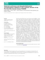
Figure 1 Right lateral (a), left lateral (b), and ventrodorsal (c) radiographic views of the thorax of a female dog (Case 13) with
mammary carcinoma. No signs of metastases were observed in the lungs.
Otoni et al. Acta Veterinaria Scandinavica 2010, 52:20
/>Page 5 of 10
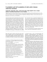
Figure 2 Right lateral (a), left lateral (b), and ventrodorsal (c) radiographic views of the thorax of a female dog (Case 15) with
mammary papillary cystadenocarcinoma. No signs of metastases were observed in the lungs.
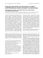
Figure 3 Transverse CT view s at the level of the thoracic diaphragmatic lobes of a female dog (Case 13) with mammary carcinom a.
No signs of metastases were observed in the lungs.
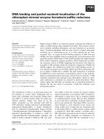
Figure 4 Transverse CT views of the thorax of a female dog (Case 11) showing small area of atelectasis (arrow).
Otoni et al. Acta Veterinaria Scandinavica 2010, 52:20
/>Page 6 of 10
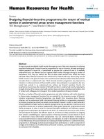
Figure 6 Transverse CT views at the level of the right medial lobe and left diaphragmatic thorax of the female dog (Case 15) with
mammary papillary cystadenocarcinoma. Observe 0.4 cm nodule in maximum dimension located on the medial portion of the left
diaphragmatic lobe showing signal of nutrient vessel (arrow).
Figure 7 Transverse CT views at the level of the right apical lobe and left thorax of a female dog (Case 17) with malignant mixed
mammary tumor. Observe 0.6 cm nodule in maximum dimension located on the left posterior cranial apical lobe (arrow).
Figure 5 Transverse CT views at the level of the right medial lobe and left apical l obe and thorax of a female dog (Case 15) with
mammary papillary cystadenocarcinoma. Observe 0.7 cm nodule in maximum dimension located on the medial portion of the left caudal
apical lobe (arrow). Note tumor present in the right caudal thoracic region.
Otoni et al. Acta Veterinaria Scandinavica 2010, 52:20
/>Page 7 of 10
eliminating one radiographic view may influence the
diagnosis in 12-15% of the patients [17]. However, three
views were not sufficient to detect the metastases, show-
ing that radiography is le ss sensitive than CT [10,11]. In
18 dogs with pulmonary metastatic neoplasia, only 9%
of the pulmonary nodules detected in CT were observed
on radiographs. Besides, pulmonary nodules were identi-
fied in a greater number of lung lobes by CT than
radiographs [8].
The dogs were maintained in spontaneous breathing
during CT examination due to the type of anesthesia
used. However, the images obtained during the apnei c
period and at the inspiration peak are considered better
to minimize the breathing artifact at the tumor location
and size [18,19]. Some autho rs have used intermittent
positive-pressure ventilation to decrease motion artifacts
[8,20]. On the other hand, in a study of dogs with meta-
static osteosarcoma using spiral CT with a collimation
of 5 mm and pitch of 2, no significant difference was
observed between normal resting respiration and hold-
ing of breath [21].
Technical modifications may b e performed to obt ain
an optimal lung CT [22]. In a study of two dogs with
pulmonary nodules evaluated by helical CT, the sug-
gested protocol included a narrow collimation (3-5
mm according to the dog’s size), pitch of 2 and inter-
val reconstruction of 1 [23]. A s imilar protocol was
used in the present study. CT examinations with intra-
venous contrast injection were performed to obtain a
better definition of the vessels and to differentiate
between normal and abnormal vascular structures
[18,24]. In addition, soft-tissue and lung windows were
utilized to enable evaluation of chest wall, and lung
parenchyma [20,18].
Thedogswerepositionedindorsalrecumbency,
although some authors suggest that sternal recumbency
provides better quality and absence of artifacts [18].
Atelectatic areas were observed by CT in 28.57% of the
cases, but these areas are probably associated with
recumbency and gravitational stasis [24] or anesthetic
procedure [25], since they are not presented radiogra-
phically. Another cause of atelectasia is the transport o f
the patient under general anesthesia to the CT gantry in
lateral recumbency [26]. In a study of dogs with meta-
static neoplas ia positioned in ventr al recumbency, when
the initial image showed ventral atelectasis, the CT was
repeated with the patient in dorsal recumbency to allow
better inflation of these lung regions [8]. The atelect atic
areas may show variable CT appeara nce such as fo cal
interstitial infiltrates, alveolar infiltrates or complete
lobar collapse [19,22]. In the present stu dy most of
these areas were peripheral small f ocal infiltrates and
apparently did not influence the results.
CT allowed detection of metastases in two cases in the
present study, thus showing greater precision than the
plain radiographic exami nation that it is considered less
sensitive for detection of small lesions [7,8,16,27]. In a
retrospective study of 18 dogs with pulmonary meta-
static neoplasia, the smallest size to detect pulmonary
nodules on CT images was 1 mm, compared with 7-9
mm on radiographs [8]. However, in a study of four
dogs presenting lung metastases of osteosarcoma con-
firmed by plain thoracic r adiographic examination, the
spiral CT did not detect 32 metastases measuring ≤1cm
in diameter [10]. Thus, the low number of lung metas-
tases observed in the present study may be underesti-
mated due to the limitation of the imaging examinations
used. Probably this fact is associated with the citation
Figure 8 Transverse CT views at the level of the right apical lobe and left thorax of a female dog (Case 17) with malignant mixed
mammary tumor. Observe 0.9 cm nodule in maximum dimension with irregular outline, clear-cut limits and surrounded by ground-glass
opacity, located on the posterior portion of the right apical lobe (arrow).
Otoni et al. Acta Veterinaria Scandinavica 2010, 52:20
/>Page 8 of 10
that most female dogs wit h malignant tumors without
metastases at the moment of the surgery will have died
or will have been euthanized on account of problems
associated with the tumor within 1 or 2 years [3].
Case no. 17 had five solid nodules with regular outline
and was well-defined at the lung parenchyma, which
characterizes lung metastases. This standard of lung
metastases is compatible with the occurrence of the
hematogenous dissemination, which can occur as single,
multiple or propagated nodules, with generally random
distribution. However, the metastatic nodules are gener-
ally circular with regular contour, especially in more
aggressive lesions, such as those of sarcomatous origin
[28]. Furthermore, the typical metastatic carcinoma
usually occurs as numerous small circular lesions [29].
Case 15 had two pulmonary nodules, one of which
presented an irregular outline, clear-cut limits and was
surrounded by ground-glass opacity. The metastatic
nodules may have irregular or lobular contour [28].
Conclusions
In this study population, spiral CT showed higher sensi-
tivity than chest radiographies to detect lung metastasis;
this indicates that CT should be performed on all female
dogs with malignant mammary tumors.
Acknowledgements
We are grateful to Fapesp for the spiral Scanner and to Capes for the
fellowship.
Author details
1
São Paulo State University (Unesp), Department of Veterinary Surgery and
Anesthesiology, School of Veterinary Medicine and Animal Science, Botucatu,
SP, Brazil.
2
Unesp, Department of Animal Reproduction and Radiology,
School of Veterinary Medicine and Animal Science, Botucatu, SP, Brazil.
3
Unesp, Department of Tropical Diseases and Diagnostic Imaging, Botucatu
Medical School, Botucatu, SP, Brazil.
4
Unesp, Department of Veterinary
Clinical Sciences, School of Veterinary Medicine and Animal Science,
Botucatu, SP, Brazil.
Authors’ contributions
CCO, SCR and KH participated in the study design, surgical procedures, and
drafted the manuscript. LCV, SMR and DPD interpreted the radiographic and
CT examinations. TG performed the anesthesia procedures. RLA performed
the microscopic examinations. All authors read and approved the final
manuscript.
Competing interests
The authors declare that they have no competing interests.
Received: 18 June 2009 Accepted: 9 March 2010
Published: 9 March 2010
References
1. Harvey JH: Mammary glands. Current techniques in small animal surgery
Baltimore: Williams & WilkinsBojrab MJ, Ellison GW, Slocum B 1998, 579-584.
2. Robbins M: Reproductive oncology. Textbook of small animal surgery
Philadelphia: SaundersSlatter D 2003, 2437-2443.
3. Hedlund CS: Mammary neoplasia. Small animal surgery St. Louis: Mosby
ElsevierFossum TW 2007, 729-735.
4. Sorenmo KU, Shofer FS, Goldschmidt MH: Effect of spaying and timing of
spaying on survival of dogs with mammary carcinoma. J Vet Intern Med
2000, 14:266-270.
5. Chang SC, Chang CC, Chang TJ, Wong ML: Prognostic factors associated
with survival two years after surgery in dogs with malignant mammary
tumours: 79 cases (1998-2002). J Am Vet Med Assoc 2005, 227:1625-1629.
6. Barthez PY, Hornof WJ, Theon AP, Craychee TJ, Morgan JP: Receiver
operating characteristic curve analysis of the performance of various
radiographic protocols when screening dogs for pulmonary metastases.
J Am Vet Med Assoc 1994, 204:237-240.
7. Baumann D, Hauser B, Hubler M, Fluckiger M: Signs of metastatic disease
on thoracic radiographs of dogs suffering from mammary gland
tumours: a retrospective study (1990-1998). Schweiz Arch Tierheilkd 2004,
146:431-435.
8. Nemanic S, London CA, Wisner ER: Comparison of thoracic radiographs
and single breath-hold helical CT for detection of pulmonary nodules in
dogs with metastatic neoplasia. J Vet Intern Med 2006, 20:508-515.
9. Schafer JF, Vollmar J, Schick F, Seemann MD, Kamm P, Erdtmann B,
Claussen CD: Detection of pulmonary nodules with breath-hold
magnetic resonance imaging in comparison with computed
tomography. Rofo 2005, 177:41-49.
10. Waters DJ, Coakley FV, Cohen MD, Davis MM, Karmazyn B, Gonin R,
Hanna MP, Knapp DW, Heifetz SA: The detection of pulmonary
metastases by helical CT: a clinicopathologic study in dogs. J Comput
Assist Tomogr 1998, 22:235-240.
11. Coakley FV, Cohen MD, Johnson MS, Gonin R, Hanna MP: Maximum
intensity projection images in the detection of simulated pulmonary
nodules by spiral CT. Br J Radiol 1998, 71:135-140.
12. Johnston SD: Reproductive system. Textbook of Small Animal Surgery San
Diego: SaundersSlatter D 2003, 2566-2583.
13. Jones CJ, Hunt RD, King NW: Genital system. Veterinary Pathology
Indianapolis: Wiley-BlackwellJones TC, Hunt RD, King NW 1997, 1169-1244.
14. Cotran RS, Kumar V, Collins T: Neoplasia. Pathologic Basis of Disease
Philadelphia: SaundresRobbins SL 1989, 233-295.
15. Rubin E, Farber JL: Neoplasia. Pathology Baltimore: Lippincott Williams &
WilkinsRubin E, Farber JL 1999, 152-209.
16. Holt D, Van Winkle T, Schelling C, Prymak C: Correlation between thoracic
radiographs and postmortem findings in dogs with hemangiosarcoma:
77 cases (1984-1989). J Am Vet Med Assoc 1992, 200:1535-1539.
17. Ober CP, Barber D: Comparison of two -vs. three-view thoracic
radiographic studies on conspicuity of structured interstitial patterns in
dogs. Vet Radiol Ultrasound 2006, 47:542-545.
18. Cardoso L, Gil F, Ramírez G, Teixeira MA, Agut A, Rivero MA, Arencibia A,
Vásquez JM: Computed tomography (CT) of the lungs of the dog using a
helical CT scanner, intravenous iodine contrast medium and different CT
windows. Anat Histo Embryol 2007, 36:328-331.
19. Morandi F, Matton JS, Lakritz J, Turki JR, Wisner ER: Correlation of helical
and incremental high-resolution thin-section computed tomographic
imaging with histomorphometric quantitative evaluation of lungs in
dogs. AJVR 2003, 64(Suppl 7):935-944.
20. Coakley FV, Cohen MD, Waters DJ, Davis MM, Karmazyn B, Gonin R,
Hanna MP: Detection of pulmonary metastases with pathological
correlation: effect of breathing on the accuracy of spiral CT. Pediatr
Radiol 1997, 27:576-579.
21. Rycle LM, Gielen IM, Simoens PJ, Van Bree H: Computed tomography and
cross-sectional anatomy of the thorax in clinically normal dogs. Am J Vet
Res 2005, 66:512-524.
22. Webb WR, Müller NL, Naidich DP: HRCT findings of lung disease. High-
resolution CT of the lung Philadelphia: Lippincott-RavenWebb WR 1996,
41-108.
23. Joly H, D’Anjou M, Alexander K, Beauchamp G: Comparison of single-slice
computed tomography protocols for detection of pulmonary nodules in
dogs. Veterinary Radiology & Ultrasound 2009, 50(Suppl 3):279-284.
24. Widmer WR: Alternate imaging for diagnosis of cancer. Cancer in dogs
and cats: medical and cirurgical management Baltimore: Teton
NewmediaMorrison WB 2002, 177-204.
25. Warner DO, Warner MA, Ritman EL: Atelectasis and chest wall shape
during anesthesia. Anestediology 1996, 85(Suppl 1):49-59.
26. Johnson EG, Wisner ER: Advances in respiratory imaging. Vet Clin Small
Anim 2007, 37:879-900.
Otoni et al. Acta Veterinaria Scandinavica 2010, 52:20
/>Page 9 of 10
27. Ketai L, Malby M, Jordan K, Meholic A, Locken J: Small nodules detected
on chest radiography: does size predict calcification?. Chest 2000,
118:610-614.
28. Oliveira AC: Metastases pulmonares. Doenças Pulmonares Rio de Janeiro:
Ganabara KooganTarantino AB 2002, 717-724.
29. Farrow CS: Lung neoplasm. Veterinary Diagnostic Imaging: the dog and the
cat New York: MosbyFarrow CS 2003, 425-437.
doi:10.1186/1751-0147-52-20
Cite this article as: Otoni et al.: Survey radiography and computerized
tomography imaging of the thorax in female dogs with mammary
tumors. Acta Veterinaria Scandinavica 2010 52:20.
Submit your next manuscript to BioMed Central
and take full advantage of:
• Convenient online submission
• Thorough peer review
• No space constraints or color figure charges
• Immediate publication on acceptance
• Inclusion in PubMed, CAS, Scopus and Google Scholar
• Research which is freely available for redistribution
Submit your manuscript at
www.biomedcentral.com/submit
Otoni et al. Acta Veterinaria Scandinavica 2010, 52:20
/>Page 10 of 10