Báo cáo thú y: " Field experience with two different vaccination strategies aiming to control infections with Actinobacillus pleuropneumoniae in a fattening pig herd" doc
Bạn đang xem bản rút gọn của tài liệu. Xem và tải ngay bản đầy đủ của tài liệu tại đây (436.96 KB, 10 trang )
RESEARC H Open Access
Field experience with two different vaccination
strategies aiming to control infections with
Actinobacillus pleuropneumoniae in a fattening
pig herd
Marie Sjölund
1,2
, Per Wallgren
1,2*
Abstract
Background: The prevalence of pleurisies recorded at slaughter is increasing in Sweden, and acute outbreaks of
actinobacillosis that require antimicrobial treatments have become more frequent. As an increased use of
antimicrobials may result in the development of antimicrobial resistance it is essential to develop alternative
measures to control the disease. Vaccinations present an appealing alternative to antimicrobial treatments. The aim
of this work was to evaluate the potential of two different vaccination strategies in a specialized fattening herd
affected by actinobacillosis.
Methods: The study was conducted in a specialized fattening herd employing age segregated rearing in eight
units. The herd suffered from infections caused by Actinobacillus pleuropneumoniae serotype 2, confirmed by
necropsy and serology. The study included 54 batches of pigs grouped into five periods. Batches of pigs of the
second period were vaccinated against actinobacillosis twice, and pigs in the fourth period were vacci nated three
times. Batches of pigs of the first, third and fifth period were not vaccinated. Concentrations of serum antibodies
to A. pleuropneumoniae and serum amyloid A (SAA) were analysed and production data were recorded.
Results: Despite vaccinating, medical treatments were required to reduce the impact of the disease. The mean
incidence of individual treatments for respiratory diseases during the rearing period ranged from 0 to 4.7 ± 1.8%,
and was greatest during the triple vaccination period (period IV; p < 0.05 when compared to other groups). A large
proportion of the vaccinated pigs seroconverted to A. pleuropneumoniae serotype 2 in the absence of a SAA-response.
The prevalence of pleuritis decreased from 25.4 ± 6.5% in the first period to 5.0 ± 3.7% in the fifth period (p < 0.001).
Conclusions: The vaccine did not effectively prevent clinical expression of A. pleuropneumoniae infections, but
seroconversion to A. pleuropneumoniae in the absence of a SAA-response in a large number pigs indicated that the
vaccine had activated the immune system. Further, the prevalence of pleuritis decreased with time. This indicates
that vaccinations together with intensified medical treatments of affected pigs could be useful in reducing the
impact of A. pleuropneumoniae serotype 2 infections.
Background
Actinobacillus pleuropneumoniae is a causative agent of
respiratory disease in pigs with symptoms ranging from
sudden deaths to subclinical disease detected as pleuri-
sies in the post m ortem inspection at slaughter [1].
Infections with A. pleuropneumoniae may cause great
economic losses due to mortality, increased feed con-
sumption, retarded growth rate and medication [1-3].
Several strategies have therefore been employed aiming
to control the effects of A. pleuropneumoniae infections
of which age segregated rearing is one [4,5]. The ban on
the use of growth promoters in Sweden in 1986 led to a
more consistent implementation of age segregated rear-
ing systems [6] which reduced the incidence
of pleurisies recorded at slaughter from 8% in 1988
* Correspondence:
1
National Veterinary Institute, Department of Animal Health and
Antimicrobial Strategies, SE-751 89 Uppsala, Sweden
Sjölund and Wallgren Acta Veterinaria Scandinavica 2010, 52:23
/>© 2010 Sjölund and Wallgren; licensee BioMed Central Ltd. This is an Open Access article distributed under the terms of the Creative
Commons Attribution License (http://cre ative commons.org/licenses/by/2.0), which permits unrestricted use, distribution, and
reproduction in any medium, provided the original work is properly cited.
to 5% in 2002 [7]. However, registrations for pleurisies
at slaughter are currently increasing and acute outbreaks
of act inobacillosis are becoming more frequent [8]. Such
outbreaks often require antibiotic treatment of entire
units with in-feed medication which has been mirrored
by an increased prescription of tetracyclines in 2007 [9].
To date, none of the tested Swedish isolates of
A. pleuropneumoniae have been resistant to the antibio-
tics tested for [9]. Despite this, it is essential to develop
antibiotic independent measures to control the disease
since an i ncreased use of antib iotics may promote the
emergence of antimicrobial resistance [10]. Antimicro-
bial resistance for A. pleuropneumoniae isolates has
been reported [11].
Vaccination presents an appealing alternative to anti-
biotics in reducing the impact of A. pleuropneumoniae.
The first generation of vaccines against A. pleuro pneu-
moniae did not provide sufficient protection against dis-
ease and were in some cases causing adverse side effects
such as depression, inappetence, fever or tissue damage
[12]. At present, one subunit vaccine is commercially
available in Sweden (Porcilis® APP, Intervet, Boxmeer,
The Netherlands). Several reports from different coun-
tries have described the efficacy of this vaccine [13,14].
According to the product details, this vaccine induces a
graduallydevelopingprotectiveimmunitywhichis
greatest two to three weeks after booster vaccination
with some protection maintained for up to seven weeks.
This work aimed at evaluating the effect over time of
two different vaccination strategies in a specialized fat-
tening herd affected by actinobacillosis.
Methods
Herd and batches followed in a longitudinal survey
The study was approved by the Ethical Committee on
Animal Experiments, Uppsala, Sweden (Licence C38/4).
It was conducted in a conventional, specialized fattening
herd producing approximately 7500 pigs per year. The
herd was free from all diseases listed by the Office Inter-
national des Epizooties, Paris, France, and also from
Aujeszky’s disease, PRRS and Salmonella. However, the
herd had suffered from infections caused by Actinobacil-
lus pleuropneumoniae serotype 2 for two years, which
had been confirmed by necropsy and serology. Batch
prevalence at slaughter for pleurisy lesions ranged from
18.7% to 26.8% and for Mycoplasma hyopneumoni ae-
like lesions from 1.7% to 19.2% during the years preced-
ing the study (see also Table 1).
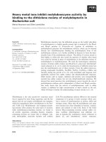
Pigs were housed in a 10 year old building with eight
units (Figure 1). Each unit housed 11 pigs per pen in 32
pens (n = 352). Strict all in - all out production with a
cycle of 16 weeks was employed in all u nits. Thus, a
new batch of pigs entered a thoroughly cleaned and dis-
infected unit every second week. The pigs arrived at an
age of 10-12 weeks with two or three suppliers per
batch. Antimicrobial substances were not routinely
added to the feed given to the pigs.
This study included 54 batches of pigs that were
grouped into five successive periods. The first period (I)
included six unvaccina ted batches. The second period
(II) included 13 batches that were vaccinated twice
against actinobacillosis (see below). The third period
(III) included 11 unvaccinated batches. The fourth per-
iod(IV)includedeightbatchesvaccinatedthreetimes
against actinobacillosis. The fifth period (V) included 16
unvaccinated batches.
Vaccine and vaccination strategies employed
A commercially available vaccine (Porcilis® APP Intervet,
Boxmeer, The Netherlands), containing three inactivated
exotoxins (ApxI, ApxII, ApxIII) and a 42 kDa outer
membrane pro tein (OMP) was used employing two dif-
ferent strategies.
Thirteen consecutive batches (period II) were vacci-
nated twice with 2 ml of Porcilis® APP. The first two
vaccinated batches were already present on the premises
when the vaccination scheme was initiated. Thus, Batch
1 and Batch 2 of period II received the first vaccination
20 and 35 days after arrival, respec tively. The other 11
batches were vaccinated on arrival. Booster vaccinations
were performed 28 days after the first vaccination.
Empl oying a second strategy (period IV), pigs were vac-
cinated three times with 2 ml of Porcilis® APP during
one turn-over of the herd, i.e. until pigs in all eight
units had been vaccinated. The first vaccination was
given on arrival to the fattening herd. Vaccinations were
repeated 28 and 56 days after arrival.
Blood sampling procedures
Blood samples without additives were collected from
pigs by jugular vein punctures using evacuated plastic
tubes (BD Vacutainer Systems, Belliver Industrial Estate,
Plymouth, United Kingdom). They were centrifuged for
10 minutes at 800 × g, after which the serum was
removed and stored at -20°C until analysed.
A cross-sectional blood sampl ing comprising six pigs
per age category/unit was performed before initiating
the vaccination strategies in order to obtain a serological
profile of the herd. This sampling was performed in
connection with an outbreak of acute pleuropneumonia.
The blood sampling procedure for the four batches of
vaccinated pigs during period II and period IV were
identical except for day 56. Blood samples were repeat-
edly collected from individually ear-tagged pigs every
fortnight. Blood samples collected on days 0, 28 and 56
were collected before pigs were vaccinated. Blood was
collected from pigs in the eighth and tenth batches of
pigs vaccinated twice (Period II: Batch 8; n = 31 and
Sjölund and Wallgren Acta Veterinaria Scandinavica 2010, 52:23
/>Page 2 of 10
Batch 10; n = 15). From pigs vaccinate d three times,
blood was collected from the first and last batch (Period
IV: Batch 1, n = 30; Batch 8, n = 21).
Detection of antibodies to A. pleuropneumoniae
serotype 2
An indirect ELISA, based on a phenol-water extraction of
the microbe as coating antigen, was used to measure serum
antibodies to A. pleuropneumoniae serotype 2 in all serum
samples collected. The cut-off value for a positive reaction
in sera diluted 1/1000 was defined as A
450
= 0.5 [15].
Serum Amyloid-A (SAA)
Serum levels of the acute phase protein Serum Amyloi d-
A (SAA) were analysed using a commerc ial kit (Serum
Amyloid A Assay TP-80 2, Tridelta, Maynooth, Ireland)
according to the instructions of the producer. The results
are presented as mg SAA per ml serum. The baselin e
serum levels of SAA were established using sera from 30
nine-week-old specific pathogen-free (SPF) pigs [16]. The
mean SAA serum levels of these pigs were 37.8 mg per
ml (Max - min range: 22.7 - 157.2 mg per ml), a nd con-
sidered as representative serum levels of SAA for healthy
pigs. As cut-off for an increased level o f SAA in the pre-
sent study the 95% percentile (70 mg per ml) of the
serum level of SAA of the SPF pigs was used.
SAA was analysed in the serum samples collected
from six pigs per age category in the cross-sectional
sampling performed before theperiodofinvestigation.
SAA was also analysed in all serum samples collected
from ten selected pigs per batch of the four sampled
vaccinated batches of period II and IV.
Clinical recordings and medical treatments
The herd veterinarian made regular visits to the herd.
Routine herd procedures included daily inspections with
disease monitoring performed by the farm manager
according to instructions from the herd veterinarian.
During the period of investigation, all pigs present on the
premises were inspected by the investigating veterinarian
on blood sampling occasions. Individual pigs with signs
of respiratory disease were treated intramuscularly with
oxytetracycline for five days (20 mg/kg body weight once
daily on days 1, 3 and 5; Engemycin® vet., Intervet, Boxm-
eer, The Netherlands) according to written instructions
from the herd veterinarian. In severe cases with many
pigs affected by respiratory disease and per- acute mortal-
ities, affected batches were in-feed medicated with either
20 mg chlortetracycline per kg body weight a nd day
(Clortetraciclina 20%, Ceva Sante Animale, Libourne
Cedex, France) or 12.5 mg doxycycline per kg body
weight and day (Pulmodox 5%, ChemVet, Silkeborg,
Denmark) for 1 0 consecutive days. In-feed medications
were initiated and prescribed by the herd veterinarian.
Registrations at slaughter and production data
The incidence of lung lesions (enzootic pneumonia,
pleuritis and necrotizing bronchopneumoni a) w as
obtained from t he regular meat inspectio n performed at
the abattoir. Data of average daily weight gain (DWG)
was obtained from the production control system used
by the herd (FarmData, BioManagement AB, Tumba,
Sweden ). These data were collected from all 54 batches
include in the study (period I to V).
Statistical analysis
All results in the text are given as mean values ± stan-
dard deviations. Continuous non-normally distributed
data was analyzed using the Wilcoxon Rank Sum test
and non-continuous data were ca tegorized and analyzed
using the Fishers Exact test.
Results
Serology
Pigs that had been at the herd for less than 80 days were
in general serologically negative to A. pleuropneumoniae
serotype 2 in the cross-sectional sampling performed
before commencing the study. However, some of the pigs
Table 1 Lesions of the respiratory tract registered at slaughter in fatteners unvaccinated or vaccinated against
Actinobacillus pleuropneumoniae in a specialized fattening herd affected by actinobacillosis
Batch Category # batches Mycoplasma-like pneumonia Pleuritis Hemmorrhagic broncho-pneumonia
(%) (%) (%)
Period I
Before vaccinations
6 7.9 ± 8.1 25.4 ± 6.5 0 ± 0
Period II
Double vaccinations
13 10.7 ± 5.6 19.7 ± 8.1 2.5 ± 5.0
Period III
In between vaccinations
11 14.8 ± 9.6 13.9 ± 3.9 0.5 ± 0.9
Period IV
Triple vaccinations
8 7.4 ± 2.3 11.1 ± 2.8 1.0 ± 1.1
Period V
After vaccinations
16 9.7 ± 4.3 5.0 ± 3.7 0.6 ± 1.0
Sjölund and Wallgren Acta Veterinaria Scandinavica 2010, 52:23
/>Page 3 of 10
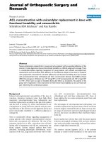
that had been at the herd for 50 days were seropositive to
A. pleuropneumoniae serotype 2. All but one of the pigs
that had been at the herd for 80 days or more were sero-
logically positive to A. pleuropneumoniae (Figure 2).
With respect to pigs vaccinated twice (period II), all
animals sampled were seronegative (A
450
<0.5)to
A. pleuropneumoniae serotype 2 on arrival to the fatten-
ing herd. Following the initial vaccination, an increase
(p < 0.001) in serum antibodies to A. pleuropneumoniae
serotype 2 was observed after 14 days in both batches
that were analysed. By this time, 11 out of 31 and 6 out
of 15 pi gs had seroconverted in Batch 8 and 10, respec-
tively (mean A
450
for seropositive pigs = 0.71 ± 0.29 in
Batch 8 and 1.06 ± 0.33 in Batch 10). The mean absor-
bance value remained at that level until 84 days after
arrival in Batch 8. In contrast, the mean absorbance
value increased continuously to A450 = 1.70 ± 0.29 at
day56inBatch10(Figure3).Atthelastsampling
Figure 1 A schematic view of a specialized fattening herd employing strict all in - all out production with a turn-over time of 16
weeks per unit. Each of the eight units housed 11 pigs per pen in 32 pens (n = 352 pigs per unit).
Sjölund and Wallgren Acta Veterinaria Scandinavica 2010, 52:23
/>Page 4 of 10
occasion, all pigs but two pigs in batch 8 were seroposi-
tive to A. pleuropneumoniae serotype 2. The mean
absorbance value for these two pigs was A
450
=0.31±
0.24.
The mean absorbance values for antibodies to
A. pleuropneumoniae serotype 2 in the first and last
batches vaccinated three times (period IV) are also
shown in figure 3. In total, two out of 30 pigs in batch 1
and four out of 21 pigs in batch 8 were sero positive to
A. pleuropneumoniae serotype 2 on arrival to the fatten-
ing herd with a mean absorbance level of A
450
=0.76±
0.29. The mean absorbance valu e increased signi ficantly
(p < 0.05) between sampling times until day 28 in both
batches, and Batch 1 continued to increase until day 42.
Following the third v accination (day 56), the amount o f
serum antibodies to A. pleuropneumoniae serotype 2
had increased (p < 0.01) at the next sampling for both
batches ( day 72). A decrease in the level of serum anti-
bodies to A. pleuropneumoniae serotype 2 was observed
at the last sampling occasion (day 112) compared to the
previous sampling occasion (day 98: Figure 3). T his
decrease was significant for Batch 1 (p < 0.001).
However, at th is occasion all but one pig (A
450
= 0.24)
were seropositive to A. pleuropneumoniae serotype 2.
SAA and the relation to seroconversion to A.
pleuropneumoniae
In total, increased serum levels of SAA were recorded in
seven out of the 48 pigs in the cross-sectional sampling.
Two of these pigs had been in the herd for 80 and 100
days, respectively. The remaining five pigs with elevated
serum levels of SAA (mean = 969 ± 618 mg per ml)
had been in the herd for 50 days. At this time point,
three of these pigs had seroconverted to A. pleuropneu-
moniae and the mean absorbance level for antibodies to
A. pleuropneumoniae was 0.38 ± 0.34 for these six pigs
(Figure 2). In contrast, pigs that had been at the herd
for 30 days had lower serum antibody levels to A. pleur-
opneumoniae (mean A
450
= 0.07 ± 0.02) and none of
these pigs had elevated serum levels of SAA.
Elevated serum co ncentrati ons of SAA were detected
in individual vaccinated pigs throughout the rearing per-
iod. Increased serum levels of SAA were demonstrated
in 16 out of 20 pigs vaccinated twice (period II), and on
Figure 2 A cross-sectional serological screening for mean serum antibody levels (A
450
)toActinobacillus pleuropneumoniae serotype 2
performed during an outbreak of actinobacillosis in a specialized fattening herd employing age segregated rearing. Six pigs per unit
were analysed. The error bars show positive standard deviations and the dotted line indicates the cut-off value for a positive reaction.
Sjölund and Wallgren Acta Veterinaria Scandinavica 2010, 52:23
/>Page 5 of 10
an individual basis, elevated SAA c oncentrations were
recorded on one to seven occasions. In pigs vaccinated
three times (period IV) increased serum levels of SAA
were demonstrated in 14 out of 20 pigs (one to three
occasions per pig).
Seroconversion to A. pleuropneumoniae serotype 2
was observed at the sampling occasion following the
sampling when elevated SAA concentrations were
observed in eight of 20 pigs that had been vaccinated
twice (40%) and in six of 20 pigs tha t had been vacci-
nated three times (30%). Thus 12 of 20 pigs vaccinated
twice (60%) and 14 of 20 pigs vaccinated three times
(70%) seroconverted to A. pleuropneumoniae serotype 2
in the absence of a SAA-response at the previous sam-
pling occasion (Table 2).
Clinical recordings and treatments
Historically, clinical symptoms of respiratory disease
had rarely been observed, and medical treatments
against respiratory diseases were generally not carried
out (Table 3). However, due to clinical disease, in-feed
medications with doxycycline were required. Five con-
secutive batches of pigs were medicated: three batches
in period I and the f irst two batches of pigs vaccinated
twice (period II). In period II, 1.3 ± 2.0% of the pigs
also required individual treatments due to respiratory
disease. The mean mortality during this period when
pigs were vaccinated twice amounted to 4.0 ± 1.3%
(Table 3).
Individual treatments for respiratory symptoms ranged
from zero to 23 pigs per batch (0 - 6.7%) for the 11
batches with unvaccinated pigs during period III (mean =
1.3 ± 2.0%). The mean mortality for these batches was
3.7 ± 1.7%.
The incidence of individual treatments for respiratory
diseases was greatest during the triple vaccination period
(period IV), (p < 0.05 when co mpared to period I, II, III
and V). Four to 22 pigs per batch were individually trea-
ted for respiratory disease symptoms (mean = 4.7 ±
1.8%). The first individual treatments were initiated 18
to 24 days after arrival to the herd. Due to the large
number of individual treatments for respiratory disease
and per acute mortalities, in-feed treatment with chlor-
tetracycline was applied at the onset of clinical signs,
appr oximately three weeks after arrival in all but two of
the eight batches. The overall mortality for the eight
batches of pigs vaccinated three times was 3.1 ± 1.4%.
After the cessation of vaccinations (period V), zero to
nine pigs per batch in 16 consecutive batches recei ved
individual treatments for respiratory disease (mean = 0.6 ±
0
0.2
0.4
0.6
0.8
1
1.2
1.4
1.6
1.8
2
2.2
0 14284256708498112
Mean absorbance (A450)
Day after arrival
Vaccination 1
Vaccination 2
Vaccination 3
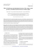
Figure 3 Mean serum antibody levels (A
450
)toActinobacillus pleuropneumoniae serotype 2 in batches of pigs vaccinated two or three
times against Actinobacillus pleuropneumoniae in a specialized fattening herd affected by the infection. The first vaccination was
performed as the pigs arrived at the specialized fattening herd. The booster vaccination(s) were carried out after intervals of 28 days, Double
vaccinated pigs are shown by filled symbols ([black diamond] = batch 8 and [black circle] = batch 10. Pigs vaccinated three times are shown by
open symbols (black triangle = batch 1 and black sqaure = batch 8). The dotted line indicates the cut-off value for a positive reaction.
Sjölund and Wallgren Acta Veterinaria Scandinavica 2010, 52:23
/>Page 6 of 10
0.9% treatments. The mean mortality for these batches
was 3.4 ± 1.3% (Table 3).
Registrations at slaughter and growth performance
The registrations at slaughter of the pigs are shown in
Table 3. Registrations for Mycoplasma-like pneumonias
and for hemorrhagic bronchopneumonias varied over
time. I n contrast, the prevalence of pleuritis was lower
(p < 0.05) for the unvaccinated pigs of period III com-
pared to pigs vaccinated twice (period II). The preva-
lence of pleuritis registered at slaughter was lower in
period V compared to all other periods (p < 0.01).
The daily growth during the rearing period ranged
from 863 ± 56 to 911 ± 34 g per day. The differences
in growth rate observed were not statistically signifi-
cant when the periods were compared to each other
(Table 1).
Discussion
This work was initiated due to an outbreak of acute
actinobacillosis confirme d through necropsy and serolo-
gical screening in a fattening herd suffering from
chronic pleuropneumoniae. However, despite double
vaccinations (period II), medical treatments were
required to reduce the impact of the disease. As the
protection of the vaccine has been reported to be of
rather short duration and greatest two to three weeks
after a booster vaccination [17], it was assumed that
three vaccinations would prolong the period of protec-
tion. Yet, individual treatments of pigs for respiratory
disease and in-feed medications were required in six of
the eight batches that were vaccinated three times (per-
iod IV). On the other hand, the pleurisy registrat ions at
slaughter decreased significantly in batches following
vaccinated ones. Similar results have also been reported
by others [18], which could be an indication of a reduc-
tion in the pathogen load, not apparent until after ter-
minating the vaccinations. However, these authors did
not observe any differences in growth performance and
pleurisy lesions recorded at slaughter between vacci-
nated and control pigs. Most likely the individual and
the in-feed medications contributed to the reduced
pathogen load, and it appears that vaccina tions together
with intensified medical treatments of affected pigs
could be useful in reducing the impact of A. pleuropneu-
moniae infections as also previously suggested [19].
The serological results from the batches vaccinated
twice (period II) indicated that transmission not only
occurred between pig s, but also between units. Airborne
transmission of A. pleuropneumoniae between closely
located units has been reported to occur under experi-
mental conditions [20,21] and A. pleuropneumoniae is
readily transmitted between pigs [20,22]. A. pleuropneu-
moniae can a lso be transmitt ed between units in large
Table 2 Elevated serum levels of SAA related to seroconversion to Actinobacillus pleuropneumoniae serotype 2 in pigs
vaccinated either two or three times against actinobacillosis
Elevated levels of SAA Period II
Vaccinated 2 times
Period IV
Vaccinated 3 times
(number of pigs = 20) (number of pigs = 20)
Not recorded 4 (20%) 6 (30%)
4 - 8 weeks prior to seroconversion 2 (10%) 4 (20%)
2 weeks prior to seroconversion 8 (40%) 6 (30%)
Simultaneous to seroconversion 6 (30%) 0
After seroconversion 0 4 (20%)
Table 3 Origin of growers, daily weight gain, medical treatments against respiratory disease and mortality in fatteners
unvaccinated or vaccinated against Actinobacillus pleuropneumoniae in a specialized fattening herd affected by
actinobacillosis
Batch Category # batches Origin of growers Weight gain Treatments Individual Treatments In feed Mortality
(Herds) (g per day) (%) (Batches) (%)
Period I
Before vaccinations
6 A, B, C 863 ± 56 0 ± 0 3 of 6 4.2 ± 1.6
Period II
Double vaccinations
13 A, B, C 911 ± 34 1.3 ± 2.0 2 of 13 4.0 ± 1.3
Period III
In between vaccinations
11 A-F 887 ± 16 1.3 ± 2.0 0 3.7 ± 1.7
Period IV
Triple vaccinations
8 D, E, F 893 ± 33 4.7 ± 1.8 6 of 8 3.1 ± 1.4
Period V
After vaccinations
16 D, E, F 883 ± 36 0.6 ± 0.9 0 3.4 ± 1.3
Sjölund and Wallgren Acta Veterinaria Scandinavica 2010, 52:23
/>Page 7 of 10
herds housing seve ral age ca te gories in t he same build-
ing even when a ll-in all-out management is e ffectuated
on room basis [23]. Indeed, a higher proportion of
batches were infected with A. pleuropneumoniae in sys-
tems employing all in-all out on room basis compared
to when all in-all out rearing was carried out by site [5].
Pigs in faci lities housing several age cate gories, as in the
herd investigated, will thereby risk to be repeatedly
exposed to the microbe.
Nevertheless, pig to pig transmission should not be
neglected. It has previously been demonstrated that A.
pleuropneumoniae is most readily isolated in pigs aged
11 to 12 weeks [24], which coincides with the mixing of
pigs from different sources on arrival to fattening herds.
At this age, serum neutralizing antibody titres are gener-
allylowwhypigsmaybesusceptibletoinfections[25].
Pigs that are seropositive to A. pleuropneumoniae have
obviously been infected and could be contagious. During
period IV, the herd received pigs that were seropositive
to A. pleuropneumoniae. Despite this, the prevalence of
pleuritis decreased during period IV. Although the pigs
originated from the same sources during period IV and
V, the prevalence of pleuritis decreased even further
during period V. It was therefore concluded that the
transmission between units was significant for maintain-
ing a high pathogen load in the herd.
Increased SAA-levels were demonstrated in pigs that
had been at the herd for 50 days in the cross-sectional
sampling. The mean absorbance value of serum antibo-
dies to A. pleuropneumoniae for these pigs (A450 = 0.38
± 0.34) indicated that the entire group was about to ser-
oconvert. A primary exposure to A. pleuropneumoniae
serotype 2 has previously been shown to induce a signif-
icant but transient SAA-response [26] with a duration
of approximately seven to ten d ays [27,28]. In contrast,
pigs do not mount SAA-responses when re-exposed to
A. pleuropneumoniae serotype 2 [28]. The interval of
two weeks between samplings may have concealed SAA-
responses in individual pigs, but the seroconversion to
A. pleuropneumoniae without a preceding SAA-response
in less than 50% of the vaccinated pigs indicated that
the vaccine had triggered the immune system. The reoc-
curring SAA-responses in individual pigs in period II
and IV i nstead indicated that other pathogens than
A. pleuropneumoniae induc ed the SAA-responses in the
vaccinated pigs.
Despite stimulating the immune system, a local IgA
response is not necessarily induced by vaccines [29].
This may explain the repeatedly occurring signs of clini-
cal disease, since IgA appears to be important in a first
line of defense against acute actinoba cillosis [30]. If so,
this highlights the importance of pathogen load reducing
efforts [31,32] including treatments of diseased pigs
[33,34] in controlling actinobacillosis. This is further
supported by the fact that vaccinations have previously
been shown not to influence whether pigs become
infected and/or infectio us [35]. The pigs in this study
did not seroconvert merely due to the vaccinations
(Batch 8, period II; Figure 3), which has also been
observed by others [36]. As pigs with low antibody levels
to the Apx toxins can be protected agai nst disease [37],
serum antibod ies may not be essential in providing pro-
tection against A. pleuropneumoniae infections. On the
other hand, specific serum IgG antibodies have been
reported t o be important in protection against pleurop-
neumonia [38], and the levels of toxin-neutralizing anti-
bodies in serum have been shown to influence the
susceptibility to A. pleuropneumoniae infections [39].
The pigs w ere vaccinated on arrival to the f attening
herd at an age of 10-12 weeks, which may have been a
suboptimal point of time as it appears to be important
to perform the first vaccination prior to exposure to a
pathogen load sufficient to cause clinical disease. Indeed,
some pigs vaccinated three times were seropositive to
A. pleuropneumoniae on arrival. Obviously these pigs
had been exposed to A. pleuropneumoniae and the
stress induced by transportation, co-mingling and
changes in f eed at a time when serum-antibody levels
generally were low made a spread of infection feasible
[1,25,40], which could have influenced the outcome of
the vaccinations.
However, similar results to ours have been reported
when three vaccinations were performed at six, 10 and
14 weeks of age [18]. These a uthors detected maternal
antibodies in serum at the age of six weeks which may
have interfered with the immune response following
vaccination. Maternally derived IgG antibodies are
reported to have a suppressing effect on the synthesis of
immunoglobulins by suckling piglets [41], and maternal
antibodies to A. pleuropneumoniae have been detected
in serum up to an age of at least eight weeks [39,42,43].
Thus, the age of six weeks may not either be an optimal
time for performing the first vaccination against
A. pleuropneumoniae. Administering the first dose at a
later time point appears to be beneficial provided that
the pigs are uninfected.
On the other hand, the presence of maternal antibo-
dies has b een shown not to hinder the ind uction of a
specific primary antibody response when administering
a low-dose infection [43]. The time point for immuniza-
tion would thus not be crucial. In an endemically
infected herd, pigs could already at the age of 11 days
have been exposed to A. Pleuropneumoniae [44]. Thus,
a carrier state can occur as the piglets harbour the
microbe in the tonsils. On the other hand, disease is
rarely seen while the piglets are still under maternal
antibody protection [37,39]. As maternal immunity
wanes, carrier pigs can transmit the infection to non-
Sjölund and Wallgren Acta Veterinaria Scandinavica 2010, 52:23
/>Page 8 of 10
immune pigs [44,45], a situation likely to occur as pigs
from several sources enter a unit at a fattening herd
[6,46].
Thus, the future demands on vaccines are high. Subu-
nit vaccines appear to convey better cross-protection
than bacterins [47]. Still, subunit vaccine s only provi de
partial clinical protection [48]. The use of live attenu-
ated vaccines might better mimic a natural course of
infection, with a potential to provide protection against
heterologous challenge [49]. Further, as an intradermal
administrat ion route can induce both mucosal and cell-
mediated immune responses [50,51], this could be a way
to enhance the response to vaccination.
Another way to reduce the impact of an A. pleurop-
neumoniae infection under field conditions could be to
ensure a high level of maternal antibodies in piglets by
vaccinating the sows [52]. Maternal antibodies combined
with a low-dose infection has been shown to be superior
in protecting pigs from a challenge infection in compari-
son to either maternal antibodies alone or a low-dose
infection in the absence of maternal immunity [43,53].
In endemically infected herds, pigs are likely to encoun-
ter a low-dose infection through asymptomatic carrier
pigs, but this can also be achieved through attenuated
live vaccines [49]. Further, an intra-na sal administration
may provide enhanced protection against disease as
local immunity has been shown to be important,
improving the clearance of bacteria from the respiratory
tract [12].
Conclusions
In conclusion, the time poi nt for immunization with the
vaccine used in this study appeared not to be crucial as
an immune response was induced, but still pigs were
not protected against disease. Thus other disease pre-
venting measures and treatments were also concluded
to be essential in controlling A. pleuropneumoniae
infections.
Acknowledgements
The authors would like to thank Intervet Schering-Plough Animal Health
Sweden for providing the vaccine used when pigs were vaccinated three
times. We would also like to thank farm manager Anders Carlsson and farm
assistant Fredrik Andersson for vaccinating the pigs, providing production
data and all practical help when collecting the samples. Maria Persson is
acknowledged for performing laboratory tests and Ann Nyman for valuable
help with the statistical analysis.
Author details
1
National Veterinary Institute, Department of Animal Health and
Antimicrobial Strategies, SE-751 89 Uppsala, Sweden.
2
Swedish University of
Agricultural Sciences, Department of Clinical Sciences, PO Box 7054, SE-750
07 Uppsala, Sweden.
Authors’ contributions
MS and PW initiated the study and deigned it. MS effectuated collection of
the blood samples and data collection. MS was head writer of the
manuscript with help from PW. Both authors have read and approve the
final manuscript.
Competing interests
The authors declare that they have no competing interests.
Received: 28 May 2009 Accepted: 25 March 2010
Published: 25 March 2010
References
1. Gottschalk M, Taylor DJ: Actinobacillus pleuropneumoniae. Diseases of Swine
Oxford: Blackwell Publishing LtdStraw B, Taylor DJ, Zimmerman JJ , 9 2006,
563-576.
2. Beskow P, Robertsson J-Å, Söderlind O: Testing of remedial measures in
fattening pig herds affected with subclinical infections of Actinobacillus
pleuropneumoniae serotype 2. Zentralbl Veterinärmed B 1993, 40(8):459-558.
3. Rohrbach BW, Hall RF, Hitchcock JP: Effect of subclinical infection with
Actinobacillus pleuropneumoniae in commingled feeder swine. JAmVet
Med Assoc 1993, 202(7):1095-1098.
4. Cleveland-Nielsen A, Nielsen EO, Ersbøll AK: Chronic pleuritis in Danish
slaughter pig herds. Prev Vet Med 2002, 55(2):121-135.
5. Busch ME, Jensen TK: The effect of all-in all-out management by site on
infection with Actinobacillus pleuropneumoniae in finishers. Proceedings of
the 19th International Pig Veterinary Society’s Congress: 16-19 July 2006;
Copenhagen Frederiksberg: Narayana PressNielsen JP, Jorsal SE 2006, 2: 198.
6. Holmgren N, Lundeheim N, Wallgren P: Infections with Mycoplasma
hyopneumoniae and Actinobacillus pleuropneumoniae in fattening pigs.
Influence on piglet production systems and influence on production
parameters. JVetMedB1999, 46:535-544.
7. Heldmer E, Lundeheim N, Robertsson J: Gross lesions at slaughter in
ecological pigs. (In Swedish). Sv Vet Tidn 2006, 58(13):13-19.
8. Beskow P, Lundeheim N, Holmgren N: Risk factors for the development of
pleuritis and pleuropneumonias in pigs. (In Swedish). Sv Vet Tidn 2008,
60(12):11-18.
9. SVARM Swedish Veterinary AntimicrobialResistance Monitoring. The
National Veterinary Institute (SVA), Uppsala, Sweden 2008 [.
se].
10. McEwen SA: Antibiotic use in animal agriculture: what have we learned
and where are we going? Anim Biotechnol 2006, 17(2):239-250.
11. Gutiérrez-Martín CB, García Del Blanco N, Blanco M, Navas J, Rodríguez-
Ferri EF: Changes in antimicrobial susceptibility of Actinobacillus
pleuropneumoniae isolated from pigs in Spain during the last decade.
Vet Microbiol 2006, 115:218-222.
12. Ramjeet M, Deslandes V, Gouré J, Jacques M: Actinobacillus
pleuropneumoniae vaccines: from bacterins to new insights into
vaccination strategies. Anim Health Res Rev 2008, 9(1):25-45.
13. Šatrán P, Nedbalcová K, Kuèerová Z: Comparison of protection efficacy of
toxoid and whole-cell vaccines against porcine pleuropneumonia
caused by endotracheal infection with Actinobacillus pleuropneumoniae.
Acta Vet Brno 2003, 72:213-219.
14. Tumamao JQ, Bowles RE, Bosch van den H, Klaasen HLBM, Fenwick BW,
Storie GJ: Comparison on the efficacy of a subunit and a live
streptomycin-dependant porcine pleuropneumoniae vaccine. Aust Vet J
2004, 82(6):370-374.
15. Wallgren P, Persson M: Relationship between the amounts of antibodies
to Actinobacillus pleuropneumoniae serotype 2, detected in blood serum
and in fluids collected from muscles of pigs. JVetMedB2000,
47:727-738.
16. Wallgren P, Vallgårda J: SPF pigs - presentation, definition and
specification of regulations. (In Swedish). Sv Vet Tidn 1993, 45:733-735.
17. Ridremont B, Kobisch M, Penning A, Schaller A, Gottschalk M: Laboratory
study of APP vaccination with a subunit vaccine on antibody serological
response in SPF piglets. Proceedings of the 19th International Pig Veterinary
Society’s Congress: 16-19 July 2006; Copenhagen Frederiksberg: Narayana
PressNielsen JP, Jorsal SE 2006, , 2: 235.
18. Jirawattanapong P, Stockhofe-Zurwieden N, Van Leengoed L, Binnendijk G,
Wisselink HJ, Raymakers R, Cruijsen T, Peet-Schwering Van Der C, Van Nes A,
Nielen M: Efficacy of a subunit vaccine against Actinobacillus
pleuropneumoniae in an endemically infected swine herd. J Swine Health
Prod 2008, 16(4):193-199.
Sjölund and Wallgren Acta Veterinaria Scandinavica 2010, 52:23
/>Page 9 of 10
19. Hofmo PO, Lium B: Attempts to establish elite breeding herds free from
Mycoplasma hyopneumoniae and Actinobacillus pleuropneumoniae by
strategic medication. Proceedings of the 15th International Pig Veterinary
Society’s Congress. 5-9 July 1998; Birmingham Nottingham: Nottingham
University PressDone S, Thomson, Varley 1998, , 2: 253.
20. Jobert JL, Savoye C, Cariolet R, Kobisch M, Madec F: Experimental aerosol
transmission of Actinobacillus pleuropneumoniae to pigs. Can J Vet Res
2000, 64:21-26.
21. Kristensen CS, Angen Ø, Andreasen M, Takai H, Nielsen JP, Jorsal SE:
Demonstration of airborne transmission of Actinobacillus
pleuropneumoniae serotype 2 between simulated pig units located at
close range. Vet Microbiol 2004, 98(3-4) :243-249.
22. Savoye C, Jobert JL, Berthelot-Hérault F, Keribin Am, Cariolet R, Morvan H,
Madec F, Kobisch M: A PCR assay used to study aerosol transmission of
Actinobacillus pleuropneumoniae from samples of live pigs under
experimental conditions. Vet Microbiol 2000, 73:337-347.
23. Nielsen JP, Hagedorn-Olsen T, Ahrens P, Dahl P, Baekbo P: Airborne A.
pleuropneumoniae infection pressure in pig fattening units. Proceedings
of the 16th International Pig Veterinary Society’s Congress: 17-20 September
2000; Melbourne Rundle Mall: Causal Productions Pty LtdCargill C, McOrist S
2006, 444.
24. Willson PJ, Falk G, Klashinsky S: Detection of Actinobacillus
pleuropneumoniae infection in pigs. Can Vet J 1987, 28:111-116.
25. Cruijsen T, Van Leengoed LAMG, Kamp EM, Hunneman WA, Riepema K,
Bartelse A, Verheijden JHM: Prevalence and development of antibodies
neutralizing the haemolysin and cytotxin of Actinobacillus
pleuropneumoniae in three infected pig herds. Vet Q 1995, 17(3):96-100.
26. Heegard PMH, Klausen J, Nielsen JP, González-Ramón N, Piñeiro M,
Lampreave F, Alava MA: The porcine acute phase response to infection
with Actinobacillus pleuropneumoniae. Haptoglobin, C-reactive protein,
major acute phase protein and serum amyloid A protein are sensitive
indicators of infection. Comp Biochem Physiol 1998, 119B(2):365-373.
27. Hultén C, Johansson E, Fossum C, Wallgren P: Interleukin 6, serum amyloid
A and haptoglobin as markers of treatment efficacy in pigs
experimentally infected with Actinobacillus pleuropneumoniae. Vet
Microbiol 2003, 95(1-2):75-89.
28. Sjölund M, Fossum C, Wallgren P: Acute-phase responses following
repeated infections with
Actinobacillus pleuropneumoniae in the
presence and absence of medical treatments. Abstract submitted to the
21st International Pig Veterinary Society’s Congress. 18-21 July 2010 .
29. Hensel A, Huter V, Katinger A, Raza P, Strnistschie C, Roesler U, Brand E,
Lubitz W: intramuscular immunization with genetically inactivated
(ghosts) Actinobacillus pleuropneumoniae serotype 9 protects pigs
against homologous aerosol challenge and prevents carrier state.
Vaccine 2000, 18(26):2945-2955.
30. Loftager MK, Eriksen L, Aasted B, Nielsen R: Protective immunity following
immunization of pigs with aerosol of Actinobacillus pleuropneumoniae
serotype 2. Res Vet Sci 1993, 55:281-286.
31. Hunneman WA: Incidence, economic effects, and control of Haemophilus
pleuropneumoniae infections in pigs. Vet Q 1986, 8(1):83-87.
32. Beskow P, Norqvist M, Wallgren P: Relationships between selected
climatic factors in fattening units and their influence on the
development of respiratory diseases in swine. Acta Vet Scand 1998,
39:49-60.
33. Wallgren P, Segall T, Pedersen Mörner A, Gunnarsson A: Experimental
infections with Actinobacillus pleuropneumoniae in pigs. I: Comparison of
five different parenteral antibiotic treatments. JVetMedB1999,
46:249-260.
34. Sjölund M, Martín de la Fuente AJ, Fossum C, Wallgren P: Responses of
pigs to a re-challenge with Actinobacillus pleuropneumoniae after being
treated with different antimicrobials following their initial exposure. Vet
Rec 2009, 164:550-555.
35. Velthuis AGJ, De Jong MCM, Kamp EM, Stockhofe N, Verheijden JH: Design
and analysis of an Actinobacillus pleuropneumoniae transmission
experiment. Prev Vet Med 2003, 60(1):53-68.
36. van Overbeke I, Chiers K, Donne E, Ducatelle R, Haesebrouck F: Effect of
endobronchial challenge with Actinobacillus pleuropneumoniae serotype
9 of pigs vaccinated with a vaccine containing Apx toxins and
transferring-binding proteins. J Vet Med B Infect Dis Vet Public Health 2001,
48(1):15-20.
37. Cruijsen T, Van Leengoed LAMG, Ham-Hoffies M, Verheijden JHM:
Convalescent pigs are protected completely against infection with a
homologous Actinobacillus pleuropneumoniae strain but incompletely
against a heterologous-serotype strain. Infect Immun 1995,
63(6):2341-2343.
38. Bossé JT, Johnson RP, Nemec M, Rosendal S: Protective local and systemic
antibody responses of swine exposed to an aerosol of Actinobacillus
pleuropneumoniae serotype 1. Infect Immun
1992, 60(2):479-484.
39. Cruijsen T, Van Leengoed LAMG, Kamp EM, Bartelse A, Korevaar A,
Verheijden JHM: Susceptibility to Actinobacillus pleuropneumoniae
infection in pigs from an endemically infected herd is related to the
presence of toxin-neutralizing antibodies. Vet Microbiol 1995, 47:219-228.
40. Wallgren P, Artursson K, Fossum C, Alm GV: Incidence of infections in pigs
bred for slaughter revealed by elevated serum levels of interferon and
development of antibodies to Mycoplasma hyopneumoniae and
Actinobacillus pleuropneumoniae. JVetMedB1993, 40:1-12.
41. Klobasa F, Butler JE, Werhahn E, Habe F: Maternal-neonatal
immunoregulation in swine. II. Influence of multiparity on de novo
immunoglobulin synthesis by piglets. Vet Immunol Immunopathol 1986,
11:149-159.
42. Vigre H, Ersbøll AK, Sørensen V: Decay of colostral antibodies to
Actinobacillus pleuropneumoniae in pigs. J Vet Med B Infect Dis Vet Public
Health 2003, 50(9):430-435.
43. Krejci J, Nechvatalova K, Kudlackova H, Faldyna M, Kucerova Z, Toman M:
Systemic and local antibody responses after experimental infection with
Actinobacillus pleuropneumoniae in piglets with passive or active
immunity. JVetMedB2005, 52:190-196.
44. Vigre H, Angen Ø, Barfod K, Thanning Lavritsen D, Sørensen V:
Transmission of Actinobacillus pleuropneumoniae in pigs under field-like
conditions: emphasis on tonsillar colonisation and passively acquired
colostral antibodies. Vet Microbiol 2002, 89:151-159.
45. Chiers K, Donné E, Van Overbeke I, Ducatelle R, Haesebrouck F:
Actinobacillus pleuropneumoniae infections in closed swine herds:
infection patterns and serological profiles. Vet Microbiol 2002,
85(4):343-352.
46. Rosendal S, Mitchell WR: Epidemiology of Haemophilus pleuropneumoniae
infection in pigs: a survey of Ontario pork producers, 1981. Can J Comp
Med 1983, 47:1-5.
47. Haesebrouk F, Chiers K, van Overbeke I, Ducatelle R: Actinobacillus
pleuropneumoniae infections in pigs: the role of virulence factors in
pathogenesis and protection. Vet Microbiol 1997, 58:239-249.
48. Chiers K, Van Overbeke I, De Laender P, Ducatelle R, Carel S, Haesebrouk F:
Effects of endobronchial challenge with Actinobacillus pleuropneumoniae
serotype 9 of pigs vaccinated with inactivated vaccines containing the
Apx toxins. Vet Q 1998, 20(2):65-69.
49. Maas A, Jacobsen ID, Meens J, Gerlach GF: Use of an Actinobacillus
pleuropneumoniae multiple mutant as a vaccine that allows
differentiation of vaccinated and infected animals.
Infect Immun 2006,
74(7):4124-4132.
50. Chin J, San Gil F, Novak M, Eamens G, Djordjevic S, Simecka J, Duncan J,
Mullbacher A: Manipulating systemic and mucosal immune responses
with skin-deliverable adjuvants. J Biotechnol 1996, 44:13-19.
51. Bernardy J, Nechvatalova K, Krejci J, Kudlacková H, Brazdova I, Kucerova Z,
Faldyna M: Comparison of different doses of antigen for intradermal
administration in pigs: The Actinobacillus pleuropneumoniae model.
Vaccine 2008, 26:6368-6372.
52. Bak H, Paul G, Jensen AW, Oever JVD: APP vaccination: influence of
vaccination on titres in sows and pigs. Proceedings of the 15th
International Pig Veterinary Society’s Congress. 5-9 July 1998; Birmingham
Nottingham: Nottingham University PressDone S, Thomson, Varley 1998, , 3:
272.
53. Nechvatalova K, Knotigova P, Krejci J, Faldyna M, Gopfert E, Šatrán P,
Toman M: Significance of different types and levels of antigen-specific
immunity to Actinobacillus pleuropneumoniae infection in piglets. Vet
Med Czech 2005, 50(2):47-59.
doi:10.1186/1751-0147-52-23
Cite this article as: Sjölund and Wallgren: Field experience with two
different vaccination strategies aiming to control infections with
Actinobacillus pleuropneumoniae in a fattening pig herd. Acta Veterinaria
Scandinavica 2010 52:23.
Sjölund and Wallgren Acta Veterinaria Scandinavica 2010, 52:23
/>Page 10 of 10