Báo cáo thú y: " Prevalence of radiographic detectable intervertebral disc calcifications in Dachshunds surgically treated for disc extrusion" pps
Bạn đang xem bản rút gọn của tài liệu. Xem và tải ngay bản đầy đủ của tài liệu tại đây (644.92 KB, 7 trang )
Rohdin et al. Acta Veterinaria Scandinavica 2010, 52:24
/>Open Access
RESEARCH
BioMed Central
© 2010 Rohdin et al; licensee BioMed Central Ltd. This is an Open Access article distributed under the terms of the Creative Commons
Attribution License ( which permits unrestricted use, distribution, and reproduction in
any medium, provided the original work is properly cited.
Research
Prevalence of radiographic detectable
intervertebral disc calcifications in Dachshunds
surgically treated for disc extrusion
Cecilia Rohdin*
1
, Janis Jeserevic
2
, Ranno Viitmaa
3
and Sigitas Cizinauskas
2
Abstract
Background: An association between the occurrence of calcified discs, visible on radiographic examination (CDVR),
and disc extrusions has been suggested in published literature over the past 10-20 years, mainly from Nordic countries.
It has also been postulated that dogs without CDVR would not develop disc extrusions. Furthermore, inheritance of
CDVR has been calculated and it has been postulated that, by selecting dogs for breeding with few, or no CDVR, the
prevalence of disc extrusions in the Dachshund population may be reduced.
Methods: The prevalence of radiographic detectable intervertebral disc calcifications was calculated from one
hundred surgeries for disc extrusion, performed in 95 Dachshunds, in order to determine if the disc causing clinically
significant IVDD, had radiographic signs of calcification at the time of confirmed disc extrusion. Inclusion criteria, for
each dog, included a complete physical, orthopedic and neurologic examination, radiographs of the entire vertebral
column, a myelogram or magnetic resonance imaging examination indicating extradural spinal cord compression, and
finally a surgical procedure confirming the diagnosis of a disc extrusion. In addition to descriptive statistics, age
correlation with number of calcifications visible at radiographic examination and with CDVR at the surgery site was
examined.
Results: We found that disc extrusions occur as frequently in discs that are found to have radiographic evidence of
calcification as those discs that do not have signs of radiographic calcification, and that IVDD (intervertebral disc
disease) requiring surgery does occur in the absence of any calcified discs on radiographic examination. We found that
calcified discs were more frequent in our Dachshund population compared to previous studies suggesting that disc
calcification might be a serious risk factor for developing disc extrusion. Further studies are needed to show,
conclusively, if selection of breeding dogs based on CDVR in the Dachshund will reduce the incidence of IVDD. The
presence of the calcifications of intervertebral disc should be evaluated with caution, as only part of the calcifications
will be detected and the real extent of the disc degeneration may be underestimated.
Background
Intervertebral disc disease (IVDD) in dogs is one of the
most common disorders in veterinary neurology. The
chondrodystophic (hypochondroplastic) breeds are
affected more frequently and in the Dachshunds the
breed prevalence of IVDD is around 19% [1-4]. Degenera-
tive changes in the intervertebral discs can be detected
already in the newborn hypochondroplastic dog and at
the age of one year most of the discs will show chondroid
metaplasia [2]. The degeneration can lead to subsequent
mineralization (calcification) of the discs. Calcifications
have been reported to be present in 46-48% of the inter-
vertebral discs on histological examination in Dachs-
hunds [2,5]. Some of the calcifications will become
extensive enough to be visible on plain radiographs, how-
ever, only part of the calcifications present on histopa-
thology will be visualized on radiographic examination
[5]. Disappearance of radiographic visible disc calcifica-
tions (CDVR), due to phagocytic activity, disc extrusion
or other process, is known to occur with time [6,7] there-
fore the occurrence and frequency of CDVR is a dynamic
process.
* Correspondence:
1
University Animal Hospital, Department of Clinical Sciences, Swedish
University of Agricultural Sciences, Box 7040, 750 07 Uppsala, Sweden
Full list of author information is available at the end of the article
Rohdin et al. Acta Veterinaria Scandinavica 2010, 52:24
/>Page 2 of 7
Some authors claim an association between the occur-
rence of CDVR and disc extrusions [7-14]. It has also
been postulated that dogs without CDVR would not
develop disc extrusions [8]. Furthermore, inheritance of
CDVR has been calculated, supporting a genetic basis for
CDVR [9]. However, it has been suggested that a com-
mon environmental factor, presumably resulting from
non-genetic causes, is significant in determining the
number of discs to undergo calcifications in affected dogs
[12]. It has been postulated that, by selecting dogs for
breeding with few, or no CDVR, the prevalence of disc
extrusions in the Dachshund population may be reduced
[8,9]. As a result of the published research over the past
10-20 years, mainly from Nordic countries, selective
breeding programs have been initiated in Norway, Den-
mark and Finland. According to the program, Dachs-
hunds with 0-2 CDVRs are accepted for breeding,
Dachshunds with 3-4 CDVRs can be accepted for breed-
ing but only to a dog with 0-1 calcifications and finally
Dachshunds with more than 5 CDVRs should not be
accepted for breeding (Norske Dachshundklubbers For-
bund prøveprosjekt 01.05.2002, Dansk Gravhundeklub
avlsanbefaling pr. 1.12.2008).
To the author's knowledge, evidence that dogs with
higher number of CDVR are predisposed to disc extru-
sion, is still missing. It is also not proven that only the
CDVRs cause disc extrusion and that discs without
CDVR do not cause disc extrusion. Therefore it is still
questionable if selection of Dachshunds with less CDVR
will reduce the incidence of the IVDD in the Dachshund
population in the future.
We postulate that intervertebral discs with or without
CDVRs can be affected by disc extrusions as the majority
of the intervertebral discs are degenerated and calcified,
as proven by histopathological studies [2,5]. The aim of
the current study is to determine if the disc causing the
clinically significant IVDD, had radiographic signs of cal-
cification at the time of confirmed disc extrusion.
Methods
Dachshunds treated surgically for intervertebal disc
extrusion at the Referral Animal Neurology Hospital,
Aisti, Helsinki, from 2005-2008, were included in the
study. Inclusion criteria, for each dog, included a com-
plete physical, orthopedic and neurologic examination,
radiographs of the entire vertebral column, a myelogram
or magnetic resonance imaging examination indicating
extradural spinal cord compression, and finally a surgical
procedure confirming the diagnosis of a disc extrusion.
A complete neurological examination, including neuro-
anatomic localization, was performed in each dog
according to Lorenz and Kornegay [15]. Neurological
assessments were made by a diplomate ECVN (SC) or a
resident in neurology (CR, JJ and RV). For neurological
assessment a modified Frankel spinal cord injury scale
was used. Dogs were classified as having spinal hyperes-
thesia (grade I), ambulatory paraparesis or tetraparesis
and ataxia (grade II), nonambulatory paraparesis or tetra-
paresis (grade III), paraplegia or tetraplegia with present
deep pain nociception (grade IV) or paraplegia or tetra-
plegia with absent deep pain nociception (grade V). A
conscious response to severe noxious stimuli by applying
a forceps across the digits of the pelvic or thoracic limbs
confirmed the presence of deep pain nociception.
Dogs were sedated with medetomidine (Domitor
®
1 mg/
ml) 10-20 μg/kg intramuscularly (IM) and methadone (L-
Polamivet
®
) 0.1-0.2 mg/kg IM for the radiographic exami-
nation. If sedation was inadequate for perpendicular
positioning of the vertebral column to the film, anesthesia
was induced with propofol (Propofol
®
) and maintained on
isofluran (Isofluran
®
) and O
2
. Lateral and ventrodorsal
survey radiographs were taken of the entire vertebral col-
umn, from C1 to S3. Eight exposures, 4 lateral and 4 ven-
trodorsal, using 15 × 30 cm flexible phosphor screen
(Kodak, GP) were taken of each column and digitally pro-
cessed with Kodak direct view CR500. When sedation
was adequate for optimal radiographic imaging, anesthe-
sia was not induced before performing the MRI or the
myelogram.
Two of the authors (JJ and RV) evaluated the radio-
graphs on E-film (Merge
®
, Healthcare) together and the
decisions if the discs were deemed calcified or not, were
made in consensus. The images were magnified and con-
trast was changed in order to achieve optimal impression
of the area of interest.
The number of calcified discs was calculated and their
localization in the vertebral column was recorded. All vis-
ible intervertebral disc calcifications including calcifica-
tions of disc material in the intervertebral disc space, the
intervertebral foramen and adjacent vertebral canal were
calculated as calcifications.
A myelogram was performed by subarachnoid injection
of contrast media, Iohexol (Omnipaque
®
240 mgl/ml) at a
dose of 0.3 ml/kg. A lumbar puncture (L5-L6) was per-
formed when the neurologic examination indicated a
thoracic or lumbar localization and a cisternal puncture
was performed when the neurologic examination indi-
cated a C1-T2 localization. Radiographs were obtained
immediately after injection in lateral, ventrodorsal, and
oblique views (45°). Myelography interpretation was per-
formed according to Sharp and Wheeler [16].
Magnetic resonance imaging (MRI) was performed
with an ESAOTE 0.18 T equipment. Dogs were posi-
tioned in left lateral recumbency in a human elbow or
shoulder coil. Sagittal and transverse images were
acquired using T1W (TR 590-750; TE 18-26; acquisition
number 2-3, FOV 192-288/192-224; slice thickness 4-5
Rohdin et al. Acta Veterinaria Scandinavica 2010, 52:24
/>Page 3 of 7
mm; gap between slices 0.4-0.5 mm) and T2W (TR 3000;
TE 80-90; acquisition number 1-2; FOV 192-288/192-
200; slice thickness 4-5 mm; gap between slices 0.4-0.5
mm) sequences. Extruded disc material was character-
ized by low signal intensity within the epidural space in
T1 and T2 weighted images [17]. MRI was the preferred
diagnostic modality. However, weather a MRI or a myelo-
gram was performed depended largely on the availability
of the MRI equipment, with myelograms commonly car-
ried out outside of normal clinic hours. MRI examina-
tions were also performed in cases when the myelogram
was inconclusive.
Following diagnostic imaging, the dogs proceeded
directly to surgery. Surgical approach depended on the
localization of the affected disc. A ventral decompression
(slot) was performed when the disc extrusion was located
in the cervical area, or a left or right sided hemilaminec-
tomy was performed when the disc extrusion was located
in the thoracic and lumbar area as described by Sharp
and Wheeler [18,19]. Disc fenestrations were not per-
formed in any of the surgeries.
In addition to descriptive statistics, age correlation with
number of CDVR and with CDVR at the surgery site was
examined. For analysis, three age groups were defined:
dogs < 5 years of age, 5-7 years of age and 8 years and
older. Non-parametric Kruskal-Wallis test followed by
Tukey test with ranked sums for pair wise comparisons
was used to compare the difference in the number of
CDVR between age groups. Differences in proportions of
dogs having CDVR at surgery site between age groups
pair wise were tested using chi-square test.
The number of CDVR was divided in two groups: 0-4
CDVR and 5 or more CDVR.
Statistical analyses were performed with Stata 10/IC
(Statacorp LP, College Station, USA) software. Statistical
software WINKS SDA 6.0 (TexaSoft, Cedar Hill, USA)
was used for Kruskal-Wallis test. The level of significance
was set at 5% (p = 0.05).
Results
Ninety five dogs were included in the study. Five dogs had
two surgeries during the time of the study and their data
were evaluated twice as two separate examinations mak-
ing together 100 patients in the study population. The
five Dachshunds, involved in two surgeries, had their sec-
ond extrusion involving a disc separate from the one pre-
viously affected. The median age of affected dogs at the
time of examination was 74 months (mean 78.08; SD ±
26.67; range 31, 158 months). The study group included
44 female and 51 male Dachshunds. One female and four
male dogs were operated and included twice in the study
population. The neuroanatomic lesion localization corre-
sponded to the area of disc extrusion in all cases. The
neuroanatomic localization was for 6 dogs at C1-C5, for
88 dogs at T3-L3 and for 6 dogs at the L4-S3 spinal cord
segments. Hyperesthesia and paraparesis with decreased
to absent postural reactions in the pelvic limbs were the
most frequent complaint. The neurological status was
assessed, and grade I was found in 3 dogs, grade II in 46
dogs, grade III in 24 dogs, grade IV in 13 dogs and finally
grade V in 14 dogs. Subsequently, 97% of the Dachshunds
of our study showed neurological deficits related to their
disc extrusion.
Two dogs had eight lumbar vertebrae, two other dogs
had an intervertebral disc present between the first and
second sacral segments making a total number of 27
intervertebral discs per dog. None of these additional
discs were calcified on radiographic examination; there-
fore they were not included in further calculations. A
block vertebrae and consequently an absent interverte-
bral disc was found in four dogs (between T13-L1 in one
and between L6-L7 in three dogs). Therefore, in our study
population, a total of 2600 disc spaces were evaluated.
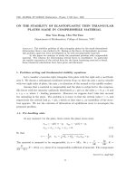
Calcified discs were found in 87 of the 100 radiographic
examinations of spines (Figure 1.). In this study popula-
tion, CDVR were found in all intervertebral disc spaces
that contain a disc. Of the 2600 discs examined, 477
(18%) showed signs of calcifications. CDVR were most
frequently found at disc-spaces T11-T12 (9.4%), C7-T1
(7.9%), T12-T13 (7.1%), T10-T11 (6.5%) (Figure 1.). The
median number of CDVR was 4 (mean 4.77; SD ± 3.77;
range 0, 19).
There was no difference in number of calcified discs in
female and male Dachshunds. Thirteen dogs had no
radiographic visible disc calcifications, 7 dogs had one
CDVR, 13 dogs had two CDVR, 10 had three CDVR, 10
had four CDVR and 47 dogs had five or more CDVR.
Hence, 53 of the Dachshunds from our study had less
than 5 CDVR.
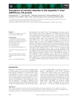
Dogs at an age of 8 years and older, showed significantly
less CDVR compared with the younger age groups (Fig-
ure 2.). Myelography was performed in 58 cases, mag-
netic resonance imaging in 35 dogs and in 7 cases both
myelography and MRI was needed to identify the site and
the side of extrusion. Hansen type I herniation, disc
extrusion, was confirmed during all 100 surgeries after
performing 6 ventral slots and 94 hemilaminectomies.
The disc space's affected with disc extrusion were found
to have radiographic calcifications in 54 of the 100 radio-
graphic examinations. Subsequently, in 46% of the Dachs-
hunds the affected disc was not calcified on radiographic
examination (Figure 3.). The discs most commonly
affected by IVD extrusions were T12-T13 (28%), followed
by T13-L1 (23%), T11-T12 (15%) and L1-L2 (10%) (Figure
1.). Dachshunds 8 years and older showed significantly
less frequently CDVR at the surgery site compared with
the 5 to 7 years old dogs (Table 1).
Rohdin et al. Acta Veterinaria Scandinavica 2010, 52:24
/>Page 4 of 7
Five dogs had two surgeries performed on two separate
occasions in the thoracolumbar spine. The mean number
of CDVR was 7.2 in these five dogs. The extruded discs
had CDVR in 6 of the 10 examinations and in all 5 dogs it
was a disc at a different location that extruded and caused
repeated signs of paralysis. The number of CDVR was the
same in two (8/8, 10/10), decreased in two (3/2, 9/5) and
increased in one (8/9) dog in the repeated radiographic
examination. The time period between the two surgeries
was less than 12 months in 3 dogs (6, 7 and 7 months), 16
Figure 1 The number of CDVR (dark grey chart) and surgical sites (pale grey chart) in 100 surgically treated Dachshunds.
Figure 2 Box plots presenting number of CDVR in Dachshunds of
different age groups. Black dot, represents outlier dog value within
the group. Asterisk, represents significant difference (p < 0.05) com-
pared with younger age groups.
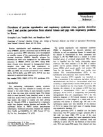
Figure 3 a. The radiographic examination of the cervical spine in
a Dachshund with a surgically confirmed disc extrusion at C4-C5
(white arrow). Notice the absence of CDVR at C4-C5.
b. A T1-weighted sagittal magnetic resonance image of the cervi-
cal spinal cord of the Dachshund in 3a. Notice the extent of calcifi-
cation of the C6-C7 disc (white arrow) compared to the affected disc at
C4-C5 (black arrow).
Rohdin et al. Acta Veterinaria Scandinavica 2010, 52:24
/>Page 5 of 7
months in one and 32 months in another patient. The
disc next to the previous operation site was extruded in
three cases (Th11-12/Th12-13, L1-2/L2-3, Th13-L1/
Th12-13). In the other two cases a more remote disc
caused the second extrusion (Th11-12/Th13-L1, Th13-
L1/L3-4).
Discussion
It has been postulated that it is usually intervertebral
discs with CDVR that are subjected to Hansen type I her-
niation [8]. Conversely, our results indicate that disc
extrusions occur as commonly in discs that are assessed
as calcified on radiographs as in discs without CDVR. In
54% of the Dachshunds in our population, the disc caus-
ing the extrusion was calcified on radiographic examina-
tion. Interestingly extruded discs were not most often
visibly calcified on radiographic examination in the
younger age group to which the radiographic evaluation
method is adapted, but was most frequent in the middle
age group (peak 5 year old dogs). The middle age group,
from 5-7 years of age, showed the highest disc extrusion
prevalence (53 dogs) in our study population. In 8 years
and older dogs extruded discs were less frequently calci-
fied on radiographic examination, following the same
trend as the general number of CDVR. It has also been
postulated that dogs with no or one radiographic disc cal-
cification might be at a reduced risk of developing disc
extrusion [7,9-14] and that disc extrusions do not occur
in Dachshunds without CDVR [8]. Our material indicates
that IVDD requiring surgery does occur also in Dachs-
hunds without any CDVR. In 13% of the dogs in our study
no disc calcifications were found on radiographic exami-
nation.
Multiple studies have suggested that screening the ver-
tebral columns of Dachshunds intended for breeding, by
radiographic examination, may be valuable in reducing
the incidence of disc extrusion [7,9-14]. According to
these suggestions, Dachshunds with 0-2 CDVR are
accepted for breeding, 3-4 CDVR may be accepted, and
dogs with > 5 CDVR should not be used for breeding pur-
poses (Norske Dachshundklubbers Forbund prøvepros-
jekt 01.05.2002, Dansk Gravhundeklub avlsanbefaling pr.
1.12.2008). In 57% of the Dachshunds treated for disc
extrusion in our study, 0-4 calcified discs were found at
the time of radiological examination, and would accord-
ingly, have been considered part of a low-risk-population
and would have been accepted for breeding purposes
according to the suggested scheme. Also the number of
CDVR showed statistically significant difference between
the three major age groups. The number of CDVR has
been reported to be a good predictor of clinically signifi-
cant IVDD in Dachshunds [9,10,12]. Our case material
clearly indicate that Dachshunds without and with rare
CDVR will be affected by disc extrusion with the same
frequency as Dachshunds with multiple calcifications vis-
ible on radiographic examination.
Absence of CDVR on radiographic examination does
not mean that the disc is neither degenerated nor calci-
fied. It has been conclusively demonstrated that radio-
graphic examination is considerably less sensitive than
histopathology in detection of calcifications within the
intervertebral disc calcifications [5]. Histopathology is
considered to be the golden standard for detection of disc
calcifications. Previous studies have found calcifications
on histopathological examination in 46-48% of the inter-
vertebral discs in Dachshunds [2,5]. This would mean
that approximately 12 intervertebral discs in the Dachs-
hunds are expected to be calcified according to the histo-
pathological study. In contrary, radiological studies were
able to detect 2.5-3.4 [20], 3.5 [21], 3.7 [13], 4.8 (current
study) of calcifications in Dachshunds (mean values).
This indicates that radiological studies are detecting only
about 20-40% of the actually existing disc calcifications
that can be identified with histopathology. Computer
tomography, a more sensitive diagnostic modality, may
detect less calcified discs not visible on conventional radi-
ography and may in this way be of advantage when con-
sidering breed screening programs.
The occurrence of intervertebral disc calcifications is
not constant throughout the hypochondroplastic dog's
Table 1: Mean number of calcified discs, visible on radiographic examination (CDVR), number of dogs having 4 or less and
5 and more CDVR and dogs having CDVR at surgery site in 100 surgeries for disc extrusion in different age groups.
≤ 4 years Age groups
5-7 years
≥ 8 years
Nr of dogs in groups 27 53 20
Mean nr of CDVR ± SD 5.74 ± 3.24
a
5.17 ± 3.83
a
2.4 ± 3.42
b
Nr of dogs with ≤ 4 CDVR 10 (37%) 27 (51%) 16 (80%)
Nr of dogs with ≥ 5 CDVR 17 (63%) 26 (49%) 4 (20%)
CDVR at surgery site 14 (52%)
ab
35 (66%)
b
5 (25%)
a
a, b
different superscript letters indicate statistically significant (p < 0.05) difference between age groups within row
Rohdin et al. Acta Veterinaria Scandinavica 2010, 52:24
/>Page 6 of 7
life. CDVR seem to be best visualized at a younger age
and later decline in frequency as the hypochondroplastic
dog matures [7]. Hansen (1952) reported an increased
frequency of histopathologically confirmed calcifications
in the chondrodystrophic dogs up to the age of 7 years
(77% calcified discs), with 100% of the discs in the tho-
racic region being histopathologically calcified at the age
of 6 years. Hansen (1952) also reported a decline in histo-
pathologically confirmed calcifications in chondrodystro-
phoid dogs older than 7 years old. These findings indicate
that although the CDVRs will decline after the Dachs-
hunds first years of life the histologically detectable disc
calcifications may increase in number up to the age of
seven. In the older Dachshunds calcifications visible on
radiographs and detectable on histopathology may disap-
pear. One could conclude that radiological examination is
not the most sensitive method to detect disc calcifica-
tions and does not necessarily reflect the real extent of
the degenerative process within the Dachshund discs.
The results of our study support the previous findings
that CDVRs are most frequently found in young adult
Dachshunds (3-4 years old, mean 5.7; SD ± 3.24) and
most rare in older Dachshunds (≥ 8 years old, mean 2.4;
SD ± 3.42).
We found a bimodal anatomical occurrence of CDVR,
with one minor peak at the transition from cervical to
thoracic spine, and a larger peak at the transition from
thoracic to lumbar spine (Figure 1.). This bimodal
appearance has already been presented by Stigen (1996).
The distribution of calcified discs on radiographs does
not entirely correlate with the site of disc extrusions but
seem to occur most frequently in areas of high mobility of
the vertebral column (Figure 1.).
Chondroid metaplasia is a unique degenerative process
of intervertebral discs seen only in hypochondroplastic
dog breeds [1,2,22]. This short legged phenotype has
recently been associated with the expression of a retro-
gene, encoding fibroblast growth factor (Fgf4) [23]. Sub-
sequently, it is likely that the intervertebral disc
calcification, part of this degenerative process, will show
high estimates of heritability [9,10,12]. The chondroid
metaplasia is inherited and all Dachshunds are affected
by this degenerative process, however, not all dogs will
have disc extrusions. The occurrence of IVDD is multi-
factorial and the fate of the degenerated hypochondro-
plastic disc will be affected by various factors, such as
body dimensions, environmental factors including
mechanics, together determining whether the disc will be
subjected to clinically significant IVDD or not [21,24].
It still needs to be proven that selection of breeding
dogs based on CDVR in the Dachshund has a potential to
reduce the incidence of IVDD in the Dachshund popula-
tion. Absence of radiological disc calcifications does not
exclude degenerative changes in the disc nor does it
exclude disc calcification.
Intervertebral disc calcification is undoubtedly a sign of
severe disc degeneration and is a serious risk factor for
the development of IVDD. Our results indicate that
CDVR were more frequent in our study of Dachshunds
surgically treated for disc extrusions compared to previ-
ous studies [9,11,13,14,21]. The occurrence of CDVR var-
ies in different studies but conclusions should be made
carefully as the study designs, inclusion criteria, age of
examined Dachshunds, absence or presence of sedation
for radiographic examination and the way radiographs
were evaluated (analog or digital) might have influenced
the final results [6,8,10,11,14,20]. The Dachshunds
included in our study were of all sizes, hair coats and var-
ied in age and the way they were used by the owners.
Conclusions
In conclusion, we found that disc extrusions occur as fre-
quently in discs that are found to have radiographic evi-
dence of calcification as those discs that do not have signs
of radiographic calcification, and that IVDD requiring
surgery does occur in the absence of any calcified discs on
radiographic examination. We found that calcified discs
were more frequent in our Dachshund population com-
pared to previous studies suggesting that disc calcifica-
tion might be a serious risk factor for developing disc
extrusion. Further studies are needed to show, conclu-
sively, if selection of breeding dogs based on CDVR in the
Dachshund will reduce the incidence of IVDD. The pres-
ence of the calcifications of intervertebral disc should be
evaluated with caution, as only part of the calcifications
will be detected and the real extent of the disc degenera-
tion may be underestimated.
Competing interests
The authors declare that they have no competing interests.
Authors' contributions
CR drafted the manuscript, JJ and RV carried out the evaluations of the radio-
graphs, SC designed the study, RV performed the statistical analysis and CR, JJ,
RV and SC were all involved in the clinical procedures involved in the study.
All authors read and approved the final manuscript.
Acknowledgements
The authors would like to thank Toomas Orro, DVM, PhD, Estonian University of
Life Sciences Tartu, Estonia for assistance with statistical analysis.
Author Details
1
University Animal Hospital, Department of Clinical Sciences, Swedish
University of Agricultural Sciences, Box 7040, 750 07 Uppsala, Sweden,
2
Referral Animal Neurology Hospital Aisti, Virtatie 9, FI-016000, Vantaa, Finland
and
3
Estonian University of Life Sciences, Tartu, Estonia
Received: 17 November 2009 Accepted: 14 April 2010
Published: 14 April 2010
This article is available from: 2010 Rohdin et al; licensee BioMed Central Ltd. This is an Open Access article distributed under the terms of the Creative Commons Attribution License ( which permits unrestricted use, distribution, and reproduction in any medium, provided the original work is properly cited.Acta Veteri naria Scandina vica 2010, 52:24
Rohdin et al. Acta Veterinaria Scandinavica 2010, 52:24
/>Page 7 of 7
References
1. Ball MU, McGuire JA, Swaim SF, Hoerlein BF: Patterns of occurrence of
disk disease among registered Dachshunds. Journal of the American
Veterinary Medical Association 1982, 180:519-522.
2. Hansen HJ: A pathologic-anatomic study on disc degeneration in dog.
Acta Orthopedica Scandinavica Supplementum 1952:1-117.
3. Hoerlein BF: Intervertebral disc disase. In Veterinary Neurology Edited by:
Oliver JE, Hoerlein BF, Mayhew IG. Philadelphia: WB Saunders;
1987:321-340.
4. Priester WA: Canine intervertebral disc disease- occurrence by age,
breed and sex among 8117 cases. Theriogenology 1976, 6:293-301.
5. Stigen Ø, Kolbjørnsen Ø: Calcification of intervertebral discs in the
dachshund: a radiographic and histopathologic study of 20 dogs. Acta
Veterinaria Scandianavica 2007, 49:1-7.
6. Jensen VF: Asymptomatic radiographic disappearance of calcified
intervertebral disc material in the dachshund. Veterinary Radiology &
Ultrasound 2001, 42:141-8.
7. Jensen VF, Arnbjerg J: Development of intervertebral disk calcification
in the dachshund: a prospective longitudinal radiographic study.
Journal of the American Animal Hospital Association 2001, 37:274-282.
8. Havranek-Balzaretti B: Beitrag zur Aetiologie der Dackellähme und Vorschlag
zur züchterischen Selektion (The etiology of intervertebral disc disease in the
dachshund and proposal of an eradication programme) Zürich: Veterinär-
Chirurgischen Klinik und Institut für Veterinärpatologie, Dissertation,
Universität, Zürich; 1980.
9. Jensen VF, Christensen KA: Inheritance of disc calcification in the
dachshund. Journal of Veterinary Medicine 2000, 47:331-340.
10. Jensen VF, Beck S, Christensen KA, Arnbjerg J: Quantification of the
association between intervertebral disk calcification and disk
herniation in Dachshunds. Journal of the American Veterinary Medical
Association 2008, 233:1090-1095.
11. Stigen Ø: Calcification of intervertebral discs in the Dachshund. A
radiographic study of 327 young dogs. Acta Veterinaria Scandinavica
1991, 32:197-203.
12. Stigen Ø, Christensen K: Calcification of intervertebral discs in the
dachshund: An estimation of heritability. Acta Veterinaria Scandinavica
1993, 34:357-361.
13. Stigen Ø: Calcification of intervertebral discs in the dachshund: a
radiographic study of 21 stud-dogs. Acta Veterinaria Scandinavica 1995,
36:329-334.
14. Stigen Ø: Calcification of intervertebral discs in the dachshund: a
radiographic study of 115 dogs at 1 and 5 years of age. Acta Veterinaria
Scandinavica 1996, 37:229-37.
15. Lorenz MD, Kornegay JN: Neurologic history and examination. In
Handbook of Veterinary Neurology 4th edition. Edited by: Lorenz MD,
Kornegay J. St. Louis: MO Elsevier Science; 2004:23-37.
16. Sharp JH, Wheeler SJ: Diagnostic aids. In Small Animal Spinal Disorders:
Diagnosis and Surgery 2nd edition. Edited by: Sharp NJH, Wheeler SJ. St.
Louis: Elsevier Mosby; Elsevier; 2005:50-51.
17. Besalti O, Pekcan Z, Sirin YS, Erbas G: Magnetic resonance imaging in
dogs with thoracomlumbar intervertebral disk disease: 69 cases (1997-
2005). Journal of the American Veterinary Medical Association 2006,
15:902-908.
18. Sharp JH, Wheeler SJ: Cervical disc disease. In Small Animal Spinal
Disorders: Diagnosis and Surgery 2nd edition. Edited by: Sharp NJH,
Wheeler SJ. St. Louis: Elsevier Mosby; Elsevier; 2005:106-120.
19. Sharp JH, Wheeler SJ: Thoracolumbar disc disease. In Small Animal
Spinal Disorders: Diagnosis and Surgery 2nd edition. Edited by: Sharp NJH,
Wheeler SJ. St. Louis: Elsevier Mosby; Elsevier; 2005:136-159.
20. Lappalainen A, Norrgård M, Alm K, Snellman M, Laitinen O: Calcification
of the intervertebral disc and curvature of the radius and ulna: a
radiographic survey of Finish miniature dachshunds. Acta Veterinaria
Scandinavia 2001, 42:229-236.
21. Jensen VF, Ersbøll AK: Mechanical factors affecting the occurrence of
intervertebral disc calcification in the dachshund- a population study.
Journal of Veterinary Medicine 2000, 47:283-296.
22. Gosh P, Taylor TKF, Yarroll JM, Braund KG, Larsen LH: Genetic factors in the
maturation of the canine intervertebral disc. Research in Veterinary
Science 1975, 19:304-311.
23. Parker HG, VonHoldt BM, Quignon P, Margulies EH, Shao SS, Mosher DS, et
al.: An expressed Fgf4 Retrogene is associated with breed-defining
chondrodysplasia in domestic dogs. Science 2009, 325:995-998.
24. Levine JM, Levine GJ, Hettlich BF, Kerwin SC, Fosgate GT: Association
between various physical factors and acute thoracolumbar
intervertebral disk extrusion or protrusion in Dachshunds. Journal of
the American Veterinary Medical Association 2006, 229:370-375.
doi: 10.1186/1751-0147-52-24
Cite this article as: Rohdin et al., Prevalence of radiographic detectable
intervertebral disc calcifications in Dachshunds surgically treated for disc
extrusion Acta Veterinaria Scandinavica 2010, 52:24