Báo cáo khoa học: " Evaluation of adverse effects in tamoxifen exposed healthy female dogs" doc
Bạn đang xem bản rút gọn của tài liệu. Xem và tải ngay bản đầy đủ của tài liệu tại đây (1019.24 KB, 6 trang )
RESEARC H Open Access
Evaluation of adverse effects in tamoxifen
exposed healthy female dogs
Wanessa LF Tavares
1
, Gleidice E Lavalle
2
, Mariana S Figueiredo
1
, Aline G Souza
1
, Angelica C Bertagnolli
1
,
Fernando AB Viana
2
, Paulo RO Paes
3
, Rubens A Carneiro
3
, Guilherme AO Cavalcanti
2
, Marilia M Melo
3
,
Geovanni D Cassali
1*
Abstract
Background: Mammary tumors are among the most frequent neoplasms in female dogs, but the strategies
employed in animal treatment are limited. In human medicine, hormone manipulation is used in cancer therapy.
Tamoxifen citrate is a selective inhibitor of oestrogen receptors and exerts a potent anti-oestrogen effect on the
mammary gland. The aim of this study was to evaluate the adverse effects when exposing healthy female dogs to
tamoxifen.
Methods: Tamoxifen was administered for 120 days at a dose of 0.5 or 0.8 mg/kg/day to either intact or spayed
female dogs. The effects were assessed through clinical examination, haematology, serum biochemistry,
ophthalmology and bone marrow aspirate examination. Ovariohysterectomy was performed and the uterus
examined by histopathology.
Results: Vulva oedema and purulent vaginal discharge developed with 10 days of tamoxifen exposure in all groups.
Pyometra was diagnosed after around 90 days of exposure in intact females with frequencies increasing during the
following 30 days of exposure. Up to 50% of dogs within the groups developed retinitis but none of the dogs had
signs of reduced visual acuity. The prevalence of retinitis in each group was similar after 120 days of exposure.
Haematological, biochemical and bone marrow changes were not observed. Due to the high risk of developing
pyometra after prolonged exposure to tamoxifen, only spayed animals should be given this medication.
Conclusions: A dose of 0.8 mg tamoxifen/kg body weight/day is recomme nded when treating tamoxifen-
responsive canine mamma ry tumors. Due to the high risk of developing pyometra, ovariohysterectomy is
recommended.
Background
The frequency of cancer and other disease s related to
aging in pet animals has increased in recent years due
to increased life expectancy [1].
Mammary tumors are among the most frequent neo-
plasms in female dogs [1-5]. Despite their importance
and high incidence, tumoral extirpation is often the pre-
ferred therapy. However, approximately 48% of dogs
with mammary carcinoma die or are euthanized within
one year after surgical remov al of the primary tumor or
recognition of metastases [6]. Thus, it is necessary to
adopt low-cost alternative therapeutic approaches that
can increase overall survival and welfare.
In human medicine, systemic therapies such as che-
motherapy and hormone manipulation are used in cancer
therapy. Tamoxifen citrate is a sel ective inhibitor of oes-
trogen receptors and exerts a potent anti-oestrogen effect
on the mammary gland [7,8]. Previous studies have eval-
uated the efficacy of tamoxifen in preventing the recur-
rence of canine mammary tumors [9]. In that study,
tamoxifen was administered orally at a mean dose of
around 1 mg/kg body weight (BW). However, 56% of the
animals developed c omplications such as pyometra, vul-
var swelling and pseudogestational behaviour thus show-
ing a need to assess drug tolerance in female dogs [9].
* Correspondence:
1
Laboratory of Comparative Pathology, Department of General Pathology,
Institute of Biological Sciences, Federal University of Minas Gerais (UFMG),
Brazil
Full list of author information is available at the end of the article
Tavares et al. Acta Veterinaria Scandinavica 2010, 52:67
/>© 2010 Tavares et al; licensee BioMed Central Ltd. This is an Open Access article distributed under the terms of the Creative Commons
Attribution License ( which permits unrestricted use, distribution, and reproduction in
any medium, provide d the original work is properly cited.
A dose of 0.5 mg/kg BW was suggested in another study
to minimize adverse effects [10].
Considering the lack of non-surgical therapeutic
resources for canine mammary neoplasms and the pro-
ven benefits of tamoxifen in treating human breast
tumors, studies of the value of tamoxifen medication i n
canine medicine is needed. The e pidemiological [11],
clinical [4,12,13], biological [4,14], and genetic similari-
ties [15] between human breast cancer and canine mam-
mary tumors allow comparisons to be made [5].
The aim of this study was to evaluate adverse effects
of tamoxifen exposure to healthy female dogs.
Methods
Animals
This study was approved by the Brazilian Committee of
Ethics in Animal Experimentation, CETEA/UFMG (Pro-
tocol number 40/2006). The dogs were forwarded for
adoption after completion of the study.
Healthy female mixed breed dogs aged 4 years ± 2.3
years, with a mean BW of 20 kg were used. Initially, the
dogs were subjected to a clinical examination including
haematology, biochemistry, ophthalmology, and bone
marrow aspirate examination as described below. Dogs
were only included if healthy. However, ophthalmologic
changes were accepted, but dogs having eye lesions were
excluded from the ophthalmology study parts.
The animals were randomly distributed into fo ur
groups:
A: 5 inta ct animals receiving 0.5 mg tamoxifen citrat e/
kg BW/day;
B: 5 spayed animals receiving 0.5 mg tamoxifen
citrate/kg BW/day;
C: 5 intact anima ls receiving 0.8 mg tamoxifen citrate/
kg BW/day;
D: 5 spayed animals receiving 0.8 mg tamoxifen
citrate/kg BW/day.
Ovariohysterectomy was performed in dogs assigned
to gro ups B and D approximately 90 days before enter-
ing the trial. The surgical procedure, anaesthetic proto-
col and immediate post-operative care were similar for
all dogs. The animals were kept in experimental kennels
from UFMG/Brazil with free access to food and water.
Drug exposure
Tamoxifen citrate (Taxofen® , Blaüsiegel, Cotia/Brazil)
was administered once a day for 120 days at the same
time during feeding at a dose of either 0.5 mg or 0.8 mg
per k g BW. The BW was determined monthly and the
total dose adjusted to the precise BW.
Examinations
Examinations were conducted with intervals of 10 days
starting with a pre-exposure examination (T00) followed
by examination after 10 days of tamoxifen exposure
(T01) and so on until 120 days of exposure (T02 to
T12). The dogs were not euthanized at the end of the
study, but ovariohyste rectomy was performed at T12 for
intact females (groups A and C) and the uterus sub-
jected to pathological examination.
Clinical examination, haematology and serum bio-
chemistry was performed at T00 to T12.
Clinical examination included evaluation of general sta-
tus, measurement of rectal temperature, heart rate , and
respiratory rate, in spection of mucosal membranes and
skin, inspection and palpation of lymph nodes, joints,
external genitalia, and mammary glands, and palpation of
the abdominal cavity with contents. Abdominal ultraso-
nography was applied if pyometra was suspected.
Haematology was based on ethylenediaminetetraacetic
acid (EDTA) stabilized blood samples taken from the
cephalic vein. Serum biochemic al analyses were done for
alanine transaminase (ALT), aspartate transaminase
(AST), gamma glutamyl transpeptidase (GGT), alkaline
phosphatase (ALP), bilirubin (direct (DB), indirect (IB)
and total (TB)), urea, creatinine, calcium and cholesterol.
Ophthalmological examinations were performed by a
specialist at T00, T06 and T12. Two drops of mydria-
tic eye solution (Mydriac yl®, Tropicamide, 1%) were
applied to each eye 15 min before examination of the
fundus. Examinations were conducted with a HEIN-
EEN 30 Indirect Ophthalmoscope and BETA 200
Direct Ophthalmoscope.
Bone marrow aspirate examinations were performed at
T00, T06 and T12. The breastbone region was trimmed
and disinfected with 70% ethanol. Lidocaine (2%) was
used for local analgesic. Bone marrow was aspirated
through a 40 × 12 hypodermic needle and a disposable
10 mL syringe. The slides were air-dried and subjected
to May-Grünewald Giemsa staining before microscopy.
Samples for histopathology were fixed in 10% neutral
buffered formalin, processed by routine methods for his-
tology and embedded in paraffin. Histological sections
of 4 μm were haemat oxylin and eosin stained. Uterine
lesions were classified as described by Dow [16].
Statistical analysis
To evaluate the parametric data from the haemograms,
biochemistry and bone marrow aspirate examination,
ANOVA with a SNK test was performed. The Kruskal-
Wallis test [17] was used to e valuate non-parametric
haemograms, biochemic al data and bone marrow aspi-
rate results.
Results
Clinical findings
All dogs were found healthy before entering the trial.
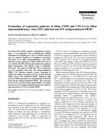
Vulva oedema and purulent vaginal d ischarge (Figure 1)
Tavares et al. Acta Veterinaria Scandinavica 2010, 52:67
/>Page 2 of 6
developed with 10 days of tamoxifen exposure in all
groups (Table 1). Pyometra ( Figure 1) was diagnosed
after around 90 days of exposure in intact females with
frequencies increasing during the following 30 days. Two
dogs develop ed pyometra in group A, while 4 dogs devel-
oped pyom etra in group C. Rare events of vomiting, diar-
rhoea and appetite loss were observed throughout the
period of the experiment. Dogs developing pyometra
were submitted to ovariohysterectomy and excluded
from the study (Table 1). Vaginal cytology was performed
to evaluate oestrus cycles, but was inconclusive.
Pathology
Two animals in group A developed cystic endometrial
hyperplasiatypeIwithproliferation of endometrial
glands, cyst formation and endometrial polyps with no
inflammatory reaction (Figure 2). The predominant
pathologic findings in cases of pyometra were symmetri-
cal distension of the uterine horns with dark-stained
sero sal surface and congestion. The uterine content was
purulent or opaque red-brown in color. The mucosa
had uneventhickness with irregular superficial haemor-
rhages, and in other portions obviously hyperplasia,
sometimes with small cysts. Microscopically, the m ost
significant feature was the endometrial hyperplasia asso-
ciated with haemorrhage and presence of polymorpho-
nuclear cells in the glandular lumen. The other three
animals in group A presented cystic endometrial hyper-
plasia type III, with mononuclear and polymorphonuc-
lear infiltration in the periglandular endometrial stroma
associated with haemorrhage. Two animals in group A
also pre sented squamous metaplasia of the endometrial
epithelium (Figure 2). Only one animal in group C pre-
sented cystic endometrial hyperplasia t ype I, while three
Figure 1 Animal: C3, Pyometra. (A) Intact female dog with purulent vaginal discharge after oral administration of tamoxifen, 0.8 mg/kg/day, for
100 days. (B) Ultrasonography revealing uterine body (1.5 cm) and uterine horns slightly dilated, thickened uterine wall with irregular mucosa
and retention of secretion within the organ. (C) Uterine histopathology after ovariohysterectomy revealing cystic endometrial hyperplasia type III
associated with hemorrhage. HE, Obj. 4×. (D) Detail showing presence of polymorphonuclear cells in glandular lumen. HE, Obj. 60×.
Tavares et al. Acta Veterinaria Scandinavica 2010, 52:67
/>Page 3 of 6
in this group developed cystic endometrial hyperplasia
type II associated with mononuclear cells (macrophages,
lymphocytes and plasmacells) with superficial subepithe-
lial haemorrhage. One animal also developed squamous
metaplasia and endometrial polyps. One animal pre-
sented cystic endometrial hyperplasia type III (Figure 1).
Ophthalmology
Three animals (A4, B3 and C 5) that had ophthalmologi-
cal alterations at T00 were excluded. Up to 50% of dogs
within the groups developed retinitis during the study
(Table 1). Once a dog had developed retinitis, this co ndi-
tion remained throughout the study. Retinitis was charac-
terised by the presence of black dots on tapetal area [18].
None of the dogs had signs of reduced visual acuity. The
prevalence of retinitis at T12 in the exposure groups (A
+B: 0.5 mg and C+D: 0.8 mg) was non-significant.
Haematology and biochemistry
Evaluation of haematological and biochemical profiles
were restricted to the period T00 to T08 as several dogs
were removed from the study during the last time of the
trial (Table 1). The mean values for erythrocytes, hae-
moglobin, globular volume, platelets, ALT, AST, GGT,
ALP, IB and calcium remained within the normal ranges
for all dogs [19].
Slight increases were observed in the mean values of
total leukocytes at T01 , T02 and T07 in group B and
T02 in group D (data not shown).
Bone marrow aspirate examination
The mean values of cellularity, megakaryocytes, metaru-
bricytes, lymphocytes, plasmacells and monocytes
remained within the normal range [20] for all dogs
throughout the trial.
Discussion
Oedema of the external genitalia as seen in dogs of all
groups develops due to the agonist action of tamoxifen
on genital hormonal receptors. After ovariohysterect-
omy, the uterine stump may develop pyometra. This
was not observed in the present study (groups B and D)
although ultrasonography was applied to all animals
having vaginal discharge. The cause of vaginal discharge
in these animals remained unsolved.
The agonist action of tamoxifen on the human uterus
promotes adverse effects such as endometritis and endo-
metr ial hyperplasia. These are considered pre-malignant
lesions thus increasing the risk for develo pment of
endometrial carcino ma [21-24]. It is evident that , as in
women, intact female dogs exposed to tamoxifen
develop endometrial cell proliferation most likel y due to
the agonist stimulation of uterine oestrogen receptors.
The res ults corroborate previous studi es [9,10] suggest-
ing that tamoxifen causes oestrogenic stimulation of
canine endometrial cells even in low doses. The number
of oestrogen receptors increases due to hormonal stimu-
lation with consequent endometrial hyperplasia and an
increased number of progesterone receptors [9]. The
serum level of this hormone remains unaltered, but as
the number of receptors is increased, l eukocyte recruit-
ment to the uterus is reduced and uterine shrinkage is
impaired. This pro cess promotes reduction of uterine
immune defences and facilitates ascending bacterial
infections, mainly caused by Escherichia coli, unleashing
pyometra [25].
Table 1 Summary of genital tract symptoms and eye lesions.
Days of tamoxyfen treatment
Group Symptom 0 10 20 30 40 50 60 70 80 90 100 110 120
A Oedema 0 5 5 5 555555 5 3* 3*
Discharge 0 3 2 1 215535 5 3* 3*
Retinitis 1 - - - - - 1 - - - - - 3
B Oedema 0 5 5 5 555555 5 5 5
Discharge 0 3 1 1 11552 5 5 5 5
Retinitis 1 - - - - - 2 - - - - - 2
C Oedema 0 5 5 5 555553*3* 1
#
1
#
Discharge 0 2 1 5 235113*3* 1
#
1
#
Retinitis 1 - - - - - 1 - - - - - 1
D Oedema 0 5 5 5 555555 5 5 5
Discharge 0 0 2 1 131321 3 2 2
Retinitis 0 - - - - - 0 - - - - - 2
* group size = 3,
#
group size = 1, - eyes not exa mined
Number of dogs having vulva oedema, purulent vaginal discharge and retinitis following oral exposure to tamoxifen at daily doses of either 0.5 mg/kg body
weight (groups A and B) or 0.8 mg/kg body weight (groups C and D). Dogs in groups B and D were spayed before the study. Each group consisted initially of 5
dogs, but animals developing pyometra or had eye lesions before tamoxifen exposure were removed from the study thus reducing the group size.
Tavares et al. Acta Veterinaria Scandinavica 2010, 52:67
/>Page 4 of 6
Three animals from groups A and C presented squa-
mous metaplasia of the endometrial epithelium, pr ob-
ably due to estrogenic stimulation as seen in women
treated with tamoxifen [26,27] and dogs with sponta-
neous cystic endometrial hyperplasia complex-pyometra
[28]. Endometrial polyps were found in one case. This
type of lesion has also been reported i n women after
treatment with tamoxifen [21,22,26].
The ophthalmological alterations are similar to those
found in women treated with tamoxifen at 20 mg dose
per kg BW for 5 years. However, the prevalence seems
to be higher in dogs even though they were exposed to
a lower dose and for a shorter period. T his discrepancy
mayberelatedtoadifferenceinspeciessensibility,
since the mechani sms of action of tamoxifen are related
to specific variant oestrogen receptor expression in dif-
ferent cell types and different mechanisms of DNA-
receptor interaction. Thus, tamoxifen acts more as
agonist than antagonist in c anines thus suggesting spe-
cies differe nces [29]. The lower dose groups (A and B)
presented ophthalmological lesions after 6 0 days of
exposure suggesting variation in sensibility not related
to dose or hormone levels (intact or spayed animal). In
human medicine, eye lesions are reversible after suspen-
sion or termination of treatment [30-33]. It is therefore
expected that such lesions will also be reversible in
canine species after treatment is suspended.
On the basis of previous studies [9,10], do ses of 0.5
and0.8mg/kgBW/daywerechosen.Consideringthe
risk of developing pyometra when administering tamoxi-
fen to intact female dogs, it is suggested that this medi-
cation be prescribed to spayed animal s only. Since there
was no difference between the lower and higher dose
groups in the other side-effects induced by tamoxifen,
the higher dose may increase the chances of therapeutic
success.
Figure 2 (A and B): Animal: A2 (intact, 0.5 mg/kg/day). (A) Cystic endometrial hyperplasia type I. Endometrial proliferation not associated
with stromal inflammatory infiltration. HE, Obj. 20×. (B) Endometrial polyp formation. HE, Obj. 4×. (C and D): Animal: A4 (intact, 0.5 mg/kg/day).
(C) Cystic endometrial hyperplasia type III with squamous metaplasia of endometrial cover epithelium and hemorrhage. HE, Obj. 20×. (D) Detail
showing squamous metaplasia of endometrial cover epithelium and haemorrhage. HE, Obj. 60×.
Tavares et al. Acta Veterinaria Scandinavica 2010, 52:67
/>Page 5 of 6
Conclusions
Tamoxifen may become an important compound in
veterinary medicine considering its therapeutic potent ial
for increasing the overall survival rate of female dogs
with mammary tumors, if its side effects are correctly
assessed and controlled. A dose of 0.8 mg tamoxifen/kg
BW/day for at least 120 days is recommended.
Acknowledgements
This work was supported by CNPq and FAPEMIG - Brazil.
Author details
1
Laboratory of Comparative Pathology, Department of General Pathology,
Institute of Biological Sciences, Federal University of Minas Gerais (UFMG),
Brazil.
2
Veterinary Teaching Hospital, College of Veterinary Medicine, Federal
University of Minas Gerais (UFMG), Brazil.
3
Department of Veterinary Clinical
Medicine and Surgery, College of Veterinary Medicine, Federal University of
Minas Gerais (UFMG), Brazil.
Authors’ contributions
WLFT was responsible for all procedures and drafted the manuscript. GEL
performed surgery and participated in the study design. MSF and AGS
participated in kennel maintenance, drug administration, sample collections
and surgical procedures. ACB conducted vaginal cytology. FABV was the
ophthalmological specialist responsible for eye examinations. PROP
performed myelogram examinations, whose samples were collected by RAC.
GAOC conduced ultrasonography examinations. MMM participated in the
study design and was the co-advisor of this work. GDC was the advisor of
this work. All authors read and approved the final manuscript.
Competing interests
The authors declare that they have no competing interest s.
Received: 6 April 2010 Accepted: 22 December 2010
Published: 22 December 2010
References
1. Paoloni M, Khanna C: Translation of new cancer treatments from pet
dogs to humans. Nat Rev Cancer 2008, 8:147-157.
2. Withrow SJ, Vail DM: In Withrow & MacEwen’s Small Animal Clinical
Oncology. Edited by: St Louis. Saunders Elsevier; 2007.
3. Benjamin A, Lee C: Classification and behavior of canine mammary
epithelial neoplasm based on life-span observations in beagles. Vet
Pathol 1999, 36:423-436.
4. Moulton E: Tumors of the mammary gland. Mammary Tumors in Domestic
Animals. 3 edition. University of California; 1990, 518-522.
5. Cassali GD, Gobbi H, Malm C, Schmitt FC: Evaluation of accuracy of fine
needle aspiration biopsy for diagnosis of canine mammary tumors:
comparative features with the human tumors. Cytopathol 2007,
18:191-196.
6. Graham JC, O’keefe DA, Gelberg HB: Immunohistochemical assay for
detecting estrogen receptors in canine mammary tumors. Am J Vet Res
1999, 60:627-630.
7. Stearns V, Gelmann P: Does tamoxifen cause cancer in humans? J Clin
Oncol 1998, 16:779-792.
8. Jordan VC: Tamoxifen (ICI46,474) as a targeted therapy to treat and
prevent breast cancer. Br J Pharmacol 2006, 147:S269-S276.
9. Morris J, Dobson J, Bostock D: Use of tamoxifen in the control of canine
mammary neoplasia. Vet Rec 1993, 27:539-541.
10. Baker R: Comments to the editor on Use of tamoxifen in the control of
canine mammary neoplasia. Vet Rec 1994, 134:24.
11. Hellmén E, Bergström R, Holmberg L: Prognostic factors in canine
mammary tumors: a multivariate study of 202 consecutives cases. Vet
Pathol 1993, 30:20-27.
12. Miller W: Oestrogens and breast cancer: biological considerations. Br Med
Bul 1991, 47:470-483.
13. Las Mulas M, Millán Y, Dios R: A prospective analysis of
immunohistochemically determined estrogen receptor alfa and
progesterone receptor expression and host and tumor factors as
predictors of disease-free period in mammary tumors of the dog. Vet
Pathol 1992, 43:243-247.
14. Geraldes M, Gärtner F, Schmitt F: Immunohistochemical study of
hormonal receptors and cell proliferation in normal canine mammary
glands and spontaneous mammary tumors. Vet Rec 2000, 146:403-406.
15. Chu L, Rutteman G, Kong J, Ghahremani M, Schmeing M, Misdorp W, Van
Garderen E, Pelletier J: Genomic organization of the canine p53 gene and
its mutational status in canine mammary neoplasia. Breast Cancer Res
Treat 1998, 50:11-25.
16. Dow C: The cystic hyperplasia-pyometra complex in the bitch. Vet Rec
1958, 70:1102-1110.
17. Sampaio I: Statistics applied to animal experimentation [in Portuguese]
Fundação de Ensino e Pesquisa em Medicina Veterinária e Zootecnia, Belo
Horizonte; 1998, 221.
18. Imperia P, Lazarus H, Lass J: Ocular complications of systemic cancer
chemotherapy. Sur ophthalmol 1989, 34:209-230.
19. Kaneko J, Harvey J, Bruss M: The erythrocyte: physiology, metabolism, and
biochemical disorders. Hepatic Function. Kidney function and damage.
Clinical Biochemistry of Domestic Animals. 6 edition. Academic Press Elsevier,
Burlington; 2008, 173-240, 379-412, 485-528.
20. Harvey J: Bone marrow examination. Atlas of Veterinary Hematology, Blood
and Bone Marrow of Domestic Animals. 1 edition. WB Saunders Company,
Philadelphia; 2001, 113-23.
21. Hulka C, Hall D: Endometrial abnormalities associated with tamoxifen
therapy for breast cancer: sonographic and pathologic correlation. Am J
Roentgenol 1993, 160:809-812.
22. Cohen I: Endometrial pathologies associated with postmenopausal
tamoxifen treatment. Gynecol Oncol 2004, 94:256-266.
23. Ganz P, Land S: Risks, benefits, and effects on quality of life of selective
estrogen-receptor modulator therapy in postmenopausal women at
increased risk of breast cancer. Menopause 2008, 15:797-803.
24. Bland A, Calingaert B, Secord A, Lee P, Valea F, Berchuck A, Soper J,
Havrilesky L: Relationship between tamoxifen use and high risk
endometrial cancer histologic types. Gynecol Oncol 2009, 112:150-154.
25. Nascimento E, Santos R: Pathology of uterus [in Portuguese]. Pathology of
Reproduction of Domestic Animals. 2 edition. Rio de Janeiro, Guanabara
Koogan; 2003, 48-69.
26. Deligdish L, Kalir T, Cohen C, De Latour M, Le Bouedec G, Penault-Llorca F:
Endometrial histopathology in 700 patients treated with tamoxifen for
breast cancer. Gynecol Oncol 2000, 78:181-186.
27. Cunha G, Cooke P, Kurita T: Role of stromal-epithelial interactions in
hormonal responses. Arc Histol Cytol 2004, 67:417-434.
28. De Cock H, Vermeirsch H, Ducatelle R, De Schepper J:
Immunohistochemical analysis of estrogen receptors in cystic-
endometritis-pyometra complex in the bitch. Theriogenol 1997,
48:1035-1047.
29. Hoffmann B, Schuler G: Receptors blockers - general aspects with respect
to their use in domestic animal reproduction. Animal Reprod Sci 2000, 60-
61:295-312.
30. Pavlidis N, Petris C, Briassoulis E: Clear evidence that long-term, low-dose
tamoxifen treatment can induce ocular toxicity. A prospective study of
63 patients. Cancer 1992, 69:2961-2964.
31. Noureddin BN, Seoud M, Bashshur Z, Salem Z, Shamseddin A, Khalili A:
Ocular toxicity in low-dose tamoxifen: a prospective study. Eye 1993,
6:729-733.
32. Tang R, Shields J, Schiffman J, Li H, Locher D, Hampton J, Prager T, Pardo G:
Retinal changes associated with tamoxifen treatment for breast cancer.
Eye 1997, 3:295-297.
33. Lazzaroni F, Scorolli L, Pizzoleo C, Savini G, De Nigris A, Giosa F, Meduri R:
Tamoxifen retinopathy: does it really exist? Gr Arc Clin Experim
Ophthalmol 1998, 236:669-673.
doi:10.1186/1751-0147-52-67
Cite this article as: Tavares et al.: Evaluation of adverse effects in
tamoxifen exposed healthy female dogs. Acta Veterinaria Scandinavica
2010 52:67.
Tavares et al. Acta Veterinaria Scandinavica 2010, 52:67
/>Page 6 of 6