Báo cáo khoa học: " Experimental infection of dogs with a feline endogenous retrovirus RD-114." pps
Bạn đang xem bản rút gọn của tài liệu. Xem và tải ngay bản đầy đủ của tài liệu tại đây (283.15 KB, 4 trang )
RESEARC H Open Access
Experimental infection of dogs with a feline
endogenous retrovirus RD-114
Rie Narushima
1*
, Noriyuki Horiuchi
1
, Tatsufumi Usui
2
, Takashi Ogawa
1
, Toshio Takahashi
3
, Tomoaki Shimazaki
4
Abstract
Background: The feline endogenous retrovirus RD114 is contained in the genome of cats. The virus may
contaminate live canine vaccines based on cultured feline cells. The in vivo infectivity, acute and subacute
pathogenicity, and viral proliferation of the RD114 virus were evaluated by experimental infection of dogs.
Methods: Nine specific pathogen free dogs were divided into three groups, with each group consisting of one
female and two male dogs. Dogs were subcutaneously inoculated in the neck with either 1 ml RD114 stock virus
(group A), inactivated RD114 virus suspension (group B), or cell culture medium (group C) as a negative control. To
assess blood cell counts and biochemical properties, blood samples from each group were collected 5 days before
inoculation, just prior to inoculation, and 1, 3, 7 and 10 days post-inoculation.
Result: During the experimental period of 51 days, none of the dogs inoculated with RD114 virus showed any
clinical signs, significant increases in rectal temperature or abnormal blood biochemical characteristics including
C-reactive protein when compared with the negative controls. We were not able to re-isolate the RD114 virus from
buffy coat cells of group A dogs. Additionally, we could not detect RD114 provirus in the genomic DNA isolated
from peripheral blood leukocytes, lymph node, spleen and sternal bone marrow cells.
Conclusions: Signs of RD114 virus proliferation were not found after subcutaneous infection of dogs. Although the
potential risk caused by infection with RD114 virus in dogs could not be assessed in this study, we suspect that
RD114 virus has little or no virulence in dogs.
Background
Domestic cats are generally assumed to harbour the infec-
tious endogenous retrovirus RD114 in their genome [1,2].
It is known that the Crandell-Rees feline kidney cell line is
contaminated with an RD114-like virus [3]. Recently,
Miyazawa et al. [4] found that certain live attenuated vac-
cines for dogs were cont amina ted with infectious RD114
virus. We also confirmed in our laboratory that infectious
RD114 virus was present in certain live attenuated canine
vaccines that were manufactured using feline cells (unpub-
lished data). The amount of infectious RD114 virus found
in manufactured live canine vaccines was as high as 1,800
50% tissue culture infective dose (TCID
50
)/vial (one vial
represents a single dose) [4]. RD114 virus can be regarded
as an ‘exogenous’ retrovirus in non-feline species including
dogs, however there is no information concerning the
etiological features of RD114 virus infection in dogs. The
present study was conducted to evaluate the in vivo infec-
tivity, acute and subacute pathogenicity, and viral pro lif-
eration of t he RD114 virus by experimental infection of
specific pathogen free (SPF) dogs.
Methods
Virus preparation
A LacZ marker rescue assay was used to detect and
titrate infectious RD114 virus [5,6]. The principle of the
assa y is based on the detection of i nfectious RD114 virus
using TE671 (human rhabdomyosarcoma) cells trans-
duced with the LacZ marker gene [TE671(LacZ) cells].
RD114 virus was prepared from the culture supernatant
of TE671 cells chronically infected with the virus [7].
Culture supernatants were filtered through a 0.45 μm
pore size membrane filters, and aliquots stored at -80 °C
until required. The titre of the stock virus was a pproxi-
mately 10
5
infectious units/ml. To prepare inactivated
RD114 virus as inocula, the stock virus was added to an
* Correspondence:
1
National Veterinary Assay Laboratory, Ministry of Agriculture, Forestry and
Fisheries, 1-15-1 Tokura, Kokubunji, Tokyo 185-8511, Japan
Full list of author information is available at the end of the article
Narushima et al. Acta Veterinaria Scandinavica 2011, 53:3
/>© 2011 Narushim a et al; lice nsee B ioMed Central Ltd. This is an Open Access article distributed under the terms of the Creative
Commons Attribution License ( which permits unrestricted use, distribution, and
reproduction in any medium, pr ovided the original work is properly cited.
equal volume of diethylether, then vigorously and inter-
mittently mixed at room temperature (around 20°C) for 3
min. Absence of infectious RD114 virus in the inactivated
viral pre paration was confirmed by the LacZ marker res-
cue assay.
Dog inoculation
Nine 10-months-old SPF beagles (six males and three
females) were divided into three groups (groups A, B
and C). Each group consisted of one female and two
male dogs with individual dogs confined to cages. The
dogs were inoculated subcutaneously in the neck with
either 1 ml of RD114 stock virus (group A), inactivated
RD114 virus suspension (group B) or cell culture med-
ium (group C) as a negativ e control. The inoculation
route was chosen according to the manufacturer’ s
instruct ions for the live canine vaccines. All animal stu-
dies were conducted in accordance with the National
Veterinary Assay Laboratory Guide for the Care and
Use of Laboratory Animals, and the relevant Animal
WelfareActs.Thedogswereeuthanized51dayspost
inoculation (PI) and autopsied. The thoracic organs,
abdominal organs and bone marrow were macroscopi-
cally examined. The axillary lymph nodes, spleen and
sternal bone marrow were collected for further virologi-
cal examinations.
Blood analyses and clinical examination
Re-isolation of the RD114 virus was attempted by co-
culturing TE671(LacZ) cells with buffy coat cells. To
assess the blood cell counts and biochemical properties,
blood samples from each group were collected 5 days
before inoculation, just prior to inoculation (day 0), and
at 1, 3, 7 and 10 days PI. White blood cells (WBC), red
blood cells, haemoglobin, haematocrit, mean cell
volume, mean corpuscular haemoglobin, mean corpus-
cular haemoglobin concentration, a nd platelet numbers
were determined in the blood cell counts. The biochem-
ical analyses included total protein, albumin, total biliru-
bin, glutamic-oxaloacetic transaminase, glutamic-pyruvic
transaminase, alkaline phosphatase, lactic dehydrogen-
ase, amylase, lipase, blood urea nitrogen, creatinine,
total cholesterol, triglyceride, sodium, potassium, chlor-
ine, calcium, inorganic phosphorous, glucose and t otal
bile acid levels. The C-reactive protein (CRP) concentra-
tion, a sensitive indicator of inflammation in dogs, was
analysed at days 0, 7, and 51 PI. Additionally, during the
experimental period, clinical signs were recorded daily,
and rectal temperature and body weight were measured
at least once a week.
Polymerase chain reaction
Genomic DNA wa s extracted from et hylenediaminete -
traacetic acid ( EDTA) stabilized whole blood, axillary
lymph nodes, spleen and bone marrow, and subjected to
polymerase chain reaction (PCR) analysis. In addition,
genomic DNA was also extracted from TE671(LacZ)
cells, 12 days after co-culture with buffy coat cells. The
PCR was conducted as described by Sakaguchi et al.[6]
by amplifying a portion of the env and pol genes of
RD114 virus. The single step PCR assay was performed
on genomic DNA extracted from TE671(LacZ) cells
infected with ten-fold serially diluted (10
0
to 10
-10
) stock
virus. The limit of detection was 10
-5
and 10
-6
for the
env and pol genes, respectively. Then, the LacZ pseudo-
type virus positive cells were detected 17, 2 and 0 cells
for 10
-4
,10
-5
and 10
-6
diluted points, respectively.
Statistical analysis
The mean values of all measured variables of each group
were calculated. Statistical analyses were performed using
SPSS software (version 13). The differences between the
three groups at each time point were compared with
P values of <0.05 considered to be significant.
Results
During the experimental period, none of the dogs in
groups A and B showed any clinical signs, including sig-
nificant increases in rectal temperature, or distinct
abnormal biochemical blood characteristics including
CRP when compared with group C dogs. In addition,
the WBC counts of group A and B dogs were nearly
equal to those of group C, and body weight increased in
all animals after inoculation (Figure 1). Statistical analy-
sis showed that the only significant difference was seen
in the potassium level in the three groups five days
before inoculation. At autopsy (51 days PI), any signifi-
cant lesions were not observe d macroscopically. Major
lymph nodes, especia lly the axil lary lymph nodes, were
small, ranging from 0.5-1 cm in diameter with distinct
cortico-medullary junctions. The spleen and bone mar-
row did not contain any significant lesions.
Infectious RD114 virus was not detected from TE671
(LacZ) cells co-cultured with buffy coat cells using the
LacZ marker rescue assay. This finding was also
confirmed by PCR assays using genomic DNA from the
co-cultured cells as a template. Additionally, RD114 pro-
virus was not detected in geno mic DNA extracted from
peripheral blo od, lymph nodes, spleen and sternal bone
marrow, using the one-step PCR assays (Figure. 2).
Discussion
It has been reported that RD114 virus infects a variety
of canine cell lines, such as t he Mardin-Darby canine
kidney line [8]. RD114 virus also actively infects cells
from cats and dogs, and may be transmitted to non-
feline species because of the xenotropic features of the
virus in vitro [9]. Therefore, if dogs are exposed to a
Narushima et al. Acta Veterinaria Scandinavica 2011, 53:3
/>Page 2 of 4
C
& C[UCHVG TKPQEWNCVKQP
͠
ITQWR#
ITQWR$
ITQWR%
D
& C[UCHVG TKPQEWNCVKQP
㧝㧜
㧟
Ǵ㨘
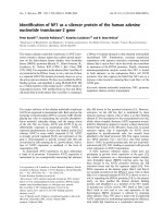
Figure 1 Mean rectal temperatures (a) and white blood cell counts (b). Significant changes in rectal temperature or white blood cell counts
in 3 groups of dogs (n = 3 for each group). Group A, RD114 virus inoculated group; B, inactivated virus group; C, control group.
M
M 1 2 3 4 5 6 7 8 9 10 11 12 13
Pol (468 bp)
Env (580 bp)
(LacZ assay 㧙 㧙 㧙 㧙 㧙 㧙 㧙 㧙 㧙 㧙 㧙 㧗 㧗 㧙 )
500 bp
(100 bp
ladder)
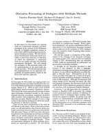
Figure 2 Polymerase chain reaction (PCR) analysis of RD114 virus in buffy coats. Genomic DNA extracted from the buffy coat samples of
dogs (n = 3 for each group) in three groups (A: RD114 virus inoculated group; B: inactivated virus group; C: control group) was tested for the
presence of RD114 virus. Lanes 1-3, group A; 4-6, group B; 7-9, group C; 10, negative control (medium); 11, positive control 1 (RD114 stock virus);
12, positive control 2 (buffy coat of group C mixed with RD114 stock virus); 13, negative control (distilled water). No PCR positives were obtained
from any of the experimental groups A-C.
Narushima et al. Acta Veterinaria Scandinavica 2011, 53:3
/>Page 3 of 4
high dose of RD114 virus, the viral genome may inte-
grate into the cells of target tissues. Viraemia is often
associate d with the acute phase of viral diseases. In clin-
ical cases caused by retroviruses, blood specimens are
mainly used for virus detection. However, we were
unable to detect RD114 provirus from blood, lymph
node, spleen and sternal b one marrow samples using a
one-step PCR assay, despite group A dogs being inocu-
lated with approximately 10
5
infectious units of RD114
virus. These results suggest that RD114 did not prolifer-
ate and disseminate in the dogs, or that RD114 virus did
not proliferate efficiently in the blood cells and hemato-
poietic system of dogs. The virus stock, neutralized with
the sera o f each dog, was inocula ted onto TE6 71(LacZ)
cells, and the number of lacZ positive foci among the
three groups were counted. In the present study, the
antibody titres remained unchanged in all dogs follow-
ing inoculation (data not sho wn), implying that RD114
virus did not proliferate in dogs.
The findings indicate that the RD114 virus has little or
no virulence in dogs. The potentia l risk caused by infec-
tion with RD114 virus in dogs cannot be accurately
assessed because a longer PI period is required for the
verification or exclusion of ret roviral infection. It is also
possible that dogs younger than 10-months might be
more susceptible to RD114 virus infection. Millions of
puppies are vaccinated worldwide on an annual basis.
Therefore it is impossible to completely rule out adverse
effects of t he infection. Many exogenous retroviruses
cause leukaemia and tumours in a wide variety of ani-
mal species, and further research is required to isolate
or detect RD114 virus in dogs that have developed leu-
kaemia and tumours following use of live canine vac-
cines manufactured using feline cells. Ho pefully, such
research would clarify the possible relationship between
the occurrence of diseases in dogs and vaccination.
Conclusions
Signs of RD114 virus proliferation were not found after
subcutaneous infection of dogs. Although the potential
risk caused by infection with RD114 virus in dogs could
not be assessed in this study because the minimum
infectious dose and pathogenicity remain unknown, we
suspect that RD114 virus has little or no virulence in
dogs.
Abbreviations
SPF: Specific pathogen free. The dogs were free from infections with canine
adenovirus (CAdV-2), canine coronavirus, canine distempervirus, canine
parainfluenzavirus, canine parvovirus, leptospira and rabies virus.
Acknowledgements
We are grateful to Dr. Yasuhiro Takeuchi, University College London, London,
UK for providing, through Dr. Takayuki Miyazawa (Kyoto University, Kyoto,
Japan), TE671 cells persistently infected with RD114 virus.
Author details
1
National Veterina ry Assay Laboratory, Ministry of Agriculture, Forestry and
Fisheries, 1-15-1 Tokura, Kokubunji, Tokyo 185-8511, Japan.
2
Avian Zoonosis
Research Center, Faculty of Agriculture, Tottori University, Koyama, Tottori
680-8553, Japan.
3
Department of Veterinary Science, Nippon Veterinary and
Life Science University 1-7-1, Kyonan-cho, Musashino-shi, Tokyo 180-8602,
Japan.
4
Animal Health Division, Food Safety and Consumer Affairs Bureau,
Ministry of Agriculture, Forestry and Fisheries, 1-2-1 Kasumigaseki, Chiyoda-
ku, Tokyo 100-8950, Japan.
Authors’ contributions
RN participated in the design of the study and performed the statistical
analysis. NH performed autopsy. TU organised basic equipment. TO
participated in collecting blood samples. TT performed finalisation of the
manuscript. TS conceived of the study, and participated in its design and
coordination. All authors read and approved the final manuscript.
Competing interests
The authors declare that they have no competing interests.
Received: 28 October 2010 Accepted: 27 January 2011
Published: 27 January 2011
References
1. McAllister RM, Nicolson M, Gardner MB, Rongey RW, Rasheed S, Sarma PS,
Huebner RJ, Hatanaka M, Oroszlan S, Gilden RV, Kabigting A, Vernon L: C-
type virus released from cultured human rhabdomyosarcoma cells. Nat
New Biol 1972, 235:3-6.
2. Fischinger PJ, Peebles PT, Nomura S, Haapala DK: Isolation of RD-114-like
oncornavirus from a cat cell line. J Virol 1973, 11:978-985.
3. Baumann JG, Gunzburg WH, Salmons B: CrFK feline kidney cells produce
an RD114-like endogenous virus that can package murine leukemia
virus-based vectors. J Virol 1998, 72:7685-7687.
4. Miyazawa T, Yoshikawa R, Golder M, Okada M, Stewart H, Palmarini M:
Isolation of an infectious endogenous retrovirus in a proportion of live
attenuated vaccines for pets. J Virol 2010, 84:3690-3694.
5. Sakaguchi S, Okada M, Shojima T, Baba K, Miyazawa T: Establishment of a
LacZ marker rescue assay to detect infectious RD114 virus. J Vet Med Sci
2008, 70:785-790.
6. Sakaguchi S, Baba K, Ishikawa M, Yoshikawa R, Shojima T, Miyazawa T:
Focus assay on RD114 virus in QN10S cells. J Vet Med Sci 2008,
70:1383-1386.
7. Stratton MR, Darling J, Pilkington GJ, Lantons PL, Reeves BR, Cooper CS:
Characterization of the human cell line TE671. Carcinogenesis 1989,
10:899-905.
8. Roth MG, Srinivas RV, Compans RW: Basolateral maturation of retroviruses
in polarized epithelial cells. J Virol 1983, 45:1065-1073.
9. Miyazawa T: Endogenous retroviruses as potential hazards for vaccines.
Biologicals 2010, 38:371-376.
doi:10.1186/1751-0147-53-3
Cite this article as: Narushima et al.: Experimental infection of dogs with
a feline endogenous retrovirus RD-114. Acta Veterinaria Scandinavica
2011 53:3.
Submit your next manuscript to BioMed Central
and take full advantage of:
• Convenient online submission
• Thorough peer review
• No space constraints or color figure charges
• Immediate publication on acceptance
• Inclusion in PubMed, CAS, Scopus and Google Scholar
• Research which is freely available for redistribution
Submit your manuscript at
www.biomedcentral.com/submit
Narushima et al. Acta Veterinaria Scandinavica 2011, 53:3
/>Page 4 of 4