Báo cáo khoa học: "Comparison of P-wave dispersion in healthy dogs, dogs with chronic valvular disease and dogs with disturbances of supraventricular conduction" pptx
Bạn đang xem bản rút gọn của tài liệu. Xem và tải ngay bản đầy đủ của tài liệu tại đây (326.29 KB, 6 trang )
RESEARCH Open Access
Comparison of P-wave dispersion in healthy dogs,
dogs with chronic valvular disease and dogs with
disturbances of supraventricular conduction
Agnieszka Noszczyk-Nowak
*
, Anna Szałas, Urszula Pasławska, Józef Nicpoń
Abstract
Background: P-wave dispersion (P
d
) is a new ECG index used in human cardiology and veterinary medicine. It is
defined as the difference between the maximum and the minimum P-wave duration recorded from multiple
different ECG leads. So far no studies were performed assessing the importance of P-wave dispersion in dogs.
Methods: The current study was aimed at determining proper value of P
d
in healthy dogs (group I), dogs with
chronic valvular disease (group II) and dogs with disturbances of supraventricular conduction (group III). The tests
were carried out in 53 healthy dogs, 23 dogs with chronic valvular disease and 12 dogs with disturbances of
supraventricular conduction of various breeds, sexes and body weight from 1,5 to 80 kg, aged between 0,5 and 17
years, submitted to the ECG examination. ECG was acquired in dogs in a standing position with BTL SD-8
electrocardiographic device and analyzed once the recording was enlarged. P-wave duration was calculated in 9
ECG leads (I, II, III, aVR, aVL, aVF, V1, V2, V4) from 5 cardiac cycles.
Results: The proper P-wave dispersion in healthy dogs was determined at up to 24 ms. P-wave dispersion was
statistically significant increased (p < 0.01) in dogs with chronic valvular disease and dogs with disturbances of
supraventricular conduction. In dogs with the atrial enlargement the P-wave dispersion is also higher than in
healthy dogs, although no significant correlation between the size of left atria and Pd was noticed (p = 0.1, r =
0,17).
Conclusions: The P-wave dispersion is a constant index in healthy dogs, that is why it can be used for evaluating
P wave change in dogs with chronic valvular disease and in dogs with disturbances of supraventricular conduction.
Background
P-wave dispersion (P
d
) is an ECG index evaluated in
human cardiology and veterinary medicine [1-3]. The
index is defined as the difference between the maximum
and minimum P-wave duration recorded from different
ECG leads. It is assumed that the duration of the P -wave
and the P
d
reflect the electrophysiological properties of
the atrium muscle. As the electrical activity of the cardiac
muscle displayed on the electrocardiogram is closely cor-
related with the conduction of specific areas of the
atrium; the regional depolarization disturbances may lead
to variety of the duration of the P-wave at different ECG
leads. Changes in the P
d
may reflect the disturbances in
the inter and intra-atrial conduction and the inhomoge-
neous propagation of the sinus impulses. It is not clearly
stated if only the conduction heterogeneity of atria (local
effect) or also the various projection of the single depo-
larization vector at different ECG leads (projection phe-
nomenon) [4,5] will have the influence on the interlead
variation of the P-wave duration. Important can be also
the obstacle in measurements, when the P-wave ampli-
tude is small and its onset and offset are difficult to
determine.
The P
d
is also evaluated in humans as a prognosis
index in case of atrial fib rillation (AF) [6-8]. It is
assumed, that this way, there will be a possibility to
detect patients that do not show visible heart disorders
although have a higher risk in developing AF [6,7]. In
veterinary medicine, up to now, the P
d
has been evalu-
ated only at healthy dogs to establish the proper values of
* Correspondence:
Department of Internal Diseases with Clinic for Horses, Dogs and Cats,
Faculty of Veterinary Medicine, Wrocław University Of Environmental And
Life Sciences, Grunwaldzki sq. 47, 50-366 Wrocław, Poland
Noszczyk-Nowak et al. Acta Veterinaria Scandinavica 2011, 53:18
/>© 2011 Noszczyk-Nowak et al; licensee BioMed Central Ltd. This is an Open Access article distributed under the terms of the Creative
Commons Attribution License (http://creativecom mons.org/licenses/by/2.0), which permits unr estricted use, distribution, and
reproduction in any medium, pro vided the original work is prope rly cited.
this index [1]. Many hopes are being placed on using P
d
,
as an indicator, in dogs predisposed to develop some
types of supra-ventricular arrhythmia, for example AF in
dogs, in dogs that are suspected to have dilated cardio-
myopathy, in dog s with enlarged atria due to mitral/tri-
cuspid insufficiency or in dogs predisposed to sinus
disorders. No research have been fulfilled to evaluate the
P
d
in dogs with supra-ventricular conduction disorders
or in dogs with enlarged atria.
The aim of this study was to evaluate the P-wave dis-
persion in healthy dogs , in dogs with mitral valve insuf-
ficiency and in dogs with supra-ventricular conduction
disorders.
Methods
The study was performed on 88 dogs, divided into three
groups. The first group included 53 dogs (22 females/31
males): 6 German Shepherds, 2 Miniature Pinschers,
3 Yorkshire Terriers, 2 Giant Schnauzers, 2 Shih-tzus,
1 Mastino Napoletano, 8 Mixed breeds, 2 Great Danes,
2 Golden Retrievers, 3 Dachshunds, 1 Irish Setter, 1 Cairn
Terrier, 1 Tibetan Mastiff, 2 Rottweilers, 1 Flat Coated
Retrie ver, 2 st. Bernards, 3 American Staffordshire Terri-
ers, 1 Bulmastiff, 2 German Pointers, 1 West Highland
White Terrier, 1 Bouvier des Flandres, 1 Beagle, 1 Border
Collie, 1 Scottish Terrier, 1 Boxer, 1 Dalmatian, 1 Chinese
Crested Dog. The body weights were between 1,5 and
80 kg, aged from 0.5 to 17 years. All dogs did not show
abnormalities in clinical exa mination, ECG and echocar-
diography (ratio LA/Ao < 1.2)
The second group included 23 dogs with mitral valve
insufficiency (5 females/18 males): 1 Shih-tzu 1 Yorkshire
Terrier, 1 M iniature Pinscher, 7 Mixed breeds, 8 Dachs-
hunds, 3 Miniature Poodles, 2 Miniature Schnauzers, body
weights between 3,3 and 38 kg, aged from 8 to 17 years.
Alldogsinthisgroupinclinical examination had heart
murmurs (level of 3 to 5) and clinical sings of heart failure
(Ib, II and IIIa, ISACHC score) [9], mitral valve insuffi-
ciency and the enlargement o f left atria confirme d in
echocardiography and ratio LA/Ao > 1.5. According to
Bonagura et al the standard for LA/Ao is 1.2, although in
the literature appear values up to 1.5 i n healthy dogs
[10-12], that is why in this study it was assumed that value
LA/Ao >1.5 (group II) indicated the atria enlargement.
Tricuspid valve insufficiency of small degree was
noticed at 5 dogs. P
d
was calculated for dogs that were
not treated earlier for cardiac disease. Group III con-
tained 12 dogs (5 females/7 males) with supra-ventricular
conduction disorders: 1 Mixed breed (sino-atrial block),
1 Great Dane (atrio-ventricular block 1’st degree),
2 Golden Retrievers (atrio-ventricular block 1’st and 2’nd
degree), 2 Dachshunds (sino-atrial block, atrio-ventricu-
lar block 1’st degree), 1 Beagle (sino-atrial block), 1 Pug
(atrio-ventricular block 1’ st degree), 2 Miniature
Schnauzers (sino-atrial block, atrio-ventricular block 1’st
degree) 1 Labrador Retriever (sino-atrial block), 1 Bull-
mastiff (atrio-ventricular block 2’ nd degree), with body
weight between 7 and 70 kg and aged from 14 m onths
till 12 years.
The animals were qualified based upon earlier investi-
gation, preliminary clinical examination and morphologi-
cal and biochemistry blood sampling (AST, ALT, urea,
creatinin e, Na
+
,K
+
,Ca
2+
,Mg
2+
,Cl
-
). No variations from
normal parameters were detected. All dogs went through
echocardiography to establish the size of the heart caves
and functions of specific structures (contractility of the
left ventricle and the function of atrio-ventricle valves).
The LA/Ao ratio was obtained by measuring the left atria
and aorta diameters in the ventricular’ s endsystolic ECG
phase [11,13]. The echocardiography examination was
performed on the echocardiograph ALOKA 4000+. The
probe used for echocardiography was sector type 5 MHz
and 7.5 MHz.
All dogs underwent ECG in standing p osition on BTL
SD08 equipped with net filter and different frequencies of
muscular filters. The ECG signals were recorded as a
direct electronic signal every 30 seconds using computer
software BTL. Additionally the computer system for ECG
record evaluation allows to reduce the interference of
muscles on the ECG record and in the same time elimi-
nate those artefacts. The system enables to enlarge the
record 200 times while using a computer display 21,3”.
The electrodes are placed accordingly: right arm (red elec-
trode), left arm (yellow electrode), right leg (black elec-
trode) and left leg (green electrode). The precordial leads
were attached as follows: V1 was placed right of sternum
at the 5
th
intercostal space, V2 - was placed just to the left
of the sternum, V4 - was placed to the left at the costo-
chondrial junction at the 6
th
intercostal space [14]. T he
record was analyzed carefully to calculate the P-wave dis-
persion. The evaluation of P-wave duration was done on 9
ECG leads (I, II, III, IV, aVR, aVL, aVF, V
1
,V
2
,V
4
) at five
cardiac cycles. The assessment was done by the means of
electronic markers on the computer screen after a 200
times enlargement of the ECG record. In every evaluated
lead the duration of P-wave was measured as a distance
between the onset (positive or negative deflection from
the isoelectric line) and the offset (return to the isoelectric
line) with precision to 1 ms. After that, minimum (P
min
)
and maximum (P
max
) values of P-wave was set. The dis-
persion of P-wave was calculated as the difference between
P
max
and P
min
and then the average from 5 measurements
have been obtained.
When the electrocardiography measurements were
completed all data were subjected to statistical analysis.
The deviation between values of P
d
were analyzed based
on Mann-Whitney U test and the correlation between
the objective index of atria’ ssize(thesizeoftheleft
Noszczyk-Nowak et al. Acta Veterinaria Scandinavica 2011, 53:18
/>Page 2 of 6
atria compared to the size of aorta - LA/Ao) and P
d
was
evaluated. We carry out multiple linear regression
dependence of P
d
from body mass, age, sex and LA/Ao.
Statistical analys is was based on program STATISTICA,
version 7.1.
The studies obtained consent of the 2nd Local Ethical
Commission No 06/2008.
Results
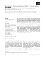
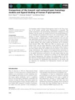
Figure 1 shows the average age of the dogs in particular
groups. The age is significantly higher at dogs showing a
degeneration of the mitral valve (p < 0.05). The average
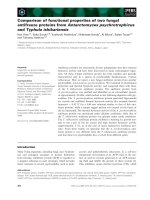
weightofthedogsinparticulargroupsisshownin
Figure 2 - dogs having the degeneration of mitra l valve
(CVD) have lower body weight than the dogs in other
groups (p < 0.05).
In all evaluated groups of dogs there were more males
then females.
Based upon the results from group I the mean values
for P
max
,P
min
,P
d
, were calculated which were accord-
ingly: P
max
- 63.4 ± 12.7 ms, P
min
- 46.6 ± 11.5 ms, P
d
-
16.8 ± 3.51 ms (range 9.2-22.6 ms, dominant = 16.2).
The correct value of P-wave dispersion of healthy dogs
was set as a mean value of P
d
± 2SD and it was less
than 24 ms.
Based on values received from healthy dogs, the
dependency of P
d
from other parameters such as: body
weight(table1),age(table2)andsex(table3)were
analyzed. No significant deviation of P
d
was noticed
according to body weight, age, and sex.
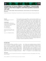
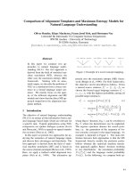
The results (P
d
) were compared between healthy dogs
(group I), dogs with mitral valve insufficiency (group II)
and dogs with supra-ventricular conduction disorders
(group III). Dogs with mitral valve insufficiency and dogs
with supra-ventricular conduction disorders had signifi-
cantly higher values of P
d
than healthy dogs (p < 0.01)
(Figure 3). The received results were also higher than
proposed norm (mean value ± SD) and were accordingly:
25,3 ± 5,1 ms (range 19.2-30.8) in group II and 24.5 ±
4.7 ms (range 15.2-30.9) in group III (table 3). The
dependency of P
d
from the level of left atria enlarge ment
were also analyzed, such as the correlation between P
d
and the LA/Ao ratio coefficient. Statistically the disper-
sion of P-wave did not differ (p = 0.86) between groups
of dogs having visible enlargement of left atria (LA/Ao
2.2 ± 1.3) or disorders of supra-ventricular conduction
(LA/Ao 1.4 ± 0.6). In the group of dogs with insufficiency
of mitral valve there were no correlations noticed with
the increase of P
d
and the level of left atria enlargement
(p = 0.1, r = 0.17). In multiple linear regression depen-
dence of P
d
from body mass, age, sex and LA/Ao was
controlled, and only P
d
is an independent parameter in
the multiple linear regression.
Discussion
In veterinary electrocardiography the gold standard is to
perform the ECG record in a recumbence, nether less
literature shows that the ECG record can be performed
also in a standing or sternum position. In many publica-
tions the ECG records were compared coming from
dogs that were in lateral recumbency or standing posi-
tion. It has been noticed that the position of the dog
does not influence the P-wave duration, P-wave ampli-
tude or PR interval [15,16]. Dogs standing position is
used also during toxico logical and pharmacological
examination. In the Hanton and Rabamampiania study
Figure 1 Avarage age of the dogs in particular groups.
Statistical significant difference (p < 0.05) between group II (n = 23)
and groups I (n = 53) and III (n = 12).
Figure 2 Aver age body mass of the dogs in particular groups.
Statistical significant difference (p < 0.05) between group II (n = 23)
and groups I (n = 53) and III (n = 12).
Table 1 P
d
in healthy dogs depending on the body mass
Body weight P
min
[ms] P
max
[ms] P
d
[ms] SD
<10 kg 36,3 52,0 15,7 4,7
10-30 kg 48,2 65,6 17,5 3,6
>30 kg 51,6 68,7 17,0 3,6
P
min
= the minimum duration of P wave, P
max
= maximum duration of P
wave, P
d
= P wave dispersion.
Noszczyk-Nowak et al. Acta Veterinaria Scandinavica 2011, 53:18
/>Page 3 of 6
it is stated that the body position of the dog during
recording of ECG had no major influence on most para-
meters. In many studies taking up the problem of the
influence of the dog’s position on the ECG record para-
meters the sufficient impact on the modified mean elec-
trical axis is underlined, what was not evaluated in this
research study [15-18]. Standing position was chosen in
this study due to lack of any documented muscle inter-
ferences with duration and amplitude of P-wave and
duration of PR interval, lower stress for the animal and
that means less heart rhythm frequency.
The frequency and degree of degeneration of atrio-ven-
tricular valves increases in older dogs. That is why the
dogs that had the insufficiency of mitral valve were, in
average, the older ones [19]. Degeneration progress with
age. In the same group of dogs the body weight was lower,
which is correlated with the predisposition of smaller and
miniature breeds to the degeneration of mitral valve [19].
No dependency between the age of healthy dogs and P
d
was noticed. A tendency for greater spread of P
d
was
observed more often in healthy dogs above 8 years old,
that was pictured by the P
d
standard deviation increase
(table 2). There is no correlation between the body weight
and P
d
in healthy dogs. There is an increase of the average
maximum and minimum duration of P-wave, correlated
with the increase of body weight, which goes together with
the increased size of the heart, particularly the size of
atrias. The ratio of these values is constant, so there are no
statistical differences betw een particular body weight
groups of healthy dogs. No correlation between sex and
the value of P
d
was noticed, even though there were more
males than females. The appearance of higher number of
males is due to preference s of the owners to have male
dogs, not due to the correlation between sex and heart dis-
orders. Received values of P
d
in healthy dogs, indepen-
dently from age, body weight and sex had small dispersion
and small standard deviation. The maximum value of P
d
in this group of dogs was 20.8 ms and was lower than
average P
d
values in the group of dogs with mitral insuffi-
ciency (group I) and dogs with supra-ventricular conduc-
tion disorders (group III).
Presented data allow to assume that P
d
value is an inde-
pendent factor from body weight and sex. It is a constant
parameter in healthy dogs, with no supra-ventricular con-
duction disturbances and changes in the atria size, result-
ing in low SD value in a big and diversified group of
healthy dogs. This allows to use the P
d
value as an inde-
pendent parameter for evaluating inter and intra-atrial
conduction.
There was noticed a significant increase in P
d
in dogs
with increased left atria due to insufficiency of the mitral
valve compared to healthy dogs. Mitral valve insufficiency
is a complex pathological process, in which takes part, in
example, the degeneration of collagen. Acid mucopolisac-
charides group around the petals of the valves which at
results in nodular thickening, deformation and weakening
of the petals which leads to valve insufficiency. Valve
insufficiency leads to the enlargement of the belonging
atria, anulus fibrosus and ventricle. In the atria appears
endocardial and atria muscular fibrosis, intraparietal
infarcts and changes in the arterial vessels caused by the
stream of regurgitation over the insufficient valve. These
processes lead to inhomogeneous propagation of the
impulses in the atria which together with the enlargement
of the atria impacts the increase o f P-wave dispersion. It
seems that Pd is more dependant from disturbances of
inter and intra-ventricular conduction and inhomoge-
neous propagation of impulses, than from the level of left
atria enlargement. Correlation wasn’t noticed between P
d
and the level of the enlargement o f left atria. Similar
results were found in humans with hypertension, who had
earlier episodes of AF or at those that had attacks of AF
shortly after P
d
measureme nts. In these tests no correla-
tion has been noticed between the value of blood pressure,
size of left atria and weight of left atria [8,19-21].
Table 2 P
d
in healthy dogs depending on the age
Age P
min
[ms] P
max
[ms] P
d
[ms] SD
<2 years 48,3 65,3 17,0 3,7
2-8 years 45,7 63 17,3 2,9
>8 years 44,3 60,5 16,2 4,6
P
min
= the minimum duration of P wave, P
max
= maximum duration of P
wave, P
d
= P wave dispersion.
Table 3 P
d
in healthy dogs depending on the sex
Sex P
min
[ms] P
max
[ms] P
d
[ms] SD
Male 47,4 64,2 17,1 4
Female 45,3 62 16,4 3,9
P
min
= the minimum duration of P wave, P
max
= maximum duration of P
wave, P
d
= P wave dispersion.
Figure 3 Average P wave dispersion (mean ± SD) in particular
groups. Statistical significant difference (p < 0.01) between group I
(n = 53) and groups II (n = 23) and III (n = 12).
Noszczyk-Nowak et al. Acta Veterinaria Scandinavica 2011, 53:18
/>Page 4 of 6
Statistically significant increase in P
d
was observed in a
group of dogs with supra-ventricular conduction disorders
compared to healthy dogs. Average value of P
d
was the
highest in this group of dogs, which is directly correlated
with improper atria conduction. The duration of P and P
d
is dependant not only from disorders in the atrium. Ane-
mia and activity of immune system can also lead to
changes in the auriculars and speed of impulse conduction
[22,23]. Infarct, dilatated cardiomyopathy, stricture of the
left atrioventricular ostium opening or congenital malfor-
mations of the heart can also lead to increased dispersion
of P-valve [23-28]. In human medicine the peculiarity and
sensitivity of P
d
has been proved and is used as a para-
meter allowing to detect patients with higher risk of
occurring or with the recurrence of atrial fibrillation [6-8].
Presented results, in this study, may also contribute to
propag ation of similar using values of P-wave dis persion
for dogs, but it still demands further research. There are
other factors, that can influ ence P
d
, that should be taken
under consideration when interpreting the results. P
d
can
increase also during endocrinology disorders such as dia-
betes and thyroid hyperfunction [29-32], but also at
patients that have the terminal phase of renal failure
[33,34]. The changes in P
d
have been noticed also in con-
nection with changes of the tension of autonomous ner-
vous system, for example while conducting Valsalva
maneuver [35,36] or in connection with panic attacks [37].
That is why it is important to interpret P
d
in connection
with other examina tion resu lts and general overview of
the patient.
Conclusions
P-wave dispersion is a constant parameter in healthy
dogs, independent from body weight, age and sex. In
dogs with inter and intra-atrial conduction disturbances
P-wave dispersion is significantly higher, that is why this
parameter can be used to evaluate the possibility of the
inter and intra-atrial conduction disturbances. In dogs
with chronic valvular disease and the atrial enlargement
the P-wave dispersion is also higher than in h ealthy
dogs, although no significant correlation between the
size of left atria and Pd was noticed. The dependency
with association between of inter and intra-atrial con-
duction disturbances with Pd in this group of dogs
demands further studies.
Authors’ contributions
ANN planned the study, carried out ECG and echocardiographic
examinations, calculated Pd and drafted the manuscript. AS calculated Pd.
UP carried out echocardiographic examinations. JN drafted the manuscript.
All authors read and approved the final manuscript.
Competing interests
The authors declare that they have no competing interests.
Received: 7 September 2010 Accepted: 11 March 2011
Published: 11 March 2011
References
1. Noszczyk-Nowak A, Pasławska U, Szałas A, Nicpoń J: P-wave dispersion in
healthy dogs. A preliminary study. Bull Vet Inst Pulawy 2008, 52:683-688.
2. Dilaveris PE, Gialafus JE: P wave dispersion: a novel predictor of
paroxysmal AF. Ann Noninvasive Electrocardiol 2001, 6:159-165.
3. Villani GQ, Piepoli M, Rosi A, Capucci A: P-wave dispersion index: a marker
of patients with paroxysmal atria fibrillation. Int J Cardiol 1996,
55:169-175.
4. Spach MS, Miller WT, Geselowitz DB, Barr RC, Kootsey JM, Johnson EA: The
discontinuous nature of propagation in normal canine cardiac muscle:
evidence for recurrent discontinuities of intracellular resistance that
affect the membrane currents. Circ Res 1981, 38:39-54.
5. Ndrepepea G, Zrenner B, Deisenhfer I, Karch M, Schneider M, Schreieck J,
Schmitt C: Relationship between surface electrocardiogram
characteristics and endocardial activation sequence in patients with
typical atrial flutter. Z Kardiol 2000, 89:527-537.
6. Dilaveris PE, Gialafos EJ, Sideris SK, Theopistou AM, Andrikopoulos GK,
Kyriakidis M, Gialafos JE, Toutouzas PK: Simple electrocardiographic
markers for the prediction of paroxysmal idiopathic atrial fibrillation. Am
Heart J 1998, 135:733-738.
7. Aytemir K, Ozer N, Atalar E, Sade E, Aksöyek S, Ovünç K, Oto A, Ozmen F,
Kes S: P wave dispersion on 12 lead electrocardiography in patients with
paroxysmal atrial fibrillation. Pacing Clin Electrophysiol 2000, 23:1109-1112.
8. Dilaveris PE, Gialafos EJ, Chrissos D, Andrikopoulos GK, Richter DJ, Lazaki E,
Gialafos JE: Detection of hypertensive patients at risk for paroxysmal
atrial fibrillation during sinus rhythm by computer-assisted P wave
analysis. J Hypertens 1999, 17:1463-1470.
9. Freeman LM, Rush JE, Farabaugh AE, Must A: Development and evaluation
of a questionnaire for assessing health-related quality of life in dogs
with cardiac disease. J Am Vet Med Assoc 2005, 226:1864-1868.
10. Koie H, Kanayama K, Sakai T, Takeuschi A: Evaluation of diagnostics
availability of continuous ANP assai and LA/AO ratio in left heart
insufficient dogs. J Vet Med Sci 2001, 63:1237-1240.
11. Bonagura JD, O’Grady MR, Herring DS: Echocardiography. Principles of
interpretation. Vet Clin North Am Small Anim Pract 1985, 15:1177-1194.
12. Takemura N, Toda N, Miyagawa Y, Asano K, Tejima K, Kanno N, Arisawa K,
Kurita T, Nunokawa K, Hirakawa A, Tanaka S, Hirose H: Evaluation of plasma
N-terminal pro-brain natriuretic peptide (NT-proBNP) concentrations in
dogs with mitral valve insufficiency. J Vet Med Sci 2009, 71:925-929.
13. Allen DG: Echocardiography as a research and clinical tool in veterinary
medicine. Can
Vet J 1982, 23:313-316.
14. Kraus MS, Moise NS, Rashniw M, Dykes N, Erb HN: Morphology of
ventricular arrhythmias in the boxer as measured by 12-lead
electrocardiography with pace-mapping comparison. J Vet Intern Med
2002, 16:153-158.
15. Coleman MG, Robson MC: Evaluation of six-lead electrocardiograms
obtained from dogs in a sitting position or sterna recumbency. Am J Vet
Res 2005, 66:233-237.
16. Rashniw M, Porciello F, Erb HN, Fruganti : Effect of body position on the
6-lead ECG of dogs. J Vet Intern Med 2002, 16:69-73.
17. Hanton G, Rabemampianina Y: The electrocardiogram of the Beagle dog:
reference values and effect of sex, genetic strain, body position and
heart rate. Lab Anim 2006, 60:123-136.
18. Pasławska U, Kurski B, Noszczyk-Nowak A, Grudziński P, Nicpoń J, Kungl K:
Analysis of mean electric al axis and amplitude of R wave in relation to
particular disorders and body position during ECG examinations [nn
Polish]. Medycyna Wet 2005, 61:1015-1017.
19. Atkins C, Bonagura J, Ettinger S, Fox P, Gordon S, Haggstrom J, Hamlin R,
Keene R, Luis-Fuentes V, Stepien R: Guidelines for the diagnosis and
treatment of canine chronic valvular heart disease. J Vet Intern Med 2009,
23:1142-1150.
20. Ciaroni S, Cuenoud L, Bloch A: Clinical study to investigate the predictive
parameters for the onset of atrial fibrillation in patients with essential
hypertension. Am Heart J 2000, 139:814-819.
21. Ozer N, Aytemir K, Atalar E, Sade E, Aksöyek S, Ovünç K, Açýl T, Nazlý N,
Ozmen F, Oto A, Kes S: P wave dispersion in hypertensive patients with
paroxysmal atrial fibrillation. Pacing Clin Electrophysiol 2000, 23:1859-1862.
Noszczyk-Nowak et al. Acta Veterinaria Scandinavica 2011, 53:18
/>Page 5 of 6
22. Dilaveris PE, Andrikopoulos GK, Metaxas G, Richter DJ, Avgeropoulou CK,
Androulakis AM, Gialafos EJ, Michaelides AP, Toutouzas PK, Gialafos JE:
Effects of ischemia on P wave dispersion and maximum P wave
duration during spontaneous angina episodes. Pacing Clin Electrophysiol
1999, 22:1640-1647.
23. Baykan M, Celik S, Erdol C, Durmuç I, Orem C, Küçükosmanoğlu M, Yilmaz R:
Effects of P wave dispersion on atrial fibrillation in patients with acute
anterior wall myocardial infarction. Ann Noninvasive Electrocardiol 2003,
8:18-21.
24. Jolda-Mydlowska B, Kobusiak-Prokopowicz M: Estimation of the P wave
and PQ interval dispersion in patients with the recent myocardial
infarction. Pol Merkuriusz Lek 2005, 18:499-502.
25. Ozer N, Yavuz B, Can I, Atalar E, Aksöyek S, Ovünç K, Ozmen F, Kes S:
Doppler tissue evaluation of intra-atrial and interatrial electromechanical
delay and comparsion with P-wave dispersion in patients with mitral
stenosis. J Am Soc Echocardiogr 2005, 18:945-948.
26. Ho TF, Chia EL, Yip WC, Chan KY: Analysis of P-wave and P dispersion in
children with secundum atrial septal defect. Ann Noninvasive Electrocardiol
2001, 6:305-309.
27. Hallioglu O, Aytemir K, Celiker A: The significance of P wave duration and
P wave dispersion for risk assessment of atrial tachyarrhytmias In
patients with corrected tetralogy of Fallot. Ann Noninvasive Electrocardiol
2004, 9:339-344.
28. Senen K, Turhan H, Riza Erbay A, Basar N, Saatci Yasar A, Sahin O, Yetkin E:
P-wave duration and P-wave dispersion in patients with dilated
cardiomyopathy. Eur J Heart Fail 2004, 6:567-956.
29. Guntekin U, Gunes Y, Simsek H, Tuncer M, Arslan S: P wave duration and
dispersion in patients with hyperthyroidism and the short-term effects
of antithyroid treatment. Indian Pacing Electrophysiol J 2009, 9:251-259.
30. Cetinarslan B, Akkoyun M, Cantürk Z, Tarkun I, Kahranman G, Komsuoglu B:
Duration of the P wave and P wave dispersion in subclinical
hyperthyroidism. Endocr Pract 2003, 9:200-203.
31. Aras D, Maden O, Ozdemir O, Aras S, Topaloglu S, Yetkin E, Demir AD,
Soylu MO, Erdogan MF, Kisacik HL, Korkmaz S: Simple electrocardiographic
markers for the prediction of paroxysmal atrial fibrillation in
hyperthyroidism. Int J Cardiol 2005, 99:59-64.
32. Yazici M, Ozdemir K, Altunkeser BB, Kayrak M, Duzenli MA, Vatankulu MA,
Soylu A, Ulgen MS: The Effect of diabetes mellitus on the P-wave
dispersion. Circ J 2007, 71:880-883.
33. Szabo Z, Kakuk G, Fülöp T, Mátyus J, Balla J, Kárpáti I, Juhász A, Kun C,
Karányi Z, Lorincz I: Effects of haemodialysis on maximum P wave
duration and P wave dispersion. Nephrol Dial Transplant 2002,
17:1634-1638.
34. Tezcan UK, Amasyali B, Can I, Aytemir K, Köse S, Yavuz I, Kursaklioglu H,
Içik E, Demirtaç E, Oto A: Increased P wave dispersion and maximum P
wave duration after hemodialysis. Ann Noninvasive Electrocardiol 2004,
9:34-38.
35. Tükek T, Akkaya V, Demirel S, Sözen AB, Kudat H, Atilgan D, Ozcan M,
Güven O, Korkut F: Effect of Valsalva maneuver on surface
electrocardiografic P wave dispersion in paroxysmal atrial fibrillation. Am
J Cardiol
2000, 85:896-899.
36. Altunkeser BB, Ozdemir K, Gok H: The effect of Valsalva maneuver on P
wave in 12-lead surface electrocardiology in patients with paroxysmal
atrial fibrillation. Angiology 2002, 53:443-449.
37. Yavuzkir M, Atmaca M, Dagli N, Balin M, Karaca I, Mermi O, Tezcan E,
Aslan IN: P-wave dispersion in panic disorder. Psychosom Med 2007,
69:344-347.
doi:10.1186/1751-0147-53-18
Cite this article as: Noszczyk-Nowak et al.: Comparison of P-wave
dispersion in healthy dogs, dogs with chronic valvular disease and dogs
with disturbances of supraventricular conduction. Acta Veterinaria
Scandinavica 2011 53:18.
Submit your next manuscript to BioMed Central
and take full advantage of:
• Convenient online submission
• Thorough peer review
• No space constraints or color figure charges
• Immediate publication on acceptance
• Inclusion in PubMed, CAS, Scopus and Google Scholar
• Research which is freely available for redistribution
Submit your manuscript at
www.biomedcentral.com/submit
Noszczyk-Nowak et al. Acta Veterinaria Scandinavica 2011, 53:18
/>Page 6 of 6