Báo cáo y học: "Association of lipid peroxidation with hepatocellular injury in preterm infan" docx
Bạn đang xem bản rút gọn của tài liệu. Xem và tải ngay bản đầy đủ của tài liệu tại đây (77.5 KB, 5 trang )
Available online />Research
Association of lipid peroxidation with hepatocellular
injury in preterm infants
Barry Weinberger
1
, Kazimierz Watorek
1
, Richard Strauss
2
, Gisela Witz
3
, Mark Hiatt
1
and Thomas Hegyi
1
1
Neonatologist, University of Medicine and Dentistry of New Jersey–Robert Wood Johnson Medical School, New Brunswick, NJ, USA
2
Gastroenterologist, University of Medicine and Dentistry of New Jersey–Robert Wood Johnson Medical School, New Brunswick, NJ, USA
3
Professor of Environmental and Community Medicine, University of Medicine and Dentistry of New Jersey-Robert Wood Johnson Medical School,
New Brunswick, NJ, USA
Correspondence: Barry Weinberger,
Introduction
The incidence of cholestasis related to total parenteral nutri-
tion (TPN) among preterm infants has been estimated to be
between 7% and 85%, depending on the population exam-
ined and the definition of cholestasis used [1]. In infants with
necrotizing enterocolitis or short bowel syndrome, the preva-
lence of TPN-related cholestasis is 60–90% [2]. Although
cholestasis is reversible in most patients after the successful
advancement of enteral feeding, progressive liver fibrosis and
cirrhosis occur in some patients even after complete enteral
521
ALT = alanine transaminase; AST = aspartate transaminase; MDA = malondialdehyde; ROIs = reactive oxygen intermediates; TBA = thiobarbituric
acid; TBARS = thiobarbituric-acid-reacting substances; TPN = total parenteral nutrition.
Abstract
Introduction We wished to determine whether cholestasis induced by total parenteral nutrition (TPN)
in preterm newborn infants is associated with increased oxidative stress secondary to increased
reactive oxygen intermediates. We hypothesized that elevated urinary thiobarbituric-acid-reacting
substances (TBARS), a marker of oxidative stress, would be associated with hepatocellular injury as
measured by serum alanine transaminase (ALT) and aspartate transaminase (AST) levels.
Materials and methods Preterm infants (< 35 weeks’ gestation) admitted to the neonatal intensive
care unit were enrolled (with their parents’ informed consent) in either the ‘cholestasis’ group (if their
direct bilirubin was > 2 mg/dl [34.2 µmol/l] and duration of TPN was ≥10 days [n = 27]) or in the
control group. Urine samples for measurement of TBARS (proportionate to lipid peroxidation) and
blood specimens for analysis of serum bilirubin, ALT, AST, and alkaline phosphatase were obtained
within 24 hours of enrollment.
Results The cholestasis and control groups were comparable with respect to gestational age, birth
weight, Apgar score, maximum F
i
O
2
, and duration of supplemental oxygen administration. Median
serum direct bilirubin concentrations in the cholestasis and control groups were, respectively,
3.3 mg/dl (56.4 µmol/l) and 1.7 mg/dl (29.1 µmol/l) (P < 0.001). Serum ALT and AST levels were also
elevated in the cholestasis group, but alkaline phosphatase levels did not differ significantly between
the groups. Urinary levels of TBARS in all the infants were correlated with ALT and AST but did not
differ significantly between cholestatic and control infants.
Discussion Our findings suggest that oxidant stress is associated with hepatocellular injury in preterm
infants. This effect is not correlated with the degree of cholestasis.
Keywords cholestasis, newborn, oxidant, peroxidation, premature
Received: 2 January 2002
Revisions requested: 26 February 2002
Revisions received: 20 June 2002
Accepted: 7 August 2002
Published: 21 August 2002
Critical Care 2002, 6:521-525 (DOI 10.1186/cc1547)
This article is online at />© 2002 Weinberger et al., licensee BioMed Central Ltd
(Print ISSN 1364-8535; Online ISSN 1466-609X). This article is
published in Open Access: verbatim copying and redistribution of this
article are permitted in all media for any non-commercial purpose,
provided this notice is preserved along with the article's original URL.
Open Access
522
Critical Care December 2002 Vol 6 No 6 Weinberger et al.
nutrition has been established [3]. Some studies have sug-
gested that excessive amino acids, hepatotoxic bile acids,
bacterial overgrowth, sepsis, micronutrient deficiency, and
TPN contaminants all contribute to cholestatic liver injury
[4–6]. Diminished volume of enteral feeds may also indepen-
dently contribute to the development of cholestasis.
However, the mechanisms of liver injury in cholestatic dis-
eases in infants remain unclear.
Recent studies have supported the hypothesis that genera-
tion of reactive oxygen intermediates (ROIs) and elevated
lipid hydroperoxides in the liver during cholestasis cause
tissue injury. Exposure of isolated hepatocytes to hydropho-
bic bile acids leads to intracellular production of oxygen free
radicals and lipid peroxides [7]. Animal studies using models
of surgically induced extrahepatic biliary obstruction have
also shown that lipid peroxidation products — specifically,
malondialdehyde (MDA) — are increased in the cholestatic
liver [8,9]. This increase is associated with decreased tissue
antioxidant activity, increased leukocyte infiltration, and early
evidence of collagen deposition; these effects are amelio-
rated by the administration of exogenous antioxidants [8–10].
Consistent with these findings, several forms of liver disease
in humans have been shown to be associated with oxidative
tissue injury. Specifically, products of lipid peroxidation are
elevated in patients with hepatic hypoxia/reperfusion,
obstructive liver disease, alcoholic liver disease, Wilson’s
disease, Alagille syndrome, sepsis, and inflammatory liver dis-
eases [11–15]. In vitro, procollagen gene expression
increases in human liver cells after exposure to lipid peroxide
breakdown products [16]. Furthermore, deposition of lipofus-
cin, a by-product of lipid peroxidation, characterizes TPN-
induced cholestasis in preterm infants [17].
The purpose of these studies was to determine whether TPN-
induced cholestasis in preterm newborn infants is associated
with increased oxidative stress secondary to increased ROIs.
We hypothesized that elevation in markers of oxidative stress
would be associated with increased liver injury, as measured
by serum alanine transaminase (ALT) and aspartate transami-
nase (AST) levels. In order to quantify ROIs in infants, we
measured urinary thiobarbituric-acid-reacting substances
(TBARS). The most abundant of these substances is MDA,
an aldehydic lipid peroxidation product formed by the action
of ROIs on lipid membranes. The identification of ROIs as
potential markers of liver injury in cholestatic preterm infants
may aid in the diagnosis and management of those at highest
risk for ongoing liver impairment.
Materials and methods
Patients
All preterm infants (< 35 weeks’ gestation) born at St Peter’s
University Hospital (New Brunswick, NJ, USA) and admitted
to the hospital’s neonatal intensive care unit between March
1997 and December 1998 were serially screened during the
course of hospitalization for entry into the ‘cholestasis’ study
group (Fig. 1). Infants with major congenital anomalies
(including all gastrointestinal and liver anomalies) or with con-
genital or acquired infection were excluded. The criteria for
entry into the cholestasis group were direct bilirubin
> 2 mg/dl (34.2 µmol/l) and duration of TPN ≥10 days. If the
infant met these criteria, the parents’ informed consent for
entry into the study was requested. During this period, 36
infants qualified for the cholestasis group, and informed
consent was obtained from the parents of 27. When each eli-
gible infant with cholestasis was enrolled, a preterm infant
without cholestasis was matched for gestational age, birth
weight, and severity of respiratory illness, and the parents’
informed consent was requested to enroll the infant as a
control subject. For the 27 infants enrolled in the cholestasis
group, matched controls were identified for 24. Parental
consent could not be obtained for 8, so 16 infants consti-
tuted the control group. Study personnel obtained demo-
graphic and medical information from infants’ medical records.
Determination of urinary thiobarbituric-acid-reacting
substances
Urine samples were obtained under sterile conditions from all
enrolled infants at the time of their entry into the study.
Informed consent was obtained from parents for the acquisi-
tion of samples, and these studies were approved by the
Committee for the Protection of Human Subjects in Research
of St Peter’s University Hospital. TBARS were measured as
previously described [18,19]. Briefly, 200 µl of urine was
combined with 10 µl of 5% butylated hydroxytoluene (in
glacial acetic acid) and 300 µl of a 0.5% aqueous thiobarbi-
turic acid (TBA) solution. The samples were vortexed and
were incubated at 100°C for 30 minutes, and the absorbance
at 532 nm was measured using a Perkin Elmer Lamba 3B
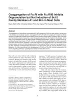
Figure 1
Patient recruitment. Preterm infants born at St Peter’s University
Hospital (New Brunswick, NJ, USA) who were admitted to the
hospital’s neonatal intensive care unit between March 1997 and
December 1998 were serially screened during hospitalization for entry
into the ‘cholestasis’ study group. Controls were matched for
gestational age, birth weight, and severity of respiratory illness. TPN,
total parenteral nutrition.
SCREEN:
Inborn, < 35 weeks gestation
No major anomalies or
congenital infection
n = 804
QUALIFIED for Cholestasis Group
n = 36
ENROLLED in Cholestasis Group
n = 27
Informed consent
Direct Bilirubin > 2 mg/dl
TPN ≥ 10 days
QUALIFIED for Control Group
n = 24
ENROLLED in Control Group
n = 16
Informed consent
M
a
t
c
h
523
spectrophotometer (PerkinElmer, Wellesley, MA, USA). The
quantity of TBARS is proportionate to the amount of MDA, a
lipid peroxidation product generated by the oxidation of mem-
brane lipids by reactive oxygen species. MDA reacts with
TBA to form a 1 : 2 MDA–TBA adduct that absorbs at
532 nm. In the present studies, MDA was confirmed to be the
predominant TBA-reacting adduct by high-performance liquid
chromatography analysis of representative samples. To
control for urine concentration, data were normalized to urine
creatinine concentrations, as previously described [20].
Determination of serum bilirubin, ALT, AST, alkaline
phosphatase
Blood specimens were obtained from subjects within
24 hours of the urine specimen. Quantitative determinations
of serum bilirubin, ALT, AST, and alkaline phosphatase levels
were performed by the clinical laboratory at St Peter’s Univer-
sity Hospital.
Statistical analysis
Direct bilirubin, ALT, AST, alkaline phosphatase, and urinary
TBARS values were not normally distributed. The data are
presented as median (25th, 75th percentile). Differences
between the groups were analyzed for significance by one-
way ANOVA using natural log transformations of the data,
which are normally distributed. Differences were regarded as
statistically significant at P values ≤ 0.05. Correlations of
bilirubin, ALT, and AST with TBARS were calculated by
regression using the log-transformed values to ensure normal
distribution of all variables in those analyses.
Results
Twenty-seven infants were enrolled in the cholestasis group
and 16 infants served as controls. The cholestasis and
control groups were not significantly different with respect to
gestational age (29.3 ± 4.7 vs 27.1 ± 3.2 weeks, respec-
tively) and birth weights (1276 ± 751 vs 1016 ± 392 g), as
well as Apgar scores, maximum F
i
O
2
, and length of time for
which supplemental oxygen was given (Table 1). Urine
samples were collected at 48.3 ± 38.2 days of age in the
cholestasis group and 38.4 ± 22.1 days in the control group
(P = 0.34). At that time, infants in the cholestasis group had
been advancing on enteral feedings in addition to parenteral
nutrition for 11.3 ± 5.5 days (range 0–23 days) and were
receiving 21.4 ± 12.3 ml/kg per day enterally. Control infants
were on full enteral feedings at the time of study.
Median serum direct bilirubin concentrations were 3.3 mg/dl
(56.4 µmol/l) in the cholestasis group and 1.7 mg/dl
(29.1 µmol/l) in the control group (P < 0.001). Median serum
ALT and AST levels were also elevated in the cholestasis
group (32 vs 9 and 71 vs 33 U/l, respectively; P < 0.01).
Values for alkaline phosphatase and mean urinary TBARS did
not differ significantly between the groups (Table 2). Urinary
TBARS were not significantly correlated with gestational age,
gender, days on TPN, indirect bilirubin, or alkaline phos-
phatase (not shown). Likewise, urinary TBARS were not
Available online />Table 1
Demographic variables of infants studied
Infants
With cholestatis Controls
Variable (n = 27) (n = 16) P
Birthweight (g) 1276 ± 751 1016 ± 392 0.62
Gestational age (weeks) 29.3 ± 4.7 27.1 ± 3.2 0.59
Apgar score (5 minutes) 7.0 ± 2.0 8.1 ± 1.3 0.07
Gender (males/females) 18/8 13/4
Maximum F
i
O
2
0.63 ± 0.31 0.58 ± 0.31 0.59
Supplemental O
2
(days) 55.7 ± 54.1 44.4 ± 36.9 0.46
TPN (days) 59.6 ± 65.6* 26.5 ± 17.0 0.04
Values are expressed as means ± standard deviation. TPN = total
parenteral nutrition. * Significantly different from control group.
Table 2
Indicators of hepatocellular injury and urinary thiobarbituric-acid-reacting substances (TBARS) in preterm infants studied
Infants
With cholestatis Controls
Variable (n = 27) (n = 16) P
Direct bilirubin (mg/dl) 3.3 (2.4,7.2)* 1.7 (1.0,1.9) <0.001
ALT (U/l) 32 (8,127)* 9 (7,16) 0.01
AST (U/l) 71 (40,189)* 33 (22,39) <0.001
Alkaline phosphatase (U/l) 383 (221,579) 269 (199,450) 0.57
Urinary TBARS 2591 (1022,6445) 3368 (1622,4625) 0.93
(ng/mg creatinine)
Values are expressed as medians (25th,75th percentiles). *Significantly different from control group (P < 0.05 using one-way analysis of variance
of natural log-transformed variables).
524
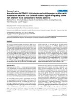
correlated with direct bilirubin (Fig. 2, top). In contrast, urinary
TBARS levels among all infants were independently corre-
lated with serum ALT and AST (Fig. 2, lower).
Discussion
We found that elevated liver transaminases are associated
with increased oxidative stress. These findings suggest that
oxidant stress (as indicated by elevated TBARS) is associ-
ated with hepatocellular injury in preterm infants. Although
there is ample evidence that oxidant stress follows cholesta-
sis, our findings suggest that oxidative injury in the liver may
be induced by mechanisms that are independent of cholesta-
sis [7–11]. For example, the production of ROIs in the liver
may be linked to inflammation, which has emerged as a
primary mechanism of liver injury after pathophysiological
insults. Activated Kupffer cells and neutrophils release ROIs
and proteases in response to inflammatory cytokines in the
liver [21]. ROIs in excess inactivate proteins, disrupt DNA,
and oxidize lipids [22]. Preterm newborn infants may be par-
ticularly susceptible to such injury because they exhibit an
imbalance between antioxidant- and oxidant-generating
systems. For example, such infants exhibit decreased levels in
the liver of superoxide dismutase, vitamin E, and β-carotene
[23–25]. Antioxidant capacity in preterm infants is also com-
promised by relative deficiencies of selenium and taurine, as
well as reduced ability to synthesize sufficient glutathione [26].
Despite longer TPN courses and elevated serum transami-
nases, infants with cholestasis or elevated serum direct biliru-
bin did not display elevated urinary TBARS. Our findings are
consistent with previous reports indicating an inconsistent
relation between bilirubin levels and the degree of histologic
liver injury [27,28]. This inconsistency suggests that cholesta-
sis and hepatic impairment in preterm infants receiving TPN
are induced by mechanisms that are not dependent on
oxidant-mediated hepatocellular injury. For example, lack of
enteral feeding plays an important role in the development of
cholestasis in the neonatal period, possibly mediated by
direct hepatotoxic activity of bile salts [5]. Bile salts are
thought to exert adverse effects on signal transduction and
gene transcription in hepatocytes and cholangiocytes and to
activate Fas-induced apoptosis [21]. TPN-induced cholesta-
sis in infants also appears to be related directly to develop-
mental immaturity of bile flow and production and to possible
infection [29].
TBARS measurements provide a measure of membrane lipid
peroxidation and, as such, may provide a direct assessment
of the progression of liver injury at the cellular level. Although
there was a statistically significant relationship between
urinary TBARS and liver transaminases, a large degree of
overlap existed between the groups. Furthermore, the associ-
Critical Care December 2002 Vol 6 No 6 Weinberger et al.
Key messages
• Serum transaminases are elevated in preterm infants
(< 35 weeks’ gestational age) with cholestasis and
prolonged administration of total parenteral nutrition
(TPN), indicating that cholestasis is associated with
hepatocellular injury
• Urinary levels of thiobarbituric-acid-reacting
substances (TBARS), which are proportional to lipid
peroxidation and oxidant stress, are correlated with
serum transaminase levels. Our findings suggest that
oxidant stress is associated with hepatocellular injury
in preterm infants
• Urinary levels of TBARS do not differ significantly
between infants with cholestasis and control infants
• Our findings suggest that cholestasis in preterm
infants receiving TPN is induced, in large part, by
mechanisms that are not dependent on
oxidant-mediated hepatocellular injury
Figure 2
Correlation of serum direct bilirubin and transaminase levels with
urinary TBARS. Urine and serum specimens were obtained from
cholestatic (n = 27) and control (n = 16) infants, as described in
Materials and methods. Urinary TBARS levels of infants (n = 43) were
plotted against (top) serum direct bilirubin (NS; analysis done after
exclusion of three outlying points) or (bottom) ALT (r = 0.43, P = 0.04)
or AST (r = 0.44, P = 0.02). Natural logarithm transformations of
variables are used, and regression lines are displayed. ALT, alanine
transaminase; AST, aspartate transaminase; TBARS, thiobarbituric-
acid-reacting substances.
5
6
7
8
9
10
0 2 4 6
Serum ALT or AST (log
n
U/l)
AST
AL
T
-0.5 0 1 2
5
6
7
8
9
10
Serum Direct Bilirubin (log
n
mg/dl)
Urinary TBARS (log
n
ng/mg creatinine)
525
ation between TBARS levels and liver injury does not neces-
sarily indicate causality. Despite physiologic evidence that
ROIs play a central role in tissue injury during inflammation, it
is possible that elevated TBARS occur secondary to other
mechanisms of hepatocellular injury [30,31]. Larger longitudi-
nal and/or interventional (e.g. antioxidant administration)
studies will be necessary to determine whether there is a
causal relationship between lipid peroxidation and TPN-
induced liver disease in preterm infants.
Competing interests
None declared.
References
1. Bell RL, Ferry GD, Smith EO, Shulman RJ, Christensen BL,
Labarthe DR, Wills CA: Total parenteral nutrition-related
cholestasis in infants. J Parenter Enteral Nutr 1986, 10:356-359.
2. Caniano DA, Starr J, Ginn-Pease ME: Extensive short-bowel
syndrome in neonates: outcome in the 1980’s. Surgery 1989,
105:119-124.
3. Dahms BB, Halpin TC: Serial liver biopsies in parenteral nutri-
tion-associated cholestasis in early infancy. Gastroenterology
1981, 81:136-144.
4. Vileises RA, Inwood RJ, Hunt CE: Prospective controlled study
of parenteral nutrition-associated cholestatic jaundice: effect
of protein intake. J Pediatr 1980, 96:893.
5. Brown MR, Thunberg BJ, Golub L, Maniscalco WM, Cox C,
Shapiro DL: Decreased cholestasis with enteral instead of
intravenous protein in the very low-birth weight infant. J Ped
Gastroenerol Nutr 1989. 9: 21-27.
6. Manginello FP, Javitt NB: Parenteral nutrition and neonatal
cholestasis. J Pediatr 1979, 94:296-298.
7. Sokol RJ, Winklhofer-Roob BM, Devereaux MW, McKim JM Jr.:
Generation of hydroperoxides in isolated rat hepatocytes and
hepatic mitochondria exposed to hydrophobic bile acids. Gas-
troenterology 1995, 109:1249-1256.
8. Parola M, Leonarduzzi G, Robino G, Albano E, Poli G, Dianzani
MU: On the role of lipid peroxidation in the pathogenesis of
liver damage induced by long-standing cholestasis. Free Rad
Biol Med 1996, 20:351-359.
9. Orellana M, Rodrigo R, Thielemann L, Guajardo V: Bile duct liga-
tion and oxidative stress in the rat: effects in liver and kidney.
Comp Biochem Physiol Part C 2000, 126:105-111.
10. Krahenbuhl S, Talos C, Lauterburg BH, Reichen J: Reduced
antioxidative capacity in liver mitochondria from bile duct
ligated rats. Hepatology 1995, 22:607-612.
11. Tsai LY, Lee KT, Tai SM, Lee SC, Yu HS: Changes of lipid per-
oxide levels in blood and liver tissue of patients with obstruc-
tive jaundice. Clin Chim Acta 1993, 215:41-50.
12. Bjorneboe A, Bjorneboe GE: Antioxidant status and alcohol-
related diseases. Alcohol Alcohol 1993, 28:111-116.
13. Sokol RJ, Twedt D, McKim JM Jr, Devereaux MW, Karrer FM, Kam
I, von Steigman G, Narkewicz MR, Bacon BR, Britton RS:
Oxidant injury to hepatic mitochondria in patients with
Wilson’s disease and Bedlington terriers with copper toxico-
sis. Gastroenterology 1994, 107:1788-1798.
14. Davit-Spraul A, Cosson C, Couturier M, Hadchouel M, Legrand A,
Lemonnier F, Therond P: Standard treatment of
αα
-tocopherol in
Alagille patients with severe cholestasis is insufficient. Pediatr
Res 2001, 49:232-236.
15. Fabris C, Pirisi M, Panozzo MP, Soardo G, Toniutto P, Hocza V,
Bartoli E: Intensity of inflammatory damage and serum lipid
peroxide concentrations in liver disease. J Clin Path 1993, 46:
364-367.
16. Parola M, Pinzani M, Casini A, Albano E, Poli G, Gentilini A, Gen-
tilini P, Dianzani MU: Stimulation of lipid peroxidation or 4-
hydroxynonenal treatment increases procollagen
αα
1(I) gene
expression in human liver fat-storing cells. Biochem Biophys
Res Commun 1993, 194:1044-1050.
17. Berger HM, Den Ouden AL, Calame JJ: Pathogenesis of liver
damage during parenteral nutrition: is lipofuscin a clue? Arch
Dis Child 1985, 60:774-776.
18. Buege JA, Aust SD: Microsomal lipid peroxidation. Methods
Enzymol 1978, 52:302-310.
19. Valenzuela A: The biological significance of malondialdehyde
determination in the assessment of tissue oxidative stress.
Life Sci 1991, 48:301-309.
20. Coulthard MG, Hey EN, Ruddock V: Creatinine and urea clear-
ances compared to inulin clearance in preterm and mature
babies. Early Hum Dev 1985, 11:11-19.
21. Jaeschke H, Gores GJ, Cederbaum AI, Hinson JA, Pessayre D,
Lemasters JJ: Mechanisms of hepatotoxicity. Toxicol Sci 2002,
65:166-176.
22. Davis KJA: Oxidative damage and repair: introduction and
overview. In Oxidative Damage and Repair: Chemical, Biological,
and Medical Aspects. Edited by Davis KJA. Elmsford, NY: Perga-
mon Press, 1991:xvii-xxvii.
23. McElroy MC, Postle AD, Kelly KG: Catalase, superoxide dismu-
tase and glutathione peroxidase activities of lung and liver
during human development. Biochim Biophys Acta 1992,
1117:153-158.
24. Lindeman JH, van Zoeren-Grobben D, Schrijver J, Speek AJ,
Poorthuis BJ, Berger HM: The total free radical trapping ability
of cord blood plasma in preterm and term babies. Pediatr Res
1989, 26:20-24.
25. Sullivan JL, Newton RB: Serum antioxidant activity in neonates.
Arch Dis Child 1988, l63:748-757.
26. Tubman TR, Halliday HL, McMaster D: Glutathione peroxidase
and selenium levels in the preterm infant. Biol Neonate 1990,
58:305-310.
27. Moss RL, Das JB, Raffensberger JG: Total parenteral nutrition-
associated cholestasis: clinical and histopathologic correla-
tion. J Pediatr Surg 1993, 28:1270-1274.
28. Beath SV, Davies P, Papadopoulou A, Khan AR, Buick RG,
Corkery JJ, Gornall P, Booth IW: Parenteral nutrition-related
cholestasis in postsurgical neonates: multivariate analysis of
risk factors. J Pediatr Surg 1996, 31:604-606.
29. Shulman RJ: New developments in total parenteral nutrition for
children. Curr Gastroenterol Rep 2000, 2:253-258.
30. Alric L, Orfila C, Carrere N, Beraud M, Carrera G, Lepert JC,
Duffaut M, Pipy B, Vinel JP: Reactive oxygen intermediates and
eicosanoid production by Kupffer cells and infiltrated
macrophages in acute and chronic liver injury induced in rats
by CCl
4
. Inflamm Res 2000, 49:700-707.
31. Laskin DL, Pendino KJ: Macrophages and inflammatory media-
tors in tissue injury. Annu Rev Pharmacol Toxicol 1995, 35:655-
677.
Available online />