Báo cáo y học: "Science review: Redox and oxygen-sensitive transcription factors in the regulation of oxidant-mediated lung injury: α role for hypoxia-inducible factor-1α" pptx
Bạn đang xem bản rút gọn của tài liệu. Xem và tải ngay bản đầy đủ của tài liệu tại đây (134.28 KB, 8 trang )
47
AM = alveolar macrophage; CF = cystic fibrosis; EPO = erythropoietin; HIF-1 = hypoxia-inducible factor-1; HS = hemorrhagic shock; IL = inter-
leukin; NF-κB = nuclear factor-κB; pO
2
= partial pressure of oxygen; redox = reduction–oxidation; ROS = reactive oxygen species; SLPI = secre-
tory leukocyte protease inhibitor; TNF = tumor necrosis factor; VEGF = vascular endothelial growth factor.
Available online />Altering gene expression is the most fundamental and effec-
tive way for a cell to respond to extracellular signals and/or
changes in its environment, in both the short term and the
long term [1]. In the short term, transcription factors are
involved in mediating responses to growth factors and a
variety of other extracellular signals [2]. In contrast, the long-
term control of gene expression induced by growth factors
and the changes in gene expression, which occur during
development, is generally (with few exceptions) irreversible.
During development, the expression of specific sets of
genes is regulated spatially (by position/morphogenetic gra-
dients) and temporally. Regulation of the signaling
responses is governed at the genetic level by transcription
factors that bind to control regions of target genes and alter
their expression [1,2]. Transcription factors are endogenous
substances, usually proteins, that are effective in the initia-
tion, stimulation or termination of the genetic transcription
process. While in the cytoplasm, the transcription factor is
incapable of promoting transcription. A signaling event
occurs, such as a change of the state of phosphorylation,
which results in protein subunit translocation into the
nucleus [3,4]. Transcription is a process in which one DNA
strand is used as a template to synthesize a complementary
RNA. Signal transduction therefore involves complex inter-
actions of multiple cellular pathways [1,2].
Review
Science review: Redox and oxygen-sensitive transcription factors
in the regulation of oxidant-mediated lung injury:
role for hypoxia-inducible factor-1
αα
John J Haddad
Severinghaus-Radiometer Research Laboratories, Molecular Neuroscience Research Division, Department of Anesthesia and Perioperative Care,
University of California at San Francisco, School of Medicine, San Francisco, California, USA
Correspondence: John J Haddad,
Published online: 14 October 2002 Critical Care 2003, 7:47-54 (DOI 10.1186/cc1840)
This article is online at />© 2003 BioMed Central Ltd (Print ISSN 1364-8535; Online ISSN 1466-609X)
Abstract
A progressive rise of oxidative stress due to altered reduction–oxidation (redox) homeostasis appears
to be one of the hallmarks of the processes that regulate gene transcription in physiology and
pathophysiology. Reactive oxygen species and reactive nitrogen species serve as signaling
messengers for the evolution and perpetuation of the inflammatory process that is often associated
with the condition of oxidative stress, which involves genetic regulation. Changes in the pattern of gene
expression through reactive oxygen species/reactive nitrogen species-sensitive regulatory transcription
factors are crucial components of the machinery that determines cellular responses to oxidative/redox
conditions. The present review describes the basic components of the intracellular oxidative/redox
control machinery and its crucial regulation of oxygen-sensitive and redox-sensitive transcription factors
within the context of lung injury. Particularly, the review discusses mechanical ventilation and NF-κB-
mediated lung injury, ischemia-reperfusion and transplantation, compromised host defense and
inflammatory stimuli, and hypoxemia and the crucial role of hypoxia-inducible factor in mediating lung
injury. Changes in the pattern of gene expression through regulatory transcription factors are therefore
crucial components of the machinery that determines cellular responses to oxidative/redox stress.
Keywords antioxidant, hypoxia-inducible factor-α, injury, lung, oxygen, redox, transcription factors
48
Critical Care February 2003 Vol 7 No 1 Haddad
In particular, reduction–oxidation/oxygen (redox)-sensitive
transcription factors have gained an overwhelming backlog of
interest momentum over the years, ever since the onset of the
burgeoning field of free radical research and oxidative stress.
The reason for this is that redox-sensitive transcription factors
are often associated with the development and progression
of many human disease states. Their ultimate regulation
therefore bears potential therapeutic intervention for possible
clinical applications [1–4].
In the present review, I will focus on elaborating a compre-
hensive overview of the current understanding of redox/
oxidative mechanisms mediating the regulation of transcrip-
tion factors. These transcription factors regulate a plethora
of cellular functions that span the range from anoxia and
hypoxia to oxidative stress within the context of oxidant-medi-
ated lung injury.
Inflammatory reactions and lung injury
Mechanical ventilation and NF-
κκ
B-mediated lung injury
Some unprecedented conditions may occur during the evolu-
tion of the inflammatory process, which can eventually lead to
dramatic changes in the progression of lung injury. For
example, positive-pressure mechanical ventilation supports
gas exchange in patients with respiratory failure but is also
responsible for significant lung injury.
Pugin and colleagues, for instance, have developed an in
vitro model in which isolated lung cells can be submitted to
a prolonged cyclic pressure-stretching strain resembling that
of conventional mechanical ventilation [5]. In this model,
cells cultured on a silastic membrane were elongated up to
7% of their initial diameter, corresponding to a 12% increase
in cell surface. The lung alveolar macrophage (AM) was iden-
tified as the main cellular source for critical inflammatory
mediators such as tumor necrosis factor (TNF)-α, the
chemokines IL-8 and IL-6, and matrix metalloproteinase-9 in
this model system of mechanical ventilation. These media-
tors were measured in supernatants from ventilated AMs,
monocyte-derived macrophages and promonocytic THP-1
cells. In addition, NF-κB was found to be activated in venti-
lated macrophages. Synergistic proinflammatory effects of
mechanical stress and molecules such as bacterial endo-
toxin were observed, suggesting that mechanical ventilation
might be particularly deleterious in pre-injured or infected
lungs. Dexamethasone, an anti-inflammatory steroid, pre-
vented IL-8 and TNF-α secretion in ventilated macrophages.
Mechanical ventilation also induced low levels of IL-8 secre-
tion by alveolar type II-like cells. Other lung cell types such
as endothelial cells, bronchial cells and fibroblasts failed to
produce IL-8 in response to a prolonged cyclic pressure-
stretching load [5]. This model is of particular value for explor-
ing physical stress-induced signaling pathways, as well as for
testing the effects of novel ventilatory strategies or adjunctive
substances aimed at modulating cell activation induced by
mechanical ventilation.
Furthermore, alterations in AM function during sepsis-induced
hypoxia may influence TNF secretion and the progression of
acute lung injury. It was proposed that acute changes in
partial pressure of oxygen (pO
2
) tension surrounding AMs
alter NF-κB activation and TNF secretion in these lung cells.
AM-derived TNF-α secretion and NF-κB expression were
determined after acute hypoxic exposure of isolated
Sprague–Dawley rat AMs. Adhered AMs (10
6
/ml) were incu-
bated (37°C at 5% CO
2
) for 2 hours with 1 µg/ml
lipopolysaccharide–endotoxin (Pseudomonas aeruginosa) in
normoxia (21% O
2
–5% CO
2
) or in hypoxia (1.8% O
2
–5%
CO
2
). The AMs exposed to lipopolysaccharide–endotoxin in
hypoxia had higher levels of TNF-α and enhanced expression
of NF-κB than those in normoxia; the predominant isoforms
were RelA (p65) and c-Rel (p75). Increased mRNA bands for
TNF-α, IL-1α and IL-1β were also observed in the hypoxic
AMs [6]. This observation demonstrates that acute hypoxia in
the lung may induce enhanced NF-κB activation in AMs,
which may result in increased production and release of
inflammatory cytokines.
Ischemia-reperfusion and transplantation
It has been reported that secretory leukocyte protease
inhibitor (SLPI) in mice regulates local and remote organ
inflammatory injury induced by hepatic ischemia-reperfusion
[7–9]. Intravenous infusion of SLPI reduced liver and lung
damage and diminished neutrophil accumulation in both
organs. These effects were accompanied by reduced serum
levels of TNF-α and macrophage inflammatory protein-2. SLPI
also suppressed activation of NF-κB in the liver. Moreover,
hepatic ischemia and reperfusion caused increased expres-
sion of SLPI mRNA and SLPI protein, which was found
specifically in hepatocytes. Furthermore, treatment of mice
with anti-SLPI antibodies enhanced serum levels of TNF-α
and macrophage inflammatory protein-2, and it increased
hepatic neutrophil accumulation and the amount of liver injury
and lung injury [7–13]. These data indicate that SLPI has pro-
tective effects against hepatic ischemia-reperfusion injury and
suggest that endogenous SLPI regulates the hepatic and
remote inflammatory responses.
In concert with these observations, attenuation of lung reper-
fusion injury after transplantation using an inhibitor of NF-κB
was achieved [14]. It was hypothesized that NF-κB is a criti-
cal early regulator of the inflammatory response in lung
ischemia-reperfusion injury and that inhibition of NF-κB acti-
vation reduces this injury and improves pulmonary graft func-
tion. With the use of a porcine transplantation model, left
lungs were harvested and stored in cold Euro-Collins preser-
vation solution for 6 hours before transplantation [14]. Activa-
tion of NF-κB occurred 30 min and 1 hour after
transplantation, and it declined to near baseline levels after
4 hours. Pyrrolidine dithiocarbamate, a potent inhibitor of NF-
κB, given to the lung graft during organ preservation
(40 mmol/l), effectively inhibited NF-κB activation and signifi-
cantly improved lung function. Compared with control lungs
49
4 hours after transplant, pyrrolidine dithiocarbamate-treated
lungs displayed significantly higher oxygenation, lower pCO
2
,
reduced mean pulmonary arterial pressure and reduced
edema and cellular infiltration [14]. This demonstrates that
NF-κB is rapidly activated and is associated with poor pul-
monary graft function in transplant reperfusion injury. Target-
ing the NF-κB pathway may therefore be a promising therapy
to reduce injury and to improve lung function.
Compromised host defense
Progressive pulmonary infection may be a prominent clinical
feature of lung injury, but the molecular basis for this suscep-
tibility remains incompletely understood.
To study this problem, Sajjan et al. developed a model of
chronic pneumonia by repeated instillation of a clinical isolate
of Burkholderia cepacia, an opportunistic Gram-negative
bacterium, from a case of cystic fibrosis (CF) into the lungs of
Cftr (m1unc
–/–
[Cftr
–/–
]) and congenic Cftr
+/+
controls [15].
Nine days after the last instillation, the CF transmembrane
regulator knockout mice showed persistence of viable bacte-
ria with chronic severe bronchopneumonia, while wild-type
mice remained healthy. A mixed population of macrophages
and neutrophils characterized the histopathological changes
in the lungs of the susceptible Cftr
–/–
mice by infiltration of a
mixed inflammatory cell population into the peribronchiolar
and perivascular spaces, by Clara cell hyperplasia, by mucus
hypersecretion in the airways and by exudation into alveolar
airspaces. An increased proportion of neutrophils was
observed in the bronchoalveolar lavage fluid from the Cftr
–/–
mice that, despite an increased bacterial load, demonstrated
minimal evidence of activation. In addition, alveolar
macrophages from Cftr
–/–
mice also demonstrated subopti-
mal activation [15].
These observations suggest that the pulmonary host
defenses are compromised in lungs from animals with CF, as
manifested by increased susceptibility to bacterial infection
and lung injury. This murine model of chronic pneumonia thus
reflects, in part, the situation in human patients and may help
to elucidate the mechanisms leading to defective host
defense in CF [16–25].
Summary
Acute lung injury therefore occurs as a result of a cascade of
cellular events initiated by either infectious or noninfectious
inflammatory stimuli. An elevated level of proinflammatory
mediators combined with a decreased expression of anti-
inflammatory molecules is a critical component of lung inflam-
mation.
Expression of proinflammatory genes is regulated by trans-
criptional mechanisms. NF-κB is one critical transcription
factor required for the expression of many cytokines involved
in the pathogenesis of acute lung injury [26–35]. In acute
lung injury caused by infection of bacteria, cytokine receptors
play a central role in initiating the innate immune system and
in activating NF-κB. Anti-inflammatory cytokines have the
ability to suppress inflammatory processes via the inhibition
of NF-κB, which can interact with other transcription factors,
and these interactions thereby lead to greater transcriptional
selectivity. Modification of transcription, and particularly of
NF-κB, is likely to be a logical therapeutic target for the
manipulation and treatment of acute lung injury [36–42].
Hypoxemia
A crucial transcription factor that is a master regulatory
element in sensing hypoxic conditions and in integrating an
adapted response via gene expression of oxygen-sensitive
and redox-sensitive enzymes and cofactors is hypoxia-
inducible factor-1 (HIF-1) (Fig. 1) [43–45]. The signal trans-
duction components that link the availability of oxygen to the
activation of these transcription factors are poorly defined,
but are broadly believed to hinge on the free abundance of
oxidants.
HIF-1 consists of two subunits: HIF-1α, which is unique to
the oxygen response; and HIF-1β (aryl hydrocarbon receptor
nuclear translocator). The stability and activity of HIF-1α, first
identified as a DNA-binding activity expressed under hypoxic
conditions, increase exponentially when pO
2
is lowered.
Whereas HIF-1β is constitutively expressed under normoxic
conditions, HIF-1α is rapidly degraded by the ubiquitin–pro-
teasome system. Under hypoxic conditions, however, HIF-1α
protein stabilizes and accumulates, thus allowing the het-
erodimer to translocate to the nucleus and to bind specific
promoter moieties of selective genes encoding erythropoietin
(EPO), vascular endothelial growth factor (VEGF), glycolytic
enzymes and glucose transporters, as well as cytokines and
other inflammatory mediators (Fig. 1) [44–46]. It is expected
that any reduction of tissue oxygenation in vivo and in vitro
would therefore provide a mechanistic stimulus for a graded
and adaptive response mediated by hypoxia-inducible factor
(Fig. 2).
Inflammatory stimuli
The role of HIF-1α in oxidant-induced lung injury is less clear,
or less prominent, than that of NF-κB. Indirect, but unprece-
dented and unequivocal, evidence was independently pro-
vided by Hellwig-Bürgel and colleagues [47–49] and by
Haddad and Land [50,51], however, to indicate HIF-1 as a
possible regulator of the evolution and propagation of the
inflammatory process. The rate of transcription of several
genes encoding proteins involved in oxygen and energy
homeostasis is controlled by HIF-1. Since EPO gene expres-
sion is inhibited by the proinflammatory cytokines, such as
IL-1β and TNF-α, while no such effect has been reported with
respect to the VEGF gene, Hellwig-Bürgel et al. investigated
the effects of these cytokines on the activation of the HIF-1
DNA-binding complex and the amount of HIF-1α protein in
human hepatoma cells in culture [47]. Under normoxic condi-
tions, both cytokines caused a moderate activation of HIF-1
Available online />50
DNA binding. In hypoxia, cytokines strongly increased HIF-1
activity compared with the effect of hypoxia alone. Only IL-1β
increased HIF-1α protein levels. In transient transfection
experiments, HIF-1-driven reporter gene expression was aug-
mented by cytokines only under hypoxic conditions. In con-
trast to their effect on EPO synthesis, neither IL-1β or TNF-α
decreased VEGF production. The mRNA levels of HIF-1α
and VEGF were unaffected. Cytokine-induced inhibition of
EPO production may thus not be mediated by impairment of
HIF-1 function [47].
Hellwig-Bürgel and colleagues subsequently proposed that
HIF-1 might be involved in modulating gene expression during
inflammation. Furthermore, since VEGF promotes angiogene-
sis and inflammatory reactions, in a parallel study VEGF
mRNA was found detectable in the proximal tubules of
inflamed kidneys but not in normal kidneys [48]. In other
organs, VEGF gene expression is induced by hypoxia and by
cytokines. To identify the cellular mechanisms in control of
tubular VEGF production, the effects of hypoxia and IL-1β on
VEGF mRNA levels, on VEGF secretion and on activity of HIF-
1 in human proximal tubular epithelial cells were assessed.
The human proximal tubular epithelial cells were grown in
monolayers from human kidneys, and hypoxia was induced by
incubation at 3% O
2
. Significant amounts of VEGF mRNA and
VEGF protein were measured in human proximal tubular
epithelial cell extracts and culture media, respectively. More-
over, stimulation of VEGF synthesis at low pO
2
tension and
following IL-1β treatment was detectable at the protein level
only. Nuclear HIF-1α protein levels and HIF-1 binding to DNA
were also increased under these conditions [48].
VEGF induction appears to increase DNA binding of HIF-1 to
hypoxia-responsive elements in the VEGF gene promoter. In
inflammatory diseases of the kidney, tubular cell-derived
VEGF may therefore contribute to microvascular leakage and
to monocyte extravasation. Regarding the mechanisms
reported, LY-294002 (an inhibitor of phosphatidylinositol 3-
kinase) suppressed HIF-1 activation in a dose-dependent
manner irrespective of the stimulus. With respect to target
proteins controlled by HIF-1, the production of EPO was fully
blocked and that of VEGF reduced following inhibition of the
phosphatidylinositol 3-kinase pathway [49]. The role of
mitogen-activated protein kinase kinases in this process
Critical Care February 2003 Vol 7 No 1 Haddad
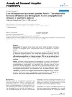
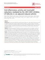
Figure 1
Oxygen-sensing proposed mechanisms for the regulation of gene transcription and the involvement of hypoxia-inducible factor-1 (HIF-1) as a
hypoxia-mediated transcriptional activity (see text for further details). AA, arachidonic acid; ARNT, aryl receptor hydrocarbon nuclear translocator;
CREB, cAMP-responsive element binding protein; CBP, CREB-binding protein; DAG, diacyl glycerol; ECF, extracellular fluid; ICF, intracellular
fluid; IP
3
, inositol triphosphate; MAPK, mitogen-activated protein kinase; NADP, nicotinamide dinucleotide oxidized; NADPH, nicotinamide
dinucleotide reduced; PKC, protein kinase C; ROS, reactive oxygen species; SAPK, stress-activated protein kinase.
C
O
Oxy
De-oxy
Low
O
2
Oxy
De-oxy
High O
2
Co
2+
Ni
2+
Oxy
De-oxy
NAD(P)H Oxidase?
ECF
ICF
O
2
O
2
O
2
.
–
NADPH
NADP
H
2
O
2
?
Fenton
Reaction
ROS
Ubiquitin Degradation
Pathway
T
1/2
HIF-1α
ARNT/
HIF-1β
HIF-1β
P
Kinase(s)
MAPK
ERK
;
MAPK
p38;
MAPK
JNK
;
SA PK;
PKC
p300
CBP
HIF-1
Site
CREB
Si
te
Modulation of
Hypoxia
Re sponsive Genes
Expression or
Suppression
IP
3
/DAG /ROS (+)
AA (+)
Hypoxia
51
remained ambiguous, because PD-98059 and U-0126
inhibitors did not significantly reduce HIF-1α levels at non-
toxic doses [49]. It was proposed that phosphatidylinositol 3-
kinase signaling is not only important in the hypoxic induction
of HIF-1, but that it is also crucially involved in the response
to insulin and IL-1.
Furthermore, evidence that reactive oxygen species (ROS) sig-
naling mediates cytokine-dependent regulation of HIF-1α has
been postulated by Haddad and Land [50,51]. In the airway
epithelium, recombinant human IL-1β and recombinant murine
TNF-α induced, in a time-dependent manner, the nuclear
translocation of HIF-1α. This translocation is an effect associ-
ated with upregulating the activity of this transcription factor
under normoxic conditions. In addition, analysis of the mode of
action of IL-1β and TNF-α revealed a novel induction of intra-
cellular ROS, including hydrogen peroxide, the superoxide
anion (O
2
−•
) and the
•
OH radical [50,51]. The antioxidants
dimethyl sulfoxide and 1,3-dimethyl-2-thiourea, purported to be
prototypical scavengers of hydrogen peroxide and
•
OH, attenu-
ated cytokine-induced HIF-1α nuclear translocation and activa-
tion in a dose-dependent manner. The NADPH-oxidase
inhibitor 4′-hydroxy-3′-methoxy-acetophenone, which may
affect mitochondrial ROS production, attenuated cytokine-
mediated nuclear translocation and activation of HIF-1α. Fur-
thermore, inhibition of the mitochondrion complex I
nicotinamide ADP-dependent oxidase by diphenylene iodo-
nium, which blocks the conversion of ubiquinone to ubiquinol,
abrogated IL-1β-dependent and TNF-α-dependent nuclear
translocation and activation of HIF-1α. Similarly, interrupting the
respiratory chain with potassium cyanide reversed the excita-
tory effect of cytokines on HIF-1α nuclear translocation and
activation [50,51]. These results indicate that a nonhypoxic
pathway mediates cytokine-dependent regulation of HIF-1α
translocation and activation in a ROS-sensitive mechanism.
Direct evidence implicating HIF-1 in lung injury emerged with
VEGF, which has been recognized as a potent mediator of
endothelial barrier dysfunction and is upregulated during
ischemia in many organs [43–46]. Because ventilated
pulmonary ischemia causes a marked increase in pulmonary
vascular permeability, it was hypothesized that VEGF would
increase during ischemic lung injury.
To test this hypothesis, VEGF expression was measured by
northern and western blot analysis in isolated ferret lungs
after 45 or 180 min of ventilated (95% or 0% O
2
) ischemia
[52]. Pulmonary vascular permeability, assessed by measure-
ment of the osmotic reflection coefficient for albumin, was
evaluated in the same lungs, as was expression of HIF-1α.
The distribution of VEGF as a function of ischemic time and
oxygen tension was also evaluated by immunohistochemical
staining in separate groups of lungs. VEGF mRNA increased
threefold by 180 min of ventilated ischemia, independent of
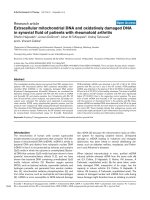
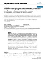
Available online />Figure 2
Potential oxygen-sensing mechanisms and the role of the transcription factor hypoxia-inducible factor-1 (HIF-1). 6GP, 6-glucose phosphate; 6PG,
6-phosphoglycerate; FAD, flavin adenine dinucleotide oxidized; FADH, flavin adenine dinucleotide reduced; NADP, nicotinamide dinucleotide
oxidized; NADPH, nicotinamide dinucleotide reduced; ROS, reactive oxygen species; VHL, von Hippel-Lindau tumor suppressor protein.
Fe
3+
Fe
2+
FADH
FAD
O
2
NADPH
NADP
G6P6PG
O
2
–•
H
2
O
2
ROS
HIF-1a
VHL
HIF-1a
VHL
Ub
Proteasome Degradation
Hypoxia
HIF-1a
HIF-1b
VHL
Cytoplasm
Nucleus
Mn
Ni
Co
52
oxygen tension. VEGF protein increased in parallel to VEGF
mRNA. Immunohistochemical staining demonstrated the
appearance of VEGF protein along alveolar septae after
180 min of hyperoxic ischemia and after 45 or 180 min of
hypoxic ischemia. In addition, albumin was not altered by
45 min of hyperoxic ischemia (0.69 ± 0.09 versus
0.50 ± 0.12, respectively), but decreased significantly after
180 min of hyperoxic ischemia and after 45 and 180 min of
hypoxic ischemia (0.20 ± 0.03, 0.26 ± 0.08 and 0.23 ± 0.03,
respectively) [52]. HIF-1α mRNA increased during both
hyperoxic and hypoxic ischemia, but HIF-1α protein increased
only during hypoxic ischemia. This implicates VEGF as a
potential mediator of increased pulmonary vascular permea-
bility in this model of acute lung injury.
Further elaborating on the mechanisms involving HIF-1 in reg-
ulating the inflammatory response, Hierholzer et al. reported
that hemorrhagic shock (HS) initiates an inflammatory
response that includes increased expression of inducible
nitric oxide synthase and production of prostaglandins [53].
Induction of inducible nitric oxide synthase during the
ischemic phase of HS may involve the activation of HIF-1.
Increased expression of cyclooxygenase-2 during HS con-
tributes to prostaglandin production. The lungs of rats sub-
jected to HS demonstrated a twofold increase in HIF-1
activation and a 7.4-fold increase in expression of cyclo-
oxygenase-2 mRNA, as compared with sham controls [53]. It
was concluded that the upregulation of inducible nitric oxide
synthase and cyclooxygenase-2 during ischemia are two
important early response genes that promote the inflam-
matory response and may contribute to organ damage
through the rapid and exaggerated production of nitric oxide
and prostaglandins.
Furthermore, in a novel study by Shoshani and colleagues,
the identification and cloning of a HIF-1-responsive gene,
designated RTP801, was recently reported. Strong upregula-
tion of RTP801 by hypoxia was detected both in vitro and in
vivo in an animal model of ischemic stroke [54]. When
induced from a tetracycline-repressible promoter, RTP801
protected MCF7 and PC12 cells from hypoxia in glucose-free
medium and from hydrogen peroxide-triggered apoptosis via
a dramatic reduction in the generation of ROS. However,
expression of RTP801 appeared toxic for nondividing neuron-
like PC12 cells and increased their sensitivity to ischemic
injury and oxidative stress. Furthermore, liposomal delivery of
RTP801 cDNA to mouse lungs also resulted in massive cell
death [54]. The biological effect of RTP801 overexpression
thus depends on the cell context and may be either protect-
ing or detrimental for cells under conditions of oxidative or
ischemic stresses. Altogether, the data suggest a complex
type of involvement of RTP801 in the pathogenesis of
ischemic diseases.
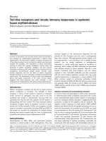
A hypothetical schematic depicting the role of HIF-1 in lung
injury is displayed in Fig. 3.
Conclusion and future prospects
The molecular response to oxidative stress is regulated by
redox-sensitive transcription factors [55–60]. The study of
gene expression and regulation is critical in the development
of novel gene therapies [61–70]. Recognition of reactive
species and redox-mediated protein modifications as poten-
tial signals may open up a new field of cell regulation via
specific and targeted genetic control of transcription factors,
and can thus provide us with a novel way of controlling
disease processes [71–75]. Dynamic variation in pO
2
and
redox equilibrium thus regulate gene expression, apoptosis
signaling and the inflammatory process, thereby bearing
potential consequences for screening emerging targets for
therapeutic intervention.
Competing interests
None declared.
Acknowledgements
The author's own publications therein cited are, in part, financially
supported by the Anonymous Trust (Scotland), the National Institute
for Biological Standards and Control (England), the Tenovus Trust
(Scotland), the UK Medical Research Council (MRC, London), the
Wellcome Trust (London) (Stephen C Land, Department of Child
Health, University of Dundee, Scotland, UK) and the National Insti-
tutes of Health (NIH; Bethesda, USA) (Philip E Bickler, Department
of Anesthesia and Perioperative Care, University of California, San
Francisco, California, USA). The work of the author was performed at
the University of Dundee, Scotland, UK. This review was written at
Critical Care February 2003 Vol 7 No 1 Haddad
Figure 3
A schematic overview of the potential signaling pathways involved in
cytokine-mediated regulation of hypoxia-induced hypoxia-inducible
factor-1α (HIF-1α) translocation and activation. Hypoxia and
inflammatory signals induce the intracellular accumulation of reactive
oxygen species (ROS), which may cause changes in the
phosphorylation state of target kinases, thereby mediating a specific
regulatory mechanism. The mitochondrion is a potential source for
cytokine-unleashed ROS, whose regulation is selectively mediated by
antioxidants. ROS-mediated signaling allows HIF-1α protein
stabilization, nuclear translocation and transcriptional activation.
Hypoxia; Inflammatory Signals
ROS
∆ Phosphorylation and Kinase Regulation
↑ HIF-1α
Protein Stabilization
↑ HIF-1α
Nuclear Translocation
↑ HIF-1α
Transcriptional Activity
Antioxidants
Mitochondrion
Hypoxia-responsive Genes; Cytokines
53
UCSF, California, USA. JJH held the Georges John Livanos prize
(London, UK) under the supervision of Stephen C Land and the NIH
award fellowship (California, USA) under the supervision of Philip E
Bickler. The author also appreciatively thanks Jennifer Schuyler
(Department of Anesthesia and Perioperative Care) for her excellent
editing and reviewing of this manuscript. I also thank my colleagues
at UCSF (San Francisco, California, USA) and the American Univer-
sity of Beirut (AUB, Beirut, Lebanon) who have criticised the work for
enhancement and constructive purposes.
References
1. D’Angio CT, Finkelstein JN: Oxygen regulation of gene expres-
sion: a study in opposites. Mol Genet Metab 2000, 71:371-380.
2. Alder V, Yin Z, Tew KD, Ronai Z: Role of redox potential and
reactive oxygen species in stress signaling. Oncogene 1999,
18:6104-6111.
3. Crapo JD, Harmsen AG, Sherman MP, Musson RA: Pulmonary
immunobiology and inflammation in pulmonary diseases. Am
J Respir Crit Care Med 2000, 162:1983-1986.
4. Haddad JJ, Olver RE, Land SC: Antioxidant/pro-oxidant equilib-
rium regulates HIF-1
αα
and NF-
κκ
B redox sensitivity: evidence
for inhibition by glutathione oxidation in alveolar epithelial
cells. J Biol Chem 2000, 275:21130-21139.
5. Pugin J, Dunn I, Jolliet P, Tassaux D, Magnenat JL, Nicod LP,
Chevrolet JC: Activation of human macrophages by mechani-
cal ventilation in vitro. Am J Physiol 1998, 275:L1040-L1050.
6. Leeper-Woodford SK, Detmer K: Acute hypoxia increases alve-
olar macrophage tumor necrosis factor activity and alters NF-
κκ
B expression. Am J Physiol 1999, 276:L909-L916.
7. Lentsch AB, Yoshidome H, Warner RL, Ward PA, Edwards MJ:
Secretory leukocyte protease inhibitor in mice regulates local
and remote organ inflammatory injury induced by hepatic
ischemia/reperfusion. Gastroenterology 1999, 117:953-961.
8. Chapman WC, Debelak JP, Wright Pinson C, Washington MK,
Atkinson JB, Venkatakrishnan A, Blackwell TS, Christman JW:
Hepatic cryo-ablation, but not radio-frequency ablation,
results in lung inflammation. Ann Surg 2000, 231:752-761.
9. Chapman WC, Debelak JP, Blackwell TS, Gainer KA, Christman
JW, Pinson CW, Brigham KL, Parker RE: Hepatic cryo-ablation-
induced acute lung injury: pulmonary hemodynamic and per-
meability effects in a sheep model. Arch Surg 2000,
135:667-672.
10. Basbaum C, Lemjabbar H, Longphre M, Li D, Gensch E, McNa-
mara N: Control of mucin transcription by diverse injury-
induced signaling pathways. Am J Respir Crit Care Med 1999,
160:S44-S48.
11. Yoshidome H, Kato A, Edwards MJ, Lentsch AB: Interleukin-10
inhibits pulmonary NF-
κκ
B activation and lung injury induced
by hepatic ischemia-reperfusion. Am J Physiol 1999, 277:-
L919-L923.
12. Armstead VE, Opentanova IL, Minchenko AG, Lefer AM: Tissue
factor expression in vital organs during murine traumatic
shock: role of transcription factors AP-1 and NF-
κκ
B. Anesthe-
siology 1999, 91:1844-1852.
13. Washington K, Debelak JP, Gobbell C, Sztipanovits DR, Shyr Y,
Olson S, Chapman WC: Hepatic cryo-ablation-induced acute
lung injury: histopathologic findings. J Surg Res 2001, 95:1-7.
14. Ross SD, Kron IL, Gangemi JJ, Shockey KS, Stoler M, Kern JA,
Tribble CG, Laubach VE: Attenuation of lung reperfusion injury
after transplantation using an inhibitor of nuclear factor-
κκ
B.
Am J Physiol Lung Cell Mol Physiol 2000, 279:L528-L536.
15. Sajjan U, Thanassoulis G, Cherapanov V, Lu A, Sjolin C, Steer B,
Wu YJ, Rotstein OD, Kent G, McKerlie C, Forstner J, Downey GP:
Enhanced susceptibility to pulmonary infection with Burk-
holderia cepacia in Cftr
–/–
mice. Infect Immun 2001, 69:5138-
5150.
16. Lentsch AB, Ward PA: Regulation of experimental lung inflam-
mation. Respir Physiol 2001, 128:17-22.
17. Fan J, Ye RD, Malik AB: Transcriptional mechanisms of acute
lung injury. Am J Physiol Lung Cell Mol Physiol 2001, 281:-
L1037-L1050.
18. Kupfner JG, Arcaroli JJ, Yum HK, Nadler SG, Yang KY, Abraham
E: Role of NF-
κκ
B in endotoxemia-induced alterations of lung
neutrophil apoptosis. J Immunol 2001, 167:7044-7051.
19. Park GY, Le S, Park KH, Le CT, Kim YW, Han SK, Shim YS, Yoo
CG: Anti-inflammatory effect of adenovirus-mediated I
κκ
B-
αα
overexpression in respiratory epithelial cells. Eur Respir J
2001, 18:801-809.
20. Semenza GL: Oxygen-regulated transcription factors and their
role in pulmonary disease. Respir Res 2000, 1:159-162.
21. Cuzzocrea S, Chatterjee PK, Mazzon E, Dugo L, Serraino I, Britti
D, Mazzullo G, Caputi AP, Thiemermann C: Pyrrolidine dithiocar-
bamate attenuates the development of acute and chronic
inflammation. Br J Pharmacol 2002, 135:496-510.
22. Sunil VR, Connor AJ, Guo Y, Laskin JD, Laskin DL: Activation of
type II alveolar epithelial cells during acute endotoxemia. Am
J Physiol Lung Cell Mol Physiol 2002, 282:L872-L880.
23. Haddad JJ, Safieh-Garabedian B, Saadé NE, Kanaan SA, Land
SC: Chemioxyexcitation (
∆∆
pO
2
/ROS)-dependent release of
IL-1
ββ
, IL-6 and TNF-
αα
: evidence of cytokines as oxygen-sensi-
tive mediators in the alveolar epithelium. Cytokine 2001, 13:-
138-147.
24. Haddad JJ, Choudhary KK, Land SC: The ex vivo differential
expression of apoptosis signaling cofactors in the developing
perinatal lung: essential role of oxygenation during the transi-
tion from placental to pulmonary-based respiration. Biochem
Biophys Res Commun 2001, 281:311-316.
25. Haddad JJ, Safieh-Garabedian B, Saadé NE, Land SC: Thiol reg-
ulation of pro-inflammatory cytokines reveals a novel
immunopharmacological potential of glutathione in the alveo-
lar epithelium. J Pharmacol Exp Ther 2001, 296:996-1005.
26. Haddad JJ, Collett A, Land SC, Olver RE, Wilson SM: NF-
κκ
B
blockade reduces the O
2
-evoked rise in Na
+
conductance in
fetal alveolar cells. Biochem Biophys Res Commun 2001, 281:-
987-992.
27. Baines DL, Ramminger SJ, Collett A, Haddad JJ, Best OG, Land
SC, Olver RE, Wilson SM: Oxygen-evoked Na
+
transport in rat
fetal distal lung epithelial cells. J Physiol 2001, 532:105-113.
28. Haddad JJ, Safieh-Garabedian B, Saadé NE, Land SC: The
biphasic immunoregulation of pyrimidylpiperazine (Y-40138)
is IL-10 sensitive and requires NF-
κκ
B targeting in the alveolar
epithelium. Br J Pharmacol 2001, 133:49-60.
29. Haddad JJ, Land SC: Nuclear factor-
κκ
B blockade attenuates
but does not abrogate lipopolysaccharide-dependent tumor
necrosis factor-
αα
biosynthesis in alveolar epithelial cells.
Biochem Biophys Res Commun 2001, 285:267-272.
30. Haddad JJ: Glutathione depletion is associated with augment-
ing a proinflammatory signal: evidence for an antioxidant/pro-
oxidant mechanism regulating cytokines in the alveolar
epithelium. Cytokines Cell Mol Ther 2000, 6:177-187.
31. Haddad JJ, Land SC: Amiloride blockades lipopolysaccharide-
induced proinflammatory cytokine biosynthesis in an I
κκ
B-
αα
/NF-
κκ
B-dependent mechanism. Evidence for the
amplification of an antiinflammatory pathway in the alveolar
epithelium. Am J Respir Cell Mol Biol 2002, 26:114-126.
32. Haddad JJ: L-Buthionine-(S,R)-sulfoximine, an irreversible
inhibitor of
γγ
-glutamylcysteine synthetase, augments LPS-
mediated pro-inflammatory cytokine biosynthesis: evidence
for the implication of an I
κκ
B-
αα
/NF-
κκ
B insensitive pathway. Eur
Cytokine Netw 2001, 12:614-624.
33. Haddad JJ, Saadé NE, Safieh-Garabedian B: Redox regulation
of TNF-
αα
biosynthesis: augmentation by irreversible inhibition
of gamma-glutamylcysteine synthetase and the involvement
of an I
κκ
B-
αα
/NF-
κκ
B-independent pathway in alveolar epithelial
cells. Cell Signal 2002, 14:211-218.
34. Haddad JJ, Land SC: Redox/ROS regulation of lipopolysac-
charide-induced mitogen-activated protein kinase (MAPK)
activation and MAPK-mediated TNF-
αα
biosynthesis. Br J Phar-
macol 2002, 135:520-536.
35. Haddad JJ: The involvement of L-
γγ
-glutamyl-L-cysteinyl-glycine
(glutathione/GSH) in the mechanism of redox signaling
mediating MAPK
p38
-dependent regulation of pro-inflamma-
tory cytokine production. Biochem Pharmacol 2002, 63:305-
320.
36. Haddad JJ, Fahlman CS: Nuclear factor-
κκ
B-independent regu-
lation of lipopolysaccharide-mediated interleukin-6 biosyn-
thesis. Biochem Biophys Res Commun 2002, 291:1045-1051.
37. Rahman I, Mulier B, Gilmour PS, Watchorn T, Donaldson K,
Jeffery PK, MacNee W: Oxidant-mediated lung epithelial cell
tolerance: the role of intracellular glutathione and nuclear
factor-
κκ
B. Biochem Pharmacol 2001, 62:787-794.
38. MacNee W, Rahman I: Is oxidative stress central to the patho-
genesis of chronic obstructive pulmonary disease? Trends
Mol Med 2001, 7:55-62.
39. Rahman I, MacNee W: Regulation of redox glutathione levels
Available online />54
and gene transcription in lung inflammation: therapeutic
approaches. Free Radic Biol Med 2000, 28:1405-1420.
40. Rahman I, van Schadewijk AA, Hiemstra PS, Stolk J, van Krieken
JH, MacNee W, de Boer WI: Localization of
γγ
-glutamylcysteine
synthetase mRNA expression in lungs of smokers and
patients with chronic obstructive pulmonary disease. Free
Radic Biol Med 2000, 28:920-925.
41. Haddad JJ: Oxygen-sensitive pro-inflammatory cytokines,
apoptosis signaling and redox-responsive transcription
factors in development and pathophysiology. Cytokines Cell
Mol Ther 2001, 7:1-14.
42. Haddad JJ: Oxygen homeostasis, thiol equilibrium and redox
regulation of signaling transcription factors in the alveolar
epithelium. Cell Signal 2002, 14:799-810.
43. Semenza GL: HIF-1 and mechanisms of hypoxia sensing. Curr
Opin Cell Biol 2001, 13:167-171.
44. Semenza GL: HIF-1 and human disease: one highly involved
factor. Genes Dev 2000, 14:1983-1991.
45. Semenza GL: Involvement of hypoxia-inducible factor 1 in
human cancer. Intern Med 2002, 41:79-83.
46. Semenza GL: HIF-1 and tumor progression: pathophysiology
and therapeutics. Trends Mol Med 2002, 8:S62-S67.
47. Hellwig-Bürgel T, Rutkowski K, Metzen E, Fandrey J, Jelkmann W:
Interleukin-1
ββ
and tumor necrosis factor-alpha stimulate DNA
binding of hypoxia-inducible factor-1. Blood 1999, 94:1561-
1567.
48. El Awad B, Kreft B, Wolber EM, Hellwig-Bürgel T, Metzen E,
Fandrey J, Jelkmann W: Hypoxia and interleukin-1
ββ
stimulate
vascular endothelial growth factor production in human proxi-
mal tubular cells. Kidney Int 2000, 58:43-50.
49. Stiehl DP, Jelkmann W, Wenger RH, Hellwig-Bürgel T: Normoxic
induction of the hypoxia-inducible factor 1
αα
by insulin and
interleukin-1
ββ
involves the phosphatidylinositol 3-kinase
pathway. FEBS Lett 2002, 512:157-162.
50. Haddad JJ, Land SC: A non-hypoxic, ROS-sensitive pathway
mediates TNF-
αα
-dependent regulation of HIF-1
αα
. FEBS Lett
2001, 505:269-274.
51. Haddad JJ: Recombinant human interleukin (IL)-1
ββ
-mediated
regulation of hypoxia-inducible factor-1
αα
(HIF-1
αα
) stabiliza-
tion, nuclear translocation and activation requires an antioxi-
dant/reactive oxygen species (ROS)-sensitive mechanism.
Eur Cytokine Netw 2002, 13:250-260.
52. Becker PM, Alcasabas A, Yu AY, Semenza GL, Bunton TE:
Oxygen-independent upregulation of vascular endothelial
growth factor and vascular barrier dysfunction during venti-
lated pulmonary ischemia in isolated ferret lungs. Am J Respir
Cell Mol Biol 2000, 22:272-279.
53. Hierholzer C, Harbrecht BG, Billiar TR, Tweardy DJ: Hypoxia-
inducible factor-1 activation and cyclooxygenase-2 induction
are early reperfusion-independent inflammatory events in
hemorrhagic shock. Arch Orthop Trauma Surg 2001, 121:219-
222.
54. Shoshani T, Faerman A, Mett I, Zelin E, Tenne T, Gorodin S,
Moshel Y, Elbaz S, Budanov A, Chajut A, Kalinski H, Kamer I,
Rozen A, Mor O, Keshet E, Leshkowitz D, Einat P, Skaliter R, Fein-
stein E: Identification of a novel hypoxia-inducible factor 1-
responsive gene, RTP801, involved in apoptosis. Mol Cell Biol
2002, 22:2283-2293.
55. Shimaoka M, Fujino Y, Taenaka N, Hiroi T, Kiyono H, Yoshiya II.
High frequency oscillatory ventilation attenuates the activa-
tion of alveolar macrophages and neutrophils in lung injury.
Crit Care 1998, 2:35-39.
56. Pinsky MR: Toward a better ventilation strategy for patients
with acute lung injury. Crit Care 2000, 4:205-206.
57. Wesselkamper SC, Prows DR, Biswas P, Willeke K, Bingham E,
Leikauf GD: Genetic susceptibility to irritant-induced acute
lung injury in mice. Am J Physiol Lung Cell Mol Physiol 2000,
279:L575-L582.
58. Leikauf GD, McDowell SA, Bachurski CJ, Aronow BJ, Gammon K,
Wesselkamper SC, Hardie W, Wiest JS, Leikauf JE, Korfhagen
TR, Prows DR: Functional genomics of oxidant-induced lung
injury. Adv Exp Med Biol 2001, 500:479-487.
59. Leikauf GD, McDowell SA, Wesselkamper SC, Hardie WD,
Leikauf JE, Korfhagen TR, Prows DR: Acute lung injury: func-
tional genomics and genetic susceptibility. Chest 2002, 121:-
70S-75S.
60. Pittet JF, MacKersie RC, Martin TR, Matthay MA: Biological
markers of acute lung injury: prognostic and pathogenetic
significance. Am J Respir Crit Care Med 1997, 155:1187-1205.
61. Kaminski N, Allard JD, Pittet JF, Zuo F, Griffiths MJ, Morris D,
Huang X, Sheppard D, Heller RA: Global analysis of gene
expression in pulmonary fibrosis reveals distinct programs
regulating lung inflammation and fibrosis. Proc Natl Acad Sci
USA 2000, 97:1778-1783.
62. Miyazaki H, Broaddus VC, Wiener-Kronish JP, Sawa T, Pittet JF,
Kravchenko V, Mathison JC, Nishizawa H, Hattori S, Yamakawa T,
Yamada H, Kudoh I: The effects of two antiinflammatory pre-
treatments on bacterial-induced lung injury. Anesthesiology
1999, 90:1650-1662.
63. Grimaldo DA, Wiener-Kronish JP, Jurson T, Shaughnessy TE,
Curtis JR, Liu LL: A randomized, controlled trial of advanced
care planning discussions during preoperative evaluations.
Anesthesiology 2001, 95:43-50.
64. Ernst EJ, Hashimoto S, Guglielmo J, Sawa T, Pittet JF, Kropp H,
Jackson JJ, Wiener-Kronish JP: Effects of antibiotic therapy on
Pseudomonas aeruginosa-induced lung injury in a rat model.
Antimicrob Agents Chemother 1999, 43:2389-2394.
65. Davis K Jr, Johannigman JA, Campbell RS, Marraccini A, Luchette
FA, Frame SB, Branson RD: The acute effects of body position
strategies and respiratory therapy in paralyzed patients with
acute lung injury. Crit Care 2001, 5:81-87.
66. Kheradmand F, Wiener-Kronish JP, Corry DB: Assessment of
operative risk for patients with advanced lung disease. Clin
Chest Med 1997, 18:483-494.
67. Song C, Al-Mehdi AB, Fisher AB: An immediate endothelial cell
signaling response to lung ischemia. Am J Physiol Lung Cell
Mol Physiol 2001, 281:L993-L1000.
68. Merker MP, Pitt BR, Choi AM, Hassoun PM, Dawson CA, Fisher
AB: Lung redox homeostasis: emerging concepts. Am J
Physiol Lung Cell Mol Physiol 2000, 279:L413-L417.
69. Dietrich M, Block G, Hudes M, Morrow JD, Norkus EP, Traber
MG, Cross CE, Packer L: Antioxidant supplementation
decreases lipid peroxidation biomarker F
2
-isoprostanes in
plasma of smokers. Cancer Epidemiol Biomarkers Prev 2002,
11:7-13.
70. van Der Vliet A, Eiserich JP, Cross CE: Nitric oxide: a pro-inflam-
matory mediator in lung disease? Respir Res 2000, 1:67-72.
71. Cross CE, van der Vliet A, Eiserich JP: Peroxidases wheezing
their way into asthma. Am J Respir Crit Care Med 2001, 164:-
1102-1103.
72. Casaburi R, Mahler DA, Jones PW, Wanner A, San PG, ZuWal-
lack RL, Menjoge SS, Serby CW, Witek T Jr: A long-term evalu-
ation of once-daily inhaled tiotropium in chronic obstructive
pulmonary disease. Eur Respir J 2002, 19:217-224.
73. Burrows B, Bloom JW, Traver GA, Cline MG: The course and
prognosis of different forms of chronic airways obstruction in
a sample from the general population. N Engl J Med 1987,
317:1309-1314.
74. Kaplan LJ, Bailey H, Formosa V: Airway pressure release venti-
lation increases cardiac performance in patients with acute
lung injury/adult respiratory distress syndrome. Crit Care
2001, 5:221-226.
75. dos Santos CC, Zhang H, Slutsky AS: From bench to bedside:
bacterial growth and cytokines. Crit Care 2002, 6:4-6.
Critical Care February 2003 Vol 7 No 1 Haddad