Báo cáo y học: "Science review: Cell membrane expression (connectivity) regulates neutrophil delivery, function and clearance" docx
Bạn đang xem bản rút gọn của tài liệu. Xem và tải ngay bản đầy đủ của tài liệu tại đây (1.32 MB, 17 trang )
291
FADD = Fas-associated death domain; FasL = Fas ligand; FMLP = f-Met-Leu-Phe; G-CSF = granulocyte colony-stimulating factor; GM-CSF =
granulocyte/macrophage colony-stimulating factor; GRO = growth-related oncogene; H
2
O
2
= hydrogen peroxide; ICAM = intercellular adhesion
molecule; IL = interleukin; MIP = macrophage inflammatory protein; NADPH = reduced nicotinamide adenine dinucleotide phosphate; NF-κB =
nuclear factor-κB; O
2
–
• = superoxide anion; PECAM = platelet–endothelial cell adhesion molecule; TNF = tumor necrosis factor.
Available online />Tissue inflammation, manifesting clinically as rubor, calor,
tumor, and dolor, has been a focus of investigation since the
beginning of medical science. Inflammation may be defined
as a condition or state that tissues enter as a response to
injury or insult. The neutrophil is the most important and the
most extensively studied cell involved in the inflammatory
response. As the principal circulating phagocyte, the neu-
trophil is the first and most abundant leukocyte to be deliv-
ered to a site of infection or inflammation, and is thus an
integral component of the innate immune system. In addition
to its role in host defense, the neutrophil is implicated in the
pathogenesis of tissue injury and of persistent inflammatory
diseases. The paradoxic roles of the neutrophil in host
defense and host injury have fueled intense scientific inquiry
into the processes of neutrophil delivery to a site of inflamma-
tion, neutrophil function within the inflammatory environment,
and neutrophil clearance from that milieu.
The aim of the present review is to highlight the importance of
neutrophil cell membrane expression in the participation and
regulation of neutrophil delivery, function, and clearance from
its environment. The relationship between altered receptor
expression and altered neutrophil function in humans and in
vivo are emphasized. The review concludes with a brief dis-
Review
Science review: Cell membrane expression (connectivity)
regulates neutrophil delivery, function and clearance
Andrew JE Seely
1
, José L Pascual
2
and Nicolas V Christou
3
1
Resident, Divisions of Thoracic Surgery and Critical Care Medicine, University of Ottawa, Ottawa, Ontario, Canada
2
Resident, Division of General Surgery, McGill University Health Center, Montreal, Quebec, Canada
3
Professor and Chief, Division of General Surgery, McGill University Health Center, Montreal, Quebec, Canada
Correspondence: Andrew JE Seely,
Published online: 9 January 2003 Critical Care 2003, 7:291-307 (DOI 10.1186/cc1853)
This article is online at />© 2003 BioMed Central Ltd (Print ISSN 1364-8535; Online ISSN 1466-609X)
Abstract
As the principal cellular component of the inflammatory host defense and contributor to host injury after
severe physiologic insult, the neutrophil is inherently coupled to patient outcome in both health and
disease. Extensive research has focused on the mechanisms that regulate neutrophil delivery, function,
and clearance from the inflammatory microenvironment. The neutrophil cell membrane mediates the
interaction of the neutrophil with the extracellular environment; it expresses a complex array of
adhesion molecules and receptors for various ligands, including mediators, cytokines,
immunoglobulins, and membrane molecules on other cells. This article presents a review and analysis
of the evidence that the neutrophil membrane plays a central role in regulating neutrophil delivery
(production, rolling, adhesion, diapedesis, and chemotaxis), function (priming and activation,
microbicidal activity, and neutrophil-mediated host injury), and clearance (apoptosis and necrosis). In
addition, we review how change in neutrophil membrane expression is synonymous with change in
neutrophil function in vivo. Employing a complementary analysis of the neutrophil as a complex system,
neutrophil membrane expression may be regarded as a measure of neutrophil connectivity, with altered
patterns of connectivity representing functionally distinct neutrophil states. Thus, not only does the
neutrophil membrane mediate the processes that characterize the neutrophil lifecycle, but
characterization of neutrophil membrane expression represents a technology with which to evaluate
neutrophil function.
Keywords apoptosis, chemotaxis, connectivity, delivery, neutrophil, receptors
292
Critical Care August 2003 Vol 7 No 4 Seely et al.
cussion and interpretation of the importance of membrane
receptor expression as a measure of cellular ‘connectivity’,
and provides suggestions for future research into the role of
neutrophils in the inflammatory response.
Neutrophil delivery to the inflammatory
microenvironment
Neutrophil production and storage
The neutrophil lifecycle begins with a bone marrow phase, fol-
lowed by a circulating phase; it ends with a tissue phase.
Within the bone marrow, neutrophils originate from self-renew-
ing myeloid stem cells; the myeloblast differentiates into the
promyloblast, and then into the myelocyte. These cells differ-
entiate into metamyelocytes as well as segmented band neu-
trophils, which are occasionally seen in circulation during a
stress response. The metamyelocyte is the precursor to poly-
morphonuclear leukocytes, which are commonly referred to as
granulocytes, including eosinophils, basophils, and neu-
trophils. The process of neutrophil maturation and differentia-
tion within the marrow takes approximately 14 days, and has
undergone considerable investigation [1]. Neutrophil produc-
tion is estimated to vary from 10
8
to 10
11
cells/day, depending
on the measurement technique used [1,2]. This is mediated by
a variety of hematopoietic growth factors, most notably granu-
locyte colony-stimulating factor (G-CSF) and granulocyte/
macrophage colony-stimulating factor (GM-CSF) [3].
Growth factors exert their effect through interaction with mem-
brane receptors, with subsequent induction of intracellular
tyrosine phosphorylation and activation of multiple signaling
cascades [4]. Variation in receptor expression and modulation
by soluble mediators occurs during cell maturation [5]. In addi-
tion to other factors, GM-CSF and G-CSF mediate prolifera-
tion and differentiation of neutrophil bone marrow stem cells,
allowing for substantial variation in neutrophil production,
which increases as much as 10-fold during a stress response
[2]. Pathologic function of growth factor receptors leads to
hematologic illness [6,7], and a reduction in marrow G-CSF
receptor expression is associated with myeloid maturation
arrest and neutropenia following severe burn injury [8]. Thus,
neutrophil production, differentiation, and maturation depend
upon physiologic interaction of growth factors with receptors
on neutrophil myeloid precursors.
After release from the bone marrow, neutrophils enter the cir-
culating compartment (i.e. the second phase of their life-
cycle). In circulation, neutrophils have a half-life of 6–9 hours.
Neutrophils comprise more than 50% of circulating leuko-
cytes and more than 90% of circulating phagocytes, and
reversibly move from circulating to marginating pools. Mar-
ginated neutrophils are those that are ‘stored’ in the capillar-
ies of certain tissues, most notably in the lung, and are much
greater in number than are those that are free in circulation at
any given time [9]. The lung harbours large numbers of mar-
ginating neutrophils because of the tremendous number of
small capillaries (with diameter less than that of the neu-
trophil), forcing neutrophils to deform in order to pass through
these capillaries [10]. The marginating pool of neutrophils
allows for rapid mobilization in response to infection or other
stresses. Despite the rapid turnover, human neutrophil counts
are relatively stable, averaging 3000–4000 neutrophils/mm
3
.
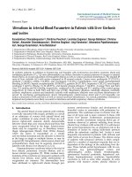
Neutrophil delivery occurs in the postcapillary venule as a
sequential series of well studied processes (Fig. 1).
Margination
Neutrophil transmigration from the intravascular to the extravas-
cular (exudate) milieu predominantly occurs in the postcapillary
venule within the systemic circulation and in the capillary in the
pulmonary circulation [11]. Neutrophil exudation is facilitated
and mediated by a combination of mechanical, chemical, and
molecular processes; these are distinct events that are linked in
a temporal sequence. The first step is ‘margination’, or move-
ment of the neutrophil from the central stream to the periphery
of a vessel. In postcapillary venules, when the vessel diameter
is 50% larger than the diameter of the leukocyte, erythrocytes
move faster than the larger leukocytes, especially in the center
of the vessel, pushing leukocytes to the vessel periphery [12].
Physical forces involved in the erythrocyte–leukocyte interac-
tions govern this radial movement of leukocytes. The impor-
tance of erythrocytes has been demonstrated in a rat
mesenteric perfusion model, in which no leukocyte margination
was observed in the absence of red cells [13]. Neutrophil mar-
gination allows for a molecular interaction between the cell sur-
faces of the neutrophil and endothelial cell to occur, resulting in
neutrophil rolling on the vessel wall.
Rolling
A state of weak adhesive interaction between the neutrophil
and endothelial cell allows the neutrophil to roll along the
surface of the postcapillary venule. ‘Rolling’ is dependent
upon both physical and molecular forces. The neutrophil’s
ability to roll and adhere to endothelial cells is inversely pro-
portional to the vessel shear rate (i.e. faster moving blood
decreases the ability of leukocytes to adhere) [14]. Neutrophil
rolling velocity is also directly proportional to luminal red
blood cell velocity [15]. Once in proximity to the endothelial
cell, a low-affinity adherence occurs and, in conjunction with
the shear stress of passing erythrocytes, the neutrophil
begins to roll along the endothelial lining of the vessel.
Selectins
Interactions between the surface of the neutrophil and the
endothelial cell allow for rolling, and subsequently adherence
and diapedesis. The low-affinity interaction involved in rolling
is largely governed by selectins and their ligands (Table 1).
Selectins are a family of glycoprotein surface adhesion mole-
cules, and include L-selectin (expressed exclusively on leuko-
cytes), E-selectin (expressed exclusively on endothelial cells),
and P-selectin (expressed on platelets and endothelial cells).
Constitutive expression of L-selectin is maintained on all cir-
culating quiescent leukocytes (except for certain subpopula-
tions of memory T cells) [16].
293
Animal intravital microscopy has demonstrated that blocking
L-selectin and/or P-selectin with high-dose selectin-binding
carbohydrate (fucoidin) decreased both neutrophil rolling and
adherence following ischemia/reperfusion [17]. L-selectin and
P-selectin gene-deficient mice exhibit diminished rolling [18].
The ligands for neutrophil L-selectin are multiple sialylated car-
bohydrate determinants, which are linked to mucin-like mole-
cules [16,19]. These selectin ligands on endothelial cells are
inducible with lipopolysaccharide or a variety of inflammatory
cytokines [20]. In addition to L-selectin mediated rolling,
endothelial cell expression of E-selectin is necessary for
normal leukocyte recruitment and may initiate leukocyte rolling
in certain models [21,22]. The rolling governed by a weak mol-
ecular interaction is a prerequisite for a stronger molecular
interaction, namely adherence. This has been demonstrated
using intravital microscopy in the rat mesenteric microcircula-
tion [23], in human neutrophils in rabbit mesenteric venules
[24], and in a cat mesenteric perfusion model [15]. However,
other investigators have demonstrated that antibodies to P-
selectin will attenuate rolling but not impact on adherence
[25]. Blocking L-selectin in animal models reduced neutrophil-
mediated tissue injury, which was believed to be dependent
upon neutrophil adherence [26]. In addition, soluble L-selectin
shed from neutrophils may attenuate TNF-α stimulated neu-
trophil adherence and subsequent vascular permeability [27].
Thus, those studies suggest that selectins not only mediate
rolling, but also impact upon ensuing leukocyte adherence.
Adherence
As with rolling, the cell surface of the neutrophil determines
its ability to undergo ‘adherence’. In contrast to rolling, which
is a dynamic low-affinity adhesive interaction, adherence is a
stationary high-affinity (strong) adhesive interaction between
the neutrophil and endothelial cell. This interaction is largely
mediated by a separate set of adhesion molecules, namely
the integrins and their ligands. The importance of integrin-
mediated adhesion to neutrophil delivery and host defense
was first demonstrated in patients with leukocyte adhesion
deficiency type 1 [28]. These patients develop life-threaten-
ing bacterial infections; this is because neutrophils are unable
to undergo transmigration to sites of inflammation as a result
of a genetic mutation in CD18, the β-subunit of the integrin
family of adhesion molecules. Neutrophils from healthy
control individuals incubated with monoclonal antibodies to
integrins, or neutrophils from patients with leukocyte adhe-
sion deficiency-1 both demonstrate deficient adhesion and
transmigration through activated endothelial monolayers [29].
Integrins and intercellular adhesion molecules
Integrins are a family of heterodimeric proteins (made up of
two different subunits, namely α-subunits and β-subunits) that
are expressed on the cell surface, and are integral to the
process of cell adhesion. Of this family, the β
2
-integrins have
attracted the most investigation; they are restricted to leuko-
cytes and are essential to normal leukocyte trafficking. They
consist of three distinct α-subunits (CD11a, CD11b, and
CD11c) that are bound to a common β-subunit (CD18).
Although the distribution of β
2
-integrins subclasses differs
among leukocyte populations, neutrophils express all three
classes. The relative contribution of each α-subunit to leuko-
cyte adherence may vary and depend upon the stimulus
leading to adherence and transmigration [30]. Neutrophil
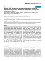
Available online />Figure 1
Neutrophil delivery in the postcapillary venule. ICAM, intercellular adhesion molecule.
294
integrins interact with complementary surface molecule
ligands on endothelial cells in order to generate the high-affin-
ity bond that characterizes adherence (Table 1). Particularly
important to neutrophils, intercellular adhesion molecule
(ICAM)-1 on endothelial cells serves as the ligand for both
CD11a/CD18 and CD11b/CD18, whereas ICAM-2 is
capable of binding CD11a only [31].
Animal intravital microscopy has demonstrated the impor-
tance of the integrin β-subunit CD18 to adhesion but not to
rolling [32,33]. Multiple studies have demonstrated that anti-
CD11/CD18 antibodies are associated with reduced inflam-
mation and injury in models of allograft rejection, endotoxin
challenge, hemorrhagic shock, aspiration pneumonia, bacter-
ial pneumonia, and ischemia/reperfusion, among others [34].
Although CD18-dependent neutrophil transmigration is
essential for physiologic neutrophil delivery, CD18-indepen-
dent neutrophil transmigration has been demonstrated in
rabbit models of respiratory and peritoneal infection, and res-
piratory and hepatic ischemia/reperfusion [35–38]; this may
depend on the type of bacteria at the site of infection [39]. In
addition to β
2
-integrin mediated adhesion, Kubes and
coworkers [40] demonstrated that expression of β
1
-integrins
(specifically α
4
β
1
) may be induced by activation or by trans-
migration in order to mediate adhesion on human neutrophils.
Notwithstanding the complexity of adhesion molecule interac-
tion, the membrane of the neutrophil and of the endothelial
cell must undergo firm adhesion in order for the process of
neutrophil transmigration to progress.
Receptor adherence in receptor molecular biology is evalu-
ated by receptor affinity, which relates to the strength of inter-
action between a single antigen-binding site and a single
antigenic determinant, as well as by receptor avidity, which
represents the strength of binding of a molecule with multiple
binding sites, such as the binding of a complex antigen with
multiple antibodies. Affinity depends upon noncovalent bonds
between binding sites and is measured using an affinity con-
stant. Avidity represents the overall binding of antibodies to
antigen, and may be greater than the sum of the affinities if
cooperative effects exist (i.e. binding at one site promotes
binding at another). Both receptor affinity and avidity may be
differentially regulated in leukocyte–endothelial cell interac-
tions involving the β
2
-integrin (CD11a/CD18) [41,42].
Both integrins on neutrophils, as well as ICAMs on endothe-
lial cells, demonstrate marked variability in expression and
adhesiveness. Augmented neutrophil expression of
CD11b/CD18 is induced from intracellular pools by various
cytokines, including f-Met-Leu-Phe (FMLP), GM-CSF, C5a,
tumor necrosis factor (TNF)-α, and others; however,
increased neutrophil adhesiveness may be more significantly
related to conformational changes in the CD11b/CD18
protein complex [43]. Chemoattractants such as the
chemokine IL-8 will activate integrin adhesiveness as well as
help to direct leukocyte migration [44,45]. In addition to con-
stitutive expression of ICAM-1 and ICAM-2 on endothelial
cells, ICAM-1 expression may be augmented by numerous
inflammatory mediators [46–48]. Thus, under the influence of
Critical Care August 2003 Vol 7 No 4 Seely et al.
Table 1
Neutrophil and endothelial cell adhesion receptors
Receptor Cell Ligand Cell type Purpose
L-selectin Neutrophil sLe
a
, sLe
x
Endothelium Rolling and weak adhesion of PMNs on EC
CD11a/CD18 Neutrophil ICAM-1, ICAM-2, ICAM-3 Endothelium Adhesion of PMNs on EC
CD11b/CD18 Neutrophil ICAM-1 Endothelium Adhesion of PMNs on EC
iC3b Complement Phagocytosis?
Fibrinogen – –
Factor X – –
CD11c/CD18 Neutrophil iC3b Complement Phagocytosis?
Fibrinogen – –
E-selectin Endothelium sLe
x
Neutrophil Firm PMN/EC adhesion
P-selectin Endothelium, platelets sLe
x
Endothelium Firm PMN/EC adhesion
PSGL-1 Neutrophil Firm PMN/EC adhesion
PECAM-1 Endothelium CD31/α
v
Leukocytes Diapedesis of PMN through EC
Neutrophil – –
ICAM-3 Neutrophil CD11a/CD18 Leukocytes Antigen presentation
ICAM, intercellular adhesion molecule; PECAM, platelet–endothelial cell adhesion molecule; PMN, polymorphonuclear leukocyte; PSGL, P-selectin
glycoprotein ligand.
295
inflammatory mediators, changes in number and conformation
of neutrophil integrins and upregulation of endothelial cell
ICAM expression will induce a transition from selectin-depen-
dent rolling to integrin/ICAM-dependent adherence [49], sub-
sequently leading to diapedesis, which is the next step in
neutrophil delivery.
Diapedesis
Following adherence, the neutrophil must pass through the
endothelial monolayer and basement membrane to enter the
extravascular inflammatory (exudate) environment. In vitro
adherence of neutrophils on activated endothelial cells will
cause a disruption in endothelial cell–cell interaction and
augment endothelial cell permeability – an effect that may be
blocked with anti-integrin monoclonal antibodies [50]. Trans-
mission electron microscopy in a human umbilical vein neu-
trophil transmigration model suggested that diapedesis of
neutrophils occurs at endothelial cell tricellular corners (the
intersection of three endothelial cells) [51]. Endothelial adhe-
sion molecules are necessary for diapedesis and transmigra-
tion. Leukocyte adherence and emigration observed after
ischemia/reperfusion and in response to leukotriene-B
4
or
platelet-activating factor is decreased with monoclonal anti-
bodies to various adhesion glycoproteins, including CD18,
CD11b, ICAM-1, and L-selectin [52,53]. Thus, membrane-
mediated adherence is a prerequisite for diapedesis – a
process that is also mediated by neutrophil–endothelial cell
membrane interaction.
Platelet–endothelial cell adhesion molecule-1
Other adhesion molecules, such as platelet–endothelial cell
adhesion molecule (PECAM)-1, are specifically involved in
the process of diapedesis. PECAM-1 is constitutively
expressed and concentrated on the lateral borders of
endothelial cells where diapedesis is observed to take place,
as well as on the surface of neutrophils, some T cells, mono-
cytes, and platelets. Blocking PECAM-1 with monoclonal
antibodies will increase neutrophil adhesion to endothelial
cells mediated by CD11b/CD18 [54,55], thus inhibiting the
ability of the neutrophil to undergo diapedesis. Monoclonal
antibodies to PECAM-1 will arrest leukocyte transmigration
by 70–90% without interfering with normal leukocyte adhe-
sion to endothelial monolayers; leukocytes remain tightly
bound to the apical surface of the endothelial cell, precisely
over the intercellular junction [56]. The importance of
endothelial and neutrophil expression of PECAM-1 was con-
firmed using in vivo murine intravital microscopy [57]. Thus,
PECAM-1 appears to allow the neutrophil to evade adhesion
at intercellular junctions so that diapedesis leading to neu-
trophil transmigration may take place.
In summary, the process of neutrophil transmigration is regu-
lated by a multistep process that involves sequential events,
each of which are necessary for progression to the next.
These cellular processes are governed by molecular interac-
tions between receptors and their ligands expressed on neu-
trophils and endothelial cells. The cell membrane of the neu-
trophil allows it to interact with endothelial cells. Leukocyte
delivery may be regulated by altering the expression and effi-
cacy of the various adhesion receptors dynamically in vivo,
leading to site-specific leukocyte accumulation. In addition to
adhesion receptors and ligands mediating neutrophil–
endothelial cell interactions, leukocyte delivery requires
further neutrophil cell membrane participation, specifically
responding to soluble mediators in the extracellular inflamma-
tory environment.
Chemotaxis
In addition to intercellular adhesion, leukocytes require a
chemoattractant gradient in order to complete the process of
transmigration. Chemoattractants are soluble molecules that
confer directionality on cell movement; cells migrate in the
direction of increasing concentration of a chemoattractant in
a process termed ‘chemotaxis’. Neutrophils have long been
known to undergo chemotaxis toward damaged or inflamed
tissue [58].
The production of chemoattractants in the inflammatory envi-
ronment is from a combination of sources, including bacterial
byproducts and cell wall constituents, complement factors,
and chemokines produced by inflammatory and noninflamma-
tory cells. For example, in addition to neutrophils themselves
[59], monocytes, smooth muscle cells, epithelial cells,
endothelial cells, and fibroblasts are capable of generating
IL-8 (a potent neutrophil chemoattractant) when they are
stimulated with an proinflammatory agonist such as IL-1 or
TNF-α [60].
Chemoattractants serve not only to direct leukocytes to spe-
cific areas of inflammation but also to recruit specific subpop-
ulations of leukocytes to inflamed tissue, such as neutrophils
in response to acute bacterial infection, eosinophils at sites of
chronic allergic inflammation or parasitic infection, and mono-
cytes in chronic inflammatory diseases. Chemoattractant
mediators may thus be classified on the basis of their spec-
trum of leukocyte activity (Table 2). Classical chemoattrac-
tants include N-formylated peptides produced by bacteria,
such as FMLP, polypeptides (e.g. C5a), and lipids (e.g.
leukotriene-B
4
), which act as chemoattractants for various
nonspecific leukocyte populations [61–63]. Chemoattractant
cytokines, or chemokines, are a novel family of chemoattrac-
tants that confer specificity to leukocyte subset responsive-
ness, and are well reviewed elsewhere [64,65]. Extensive
in vitro and in vivo investigation has identified IL-8 as a princi-
pal factor in neutrophil delivery [66–69]. Other chemokines
that are specific for neutrophils include epithelial cell derived
neutrophil activating peptide; neutrophil activating peptide-2;
growth-related oncogene (GRO)-α, GRO-β and GRO-δ; and
macrophage inflammatory protein (MIP)-2α and MIP-2β.
These chemokines are structurally similar, and consist of the
first two cysteine (C) amino acid residues separated by a
separate amino acid (X), and are referred to as CXC
Available online />296
chemokines or α chemokines. A separate family of
chemokines are known as CC chemokines, because the first
two cysteine residues are in juxtaposition. Monocyte
chemoattractant protein-1, -2 and -3; MIP-1α and MIP-1β;
and RANTES (regulated upon activation, normal T cell
expressed and secreted) are members of the CC family, or
β chemokines. The activity of the CC supergene family of
chemokines is predominantly oriented toward monocytes
[70]. Thus, chemoattractants help to explain how leukocytes
localize to specific inflammatory sites, and how specific
leukocyte populations are recruited to those sites.
Chemoattractant receptors
Leukocyte delivery is further regulated by chemoattractant
receptors that exhibit specificity for both the type of leukocyte
on which they are expressed and the ligand to which they
bind. The specificity of chemoattractant-induced leukocyte
chemotaxis is related to differential expression of chemokine
receptors, a superfamily of G-protein-coupled receptors with
seven transmembrane regions [71,72]. Although chemokine
receptors share similar structures, they differ in their ligand
specificity (Table 3). For example, IL-8 receptor A (CXC R1)
and IL-8 receptor B (CXC R2) have a 78% identical amino
acid sequence, and both bind IL-8; however, although IL-8
receptor A is specific for IL-8, IL-8 receptor B has multiple
agonists, including other CXC chemokines such GRO-α,
GRO-β, GRO-δ, neutrophil-activating peptide-2, and epithe-
lial cell-derived neutrophil activating peptide-78 [73]. Neu-
trophil transmigration appears to depend to a greater degree
on IL-8 receptor A than on IL-8 receptor B, because antibod-
ies directed against IL-8 receptor A inhibited the majority
(78%) of IL-8 induced chemotaxis [74]. In contrast, IL-8
receptor B has been implicated in transendothelial migration
of T cells [75]. In addition, chemoattractant receptors are
expressed on specific leukocyte subsets (Table 3); whereas
receptors to the classical chemoattractants are expressed on
monocytes, neutrophils, eosinophils and basophils, CXC
chemokine receptors are primarily restricted to neutrophils [16].
Thus, chemokine receptors display both ligand and leukocyte
specificity. These complex rules defining the interactions
between specific chemoattractants and leukocytes are the
mechanisms that allow the host response to deliver specific
subsets of leukocytes to localized areas of infection or inflam-
mation. Chemoattractant receptors not only mediate the
process of chemotaxis, but changes in receptor expression
within the inflammatory environment confer changes on cell
function. Before discussing changes in neutrophil cell surface
expression, we consider neutrophil function and clearance
from the inflammatory microenvironment.
Neutrophil function in the inflammatory
microenvironment
Neutrophil priming and activation
Neutrophils can exist in various stable functional states. The
different states are associated with different patterns of
altered membrane expression (Table 4). Quiescent neu-
trophils can be ‘activated’ by various inflammatory mediators
in order to produce reactive oxygen metabolites (the respira-
tory burst) and destructive proteolytic enzymes (see below).
In addition to being activated, the neutrophil can be ‘primed’
to produce an augmented or exaggerated response to an
activating stimulus. Priming is defined as an enhancement or
amplification of the neutrophil respiratory burst in response to
a given activating stimulus following exposure to the priming
agent [76]. Altering the neutrophil from a ‘resting’ state to a
‘primed’ state does not activate the respiratory burst directly
but will potentiate the neutrophil response to a subsequent
stimulus [77].
Various mediators have been found to cause neutrophil
priming, including adenosine triphosphate [78], platelet-acti-
Critical Care August 2003 Vol 7 No 4 Seely et al.
Table 2
Neutrophil chemoattractants
Neutrophil specific Leukocyte nonspecific
IL-8 C5a
Granulocyte chemotactic protein (GCP)-2 Tumor necrosis factor (TNF)
Epithelial cell-derived neutrophil attractant (ENA)-78 Monocyte chemoattractant protein (MCP)-1, MCP-2, MCP-3, MCP-4
Neutrophil-activating peptide (NAP)-2 f-Met-Leu-Phe (FMLP)
Growth-related oncogene (GRO)-α, GRO-β, GRO-γ Macrophage chemotactic and activating factor (MCAF)
Macrophage inflammatory protein (MIP)-1, MIP-2 Platelet-activating factor (PAF)
Regulated upon activation, normal T cell expressed and secreted (RANTES)
Platelet factor (PF)-4 I-309
Mast cell-derived chemotactic factor Casein
5-Hydroxyeicosatetraenoic acid Leukotriene-B
4
(LTB
4
)
297
vating factor [79], IL-8 [80], IL-6 [81], lipopolysaccharide
[82], and leukotriene-B
4
[83]. An alteration in cell surface
receptor expression has been proposed to mediate the
priming phenomenon; for example, GM-CSF and TNF cause
an increase in neutrophil FMLP receptor expression when
primed [84,85]. However, other investigators have demon-
strated diminished or unchanged numbers of receptors with
other priming agents, or that the priming effect was tempo-
rally unrelated to increase in receptor numbers [86–88].
Other groups found that the priming effect altered the signal
transduction cascade distal to the FMLP receptor, involving a
direct activation of G-proteins [89]. An immediate and rapid
rise in intracellular [Ca
2+
] is implicated in the ability of a group
of agents to cause priming, including IL-8, adenosine triphos-
phate, leukotriene-B
4
, and platelet-activating factor [78,90].
Under certain conditions, however, priming secondary to
FMLP occurs without any rise in [Ca
2+
] [91]. Other priming
agents, such as TNF-α, GM-CSF, and lipopolysaccharide, are
associated with less rapid rises in intracellular [Ca
2+
], and
require longer incubation periods to achieve the priming
effect [92]. Priming effects are further complicated by the fact
that priming agents exhibit synergy [93,94]. Neutrophil
priming and subsequent activation has been hypothesized to
play an important role in endothelial cell and end-organ injury
and in the pathogenesis of multiple organ dysfunction [95],
which is supported by data from an animal ischemia/reperfu-
sion model [96] and observations in human neutrophils fol-
lowing trauma [97].
In summary, neutrophil priming occurs through different, inter-
connected pathways marked by redundancy and synergy, is
mediated by intracellular pathways, and is characterized by
alteration in surface receptor expression.
Strongly related to priming, neutrophil activation is an integral
component of the systemic host response. Neutrophils are
the most abundant inflammatory cells, and their activation is
essential for host defense against bacterial or fungal infec-
tion, as well as being principally involved in host injury in
states of persistent inflammation. Our patients live to survive
the balance between the paradoxic roles of the neutrophil.
Although this subject has been comprehensive reviewed
[98,99], the physiologic and pathologic roles of the neu-
trophil are presented, highlighting the role of the neutrophil
cell membrane. Both neutrophil microbicidal activity and neu-
trophil-induced tissue injury are representative of the function
of the activated neutrophil within the exudate inflammatory
microenvironment.
Neutrophil microbicidal activity and neutrophil-
induced tissue injury
The neutrophil is the principal phagocyte delivered to inflam-
matory sites; its role is to destroy and ingest pathogens in the
circulating and exudate milieu, which is an important compo-
nent of nonspecific immunity. Deficiencies in neutrophil func-
tion are well studied and are clearly linked to increased
frequency and severity of bacterial and fungal infections
Available online />Table 3
Neutrophil chemoattractant receptors and their ligands
Class Receptors Ligands
C-X-C receptors CXCR1 (IL-8 receptor A) IL-8
CXCR2 (IL-8 receptor B) IL-8, GRO, NAP-2, ENA-78, GCP-2
CXCR3 Mig, IP-10
CXCR4 SDF-1
C-C receptors CCR1 MIP-1α, MIP-1β, MCP-3
CCR2A, CCR2B MCP-1, MCP-3
CCR3 Eotaxin, RANTES, MCP-3
CCR4 MIP-1α, RANTES, MCP-1
CCR5 MIP-1α, MIP-1β, RANTES
CCR6 MIP-3α
CCR7 ELC
CCR8 I-309
Non-C-X-C C5aR C5a
FMLPr FMLP
ELC, Epstein-Barr virus-induced molecule 1 ligand chemokine (CCL19); ENA, epithelial cell derived neutrophil activating peptide; FMLP, f-Met-Leu-
Phe; GCP, granulocyte chemotactic protein; GRO, growth-related oncogene; IP, inducible protein; IP-10, interferon-gamma inducible protein;
MCP, monocyte chemoattractant protein; Mig, monokine induced by interferon-gamma (CXCL9); MIP, macrophage inflammatory protein; NAP,
neutrophil-activating peptide; RANTES, regulated upon activation, normal T cell expressed and secreted; SDF, stromal derived factor.
298
[100]. Simultaneously, the neutrophil’s destructive capacity
leads to host injury in numerous disease states [101]. This
paradox is at the heart of the difficulty in creating effective
immunomodulation for critically ill patients.
Cell surface receptors on the neutrophil are essential to the
process of phagocytosis and simultaneous activation of
microbicidal mechanisms. Using mechanisms similar to those
used in chemotactic movement, the membrane of the neu-
Critical Care August 2003 Vol 7 No 4 Seely et al.
Table 4
Human neutrophil states: adhesion, chemotaxis, apoptosis and function
PMN state PMN receptors PMN functions
Circulating PMN Adhesion receptors: constitutive expression PMN–EC interactions: baseline PMN rolling,
(resting bloodstream PMN, of L-selectin, PECAM-1 adhesion on activated endothelium and transmigration
collected by venipuncture) Chemoattractant receptors: constitutive Chemotaxis: will undergo chemotaxis to PMN-specific
expression of IL-8 receptor A, IL-8 receptor B, and leukocyte nonspecific chemoattractants
C5aR Function: minimal PMN respiratory burst (ROI
•
) and
Apoptosis receptors: constitutive expression microbicidal activity (proteolytic enzymes)
of TNF-α receptor I, Fas, FasL Apoptosis: constitutive apoptosis (PMN half-life ~6 h)
Primed PMN (PMN stimulated Adhesion receptors: increased expression of PMN–EC interactions: unclear impact on rolling,
with priming agent in vitro) CD11b, L-selectin, PECAM-1, ↔FMLPr adhesion, diapedesis
Chemoattractant receptors: ?IL-8 receptor A, Chemotaxis: no change in chemotaxis
?IL-8 receptor B, ↔C5aR Function: when activated, display increased respiratory
Apoptosis receptors: ?TNF-α receptor I, burst and microbicidal activity after activation
?Fas, ?FasL Apoptosis: delayed constitutive apoptosis
Other: CD14, ↑LTB
4
r, ↑PAFr
Activated PMN (PMN stimulated Adhesion receptors: ↑CD11b, ↑FMLPr, PMN–EC interactions: ↑PMN rolling and adhesion,
with activating agent in vitro) ?L-selectin, PECAM-1 ?transmigration
Chemoattractant receptors: ↓IL-8 receptor A, Chemotaxis: ↔chemotaxis to C5a, LTB
4
/ZAS;
↓IL-8 receptor B, ↔C5aR ↑?chemotaxis to FMLP
Apoptosis receptors: unknown Function: ↑respiratory burst (ROI
•
) and microbicidal
Other: ↓C3br, ↓1C3b activity (proteolytic enzymes); ↑phagocytosis
Apoptosis: delayed apoptosis
Exudate PMN (PMN collected from Adhesion receptors: ↑CD11b, ↑Mac-1, PMN–EC interactions: unknown
dermal exudate milieu in vivo) ↓L-selectin, ↓PECAM-1 Chemotaxis: ↑baseline chemotaxis, ↓chemotaxis to IL-8,
Chemoattractant receptors: ↓IL-8 receptor A, ↑chemotaxis to C5a
↓IL-8 receptor B, ↑C5ar Function: ↑respiratory burst (ROI
•
), ↑microbicidal
Function: ↑FMLPr activity and phagocytosis
Apoptosis receptors: ↓binding to TNF-α, Apoptosis: ↓constitutive apoptosis; ↓TNF-α-induced,
?↓TNF receptor I, ↔Fas, FasL but not Fas-induced apoptosis
Septic PMN (PMN collected from Adhesion receptors: ↓L-selectin, ?CD11b, PMN–EC interactions: unknown
circulation in septic patients in vivo) ?FMLPr, ?PECAM-1 Chemotaxis: ↓chemotaxis to IL-8 and C5a
Chemoattractant receptors: ↓IL-8 receptor A, Function: ? ↑respiratory burst (ROI
•
), ?↑microbicidal
↓IL-8 receptor B, ↓C5aR activity and phagocytosis
Apoptosis receptors: ↓TNF-α receptor I, ?Fas, Apoptosis: ↓constitutive apoptosis; ↓TNF-α-induced,
?FasL but not Fas induced, apoptosis
Unresponsive or apoptotic PMN Adhesion receptors: ↓L-selectin, ?CD11b, PMN–EC interactions: no interaction
?PECAM-1 Chemotaxis: ↓chemotaxis
Chemoattractant receptors: unknown Function: ↓respiratory burst (ROI
•
), ↓phagocytosis
Apoptosis receptors: ? ↓TNF receptor I, ?Fas, Apoptosis: unresponsive PMN undergo apoptosis.
?FasL and apoptotic PMN are unresponsive
Other: ↓PAFr
?, unknown/controversial; EC, endothelial cell; FasL, Fas ligand; FMLP, f-Met-Leu-Phe; LT, leukotriene; PAF, platelet-activating factor; PECAM,
platelet–endothelial cell adhesion molecule; PMN, polymorphonuclear leukocyte; ROI, reactive oxygen intermediates; TNF, tumor necrosis factor;
ZAS, zymosan activated serum.
299
trophil is capable of extending pseudopodia and engulfing
micro-organisms. Opsonins will bind to neutrophil receptors
and trigger phagocytosis. Opsonins principally include com-
plement fragments and antibodies. IgG, which comprises
85% of circulating immunoglobulin, will bind to IgG recep-
tors. These membrane-bound glycoprotein complexes are
expressed on hematopoietic and endothelial cells, consist of
three classes (FcγI, FcγII, FcγIII, and FcRB), and when bound
to IgG they cause tyrosine kinase mediated alteration in cell
function [102].
Human neutrophils constitutively express two distinct Fcγ
receptors, namely FcγRIIa (CD32) and FcγRIIIb (CD16), both
of which cause cell activation through the same intracellular
pathways [103]. Changes in receptor expression alter the
ability of neutrophils to respond to opsonins. For example,
although FcγRIIIb and FcγRIIa are low-affinity, constitutively
expressed receptors on circulating neutrophils in healthy
control individuals, FcγRI (CD64) is a high-affinity IgG recep-
tor, which is induced by inflammatory cytokines [104] and is
expressed in circulating neutrophils in patients with bacterial
infections [105] and septic shock [106].
When opsonized particulate matter is encountered by the
neutrophil, the plasma membrane flows around the offending
agent, engulfing it completely with minimal extracellular fluid.
Phagocytosis is immediately followed by release of cytosolic
granules into the phagocytic vacuoles, converting the phago-
some into a phagolysosome. A synergistic combination of
potent oxidants and enzymes serve to destroy the targets
ingested by the neutrophil within the phagosome [107]. In
addition, neutrophils may be activated by soluble stimuli, an
interaction that is again mediated by the neutrophil mem-
brane, through cytokine and chemokine receptors,
immunoglobulin (Fc) receptors, and adhesion molecules,
among others. In contrast to ingestion of particulate stimuli,
activation of a neutrophil by soluble stimuli will yield release of
its toxic components into the extracellular space; this process
is of clinical significance in inflammatory disease states.
Neutrophil toxins are divided into two groups based on their
localization within the cell: intracellular granules and plasma
membrane [101]. At least four distinct classes of intracellular
granules have been characterized within neutrophils, contain-
ing microbicidal peptides, proteins, and enzymes such as elas-
tase, proteinases and myeloperoxidase [108]. These enzymes
are released into phagocytic vacuoles or into the extracellular
environment, depending upon the stimulus. Concurrently, neu-
trophil membrane reduced nicotinamide adenine dinucleotide
phosphate (NADPH) oxidase is activated. The activated
NADPH oxidase converts oxygen to the superoxide anion
(O
2
–•
), a process known as the respiratory burst. The majority
of O
2
–•
then dismutates to hydrogen peroxide (H
2
O
2
). In addi-
tion to residing on the surface of the neutrophil, NADPH
oxidase is assembled intracellularly in stimulated neutrophils
[109]. Hypochlorous acid is formed when myeloperoxidase
oxidases chlorine in the presence of H
2
O
2
. In addition to the
direct toxic effects of O
2
–•
, proteolytic enzymes and hypochlor-
ous acid, neutrophil endothelial cell injury may also occur
through combination of H
2
O
2
with reduced iron within the
endothelial cell, forming the highly reactive and toxic hydroxyl
radical [110]. Reactive nitrogen species, including nitric oxide,
act independently and synergistically with reactive oxygen
species to augment neutrophil delivery, and form secondary
cytotoxic species [98]. Thus, neutrophil microbicidal activity is
mediated by a synergistic combination of membrane respira-
tory burst and intracellular granules.
Neutrophil-mediated tissue injury is dependent upon a
balance of competing protective and destructive pathways.
To protect the host against the damaging products generated
by neutrophils, there exist antioxidants and powerful protease
inhibitors within the extracellular matrix, such as α
1
-protease
inhibitor, α
2
-macroglobulin, and secretory leukoproteinase
inhibitor [111]. To counteract the neutralizing effect of the
protease inhibitors, hypochlorous acid will inactivate the
antiproteases in the immediate vicinity of the neutrophil [101].
Neutrophils also contain an endogenous supply of anti-
oxidants, protecting themselves and the surrounding tissue.
Also contributing to the balance of inflammation, the rate of
clearance of neutrophils through apoptosis correlates with
degree and resolution of inflammation, and is discussed
below in greater depth. The balance of inflammatory and anti-
inflammatory mediators is coupled with the neutrophil’s para-
doxic roles. An inflammatory response associated with severe
sepsis may be harmful, whereas the inflammatory response is
necessary to clear infection, as demonstrated in an elegant
murine cecal ligation and puncture model utilizing variable
caliber of puncture. Inflammatory responses may be localized
or systemic, and interventions that yield a reduction in neu-
trophil-mediated inflammatory injury in one organ may predis-
pose to infection at other sites. Genetic factors are clearly
involved in determining host response to physiologic insult,
and have only recently been subjected to active investigation.
Improved understanding of these factors are essential if we
are to understand better how to intervene effectively in
patients with overwhelming persistent inflammation.
Neutrophil clearance from the inflammatory
microenvironment: apoptosis and necrosis
Apoptosis is the principal means by which physiologic cell
death occurs (Fig. 2), although abnormal apoptosis is associ-
ated with various pathologic illness states. It is a highly
orchestrated, much studied form of cell death in which cells
commit suicide by cleaving their DNA into relatively uniform
short segments, dividing the cell into membrane-packaged
parcels of intracellular contents (including intact organelles)
that are then phagocytosed by surrounding cells. Physiologic
cell death is crucial to the varied functions of multicellular
organisms, including normal tissue development, homeosta-
sis, and neural and immune system development [112].
Because illness may reflect an altered balance between cell
Available online />300
proliferation and cell death, too little or too much apoptosis
has been implicated in human diseases such as Alzheimer’s
disease and cancer [113].
Apoptosis, a term introduced by Kerr in 1972 [114], denotes
a form of cell death under genetic control that results in
removal of a cell with no inflammatory reaction. A cell under-
going apoptosis will shrink. Its nucleus will undergo karyor-
rhexis (fragmentation) and karyolysis (dissolution), its DNA
undergoes specific internucleosomal cleavage (resulting in
DNA segments of approximately 185 base pairs in length),
and the cell will ultimately break up into apoptotic bodies con-
taining pyknotic nuclear debris [115]. Surrounding cells, even
those that are not ‘professional phagocytes’ such as epithelial
cells, will phagocytose the apoptotic bodies. The phagocyto-
sis of apoptotic bodies containing intact cellular organelles
allows for efficient recycling of valuable intracellular contents,
without causing an inflammatory response.
The lack of inflammation associated with apoptosis is crucial
to the distinction between apoptosis and other forms of cell
death. For example, ischemic cell death (termed oncosis) is
characterized by cellular swelling, organelle swelling, bleb-
bing and increased membrane permeability, and nonspecific
DNA breakup, which will evolve to cell membrane dissolution,
or necrosis [115]. Particularly important to the neutrophil,
oncosis and necrosis involve the spillage of intracellular con-
tents into the extracellular environment, with resultant inflam-
mation. The lack of inflammation associated with neutrophil
clearance through apoptosis has led to intensive investigation
regarding the regulation of neutrophil apoptosis. Here we
focus on the role of neutrophil membrane expression in the
process of apoptosis. First, alteration in receptor expression
occurs during the process of apoptosis, providing a means to
detect apoptosis; second, the neutrophil membrane mediates
the activation of apoptosis through death receptors.
Alterations in cell membrane expression in apoptotic cells
may be used to detect apoptosis in the laboratory. It was
noted that phagocytosis is inhibited by phosphatidylserine,
regardless of species (human or murine) or type of apoptotic
cell (lymphocyte or neutrophil) [116]. Phosphatidylserine nor-
mally resides on the inner membrane leaflet, but is expressed
on the outer membrane as an early feature of apoptosis [117]
and is implicated in macrophage recognition of apoptotic
cells [118]. Flow cytometry analysis using a fluorescent-
labeled molecule (annexin V) that specifically binds to phos-
phatidylserine facilitates the quantification of cells that
express phosphatidylserine and thus are undergoing apopto-
sis [119,120]. The phosphatidylserine-binding technique
detects early apoptosis, and provides clear differentiation
between necrotic and apoptotic cells.
Death receptors
In addition to genetically controlled, pre-programmed apopto-
sis, cells may be instructed to undergo apoptosis by the
binding of neutrophil membrane death receptors, which trans-
mit signals initiated by the binding of a death ligand [121].
Death receptors are part of the TNF receptor gene superfam-
ily, and contain a cytoplasmic sequence that has been named
the ‘death domain’ – a sequence of approximately 80 base
pairs near the carboxyl-terminus that is located within the
intracellular region of the receptor and mediates its cytotoxic-
ity [122,123]. The best characterized and presumably most
important death receptors are Fas (CD95) and TNF receptor I
(the p55 or 55 kDa TNF receptor) [123,124]. Neutrophils
express both of these receptors, which may be activated by
their ligands to induce rapid cell death. Other more recently
discovered death receptors include death receptor-3, -4,
and -5; these receptors are not expressed on neutrophils,
have not yet been investigated with respect to neutrophil
apoptosis, or are not recognized as significant to neutrophil
homeostasis [121]. Following activation of a death receptor,
a receptor-specific complex cascade of intracellular events
results in apoptosis.
Fas
When Fas ligand (FasL) interacts with Fas (a death receptor),
the cell expressing the Fas will undergo rapid apoptosis
[125,126]. The Fas–FasL apoptotic pathway has been
demonstrated to play important roles in immune system
Critical Care August 2003 Vol 7 No 4 Seely et al.
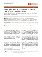
Figure 2
Neutrophil apoptosis pathways. Note that Fas, FADD, and FLICE are
also known as APO-1, MORT-1, and MACH, respectively. FADD, Fas-
associated death domain; FasL, Fas ligand; FLICE, FADD-like IL-1β
converting enzyme (ICE); IAP, inhibitor of apoptosis protein; NF-κB,
nuclear factor-κB; TNFR, tumor necrosis factor receptor; TRADD,
TNFR-associated protein death domain; TRAF, TNFR-associated
factor.
301
development and function, including the regulation of T-cell
development and apoptosis, and killing of inflammatory cells
at ‘immune-privileged’ sites [127–130]. Fas and FasL are of
crucial importance to initiation of apoptosis in human neu-
trophils. Anti-Fas antibodies accelerate neutrophil apoptosis
to a greater degree than do lymphocytes and monocytes
[131]. FasL exists either in soluble form or as a cell surface
molecule, forming part of the TNF family. FasL can bind three
Fas molecules simultaneously, causing clustering of the
death domains and leading to binding of specific intracellular
proteins. Fas-associated death domain (FADD) and FADD-
like IL-1β converting enzyme bind to the death domain, and
activate a family of specific cysteine proteases called ‘cas-
pases’ [121]. Caspases represent the machinery of cell
death: they inactivate proteins that protect against apoptosis;
they disable and deregulate proteins in general; and they par-
ticipate in direct disassembly of cell structures, including the
reorganization of the cytoskeleton and disruption of the
nucleus [132].
Tumor necrosis factor receptor I
TNF-α dramatically increases apoptosis rates in circulating
neutrophils of healthy human controls [133,134]. Similar to
FasL, three TNF-α molecules can trimerize on TNF receptor I,
leading to clustering of death domains and to binding by TNF
receptor associated death domain. Two distinct and indepen-
dent signaling pathways then proceed [135]: activation of
nuclear factor-κB (NF-κB); and activation of the caspase
pathway, leading to apoptosis (mediated through FADD,
similar to the Fas pathway) [135]. NF-κB regulates a wide
variety of genes that are involved in the synthesis of
hematopoietic growth factors, chemokines, and leukocyte
adhesion molecules [136–138]. Recent evidence also impli-
cates NF-κB activation as an important survival mechanism in
granulocytes. It has been shown to downregulate TNF-medi-
ated apoptosis in a negative feedback mechanism
[139–141]. The survival mechanism mediated by NF-κB
explains why TNF-α may not trigger apoptosis unless protein
synthesis is blocked. Given that activation of the death recep-
tor TNF receptor I leads to competing pathways, TNF-α will
have differential effects on neutrophil apoptosis, depending
on the activation state of the neutrophil [142].
The signaling pathways initiated by both TNF and FasL may
be ‘modulated’ by a variety of mediators in the inflammatory
environment. Specifically, delayed apoptosis in states of per-
sistent inflammation has been extensively investigated. Many
inflammatory mediators cause a delay in constitutive neu-
trophil apoptosis, and include IL-2 [143], IL-6 [144], IL-8
[145], G-CSF [146], GM-CSF [147], C5a, and lipopolysac-
charide [134,148]. In addition to constitutive apoptosis,
inducible apoptosis mediated by the Fas pathway is sup-
pressed by a variety of inflammatory mediators, including IL-8
[149], G-CSF, GM-CSF, interferon-γ, and TNF-α [150]. This
delay in Fas-mediated apoptosis secondary to inflammatory
cytokines may be diminished in elderly persons [151]. In addi-
tion, inflammatory mediators may alter intracellular factors
within neutrophils in order to delay apoptosis; these factors
include mitochondrial stability and caspases activity [152], in
addition to NF-κB activation. Other agents in the inflammatory
microenvironment that have been demonstrated to modulate
neutrophil apoptosis include immune complexes [153], reac-
tive oxygen intermediates [154], and red blood cells (possibly
secondary to scavenging oxidants) [155]. In addition,
engagement of neutrophil adhesion receptors will delay
apoptosis [156]. Thus, through alterations that occur during
and after neutrophil delivery to the exudate environment,
numerous agents modulate the rate of constitutive and
inducible neutrophil apoptosis.
Neutrophil cell surface expression in the
exudate environment
Neutrophils display altered membrane expression and cell
function following transmigration. Using monoclonal antibod-
ies directed toward surface molecules, characterization of the
neutrophil cell surface reveals significant and consistent alter-
ation in exudate neutrophil membrane expression. Our labora-
tory has previously demonstrated that exudate
polymorphonuclear neutrophils have enhanced microbicidal
activity, superoxide production, and augmented expression of
CD16 and the FMLP receptor, and are refractory to further
stimulation with TNF [157]. Multiple studies have confirmed
that human exudate neutrophils collected in skin windows are
primed for enhanced metabolic activation and phagocytic
activity [158–161]. In addition to altered function within the
inflammatory environment, exudate neutrophils demonstrate
altered membrane expression, including receptors that
mediate adhesion, chemotaxis, and function.
Adhesion receptors are altered after transmigration. Our labo-
ratory and others have found increased expression of CD11b,
decreased L-selectin, and decreased PECAM-1 expression in
exudate neutrophils following transmigration [57,157,162,163].
The loss of PECAM-1 is particularly interesting because it
mediates adhesion to endothelial cell corners and is neces-
sary for diapedesis (see discussion above) [56,164]. The
alteration in adhesion molecule expression may allow the neu-
trophil to complete the process of diapedesis, and undergoes
chemotaxis to a site of inflammation or infection.
Evidence suggests that change in the membrane expression
in exudate neutrophils is closely tied to the mobilization of
secretory vesicles. Exudate neutrophils collected in skin
windows displayed increased surface expression of alkaline
phosphatase, complement receptor 1, and CD11b/CD18,
but a complete loss of L-selectin following transmigration,
and the increase in the content of surface molecules in the
plasma membrane correlated with complete mobilization of
secretory vesicles [165]. Loss of specific granules also corre-
lated with increased number of FMLP receptors in exudate
neutrophils [161]. Thus, the changes to membrane expres-
sion are intrinsic to the change in neutrophil function.
Available online />302
Exudate neutrophils exhibit a reduced number of chemo-
attractant receptors, along with reduced chemotaxis. In
animal models, exudate neutrophils demonstrate reduced
chemotactic response [166]. In humans, neutrophils isolated
from skin windows have diminished chemotactic ability when
compared with circulating neutrophils [159]. Exudate neu-
trophils from pustules in a single patient exhibited markedly
reduced chemotaxis to C5a, FMLP, leukotriene-B
4
, and IL-8
when compared with circulating neutrophils [167]. Bron-
choalveolar lavage neutrophils in patients with chronic respi-
ratory tract infections have reduced IL-8 receptor A and IL-8
receptor B when compared with circulating neutrophils [168].
In addition to demonstrating alterations in chemoattractant
receptor expression, dynamic and variable alterations in neu-
trophil chemoattractant receptors in vivo correlate with
changes in cell function (chemotaxis). We have shown that
compared to control circulating neutrophils, exudate neu-
trophils (i.e. neutrophils that have undergone transmigration
to the extravascular inflammatory environment from healthy
subjects) simultaneously exhibit increased C5a receptors,
increased C5a chemotaxis, reduced IL-8 receptors (both IL-8
receptor A and B), and reduced IL-8 chemotaxis. In a sepa-
rate but related experiment again comparing to control circu-
lating neutrophils, circulating neutrophils isolated from septic
patients (APACHE II 23.6 ± 7.8) displayed reduced C5a
receptors, reduced C5a chemotaxis, a lesser decrease in IL-8
receptors with no change in IL-8 chemotaxis. These observa-
tions of in vivo receptor alteration and cell function suggest
both specific and generalizable conclusions, including: (1)
diminished chemoattractant receptors and chemotaxis in
septic neutrophils may account for decreased neutrophil
delivery to peripheral sites observed in these patients [169];
(2) exudate neutrophil chemotaxis may depend more on C5a
than on IL-8; and (3) change in neutrophil chemoattractant
receptor expression appears to regulate neutrophil chemo-
taxis in vivo.
Exudate neutrophils have delayed rates of physiologic cell
death, or apoptosis. In humans, exudate neutrophils have
decreased surface expression of FcγRIII [157] and FcγRIII is
known to be decreased in apoptosis [170,171]. In an animal
model, pulmonary exudate neutrophils exhibited delayed con-
stitutive and induced apoptosis [172]. In humans, using a
sterile skin blister skin window technique, we found that
exudate neutrophils had delayed apoptosis, a reduction in
TNF-α membrane binding, and a decreased susceptibility to
TNF-α-induced apoptosis [173]. Human salivary neutrophils
do not respond to a combination of TNF-α and cyclohex-
imide, unlike circulating neutrophils [174]. Thus, exudate neu-
trophils have delayed apoptosis, and will be less responsive
to certain apoptotic stimuli such as TNF.
In summary, following delivery to the inflammatory environ-
ment, neutrophils are primed for enhanced bactericidal activ-
ity, have altered expression of chemoattractant receptors and
chemotaxis, and are refractory to cell death. These mecha-
nisms have presumably developed in order to facilitate neu-
trophil effector function in host defense. Participating in and
being altered by the multiple sequential steps that are
involved in the neutrophil’s path from the circulation to the
inflammatory environment, the neutrophil membrane is
changed into a new configuration, reflecting the fact that the
function of the neutrophil (its overall properties) have
changed also.
Neutrophil connectivity
The cell membrane of the neutrophil is the principal means by
which the neutrophil interacts and communicates with its
environment, and it is not surprising that the membrane medi-
ates the processes that are inherent to the neutrophil’s life-
cycle. As a complementary interpretation of this observation,
the neutrophil membrane offers a measure of neutrophil ‘con-
nectivity’, that is, the degree and nature of its interconnected-
ness with other elements within the host response. This
concept becomes essential when evaluating a complex non-
linear system, such the systemic host response to trauma,
shock, or sepsis [175].
A complex system may be thought of as one that is able to
exist in stable states, with systemic properties and functions
that are wholly distinct from the innumerable, interconnected,
interdependent parts of the system, which are continuously
engaged in a dynamic web of nonlinear relationships. The
systemic host response, with its interdependent metabolic,
neural, endocrine, immune, and inflammatory systems, may be
regarded as a complex system [175]. In addition, the neu-
trophil itself may be regarded as a complex system in its own
right, with its own systemic or ‘emergent’ properties. Emer-
gent properties of the neutrophil might include adhesiveness,
chemotactic ability, activation state, and rate of cell death, all
of which represent a measure of cell function. We previously
observed that changes in variability (patterns of change over
time) and connectivity (patterns of interconnection over
space) of the elements of a complex system may be utilized
as a measure of changes in the systemic properties of that
complex system, which define whether a patient is healthy or
ill [175]. The demonstration that altered connectivity (i.e. neu-
trophil membrane expression) is associated with altered
emergent properties (i.e. function) of the neutrophil repre-
sents a demonstration of this hypothesis on a smaller scale.
These observations suggest further hypotheses for investiga-
tion. For example, utilizing dynamic measurement of neu-
trophil membrane expression as a technology to analyze
neutrophil function, it may be possible to identify which
patients might benefit from attempted immunomodulation,
and when an intervention should be performed.
Conclusion
As the foremost circulating phagocyte, which is essential to
normal effective host defense and is responsible for host tissue
injury in states of persistent inflammation, the neutrophil has
undergone extensive investigation. The present review arrived
Critical Care August 2003 Vol 7 No 4 Seely et al.
303
at two principal conclusions: the neutrophil membrane medi-
ates the processes that are integral to neutrophil delivery, func-
tion, and clearance; and alterations in membrane expression
occur with changes in cell function. The neutrophil membrane
mediates neutrophil delivery, including neutrophil–endothelial
cell interactions, rolling, adhesion, and diapedesis. During this
process, and in the interstitial inflammatory environment, the
neutrophil responds to various chemoattractants, based on the
presence and binding capacity of the appropriate receptors. In
the inflammatory environment, neutrophil membrane receptors
participate in phagocytosis, priming, and activation, leading to
release of a toxic arsenal of granules and activation of the mem-
brane-bound respiratory burst. Following completion of its func-
tion, the neutrophil is cleared via physiologic cell death or
apoptosis, a process that is activated by membrane-bound
death receptors. In summary, because the neutrophil mem-
brane is the principal means by which the cell interacts with its
surroundings, it is the principle mediator of neutrophil develop-
ment during the neutrophil lifecycle. The second principal con-
clusion that may be derived from the evaluation of neutrophil
membrane expression and function is that alterations in the
neutrophil membrane are synonymous with alterations in cell
function, and observation whose clinical significance merits
exploration. Thus, in addition to the neutrophil membrane medi-
ating cell processes during the neutrophil lifecycle, changes in
membrane expression allows for in vivo regulation of cellular
function.
Competing interests
None declared.
References
1. Baehner RL (editor). Neutrophil structure and function in Hema-
tology: Basic Principles and Practice. New York: Churchill-Living-
stone 2000, Chapter 38: page 667-669.
2. Cannistra SA, Griffin JD: Regulation of the production and
function of granulocytes and monocytes. Semin Hematol
1988, 25:173-188.
3. Lieschke GJ, Burgess AW: Granulocyte colony-stimulating
factor and granulocyte-macrophage colony-stimulating factor
(1). N Engl J Med 1992, 327:28-35.
4. Tidow N, Welte K: Advances in understanding postreceptor
signaling in response to granulocyte colony-stimulating
factor. Curr Opin Hematol 1997, 4:171-175.
5. Khwaja A, Carver J, Jones HM, Linch DC: Dynamic modulation
of the cell surface expression of the granulocyte-macrophage
colony-stimulating factor receptor. Br J Haematol 1993, 85:42-
49.
6. Hermans MH, Antonissen C, Ward AC, Mayen AE, Ploemacher
RE, Touw IP: Sustained receptor activation and hyperprolifera-
tion in response to granulocyte colony-stimulating factor (G-
CSF) in mice with a severe congenital neutropenia/acute
myeloid leukemia-derived mutation in the G-CSF receptor
gene. J Exp Med 1999, 189:683-692.
7. Ward AC, van Aesch YM, Schelen AM, Touw IP: Defective inter-
nalization and sustained activation of truncated granulocyte
colony-stimulating factor receptor found in severe congenital
neutropenia/acute myeloid leukemia. Blood 1999, 93:447-458.
8. Shoup M, Weisenberger JM, Wang JL, Pyle JM, Gamelli RL,
Shankar R: Mechanisms of neutropenia involving myeloid mat-
uration arrest in burn sepsis. Ann Surg 1998, 228:112-122.
9. Boggs DR: The kinetics of neutrophilic leukocytes in health
and in disease. Semin Hematol 1967, 4:359-386.
10. Hogg JC: Neutrophil kinetics and lung injury. Physiol Rev
1987, 67:1249-1295.
11. Downey GP, Worthen GS, Henson PM Hyde DM: Neutrophil
sequestration and migration in localized pulmonary inflamma-
tion. Capillary localization and migration across the inter-
alveolar septum. Am Rev Respir Dis 1993, 147:168-176.
12. Schmid-Schonbein GW, Usami S, Skalak R, Chien S: The inter-
action of leukocytes and erythrocytes in capillary and post-
capillary vessels. Microvasc Res 1980, 19:45-70.
13. Blixt A, Jonsson P, Braide M, Bagge U: Microscopic studies on
the influence of erythrocyte concentration on the post-junc-
tional radial distribution of leukocytes at small venular junc-
tions. Int J Microcirc Clin Exp 1985, 4:141-156.
14. Firrell JC, Lipowsky HH: Leukocyte margination and deforma-
tion in mesenteric venules of rat. Am J Physiol 1989, 256:
H1667-H1674.
15. Perry MA, Granger DN: Role of CD11/CD18 in shear rate-
dependent leukocyte-endothelial cell interactions in cat
mesenteric venules. J Clin Invest 1991, 87:1798-1804.
16. Springer TA: Traffic Signals for Lymphocyte recirculation and
leukocyte emigration: the multistep paradigm. Cell 1994, 76:
301-314.
17. Kubes P, Jutila M, Payne D: Therapeutic potential of inhibiting
leukocyte rolling in ischemia/reperfusion. J Clin Invest 1995,
95:2510-2519.
18. Ley K, Bullard DC, Arbones ML, Bosse R, Vestweber D, Tedder
TF, Beaudet AL: Sequential contribution of L- and P-selectin to
leukocyte rolling in vivo. J Exp Med 1995, 181:669-675.
19. Rosen SD: Cell surface lectins in the immune system. Semin
Immunol 1993, 5:237-247.
20. Spertini O, Luscinskas FW, Kansas GS, Munro JM, Griffin JD, Gim-
brone MA Jr, Tedder TF: Leukocyte adhesion molecule-1 (LAM-1,
L-selectin) interacts with an inducible endothelial cell ligand to
support leukocyte adhesion. J Immunol 1991, 147:2565-2573.
21. Kanwar S, Bullard DC, Hickey MJ, Smith CW, Beaudet AL,
Wolitzky BA, Kubes P: The association between alpha4-inte-
grin, P-selectin, and E-selectin in an allergic model of inflam-
mation. J Exp Med 1997, 185:1077-1087.
22. Mulligan MS, Varani J, Dame MK, Lane CL, Smith CW, Anderson
DC, Ward PA: Role of endothelial-leukocyte adhesion mole-
cule 1 (ELAM-1) in neutrophil-mediated lung injury in rats. J
Clin Invest 1991, 88:1396-1406.
23. Lindbom L, Xie X, Raud J, Hedqvist P: Chemoattractant-induced
firm adhesion of leukocytes to vascular endothelium in vivo is
critically dependent on initial leukocyte rolling. Acta Physiol
Scand 1992, 146:415-421.
24. Von Andrian UH, Hansell P, Chambers JD, Berger EM, Torres
Filho I, Butcher EC, Arfors KE: L-selectin function is required for
beta 2-integrin-mediated neutrophil adhesion at physiological
shear rates in vivo. Am J Physiol 1992, 263:H1034-H1044.
25. Bienvenu K, Granger DN: Molecular determinants of shear
rate-dependent leukocyte adhesion in postcapillary venules.
Am J Physiol 1993, 264:H1504-H1508.
26. Mulligan MS, Miyasaka M, Tamatani T, Jones ML, Ward PA:
Requirements for L-selectin in neutrophil-mediated lung
injury in rats. J Immunol 1994, 152:832-840.
27. Ferri LE, Pascual J, Seely AJ, Chaudhury P, Christou NV: Soluble
L-selectin attenuates tumor necrosis factor-alpha-mediated
leukocyte adherence and vascular permeability: a protective
role for elevated soluble L-selectin in sepsis. Crit Care Med
2002, 30:1842-7.
28. Anderson DC, Springer TA: Leukocyte adhesion deficiency: an
inherited defect in the Mac-1, LFA-1, and p150,95 glycopro-
teins. Annu Rev Med 1987, 38:175-194.
29. Smith CW, Rothlein R, Hughes BJ, Mariscalco MM, Rudloff HE,
Schmalstieg FC, Anderson DC: Recognition of an endothelial
determinant for CD 18-dependent human neutrophil adherence
and transendothelial migration. J Clin Invest 1988, 82:1746-1756.
30. Granger DN, Kubes P: The microcirculation and inflammation:
modulation of leukocyte-endothelial cell adhesion. J Leuko-
cyte Biol 1994, 55:662-675.
31. Zimmerman GA, Prescott SM, McIntyre TM: Endothelial cell
interactions with granulocytes: tethering and signaling mole-
cules. Immunol Today 1992, 13:93-100.
32. von Andrian UH, Chambers JD, McEvoy LM, Bargatze RF, Arfors
KE, Butcher EC: Two-step model of leukocyte-endothelial cell
interaction in inflammation: distinct roles for LECAM-1 and the
leukocyte beta 2 integrins in vivo. Proc Natl Acad Sci USA
1991, 88:7538-7542.
Available online />304
33. Argenbright LW, Letts LG, Rothlein R: Monoclonal antibodies to
the leukocyte membrane CD18 glycoprotein complex and to
intercellular adhesion molecule-1 inhibit leukocyte-endothe-
lial adhesion in rabbits. J Leukoc Biol 1991, 49:253-257.
34. Smith CW: Endothelial adhesion molecules and their role in
inflammation. Can J Physiol Pharmacol 1993, 71:76-87.
35. Doerschuk CM, Winn RK, Coxson HO, Harlan JM: CD18-depen-
dent and -independent mechanisms of neutrophil emigration
in the pulmonary and systemic microcirculation of rabbits. J
Immunol 1990, 144:2327-2333.
36. Winn RK, Harlan JM: CD18-independent neutrophil and
mononuclear leukocyte emigration into the peritoneum of
rabbits. J Clin Invest 1993, 92:1168-1173.
37. Thomas DD, Sharar SR, Winn RK, Chi EY, Verrier ED, Allen MD,
Bishop MJ: CD18-independent mechanism of neutrophil emi-
gration in the rabbit lung after ischemia-reperfusion. Ann
Thorac Surg 1995, 60:1360-1366.
38. Langdale LA, Flaherty LC, Liggitt HD, Harlan JM, Rice CL, Winn
RK: Neutrophils contribute to hepatic ischemia-reperfusion
injury by a CD18- independent mechanism. J Leukoc Biol
1993, 53:511-517.
39. Conlan JW, North RJ: Listeria monocytogenes, but not Salmo-
nella typhimurium, elicits a CD18-independent mechanism of
neutrophil extravasation into the murine peritoneal cavity.
Infect Immun 1994, 62:2702-2706.
40. Kubes P, Niu XF, Smith CW, Kehrli ME Jr, Reinhardt PH,
Woodman RC: A novel beta 1-dependent adhesion pathway
on neutrophils: a mechanism invoked by dihydrocytocha-
lasin B or endothelial transmigration. FASEB J 1995, 9:
1103-1111.
41. Krauss K, Altevogt P: Integrin leukocyte function-associated
antigen-1-mediated cell binding can be activated by cluster-
ing of membrane rafts. J Biol Chem 1999, 274:36921-36927.
42. Hermanowski-Vosatka A, Van Strijp JA, Swiggard WJ, Wright SD:
Integrin modulating factor-1: a lipid that alters the function of
leukocyte integrins. Cell 1992, 68:341-352.
43. Carlos TM, Harlan JM: Membrane proteins involved in phago-
cyte adherence to endothelium. Immunol Rev 1990, 114:5-28.
44. Carveth HJ, Bohnsack JF, McIntyre TM, Baggiolini M, Prescott
SM, Zimmerman GA: Neutrophil activating factor (NAF)
induces polymorphonuclear leukocyte adherence to endothe-
lial cells and to subendothelial matrix proteins. Biochem
Biophys Res Commun 1989, 162:387-393.
45. Detmers PA, Lo SK, Olsen-Egbert E, Walz A, Baggiolini M, Cohn
ZA: Neutrophil-activating protein 1/interleukin 8 stimulates
the binding activity of the leukocyte adhesion receptor
CD11b/CD18 on human neutrophils. J Exp Med 1990, 171:
1155-1162.
46. Pober JS, Cotran RS: The role of endothelial cells in inflamma-
tion. Transplantation 1990, 50:537-544.
47. Pober JS, Cotran RS: Cytokines and endothelial cell biology.
Physiol Rev 1990, 70:427-451.
48. Yan HC, Juhasz I, Pilewski J, Murphy GF, Herlyn M, Albelda SM:
Human/severe combined immunodeficient mouse chimeras.
An experimental in vivo model system to study the regulation
of human endothelial cell-leukocyte adhesion molecules. J
Clin Invest 1993, 91:986-996.
49. Smith CW: Leukocyte-endothelial cell interactions. Semin
Hematol 1993, 30:45-53; discussion 54-55.
50. Del Maschio A, Zanetti A, Corada M, Rival Y, Ruco L, Lampugnani
MG, Dejana E: Polymorphonuclear leukocyte adhesion trig-
gers the disorganization of endothelial cell-to-cell adherens
junctions. J Cell Biol 1996, 135:497-510.
51. Burns AR, Walker DC, Brown ES, Thurmon LT, Bowden RA,
Keese CR, Simon SI, Entman ML, Smith CW: Neutrophil
transendothelial migration is independent of tight junctions
and occurs preferentially at tricellular corners. J Immunol
1997, 159:2893-2903.
52. Kurose I, Anderson DC, Miyasaka M, Tamatani T, Paulson JC,
Todd RF, Rusche JR, Granger DN: Molecular determinants of
reperfusion-induced leukocyte adhesion and vascular protein
leakage. Circ Res 1994, 74:336-343.
53. Zimmerman BJ, Holt JW, Paulson JC, Anderson DC, Miyasaka M,
Tamatani T, Todd RF III, Rusche JR, Granger DN: Molecular
determinants of lipid mediator-induced leukocyte adherence
and emigration in rat mesenteric venules. Am J Physiol 1994,
266:H847-H853.
54. Berman ME, Muller WA: Ligation of platelet/endothelial cell
adhesion molecule 1 (PECAM- 1/CD31) on monocytes and
neutrophils increases binding capacity of leukocyte CR3
(CD11b/CD18). J Immunol 1995, 154:299-307.
55. Kuwabara H, Tanaka S, Sakamoto H, Oryu M, Uda H: Antibody
mediated ligation of platelet/endothelial cell adhesion mole-
cule-1 (PECAM-1) on neutrophils enhances adhesion to cul-
tured human dermal microvascular endothelial cells. Kobe J
Med Sci 1996, 42:233-241.
56. Muller WA, Weigl SA, Deng X, Phillips DM: PECAM-1 is required
for transendothelial migration of leukocytes. J Exp Med 1993,
178:449-460.
57. Christofidou-Solomidou M, Nakada MT, Williams J, Muller WA,
DeLisser HM: Neutrophil platelet endothelial cell adhesion
molecule-1 participates in neutrophil recruitment at inflam-
matory sites and is down-regulated after leukocyte extravasa-
tion. J Immunol 1997, 158:4872-4878.
58. Ryan GB, Hurley JV: The chemotaxis of polymorphonuclear
leucocytes towards damaged tissue. Br J Exp Pathol 1966, 47:
530-536.
59. Cassatella MA, Bazzoni F, Ceska M, Ferro I, Baggiolini M, Berton
G: IL-8 production by human polymorphonuclear leukocytes.
The chemoattractant formyl-methionyl-leucyl-phenylalanine
induces the gene expression and release of IL-8 through a
pertussis toxin-sensitive pathway. J Immunol 1992, 148:3216-
3220.
60. Kunkel SL, Lukacs NW, Strieter RM: The role of interleukin-8 in
the infectious process. Ann N Y Acad Sci 1994, 730:134-143.
61. Ford-Hutchinson AW, Bray MA, Doig MV, Shipley ME, Smith MJ:
Leukotriene B, a potent chemokinetic and aggregating sub-
stance released from polymorphonuclear leukocytes. Nature
1980, 286:264-265.
62. Schiffmann E, Corcoran BA, Wahl SM: N-formylmethionyl pep-
tides as chemoattractants for leucocytes. Proc Natl Acad Sci
USA 1975, 72:1059-1062.
63. Fernandez HN, Henson PM, Otani A, Hugli TE: Chemotactic
response to human C3a and C5a anaphylatoxins. I. Evaluation
of C3a and C5a leukotaxis in vitro and under stimulated in
vivo conditions. J Immunol 1978, 120:109-115.
64. Oppenheim JJ, Zachariae CO, Mukaida N, Matsushima K: Proper-
ties of the novel proinflammatory supergene “intercrine”
cytokine family. Annu Rev Immunol 1991, 9:617-648.
65. Kunkel SL, Lukacs N, Strieter RM: Expression and biology of
neutrophil and endothelial cell-derived chemokines. Semin
Cell Biol 1995, 6:327-336.
66. Huber AR, Kunkel SL, Todd RFD, Weiss SJ: Regulation of
transendothelial neutrophil migration by endogenous inter-
leukin-8. Science 1991, 254:99-102. (published errata appear in
Science 1991, 254:631 and 1435.)
67. Smart SJ, Casale TB: TNF-alpha-induced transendothelial neu-
trophil migration is IL-8 dependent. Am J Physiol 1994, 266:
L238-L245.
68. Smart SJ, Casale TB: Pulmonary epithelial cells facilitate TNF-
alpha-induced neutrophil chemotaxis. A role for cytokine net-
working. J Immunol 1994, 152:4087-4094.
69. Mulligan MS, Jones ML, Bolanowski MA, Baganoff MP, Deppeler
CL, Meyers DM, Ryan US, Ward PA: Inhibition of lung inflam-
matory reactions in rats by an anti-human IL-8 antibody. J
Immunol 1993, 150:5585-5595.
70. Strieter RM, Koch AE, Antony VB, Fick RB Jr, Standiford TJ,
Kunkel SL: The immunopathology of chemotactic cytokines:
the role of interleukin-8 and monocyte chemoattractant
protein-1. J Lab Clin Med 1994, 123:183-197.
71. Kupper RW, Dewald B, Jakobs KH, Baggiolini M, Gierschik P: G-
protein activation by interleukin 8 and related cytokines in
human neutrophil plasma membranes. Biochem J 1992, 282:
429-434.
72. Kelvin DJ, Michiel DF, Johnston JA, Lloyd AR, Sprenger H, Oppen-
heim JJ, Wang JM: Chemokines and serpentines: the molecu-
lar biology of chemokine receptors. J Leukoc Biol 1993, 54:
604-612.
73. Ahuja SK, Murphy PM: The CXC chemokines growth-regulated
oncogene (GRO) alpha, GRObeta, GROgamma, neutrophil-
activating peptide-2, and epithelial cell-derived neutrophil-
activating peptide-78 are potent agonists for the type B, but
not the type A, human interleukin-8 receptor. J Biol Chem
1996, 271:20545-20550.
Critical Care August 2003 Vol 7 No 4 Seely et al.
305
74. Hammond ME, Lapointe GR, Feucht PH, Hilt S, Gallegos CA,
Gordon CA, Giedlin MA, Mullenbach G, Tekamp-Olson P: IL-8
induces neutrophil chemotaxis predominantly via type I IL-8
receptors. J Immunol 1995, 155:1428-1433.
75. Santamaria Babi LF, Moser B, Perez Soler MT, Moser R,
Loetscher P, Villiger B, Blaser K, Hauser C: The interleukin-8
receptor B and CXC chemokines can mediate transendothe-
lial migration of human skin homing T cells. Eur J Immunol
1996, 26:2056-2061.
76. Botha AJ, Moore FA, Moore EE, Fontes B, Banerjee A, Peterson
VM: Postinjury neutrophil priming and activation states: thera-
peutic challenges [editorial]. Shock 1995, 3:157-166.
77. Guthrie LA, McPhail LC, Henson PM, Johnston RB Jr: Priming of
neutrophils for enhanced release of oxygen metabolites by
bacterial lipopolysaccharide. Evidence for increased activity of
the superoxide-producing enzyme. J Exp Med 1984, 160:
1656-1671.
78. Kuhns DB, Wright DG, Nath J, Kaplan SS, Basford RE: ATP
induces transient elevations of [Ca2+]i in human neutrophils
and primes these cells for enhanced O
2
–
generation. Lab
Invest 1988, 58:448-453.
79. Vercellotti GM, Yin HQ, Gustafson KS, Nelson RD, Jacob HS:
Platelet-activating factor primes neutrophil responses to ago-
nists: role in promoting neutrophil-mediated endothelial
damage. Blood 1988, 71:1100-1107.
80. Wozniak A, Betts WH, McLennan G, Scicchitano R: Activation of
human neutrophils by tachykinins: effect on formyl-methionyl-
leucyl-phenylalanine- and platelet-activating factor-stimulated
superoxide anion production and antibody-dependent cell-
mediated cytotoxicity. Immunology 1993, 78:629-634.
81. Biffl WL, Moore EE, Moore FA, Carl VS, Kim FJ, Franciose RJ:
Interleukin-6 potentiates neutrophil priming with platelet-acti-
vating factor. Arch Surg 1994, 129:1131-1136.
82. Forehand JR, Pabst MJ, Phillips WA, Johnston RB Jr:
Lipopolysaccharide priming of human neutrophils for an
enhanced respiratory burst. Role of intracellular free calcium.
J Clin Invest 1989, 83:74-83.
83. Gay JC, Beckman JK, Brash AR, Oates JA, Lukens JN: Enhance-
ment of chemotactic factor-stimulated neutrophil oxidative
metabolism by leukotriene B4. Blood 1984, 64:780-785.
84. Weisbart RH, Golde DW, Gasson JC: Biosynthetic human GM-
CSF modulates the number and affinity of neutrophil f-Met-
Leu-Phe receptors. J Immunol 1986, 137:3584-3587.
85. Atkinson YH, Marasco WA, Lopez AF, Vadas MA: Recombinant
human tumor necrosis factor-alpha. Regulation of N- formyl-
methionylleucylphenylalanine receptor affinity and function on
human neutrophils. J Clin Invest 1988, 81:759-765.
86. O’Flaherty JT, Rossi AG, Redman JF, Jacobson DP: Tumor necrosis
factor-alpha regulates expression of receptors for formyl-
methionyl-leucyl-phenylalanine, leukotriene B4, and platelet-
activating factor. Dissociation from priming in human poly-
morphonuclear neutrophils. J Immunol 1991, 147:3842-3847.
87. Zimmerli W, Reber AM, Dahinden CA: The role of formylpeptide
receptors, C5a receptors, and cytosolic-free calcium in neu-
trophil priming. J Infect Dis 1990, 161:242-249.
88. Tennenberg SD, Fey DE, Lieser MJ: Oxidative priming of neu-
trophils by interferon-gamma. J Leukoc Biol 1993, 53:301-308.
89. McColl SR, Beauseigle D, Gilbert C, Naccache PH: Priming of
the human neutrophil respiratory burst by granulocyte-
macrophage colony-stimulating factor and tumor necrosis
factor-alpha involves regulation at a post-cell surface recep-
tor level. Enhancement of the effect of agents which directly
activate G proteins. J Immunol 1990, 145:3047-3053.
90. Wozniak A, Betts WH, Murphy GA, Rokicinski M: Interleukin-8
primes human neutrophils for enhanced superoxide anion
production. Immunology 1993, 79:608-615.
91. Walker BA, Hagenlocker BE, Ward PA: Superoxide responses
to formyl-methionyl-leucyl-phenylalanine in primed neu-
trophils. Role of intracellular and extracellular calcium. J
Immunol 1991, 146:3124-3131.
92. Richter J, Andersson T, Olsson I: Effect of tumor necrosis factor
and granulocyte/macrophage colony-stimulating factor on
neutrophil degranulation. J Immunol 1989, 142:3199-3205.
93. Dewald B, Baggiolini M: Activation of NADPH oxidase in
human neutrophils. Synergism between fMLP and the neu-
trophil products PAF and LTB4. Biochem Biophys Res
Commun 1985, 128:297-304.
94. Robinson JM, Badwey JA, Karnovsky ML, Karnovsky MJ: Super-
oxide release by neutrophils: synergistic effects of a phorbol
ester and a calcium ionophore. Biochem Biophys Res Commun
1984, 122:734-739.
95. Partrick DA, Moore FA, Moore EE, Barnett CC Jr, Silliman CC:
Neutrophil priming and activation in the pathogenesis of
postinjury multiple organ failure. New Horizons 1996, 4:194-
210.
96. Moore EE, Moore FA, Franciose RJ, Kim FJ, Biffl WL, Banerjee A:
The postischemic gut serves as a priming bed for circulating
neutrophils that provoke multiple organ failure. J Trauma
1994, 37:881-887.
97. Tanaka H, Ogura H, Yokota J, Sugimoto H, Yoshioka T, Sugimoto
T: Acceleration of superoxide production from leukocytes in
trauma patients. Ann Surg 1991, 214:187-192.
98. Smith JA: Neutrophils, host defense, and inflammation: a
double-edged sword. J Leukoc Biol 1994, 56:672-686.
99. Haslett C: Introduction: the paradox of inflammation. Semin
Cell Biol 1995, 6:315-316.
100. Malech HL, Gallin JI: Current concepts: immunology. Neu-
trophils in human diseases. N Engl J Med 1987, 317:687-694.
101. Weiss SJ: Tissue destruction by neutrophils. N Engl J Med
1989, 320:365-376.
102. Heijnen IA, van de Winkel JG: Human IgG Fc receptors. Int Rev
Immunol 1997, 16:29-55.
103. Chuang FY, Sassaroli M, Unkeless JC: Convergence of Fc
gamma receptor IIA and Fc gamma receptor IIIB signaling
pathways in human neutrophils. J Immunol 2000, 164:350-
360.
104. Repp R, Valerius T, Sendler A, Gramatzki M, Iro H, Kalden JR,
Platzer E: Neutrophils express the high affinity receptor for
IgG (Fc gamma RI, CD64) after in vivo application of recombi-
nant human granulocyte colony- stimulating factor. Blood
1991, 78:885-889.
105. Fjaertoft G, Hakansson L, Ewald U, Foucard T, Venge P: Neu-
trophils from term and preterm newborn infants express the
high affinity Fcgamma-receptor I (CD64) during bacterial
infections. Pediatr Res 1999, 45:871-876.
106. Fischer G, Schneider EM, L Moldawer LL, Karcher C, Barth E,
Suger-Wiedeck H, Georgieff M, Weiss M: CD64 surface expres-
sion on neutrophils is transiently upregulated in patients with
septic shock. Intensive Care Med 2001, 27:1848-1852.
107. Hampton MB, Kettle AJ, Winterbourn CC: Inside the neutrophil
phagosome: oxidants, myeloperoxidase, and bacterial killing.
Blood 1998, 92:3007-3017.
108. Borregaard N, Lollike K, Kjeldsen L, Sengelov H, Bastholm L,
Nielsen MH, Bainton DF: Human neutrophil granules and
secretory vesicles. Eur J Haematol 1993, 51:187-198.
109. Vaissiere C, Le Cabec V, Maridonneau-Parini I: NADPH oxidase
is functionally assembled in specific granules during activa-
tion of human neutrophils. J Leukoc Biol 1999, 65:629-634.
110. Varani J, Ward PA: Mechanisms of neutrophil-dependent and
neutrophil-independent endothelial cell injury. Biol Signals
1994, 3:1-14.
111. Travis J, Salvesen GS: Human plasma proteinase inhibitors.
Annu Rev Biochem 1983, 52:655-709.
112. Vaux DL, Haecker G, Strasser A: An evolutionary perspective
on apoptosis. Cell 1994, 76:777-779.
113. Thompson CB: Apoptosis in the pathogenesis and treatment
of disease. Science 1995, 267:1456-1462.
114. Kerr JF, Wyllie AH, Currie AR: Apoptosis: a basic biological
phenomenon with wide-ranging implications in tissue kinet-
ics. Br J Cancer 1972, 26:239-257.
115. Majno G, Joris I: Apoptosis, oncosis, and necrosis. An overview
of cell death. Am J Pathol 1995, 146:3-15.
116. Fadok VA, Savill JS, Haslett C, Bratton DL, Doherty DE, Campbell
PA, Henson PM: Different populations of macrophages use
either the vitronectin receptor or the phosphatidylserine
receptor to recognize and remove apoptotic cells. J Immunol
1992, 149:4029-4035.
117. Martin SJ, Reutelingsperger CP, McGahon AJ, Rader JA, van Schie
RC, LaFace DM, Green DR: Early redistribution of plasma
membrane phosphatidylserine is a general feature of apopto-
sis regardless of the initiating stimulus: inhibition by overex-
pression of Bcl-2 and Abl. J Exp Med 1995, 182:1545-1556.
118. Fadok VA, Voelker DR, Campbell PA, Bratton DL, Cohen JJ, Noble
PW, Riches DW, Henson PM: The ability to recognize phos-
Available online />306
phatidylserine on apoptotic cells is an inducible function in
murine bone marrow-derived macrophages. Chest 1993, 103
(suppl):102S.
119. Koopman G, Reutelingsperger CP, Kuijten GA, Keehnen RM, Pals
ST, van Oers MH: Annexin V for flow cytometric detection of
phosphatidylserine expression on B cells undergoing apopto-
sis. Blood 1994, 84:1415-1420.
120. Vermes I, Haanen C, Steffens-Nakken H, Reutelingsperger C: A
novel assay for apoptosis. Flow cytometric detection of phos-
phatidylserine expression on early apoptotic cells using fluo-
rescein labelled Annexin V. J Immunol Methods 1995, 184:
39-51.
121. Ashkenazi A, Dixit VM: Death receptors: signaling and modula-
tion. Science 1998, 281:1305-1308.
122. Tartaglia LA, Ayres TM, Wong GH, Goeddel DV: A novel domain
within the 55 kd TNF receptor signals cell death. Cell 1993,
74:845-853.
123. Nagata S: Apoptosis by death factor. Cell 1997, 88:355-365.
124. Smith CA, Farrah T, Goodwin RG: The TNF receptor superfam-
ily of cellular and viral proteins: activation, costimulation, and
death. Cell 1994, 76:959-962.
125. Nagata S: Fas and Fas ligand: a death factor and its receptor.
Adv Immunol 1994, 57:129-144.
126. Lynch DH, Ramsdell F, Alderson MR: Fas and FasL in the home-
ostatic regulation of immune responses. Immunology Today
1995, 16:569-574.
127. Nagata S, Golstein P: The Fas death factor. Science 1995, 267:
1449-1456.
128. Dhein J, Walczak H, Baumler C, Debatin KM, Krammer PH:
Autocrine T-cell suicide mediated by APO-1/(Fas/CD95).
Nature 1995, 373:438-441.
129. Alderson MR, Tough TW, Davis-Smith T, Braddy S, Falk B,
Schooley KA, Goodwin RG, Smith CA, Ramsdell F, Lynch DH:
Fas ligand mediates activation-induced cell death in human T
lymphocytes. J Exp Med 1995, 181:71-77.
130. Griffith TS, Brunner T, Fletcher SM, Green DR, Ferguson TA: Fas
ligand-induced apoptosis as a mechanism of immune privi-
lege. Science 1995, 270:1189-1192.
131. Iwai K, Miyawaki T, Takizawa T, Konno A, Ohta K, Yachie A, Seki H,
Taniguchi N: Differential expression of bcl-2 and susceptibility
to anti-Fas-mediated cell death in peripheral blood lympho-
cytes, monocytes, and neutrophils. Blood 1994, 84:1201-1208.
132. Thornberry NA, Lazebnik Y: Caspases: enemies within. Science
1998, 281:1312-1316.
133. Takeda Y, Watanabe H, Yonehara S, Yamashita T, Saito S, Sendo
F: Rapid acceleration of neutrophil apoptosis by tumor necro-
sis factor-alpha. Int Immunol 1993, 5:691-694.
134. Watson RW, Redmond HP, Wang JH, Bouchier-Hayes D: Bacte-
rial ingestion, tumor necrosis factor-alpha, and heat induce
programmed cell death in activated neutrophils. Shock 1996,
5:47-51.
135. Hsu H, Xiong J, Goeddel DV: The TNF receptor 1-associated
protein TRADD signals cell death and NF-kappa B activation.
Cell 1995, 81:495-504.
136. Baeuerle PA, Henkel T: Function and activation of NF-kappa B
in the immune system. Annu Rev Immunol 1994, 12:141-179.
137. Baldwin AS Jr: The NF-kappa B and I kappa B proteins: new
discoveries and insights. Annu Rev Immunol 1996, 14:649-683.
138. Schutze S, Wiegmann K, Machleidt T, Kronke M: TNF-induced
activation of NF-kappa B. Immunobiology 1995, 193:193-203.
139. Van Antwerp DJ, Martin SJ, Kafri T, Green DR, Verma IM: Sup-
pression of TNF-alpha-induced apoptosis by NF-kappaB.
Science 1996, 274:787-789.
140. Wang CY, Mayo MW, Baldwin AS Jr: TNF- and cancer therapy-
induced apoptosis: potentiation by inhibition of NF-kappaB.
Science 1996, 274:784-787.
141. Van Antwerp DJ, Martin SJ, Verma IM, Green DR: Inhibition of
TNF-induced apoptosis by NF-kappa B. Trends Cell Biol 1998,
8:107-111.
142. Homburg CH, Roos D: Apoptosis of neutrophils. Curr Opin
Hematol 1996, 3:94-99.
143. Pericle F, Liu JH, Diaz JI, Blanchard DK, Wei S, Forni G, Djeu JY:
Interleukin-2 prevention of apoptosis in human neutrophils.
Eur J Immunol 1994, 24:440-444.
144. Biffl WL, Moore EE, Moore FA, Barnett CC Jr, Carl VS, Peterson
VN: Interleukin-6 delays neutrophil apoptosis. Arch Surg
1996, 131:24-29; discussion 29-30.
145. Kettritz R, Gaido ML, Haller H, Luft FC, Jennette CJ, Falk RJ:
Interleukin-8 delays spontaneous and tumor necrosis factor-
alpha-mediated apoptosis of human neutrophils. Kidney Int
1998, 53:84-91.
146. Adachi S, Kubota M, Lin YW, Okuda A, Matsubara K, Wakazono
Y, Hirota H, Kuwakado K, Akiyama Y: In vivo administration of
granulocyte colony-stimulating factor promotes neutrophil
survival in vitro. Eur J Haematol 1994, 53:129-134.
147. Cox G, Gauldie J, Jordana M: Bronchial epithelial cell-derived
cytokines (G-CSF and GM-CSF) promote the survival of
peripheral blood neutrophils in vitro. Am J Respir Cell Mol Biol
1992, 7:507-513.
148. Lee A, Whyte MK, Haslett C: Inhibition of apoptosis and pro-
longation of neutrophil functional longevity by inflammatory
mediators. J Leukocyte Biol 1993, 54:283-288.
149. Leuenroth S, Lee C, Grutkoski P, Keeping H, Simms HH: Inter-
leukin-8-induced suppression of polymorphonuclear leuko-
cyte apoptosis is mediated by suppressing CD95 (Fas/Apo-1)
Fas-1 interactions. Surgery 1998, 124:409-417.
150. Liles WC, Kiener PA, Ledbetter JA, Aruffo A, Klebanoff SJ: Differ-
ential expression of Fas (CD95) and Fas ligand on normal
human phagocytes: implications for the regulation of apopto-
sis in neutrophils. J Exp Med 1996, 184:429-440.
151. Tortorella C, Piazzolla G, Spaccavento F, Pece S, Jirillo E,
Antonaci S: Spontaneous and Fas-induced apoptotic cell
death in aged neutrophils. J Clin Immunol 1998, 18:321-329.
152. Watson RW, O’Neill A, Brannigen AE, Coffey R, Marshall JC,
Brady HR, Fitzpatrick JM: Regulation of Fas antibody induced
neutrophil apoptosis is both caspase and mitochondrial
dependent. FEBS Lett 1999, 453:67-71.
153. Gamberale R, Giordano M, Trevani AS, Andonegui G, Geffner JR:
Modulation of human neutrophil apoptosis by immune com-
plexes. J Immunol 1998, 161:3666-3674.
154. Kasahara Y, Iwai K, Yachie A, Ohta K, Konno A, Seki H, Miyawaki
T, Taniguchi N: Involvement of reactive oxygen intermediates
in spontaneous and CD95 (Fas/APO-1)-mediated apoptosis
of neutrophils. Blood 1997, 89:1748-1753.
155. Aoshiba K, Nakajima Y, Yasui S, Tamaoki J, Nagai A: Red blood
cells inhibit apoptosis of human neutrophils. Blood 1999, 93:
4006-4010.
156. Watson RW, Rotstein OD, Nathens AB, Parodo J, Marshall JC:
Neutrophil apoptosis is modulated by endothelial transmigra-
tion and adhesion molecule engagement. J Immunol 1997,
158:945-953.
157. Yee J, Giannias B, Kapadia B, Chartrand L, Christou NV: Exuda-
tive neutrophils. Modulation of microbicidal function in the
inflammatory microenvironment. Arch Surg 1994, 129:99-105.
158. Coble BI, Briheim G, Dahlgren C, Molin L: Function of exudate
neutrophils from skin in psoriasis. Int Arch Allergy Appl
Immunol 1988, 85:398-403.
159. Wandall JH: Function of polymorphonuclear neutrophilic leu-
cocytes. Comparison of leucocytes from blood and exudate in
healthy volunteers. Acta Pathol Microbiol Immunol Scand [C]
1982, 90:7-13.
160. Zimmerli W, Seligmann BE, Gallin JI: Neutrophils are hyperpo-
larized after exudation and show an increased depolarization
response to formyl-peptide but not to phorbol myristate
acetate. Eur J Clin Invest 1987, 17:435-441.
161. Zimmerli W, Seligmann B, Gallin JI: Exudation primes human
and guinea pig neutrophils for subsequent responsiveness to
the chemotactic peptide N-formylmethionylleucylphenylala-
nine and increases complement component C3bi receptor
expression. J Clin Invest 1986, 77:925-933.
162. Ahmed NA, Christou NV: Decreased neutrophil L-selectin
expression in patients with systemic inflammatory response
syndrome. Clin Invest Med 1996, 19:427-434.
163. Kuhns DB, Long Priel DA, Gallin JI: Loss of L-selectin (CD62L)
on human neutrophils following exudation in vivo. Cell
Immunol 1995, 164:306-310.
164. Muller WA: The use of anti-PECAM reagents in the control of
inflammation. Agents Actions Suppl 1995, 46:147-157.
165. Sengelov H, Follin P, Kjeldsen L, Lollike K, Dahlgren C, Borre-
gaard N: Mobilization of granules and secretory vesicles
during in vivo exudation of human neutrophils. J Immunol
1995, 154:4157-4165.
166. Biggar WD, Barker C, Hamilton G, Crawford L, Bohn D, Kent G:
Migration in vitro by blood and exudate neutrophils assessed
Critical Care August 2003 Vol 7 No 4 Seely et al.
307
serially during an inflammatory response. Immunol Invest
1986, 15:431-438.
167. Mrowietz U, Schroder JM, Brasch J, Christophers E: Infiltrating
neutrophils differ from circulating neutrophils when stimu-
lated with C5a, NAP-1/IL-8, LTB4 and FMLP. Scand J Immunol
1992, 35:71-78.
168. Soejima K, Fujishima S, Nakamura H, Waki Y, Nakamura M, Mat-
subara H, Tasaka S, Sayama K, Ishizaka A, Kanazawa M: Down-
modulation of IL-8 receptors, type A and type B, on human
lung neutrophils in vivo. Am J Physiol 1997, 273:L618-L625.
169. Seely AJE, Naud JF, Campisi G, Giannias B, Liu S, DiCarlo A,
Ferri LE, Pascual JL, Tchervenkov J, Christou NV: Alteration of
chemoattractant receptor expression regulates neutrophil
chemotaxis in vivo. Ann Surg 2002, 235:550-559.
170. Dransfield I, Buckle AM, Savill JS, McDowall A, Haslett C, Hogg
N: Neutrophil apoptosis is associated with a reduction in
CD16 (Fc gamma RIII) expression. J Immunol 1994, 153:1254-
1263.
171. Homburg CH, de Haas M, von dem Borne AE, Verhoeven AJ,
Reutelingsperger CP, Roos D: Human neutrophils lose their
surface Fc gamma RIII and acquire Annexin V binding sites
during apoptosis in vitro. Blood 1995, 85:532-540.
172. Watson RW, Rotstein OD, Parodo J, Jimenez M, Soric I, Bitan R,
Marshall JC. Impaired apoptotic death signaling in inflamma-
tory lung neutrophils is associated with decreased expres-
sion of interleukin-1 beta converting enzyme family proteases
(caspases). Surgery 1997, 122:163-171; discussion 171-172.
173. Seely AJ, Swartz DE, Giannias B, Christou NV: Reduction in neu-
trophil cell surface expression of tumor necrosis factor recep-
tors but not Fas after transmigration: implications for the
regulation of neutrophil apoptosis. Arch Surg 1998, 133:1305-
1310.
174. Niwa M, Hara A, Kanamori Y, Kohno K, Yoshimi N, Mori H,
Uematsu T: Comparison of susceptibility to apoptosis induced
by rhTNF-alpha and cycloheximide between human circulat-
ing and exudated neutrophils. Life Sci 1997, 61:205-215.
175. Seely AJ, Christou NV: Multiple organ dysfunction syndrome:
exploring the paradigm of complex nonlinear systems. Crit
Care Med 2000, 28:2193-2200.
Available online />