Báo cáo khoa học: " Procalcitonin as a marker of bacterial infection in the emergency department: an observational study" pptx
Bạn đang xem bản rút gọn của tài liệu. Xem và tải ngay bản đầy đủ của tài liệu tại đây (111.67 KB, 9 trang )
R12
Critical Care February 2004 Vol 8 No 1 Chan et al.
Research
Procalcitonin as a marker of bacterial infection in the emergency
department: an observational study
Yi-Ling Chan
1
, Ching-Ping Tseng
2
, Pei-Kuei Tsay
3
, Shy-Shin Chang
1
, Te-Fa Chiu
1
and Jih-Chang Chen
4
1
Attending Physician, Department of Emergency Medicine, Chang Gung Memorial Hospital Linkou Medical Center, Taoyuan, Taiwan
2
Associated Professor, The School of Medical Technology, Chang Gung University, Taoyuan, Taiwan
3
Assistant Professor, Center of Biostatistics, Chang Gung University, Taoyuan, Taiwan
4
Chief, Department of Emergency Medicine, Chang Gung Memorial Hospital Linkou Medical Center, Taoyuan, Taiwan
Correspondence: Shy-Shin Chang,
Introduction
Bacterial infection can cause sepsis [1]. Sepsis with acute
organ dysfunction, namely severe sepsis [1], is a major threat
to life [2]. Early institution of an appropriate antimicrobial
regimen in infected patients is associated with a better
outcome [3], and hence early diagnosis of bacterial infection
is of primary importance. However, some patients with an
infection have minimal or even no symptoms or signs. Not all
patients who appear septic demonstrate an infection, and the
widespread administration of antibiotics to all these patients
APACHE = Acute Physiology and Chronic Health Evaluation; AUC = area under the receiver operating characteristic curve; BT = body tempera-
ture; CRP = C-reactive protein; ED = emergency department; IL = interleukin; NPV = negative predictive value; PCT = procalcitonin; PPV = posi-
tive predictive value; SIRS = systemic inflammatory response syndrome; TNF-α = tumor necrosis factor alpha; WBC, white blood cell.
Abstract
Introduction Procalcitonin (PCT) has been proposed as a marker of infection in critically ill patients; its
level is related to the severity of infection. We evaluated the value of PCT as a marker of bacterial
infection for emergency department patients.
Methods This prospective observational study consecutively enrolled 120 adult atraumatic patients
admitted through the emergency department of a 3000-bed tertiary university hospital in May 2001.
Fifty-eight patients were infected and 49 patients were not infected. The white blood cell counts, the
serum C-reactive protein (CRP) level (mg/l), and the PCT level (ng/ml) were compared between the
infected and noninfected groups of patients.
Results A white blood cell count >12,000/mm
3
or <4000/mm
3
was present in 36.2% of the infected
patients and in 18.4% of the noninfected patients. The best cut-off serum levels for PCT and CRP,
identified using the Youden’s Index, were 0.6 ng/ml and 60 mg/l, respectively. Compared with CRP,
PCT had a comparable sensitivity (69.5% versus 67.2%), a lower specificity (64.6% versus 93.9%),
and a lower area under the receiver operating characteristic curve (0.689 versus 0.879). PCT levels,
but not CRP levels, were significantly higher in bacteremic and septic shock patients. Multivariate
logistic regression identified that a PCT level ≥ 2.6 ng/ml was independently associated with the
development of septic shock (odds ratio, 38.3; 95% confidence interval, 5.6–263.5; P < 0.001).
Conclusions PCT is not a better marker of bacterial infection than CRP for adult emergency
department patients, but it is a useful marker of the severity of infection.
Keywords bacterial infection, C-reactive protein, emergency department, procalcitonin, sepsis
Received: 23 September 2003
Accepted: 16 October 2003
Published: 20 November 2003
Presented in part at the 31st Critical Care Congress of the Society of
Critical Care Medicine, San Diego, CA, January 2002.
Critical Care 2004, 8:R12-R20 (DOI 10.1186/cc2396)
This article is online at />© 2004 Chan et al., licensee BioMed Central Ltd
(Print ISSN 1364-8535; Online ISSN 1466-609X). This is an Open
Access article: verbatim copying and redistribution of this article are
permitted in all media for any purpose, provided this notice is
preserved along with the article's original URL.
Open Access
R13
Available online />carries problems of antibiotic resistance, of drug toxicity, and
of increased medical costs. There is a need for an effective
and accurate biochemical marker to support, or exclude, the
diagnosis of infection.
The host response to bacterial infection involves the activa-
tion of complex immune mechanisms and the release of a
wide array of inflammatory mediators [4], which has led to the
suggestion that some of these mediators could be used as
markers of infection or its severity [5]. Previous studies
addressed the use of tumor necrosis factor alpha (TNF-α),
IL-6 [5,6], and C-reactive protein (CRP) [7,8] to identify infec-
tion and to predict the presence of bacteremia, the severity of
disease, and mortality. The common problem for these media-
tors is their nonspecific nature, and the correlation between
CRP and the severity of disease is not always clear [9,10].
Procalcitonin (PCT) has recently been proposed as a marker of
bacterial infection in critically ill patients [10,11]. PCT is a 116
amino acid peptide with a sequence identical to that of the pro-
hormone of calcitonin [12], but PCT itself has no known hor-
monal activity. Under normal metabolic conditions, PCT is only
present in the C cell of the thyroid gland. In bacterial infection
and sepsis, however, intact PCT is found in the blood and,
more importantly, its level is related to the severity of sepsis
[10,11,13]. We evaluated the value of PCT as a marker of bac-
terial infection in emergency department (ED) patients. We
hypothesized that, for ED patients, PCT is a more sensitive and
specific marker of bacterial infection compared with CRP and
the white blood cell (WBC) count. We also hypothesized that
the PCT level is related to the severity of infection.
Materials and methods
Study design
The present study was a prospective observational study
using a consecutive sample of adult atraumatic patients
admitted through the ED of a tertiary university hospital. The
primary outcome was the infection status of the patients. The
study was approved by the Institutional Review Board of the
hospital, and informed consent was waived in view of the lack
of need for additional blood sampling.
Study population and setting
The study was performed from 16 to 20 May 2001 in the ED
of a 3000-bed tertiary university hospital with about 150,000
visits annually. All adult atraumatic patients admitted through
the ED of the hospital, except for those who were dead on
arrival and those who were referred from a ward or an inten-
sive care unit of other hospitals, were included in the study.
Study protocols
All patients were examined for signs and symptoms of infec-
tion on ED admission. Samples were collected for cultures of
blood and of other body fluids, depending on the clinical
symptoms. There were no protocol-driven decisions regard-
ing disposition from the ED or specimen collections other
than phlebotomy for the study proteins. Three groups of
patients were defined based on clinical findings, on labora-
tory findings, and on bacteriologic findings throughout the
admission course. The WBC count and the serum CRP and
the serum PCT levels were compared between infected and
noninfected patients [14].
Infected patients
Patients had a definable source of infection and/or positive
blood cultures and received antibiotic treatment. A patient
was considered to have bacteremia if he/she had a clinical
infection and a positive blood culture. The diagnosis of
urinary tract infection required the presence of symptoms
such as urinary frequency, dysuria, costovertebral angle ten-
derness, and a significant growth of 10
4–5
cfu/ml bacteria in
urine culture. The diagnosis of pneumonia was based on both
respiratory symptoms such as a productive cough, dyspnea
and chest pain, and a pneumonic infiltrate that disappeared
during the antibiotic treatment while the patient recovered.
For other foci, distinct radiological or microbiological docu-
mentation of the foci and recovery during the antimicrobial
treatment were required.
Noninfected patients
Noninfected patients were those who, throughout their
course of admission to the hospital or in the examinations
performed, had no evidence of infection clinically. The
patients did not receive antibiotic therapy.
Possibly infected patients
Thirteen patients had an uncertain diagnosis of infection. Two
patients suffered from cholecystitis, two patients suffered
from hollow organ perforation, and one patient suffered from
appendicitis. These patients’ blood cultures were either nega-
tive or unchecked, and none had an ascites culture. Three
female patients had asymptomatic urinary tract infection,
along with other diagnoses that became their principal reason
for admission. All three patients had negative blood culture,
and none received antibiotic therapy. Two patients had suspi-
cious nosocomial infection. Eleven patients had systemic
inflammatory response syndrome (SIRS) [1]. Since the diag-
nosis of infection in this group was doubtful, all 13 patients
were excluded from analysis.
Measurements
All data were collected by two of the authors (YLC and SSC)
themselves. The clinical and laboratory data collected included
age, sex, admission diagnosis, patient disposition, body tem-
perature (BT), WBC count, CRP levels, and other available
information required for the calculation of the Acute Physiology
and Chronic Health Evaluation (APACHE II) score [15].
The American College of Chest Physicians/Society of Critical
Care Medicine Consensus Conference definitions of sepsis
[1] were used to identify patients with sepsis, with severe
sepsis, and with septic shock in the infected group, and to
R14
Critical Care February 2004 Vol 8 No 1 Chan et al.
identify SIRS in the noninfected group of patients. The infec-
tious status of the patients, the presence of bacteremia, the
development of severe sepsis or septic shock, and the mor-
tality were documented by chart review.
The serum CRP level was measured by laser immunoneph-
elometry (Wako Pure Chemical Industries, Osaka, Japan).
Serum samples for PCT determination were collected and
stored at –20°C for less than 2 weeks before assay. The PCT
level was measured by immunoluminometric assay (Brahms
Diagnostica, Berlin, Germany). The detection limit of this test
is 0.5 ng/ml. The serum samples were processed by the
same person without prior knowledge of the patient outcome
and miscellaneous laboratory data. All samples were tested in
duplicate.
Data analysis
The best cut-off value was chosen using Youden’s Index [16].
The Mann–Whitney U test was used to compare indepen-
dent samples, and the chi-square test (or Fisher’s exact test
when appropriate) was used to compare proportions. All vari-
ables were expressed as the median. All tests were two-
sided, and P < 0.05 was considered significant. The receiver
operating characteristic curve and the respective areas under
the curve (AUCs) [17] were calculated. The correlations
between age, APACHE II score, BT, WBC count, and serum
CRP and PCT levels were tested using the Spearman corre-
lation coefficients.
A multiple logistic regression model [18] was used to identify
variables (age, gender, APACHE II score, BT, WBC count,
serum CRP and PCT levels, the presence of SIRS, a BT or a
WBC count that fulfilled the SIRS criteria [1], and a CRP or a
PCT level that was higher than the cut-off value for identifying
infection and predicting the development of septic shock)
independently associated with outcome variables; namely,
the presence of bacterial infection, the development of septic
shock, and mortality. For all models, both forward and back-
ward selection procedures were employed. The model of
best fit was determined by the log-likelihood estimate. Vari-
ables with P ≤ 0.15 were included in the model. Data were
computed with the Statistical Program for Social Science
(SPSS, Chicago, IL, USA).
Results
Admission diagnoses of the 107 patients included in the
analysis are presented in Table 1. The age ranged from 19 to
90 years (median, 66 years), and 59.8% (n = 64) of the
patients were male. The median APACHE II score was 10.
Six patients died, giving a crude mortality rate of 5.6%. Fifty-
eight (54.2%) of the 107 patients were infected and 49
patients (45.8%) were not infected (Table 2). In the 58
infected patients, 45 patients (77.6%) had an identified etio-
logical microorganism, and 11 patients (19.0%) had bac-
teremia. The commonest site of infection was the urinary
tract, followed by the lung, wounds and soft tissue, and the
biliary system (Tables 1 and 3). Seven patients had more than
one infection site, and 18 patients had more than one kind of
infected microorganisms (Table 3). Seventeen (29.3%)
patients had severe sepsis, and 11 patients (19.0%) devel-
oped septic shock.
SIRS was present in 82.8% of the infected patients and in
42.9% of the noninfected patients (P < 0.001). A WBC
count >12,000/mm
3
or < 4000/mm
3
, fulfilling the SIRS crite-
ria, was present in 36.2% of the infected patients and in
18.4% of the noninfected patients (P < 0.001) (Table 4). In
patients with SIRS (n = 69), there was no difference in the
proportion fulfilling the WBC count criteria between infected
and noninfected patients (P = 0.661).
Using Youden’s Index, the best cut-off values for CRP and
PCT levels were 60 mg/l and 0.6 ng/ml, respectively. CRP
levels were ≥ 60 mg/l in 67.2% of the infected patients, and
in only 6.1% of the noninfected patients (P < 0.001). PCT
Table 1
Clinical diagnoses of the patients
Number of
patients
a
%
Infected (n = 58)
Urinary tract infection 26 44.8
Pneumonia, empyema, and lung abscess 17 29.3
Wound and soft tissue infection 11 19.0
Biliary tract infection and cholecystitis 5 8.6
Bacteremia 2 3.4
Epiglottitis 1 1.7
Appendicitis, with positive ascites culture 1 1.7
Central venous catheter 1 1.7
Perianal abscess 1 1.7
Noninfected
Cerebrovascular accident 11 22.4
Malignancy 8 16.3
Gastrointestinal bleeding 7 14.3
Liver cirrhosis 5 10.2
Chronic renal insufficiency and end stage 4 8.2
renal disease
Congestive heart failure 3 6.1
Ischemic heart disease 3 6.1
Chronic lung disease 3 6.1
Diabetic ketoacidosis and nonketotic 2 4.1
hyperosmolar syndrome
Miscellaneous 3 6.1
a
Seven patients had more than one site of infection.
R15
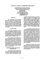
levels were ≥ 0.6 ng/ml in 69.5% of the infected patients, but
also in 35.3% of the noninfected patients (P < 0.001). The
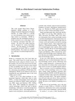
median serum CRP and PCT concentrations in infected and
noninfected patients were 102.75 and 5.30 mg/l (P < 0.001)
and 0.93 and 0.47 ng/ml (P = 0.001), respectively (Fig. 1).
There was no relationship between the serum CRP and PCT
levels and the type of infecting bacteria (data not shown). A
serum PCT level > 2 ng/ml is 100% specific for infection in
patients with SIRS (n = 69), but only 35.4% patients reached
such a high level. The negative predictive value (NPV) for
CRP in this group of patients was only 50.9%, and that for
PCT was only 50.0% (Table 4). The BT and the CRP level
(r = 0.37, P < 0.001), the WBC count and the CRP level
(r = 0.32, P = 0.001), and the CRP and PCT levels (r = 0.38,
P < 0.001) were significantly correlated.
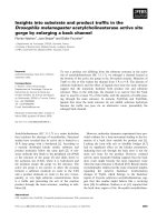
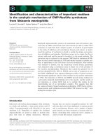
Figure 2 shows the receiver operating characteristic curves
predicting the presence of bacterial infection and the devel-
opment of septic shock. The AUC for infection identification
was greatest for CRP, followed by PCT and then WBC
(0.879 versus 0.689 versus 0.627; all P < 0.05). In predicting
the development of septic shock, the AUC for PCT was
greater than that for CRP (0.911 versus 0.767; both
P < 0.05).
Available online />Table 2
Median age, sex, median Acute Physiology and Chronic Health Evaluation (APACHE) II score, mortality, median white blood cell
count, C-reactive protein level and procalcitonin level for each group
Infected (n = 58) Noninfected (n = 49) P value
Age (years) 66.5 (42.8–75.3)
a
65.0 (46.5–70.5)
a
0.812
Sex (male/female) 30/28 34/15 0.063
APACHE II score 13.0 (7.4–18.6)
a
10.0 (6.8–18.0)
a
0.045
Mortality 5 (8.6%) 1 (2.0%) 0.216
White blood cell count (/mm
3
) 10,650 (7375–12,700)
a
8,100 (6,250–10,150) 0.024
C-reactive protein level (mg/l) 102.75 (32.35–169.63)
a
5.30 (2.00–17.65)
a
< 0.001
Procalcitonin level (ng/ml) 0.93 (0.48–2.45) 0.47 (0.29–0.92) < 0.001
a
Data presented as median (interquartile range).
Table 3
Site of infection and microbiology
Site of infection Microbiology
Urinary tract (n = 26) Escherichia coli (7), Pseudomonas aeruginosa (4), Enterococcus faecalis (3),
Staphylococcus aureus (2), Serratia marcescens (2), Klebsiella pneumoniae (1),
Proteus mirabilis (1), Citrobacter diversus (1), Pseudomonas species (1), Candida albicans (1),
unknown (4)
Lung (n = 17) Staphylococcus aureus (6), Pseudomonas aeruginosa (5), Streptococcus pneumoniae (2),
Klebsiella pneumoniae (2), Acinetobacter baumanii (2), Hemophilus influenzae (1),
Escherichia coli (1), Proteus mirabilis (1), Serratia marcescens (1), unknown (5)
Wound and soft tissue (n = 10) Staphylococcus aureus (2), Viridans streptococcus (2), Prevotella species (2), Escherichia coli (1),
Klebsiella pneumoniae (1), Proteus vulgaris (1), Bacteroides species (1),
Peptostreptococcus (1), unknown (1)
Abdominal (gastrointestinal tract and Escherichia coli (4), Proteus vulgaris (2), Citrobacter freundii (2), Enterococcus faecalis (2),
biliary system) (n = 5) Morganella morganii (1), Klebsiella oxytoca (1), Pseudomonas aeruginosa (1),
Enterococcus avium (1), Clostridium perfringens (1), Gemella morbillirum (1),
Viridans streptococcus (1), unknown (1)
Bacteremia (n = 2) Viridans streptococcus (1), Aeromonas caviae (1)
Miscellaneous
Central venous catheter (n = 1) Staphylococcus aureus
Perianal abscess (n = 1) Staphylococcus aureus
Unknown (n = 2)
R16
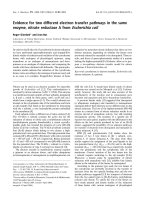
The median PCT levels were 0.50 ng/ml in noninfected
patients without SIRS, 0.47 ng/ml in noninfected patients
with SIRS, 0.67 ng/ml in septic patients, and 3.13 ng/ml in
septic shock patients. The PCT levels in the septic shock
patients were significantly higher than in the sepsis subgroup
(P < 0.001). The median CRP levels in each group were
3.20, 9.70, 75.60, and 106.35 mg/l, respectively. The CRP
levels in the sepsis subgroup were significantly higher than in
the noninfected SIRS subgroup (P < 0.001) (Fig. 3). In
infected patients (n = 58), the best cut-off value of PCT levels
predicting the development of septic shock was 2.6 ng/ml
(sensitivity, 72.7%; specificity, 91.5%; positive predictive
value [PPV], 66.7%; NPV, 93.5%; P < 0.001), and that for
CRP was 142 mg/l (P = 0.151). The median PCT level of the
bacteremic and nonbacteremic groups was 2.51 and
0.70 ng/ml, respectively (P = 0.006), and the median CRP
level for each group was 84.1 and 114.0 mg/l, respectively
(P = 0.613) (Fig. 3). Using Youden’s Index, the best cut-off
level for PCT to predict the presence of bacteremia was
1 ng/ml. The specificity was only 63.8%, but the NPV was
96.8% (P = 0.001).
Multivariate logistic regression identified a BT that fulfills the
SIRS criteria [1] (odds ratio, 11.9; 95% confidence interval,
3.2–44.5; P < 0.001), a CRP level ≥ 60 mg/l (odds ratio,
27.3; 95% confidence interval, 6.7–110.6; P < 0.001) and a
PCT level ≥ 0.6 ng/m (odds ratio, 3.5; 95% confidence inter-
val, 1.1–11.1; P = 0.033), were independently associated
with the presence of bacterial infection. The APACHE II score
(odds ratio, 1.1; 95% confidence interval, 1.0–1.3;
P = 0.058) and a PCT level ≥ 2.6 ng/ml (odds ratio, 38.3,
95% confidence interval, 5.6–263.5; P < 0.001) were inde-
pendently associated with the development of septic shock.
The APACHE II score (odds ratio, 1.2; 95% confidence inter-
val 1.0–1.4; P = 0.04) was the only variable independently
associated with mortality.
Discussion
The diagnosis of bacterial infection in acutely ill patients is not
always straightforward. Infection, in contrast with coloniza-
tion, involves some degree of host response. It is the manifes-
tations of the host response that cause us to presume a
patient has an infection and to proceed to search for the
infection focus and administer antimicrobial agents. In some
elderly patients, neonate patients, and immunosuppressed
patients, however, the manifestations may be absent, and
these patients turn out to be most vulnerable to the compli-
cated courses of infection. Similar manifestations, on the con-
trary, may be induced by stimuli other than bacterial infection
(e.g. trauma, pancreatitis, burn, etc.).
Routine laboratory tests in patients presenting with SIRS fre-
quently lack both sensitivity and specificity in differentiating
which patients should receive antibiotics, and most confirma-
tory microbiological tests results are not immediately avail-
able. In today’s climate of escalating medical costs and
increasing antibiotic resistance, it is perhaps even more
important to be able to identify those patients in whom an
antimicrobial agent is likely to be of benefit. Many rapid diag-
nostic methods for detecting infection have been developed
in recent years [9], and much effort has gone into finding bio-
chemical markers of infection; for example, finding markers
like cardiac troponin used as the marker of myocardial injury.
The ideal biochemical marker of bacterial infection, if any,
should be sensitive enough to detect the presence of infec-
tion in patients with minimal or even no host response, should
be specific enough to discriminate infection from other stimuli
that may induce SIRS, should be present early in the course
Critical Care February 2004 Vol 8 No 1 Chan et al.
Table 4
Clinical parameters, serum C-reactive protein and procalcitonin levels for diagnosing adult infection in an emergency department (%)
Positive Negative
Parameter Sensitivity Specificity predictive value predictive value
SIRS 82.8 (48/58) 57.1 (28/49) 69.6 (48/69) 73.7 (28/38)
White blood cell count >12,000/mm
3
36.2 (21/58) 81.6 (40/49) 70.0 (21/30) 51.9 (40/77)
or <4000/mm
3
C-reactive protein (cut-off 60 mg/l)
All 67.2 (39/58) 93.9 (46/49) 92.9 (39/42) 70.8 (46/65)
SIRS 68.8 (33/48) 90.5 (19/21) 94.3 (33/35) 50.9 (19/34)
No SIRS 60.0 (6/10) 96.4 (27/28) 85.7 (6/7) 87.1 (27/31)
Procalcitonin (cut-off 0.6 ng/ml)
All 70.7 (41/58) 63.3 (31/49) 69.5 (41/59) 64.6 (31/48)
SIRS 70.8 (34/48) 66.7 (14/21) 82.9 (34/41) 50.0 (14/28)
No SIRS 70.0 (7/10) 60.7 (17/28) 38.9 (7/18) 85.0 (17/20)
Numbers in parentheses indicate patient numbers. SIRS, systemic inflammatory response syndrome.
R17
of the disease, should be rapidly and conveniently measured,
and should be of prognostic significance.
TNF-α and IL-6 have been studied as markers of bacterial
infection for ED patients [4,5]. Moscovitz and colleagues [5]
collected 100 patients admitted through the ED with signs of
infection and reported that plasma IL-6 concentrations were
able to predict bacteremia and death from infection. A plasma
IL-6 concentration ≥ 2.0 ng/ml detected bacteremia with a
sensitivity of 42.1%, with a specificity of 96.7%, and with a
PPV of 72.7%. Plasma TNF-α concentrations predicted mor-
tality from all causes. The results just reflected the nonspe-
cific nature of TNF-α for identifying infection, and disclosed
the potential usefulness of IL-6 as a marker of severe infec-
tion. A cut-off level of 2.0 ng/ml, however, is too high to be
clinically useful.
Terregino and colleagues [6] collected 180 ED patients with
SIRS and reported TNF-α to be a more important predictor of
disease progression to severe sepsis than IL-6. Both
cytokines had low PPVs but high NPVs for severe sepsis, for
bacteremia, and for death at various cut-off levels. The cut-off
values of the two mediators for diagnosing infection were not
reported. In their study 108 patients were presumed infec-
tious and were admitted, but only 33 (30.6%) had recovery of
organisms from sites. Could it be that some patients with
high TNF-α and IL-6 levels had SIRS caused by stimuli other
than bacterial infection, and certainly did not progress to
severe sepsis, bacteremia, and death, thus giving rise to low
PPVs? If this was the case, then again it reflected the non-
Available online />Figure 1
C-reactive protein (CRP) and procalcitonin (PCT) concentrations in
infected and noninfected patients. Bar represents the median.
CRP (mg/l)
500
400
300
200
100
50
40
30
20
10
5
4
3
2
1
Noninfected Infected
Cut-off 60 mg/l
PCT (ng/ml)
300
200
100
50
40
30
20
10
5
4
3
2
1
0.5
0.4
0.3
0.2
0.1
Noninfected Infected
Cut-off 0.6 ng/ml
Cut-off 2.0 ng/ml
Figure 2
Receiver operating characteristic curves of C-reactive protein (open
circle), of procalcitonin (solid triangle), and of the white blood cell
count (open triangle) in (a) the diagnosis of infection and
(b) predicting septic shock.
1 – Specificity
10
Sensitivity
1
0
1 – Specificity
10
Sensitivity
1
0
(a)
(b)
R18
specific nature of the two mediators to be used as markers of
infection.
CRP is an acute phase protein [19]. In contrast to most acute
phase proteins for which there are wide plasma level varia-
tions (which depend on synthesis, consumption, and cata-
bolic rates), CRP has a plasma half-life that is constant under
almost all conditions [20]. Its plasma level is determined
exclusively by its rate of synthesis, which reflects the pres-
ence and extent of disease activity. CRP has been widely
used clinically as a diagnostic tool for infection identification
[7,8,21]. Some authors even advocate using CRP as one of
the criteria of sepsis [22]. Our results showed that CRP is a
good marker of bacterial infection in adult atraumatic ED
patients. This is in contrast to the results from the study by
Ugarte and colleagues [10], who investigated 190 critically ill
patients and reported a sensitivity of 71.8% and a specificity
of 66.6% for infection identification at the cut-off CRP level of
79 mg/l. There was a comparable sensitivity (67.2%) but a
much higher specificity (93.9%) according to our data. The
difference in specificity between the two studies is reason-
able because critically ill patients generally maintained higher
‘normal’ CRP levels than did the ED patients, which is imme-
diately evident by the marked difference in median CRP levels
(56 mg/l versus 5.3 mg/l) between noninfected patients of
the two studies. CRP does have shortcomings. Our data
showed that CRP is unable to discriminate bacteremic and
septic shock patients, and the low NPV, especially in patients
with SIRS, limited its use as a means to exclude the presence
of infection.
Remarkably little is known about the process by which PCT is
released in sepsis; even the source of its generation during
bacterial infection is not well defined. Plasma concentrations
of PCT are substantially below 0.1 ng/ml in healthy individu-
als. The most potent stimulator for PCT induction under
experimental conditions is the systemic effect of bacterial
endotoxins [23]. Viral and localized bacterial infections have
Critical Care February 2004 Vol 8 No 1 Chan et al.
Figure 3
Median C-reactive protein (CRP) and procalcitonin (PCT) concentrations in bacteremic patients, nonbacteremic patients, noninfected patients,
systemic inflammatory response syndrome (SIRS) patients, sepsis patients, and septic shock patients. * P < 0.001.
BacteremiaNo bacteremia
CRP (mg/l)
120
100
80
60
40
20
0
Septic shockSepsisSIRSNo infection
CRP (mg/l)
160
140
120
100
80
60
40
20
0
Septic shockSepsisSIRSNo infection
PCT (ng/ml)
3.5
3.0
2.5
2.0
1.5
1.0
.5
0.
0
BacteremiaNo bacteremia
PCT (ng/ml)
3.0
2.5
2.0
1.5
1.0
.5
0.
0
*
**
R19
lower plasma PCT levels than patients with systemic infec-
tions [24]. Autoimmune diseases [25] and neoplastic dis-
orders [26] do not induce PCT. If systemic infection is
present in immunosuppressed patients, PCT remains
elevated [27]. Plasma PCT is very stable and is not degraded
to hormonally active calcitonin [28].
The actual pathophysiologic role of PCT is still under investi-
gation, and it was speculated that PCT might also be an
acute phase protein [29]. Our data showed that PCT might
be used not just as a marker of infection, but, more impor-
tantly, that it is a good marker of the severity of infection. Our
results were comparable with those from the study by Ugarte
and colleagues [10]; at a cut-off value of 0.6 ng/ml, PCT had
a sensitivity of 67.6%, a specificity of 61.3%, a PPV of
71.0%, and a NPV of 57.5%. In patients with SIRS, our data
showed a NPV for PCT that is too low to be safely used to
exclude the presence of infection.
Hausfater and colleagues [30] collected 195 ED patients
with suspected infectious or inflammatory disease and found
that 24 (35%) of 68 patients with systemic infection had
serum PCT levels > 0.5 ng/ml (specificity, 99%; PPV, 96%;
NPV, 74%). The mean PCT level was 5.3 ng/ml (range
0.5–98.5 ng/ml) for infected patients and 0.09 ng/ml for non-
infected patients (P < 0.001), and the mean CRP level for
each group was 88 mg/l and 80 mg/l, respectively (P = 0.9).
The mean APACHE II score of the study subjects and the
number of patients progressing to septic shock were not
reported. Our study subjects had a higher value
(mean ± standard deviation, 8.98 ± 32.02 ng/ml in infected
patients and 0.75 ± 0.73 ng/ml in noninfected patients) and a
broader range (0.10–210.55 ng/ml) of PCT levels; the ability
of PCT in discriminating the presence of infection was lower.
The subjects of the present study were patients admitted to
the hospital through the ED, and theoretically the mean PCT
level should be lower compared with that of Hausfater and
colleagues (indeed, the median PCT level of infected patients
was lower than that from Ugarte and colleagues [10]).
Whether the overall PCT levels in our study subjects were
significantly higher, and whether the differences were related
to patient characteristics, disease courses, and severity, other
factors such as racial difference remain uncertain.
Guven and colleagues [31] collected 34 patients with SIRS
and reported an AUC for predictive accuracy of sepsis for a
PCT level, a WBC count and a CRP level of 0.88, 0.44 and
0.34, respectively. So is CRP good or bad? How about
PCT? We believe the performance of each marker in different
studies is closely related to the characteristics of the study
subjects. In most studies the levels of the inflammatory medi-
ators produced are far higher when the initial trigger is infec-
tious, and it seems obvious that, although the same pathway
is probably involved, there is a clear quantitative difference in
the activation of the inflammatory network for septic versus
nonseptic insult [9]. Our study subjects were heterogeneous
ED patients, in which the average inflammatory status was
less severe than critically ill patients and the most frequent
cause of acute inflammation was ultimately infection. It might
be that elevated CRP levels in ED patients are most fre-
quently induced by an infection, and thus CRP becomes a
good marker of infection in ED. However, many other stimuli
also cause inflammation. An elevated serum CRP level in indi-
vidual patients should hence be interpreted with caution, and
an underlying cause of inflammation other than infection
should always be considered. It seems that PCT is more spe-
cific to infectious stimuli compared with CRP [13,23,24]. We
recommend that in patients with elevated CRP levels, PCT
may be used as a measure to further support the diagnosis of
infection, and as a marker of disease severity.
There were limitations with the present study. The first limita-
tion is that patients who were directly discharged from the ED
were not included. The effects may be twofold. Certain
patients were not enrolled, especially those with elevated
CRP levels induced by stimuli other than bacterial infection.
This might result in an overestimation of the specificity of
CRP, and a possible underestimation of the specificity of
PCT if the serum levels of these patients were within the
normal range. The subjects in the present study, on the con-
trary, were all admitted patients who might have more serious
diseases and higher ‘normal’ CRP levels than the general ED
patient population. This might result in an underestimation of
both the specificity and the PPV of CRP. Which effect pre-
dominated is difficult to predict. Further studies of larger
sample size to recruit all ED patients are needed to obtain
more convincing results.
A second limitation is that some authors advocated close
follow-ups of the PCT levels in infected patients, as the peak
level correlated best with prognosis [32]. The present study
used only a single PCT level on admission to the ED, and the
correlation with the outcomes may be suboptimal.
The final limitation of the study is that the number of patients
at risk of having infection without developing SIRS is limited.
PCT levels were reported to elevate normally in infected
immunosuppressed patients [27]. Failure to recruit these
patients may lead to an underestimation of sensitivity. Now
that PCT showed convincing results in detecting infection in
immunosuppressed and leukopenic patients, shall we assay
PCT routinely in all these patients? Further studies with a
special focus to the immunosuppressed patients are needed.
Conclusions
PCT is not a better marker of bacterial infection than CRP in
adult ED patients, but it is a useful marker of the severity of
infection. An elevated CRP level in ED patients has a high
PPV for infection, but the absence of CRP elevation cannot
be used safely to exclude the presence of infection, espe-
cially in patients with SIRS. In patients with elevated serum
Available online />R20
CRP levels, PCT might be used to further support the pres-
ence of infection and to predict the disease severity.
Competing interests
None declared.
Acknowledgements
The authors are indebted to the staff of the Emergency Department,
Linkou Chang Gung Memorial Hospital, Taoyuan. They thank the
Nursing Specialists Ms Yu-Mei Chang, Shu-Chin Tsai, Mei-Chuang
Chang, and Li-Ling Lin for their help, and thank Mr Jackson Lin and
Level Biotech Co. for technical assistance and support. Dr Chan, Dr
Chang and Dr Tseng contributed equally to the present work.
References
1. Members of the American College of Chest Physicians/Society of
Critical Care Medicine Consensus Conference Committee:
American College of Chest Physicians/Society of Critical Care
Medicine Consensus Conference: definitions for sepsis and
organ failure and guidelines for the use of innovative thera-
pies in sepsis. Crit Care Med 1992, 20:864-874.
2. Angus DC, Linde-Zwirble WT, Lidicker J, Clermont G, Carcillo J,
Pinsky MR: Epidemiology of severe sepsis in the United
States: analysis of incidence, outcome, and associated costs
of care. Crit Care Med 2001, 29:1303-1310.
3. Kollef MH, Sherman G, Ward S, Fraser VJ: Inadequate antimi-
crobial treatment of infections: a risk factor for hospital mor-
tality among critically ill patients. Chest 1999, 115:462-474.
4. Galley HF, Webster NR: The immuno-inflammatory cascade.
Br J Anaesth 1996, 77:11-16.
5. Moscovitz H, Shofer F, Mignott H, Behrman A, Kilpatrick L:
Plasma cytokine determinations in emergency department
patients as a predictor of bacteremia and infectious disease
severity. Crit Care Med 1994, 22:1102-1107.
6. Terregino CA, Lopez BL, Karras DJ, Killian AJ, Arnold GK:
Endogenous mediators in emergency department patients
with presumed sepsis: are levels associated with progression
to severe sepsis and death? Ann Emerg Med 2000, 35:26-34.
7. Matson A, Soni N, Sheldon J: C-reactive protein as a diagnostic
test of sepsis in the critically ill. Anaesth Intensive Care 1991,
19:182-186.
8. Povoa P, Almeida E, Moreira P, Fernades A, Mealha R, Aragao A,
Sabino H: C-reactive protein as an indicator of sepsis. Inten-
sive Care Med 1998, 24:1052-1056.
9. Carlet J: Rapid diagnostic methods in the detection of sepsis.
Infect Dis Clin North Am 1999, 13:483-494.
10. Ugarte H, Silva E, Mercan D, De Mendonca A, Vincent JL: Procal-
citonin used as a marker of infection in the intensive care unit.
Crit Care Med 1999, 27:498-504.
11. Muller B, Becker KL, Schachinger H, Rickenbacher PR, Huber
PR, Zimmerli W, Ritz R: Calcitonin precursors are reliable
markers of sepsis in a medical intensive care unit. Crit Care
Med 2000, 28:977-983.
12. Le Moullec JM, Jullienne A, Chenais J, Lasmoles F, Guliana JM,
Milhaud G, Moukhtar MS: The complete sequence of human
preprocalcitonin. FEBS 1984, 167:93-97.
13. De Werra I, Jaccard C, Corradin SB, Chiolero R, Yersin B, Gallati
H, Assicot M, Bohuon C, Baumgartner JD, Glauser MP, Heumann
D: Cytokines, nitrite/nitrate, soluble tumor necrosis factor
receptors, and procalcitonin concentrations: comparisons in
patients with septic shock, cardiogenic shock, and bacterial
pneumonia. Crit Care Med 1997, 25:607-613.
14. Anonymous: The problem of sepsis. An expert report of the
European Society of Intensive Care Medicine. Intensive Care
Med 1994, 20:300-304.
15. Knaus WA, Draper EA, Wagner DP, Zimmerman JE: APACHE II:
a severity of disease classification system. Crit Care Med
1985, 3:818-829.
16. Youden WJ: Index rating for diagnostic tests. Cancer 1950, 3:
32-35.
17. Beck JR, Shultz EK: The use of receiver operating characteris-
tic (ROC) curves in test performance evaluation. Arch Pathol
Lab Med 1986, 110:13-20.
18. Walker SH, Duncan DB: Estimation of the probability of an
event as a function of several independent variables. Biometrika
1967, 55:167-179.
19. Pepys MB, Baltz ML: Acute phase proteins with special refer-
ence to C-reactive protein and related proteins (pentraxins)
and serum amyloid A protein. Adv Immunol 1983, 34:141-212.
20. Vigushin DM, Pepys MB, Hawkins PN: Metabolic and scinti-
graphic studies of radioiodinated human C-reactive protein in
health and disease. J Clin Invest 1993, 90:1351-1357.
21. Ballou SP, Kushner I: C-reactive protein and the acute phase
response. Adv Int Med 1992, 37:313-316.
22. Bota DP, Ferreira FL, Bross A, Melot C, Vincent JL: Sepsis crite-
ria in critically ill patients [abstract]. Crit Care Med 1999, 27:
A142.
23. Dandona P, Nix D, Wilson MF, Aljada A, Love J, Assicot M,
Bohuon C: Procalcitonin increase after endotoxin injection in
normal subjects. J Clin Endocrinol Metab 1994, 79:1605-1608.
24. Assicot M, Gendrel D, Carsin H, Raymond J, Guilbaud J, Bohuon
C: High serum procalcitonin concentrations in patients with
sepsis and infection. Lancet 1993, 27:515-518.
25. Eberhard OK, Haubitz M, Brunkhorst FM, Kliem V, Koch KM,
Brunkhorst R: Usefulness of procalcitonin for differentiation
between activity of systemic autoimmune disease (systemic
lupus erythematosus/systemic antinutrophil cytoplasmic anti-
body-associated vasculitis) and invasive bacterial infection.
Arthritis Rheum 1997, 40:1250-1256.
26. Fleischhack G, Kambeck I, Cipic D, Hasan C, Bode U: Procalci-
tonin in paediatric cancer patients: its diagnostic relevance is
superior to that of C-reactive protein, interleukin 6, interleukin
8, soluble interleukin 2 receptor and soluble tumour necrosis
factor receptor II. Br J Haematol 2000, 111:1093-1102.
27. Al-Nawas B, Shah PM: Procalcitonin in patients with and
without immunosuppression and sepsis. Infect 1996, 24:434-
436.
28. Snider RH Jr, Nylen ES, Becker KL: Procalcitonin and its com-
ponent peptides in systemic inflammation: immunochemical
characterization. J Investig Med 1997, 45:552-560.
29. Nijsten MWN, Olinga P, Hauw The T, De Vries EGE, Koops HS,
Groothuis GMM, Limburg PC, Ten Duis HJ, Moshage H, Hoekstra
HJ, Bijzet J, Zwaveling JH: Procalcitonin behaves as a fast
responding acute phase protein in vivo and in vitro. Crit Care
Med 2000, 28:458-461.
30. Hausfater P, Garric S, Ben Ayed S, Rosenheim M, Bernard M,
Riou B: Usefulness of procalcitonin as a marker of systemic
infection in emergency department patients: a prospective
study. Clin Infect Dis 2002, 34:895-901.
31. Guven H, Altintop L, Baydin A, Esen S, Aygun D, Hokelek M,
Doganay Z, Bek Y: Diagnostic value of procalcitonin levels as
an early indicator of sepsis. Am J Emerg Med 2002, 20:202-
206.
32. Whang KT, Steinwald PM, White JC, Nylen ES, Snider RH, Simon
GL, Goldberg RL, Becker KL: Serum calcitonin precursors in
sepsis and systemic inflammation. J Clin Endocrinol Metab
1998, 83:3296-3301.
Critical Care February 2004 Vol 8 No 1 Chan et al.
Key messages
• Using a cut-off level chosen by Youden’s Index, PCT is
not a better marker of bacterial infection than CRP for
adult emergency department patients. Yet high serum
PCT level is highly specific for infection
• A low serum CRP or PCT level cannot be used safely
to exclude the presence of infection, especially in
patients with SIRS
• In patients with elevated serum CRP levels, PCT may
be used as a measure to further support the diagnosis
of infection, and as a marker of disease severity