Báo cáo khoa học: "Clinical review: Influence of vasoactive and other therapies on intestinal and hepatic circulations in patients with septic shock" ppsx
Bạn đang xem bản rút gọn của tài liệu. Xem và tải ngay bản đầy đủ của tài liệu tại đây (94.87 KB, 10 trang )
170
ICG = indocyanine green; ICU = intensive care unit; MEGX = monoethylglycinexylidide; NAC = N-acetyl cysteine; PCO
2
= partial carbon dioxide
tension; pHi = intramucosal pH.
Critical Care June 2004 Vol 8 No 3 Asfar et al.
Introduction
Research interest has focused on the intestinal and hepatic
circulations in various models of shock, and particularly in
septic shock. The splanchnic area is reported to be the ‘motor’
of multiple organ failure [1] and the ‘canary’ of the body [2]. In
fact, because of its peculiar vascular anatomy, the hepato-
splanchnic area is jeopardized during septic shock, which may
potentially lead to a vicious circle of inflammatory responses,
culminating in multiple organ failure syndrome.
The present clinical review briefly discusses the splanchnic
vascular anatomy and focuses on the different therapeutic
approaches that have been proposed to promote perfusion of
the gastrointestinal tract during resuscitation of patients with
septic shock. When possible and reasonable, we propose
therapeutic recommendations.
References were obtained from Medline database (from the
earliest records to 2003). We used the following keywords:
gastric mucosal pH or pHi, splanchnic, haemodynamics,
microcirculation, sepsis, septic shock, vasoactive drugs,
dobutamine, dopamine, norepinephrine, epinephrine, dopex-
amine vasopressin, terlipressin, prostacyclin, N-acetyl cys-
teine, dialysis and haemofiltration. We also reviewed the
reference lists of all available review articles and primary
studies to identify references not found in computerized
searches. We placed emphasis on prospective, randomized,
controlled clinical trials.
Anatomy of hepato-splanchnic vascular bed
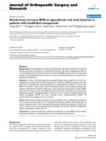
The splanchnic vasculature includes both serial and parallel
vascular beds (Fig. 1). The gut is perfused by the coeliac
trunk and mesenteric arteries, and is drained via the portal
Review
Clinical review: Influence of vasoactive and other therapies on
intestinal and hepatic circulations in patients with septic shock
Pierre Asfar
1
, Daniel De Backer
2
, Andreas Meier-Hellmann
3
, Peter Radermacher
4
and
Samir G Sakka
5
1
Staff Physician, Département de Réanimation Médicale, Centre Hospitalier Universitaire, Angers, France
2
Staff Physician, Département de Réanimation Médicale, Hôpital Erasme, Université Libre, Bruxelles, Belgium
3
Head, Klinik für Anästhesie, Intensivmedizin und Schmerztherapie, Helios Klinikum, Erfurt, Germany
4
Section Head, Sektion Anästhesiologische Pathophysiologie und Verfahrensentwicklung, Universitätsklinikum, Ulm, Germany
5
Staff Physician, Department of Anesthesiology and Intensive Care Medicine, Friedrich-Schiller University, Jena, Germany
Correspondence: Peter Radermacher,
Published online: 29 December 2003 Critical Care 2004, 8:170-179 (DOI 10.1186/cc2418)
This article is online at />© 2004 BioMed Central Ltd
Abstract
The organs of the hepato-splanchnic system are considered to play a key role in the development of
multiorgan failure during septic shock. Impaired oxygenation of the intestinal mucosa can lead to
disruption of the intestinal barrier, which may promote a vicious cycle of inflammatory response, increased
oxygen demand and inadequate oxygen supply. Standard septic shock therapy includes supportive
treatment such as fluid resuscitation, administration of vasopressors (adrenergic and nonadrenergic
drugs), and respiratory and renal support. These therapies may have beneficial or detrimental effects not
only on systemic haemodynamics but also on splanchnic haemodynamics, at both the macrocirculatory
and microcirculatory levels. This clinical review focuses on the splanchnic haemodynamic and metabolic
effects of standard therapies used in patients with septic shock, as well as on the recently described
nonconventional therapies such as vasopressin, prostacyclin and N-acetyl cysteine.
Keywords adrenergic drugs, nonconventional treatments, septic shock, splanchnic circulation, supportive treatment
171
Available online />system. The liver has a unique and special blood supply that
includes both arterial (the common hepatic artery) and venous
(the portal vein) inflow. The portal vein supplies 75–80% of
the liver blood flow and the hepatic artery supplies 20–25%.
Physiologically, there is an interdependent response with a
compensatory blood flow between the portal vein and the
hepatic artery called the hepatic arterial buffer response [3].
The hepato-splanchnic blood flow accounts for 25–30% of
the cardiac output [4], and the regional oxygen extraction is
slightly higher than the whole body oxygen extraction. During
sepsis or septic shock, splanchnic oxygen extraction is
increased compared with nonseptic patients (44% versus
30%), which leads to an increase in the hepatic venous/mixed
venous haemoglobin oxygen saturation gradient [4]. In clinical
practice it is generally not possible to determine portal venous
flow in isolation, and measurements are taken from the hepato-
splanchnic region as a whole. The flow is estimated at bedside
by the method of primed, constant infusion of indocyanine
green (ICG) with hepatic venous catheterization [5].
The intestinal villus is supplied by a single, unbranched arterial
vessel that arborizes at the villus tip into a network of surface
capillaries drained by a central villus vein. This anatomical
arrangement allows countercurrent exchange and shunting of
diffusible molecules such as oxygen, and hypoxia may occur at
the tip of the villus even during moderate decreases in macro-
circulatory flow [6]. In addition, intestinal villi perfusion is highly
heterogeneous, as suggested by the wide range of intestinal
surface oxygen saturation [7].
In patients with sepsis, splanchnic blood flow usually
increases in proportion to cardiac output [8] and is associ-
ated with decreased hepatic vein oxygen haemoglobin satu-
ration. Two different interpretations are possible: first, the
increase in splanchnic blood flow is insufficient to meet the
increased oxygen consumption; and second, hepatic arterial
blood flow is reduced as a consequence of the hepatic arter-
ial buffer response. The latter hypothesis is supported by the
observations of De Backer and coworkers [9], who demon-
strated that there is usually no net lactate production from the
hepato-splanchnic area. In addition, the observation that
splanchnic blood flow is increased does not rule out an
impairment in microvascular blood flow [10–12] or the pres-
ence of cytopathic hypoxia [13].
In normal conditions the partial carbon dioxide tension (P
CO
2
)
gap, which is defined as the difference between mucosal
P
CO
2
measured with a tonometer and arterial PCO
2
, is low. In
case of inadequate mucosal blood flow, whether tissue
hypoxia is present or not, the P
CO
2
gap increases. Levy and
coworkers [14] recently reported that a P
CO
2
gap greater
than 20 mmHg was associated with poor outcome in patients
with septic shock. Unfortunately, there is no apparent correla-
tion between P
CO
2
gap and global or regional haemodynamic
measurements in septic patients [15] because the P
CO
2
gap
mirrors both variations in microvascular flow [10] and in
carbon dioxide metabolism [16]. For these reasons variations
in P
CO
2
gap must be interpreted with caution.
Therapeutic strategies
Fluid challenge
The mainstay of supportive treatment in patients with severe
sepsis or septic shock is maintenance of adequate fluid
balance, titration of appropriate oxygen delivery, and ade-
quate perfusion pressure [17]. Hypovolaemia is a common
clinical occurrence in intensive care medicine and results
from several mechanisms such as fluid loss, haemorrhage,
vasoplegia and capillary leak syndrome. This explains why
fluid replacement therapy is a key component in the treatment
of severe sepsis and septic shock. Although there is no con-
sensus regarding the ideal type of fluid replacement, colloids
are efficient in this indication [18].
There are few clinical studies focusing on the effects of col-
loids on splanchnic haemodynamics. In a randomized study
conducted in patients with sepsis, Boldt and coworkers [19]
assessed the effects on tonometric gastric mucosal acidosis
of hydroxyethyl starch and albumin targeted to maintain pul-
monary artery occlusion pressure between 12 and 18 mmHg.
In hydroxyethyl starch treated patients cardiac index, oxygen
delivery and consumption increased, and gastric intramucosal
pH (pHi) remained stable whereas it decreased in albumin
treated patients. In three other studies [20–22] conducted in
patients with sepsis and septic shock, fluid challenges per-
formed with hydroxyethyl starch neither altered the P
CO
2
gap
nor influenced splanchnic haemodynamics. Moreover, a ran-
domized comparison of hydroxyethyl starch and gelatin in
haemodynamically stable septic patients revealed a beneficial
effect of gelatin on the P
CO
2
gap [20]. These studies sug-
gested no better effect of one colloid over the others on
splanchnic haemodynamics, and the use of colloids must be
weighed against their side effects [23].
Figure 1
Splanchnic anatomy and flows in healthy volunteers.
Stomach
Spleen
Pancreas
Small
Intestine
Colon
LIVER
Hepatic
Vein
s
Celiac artery
700 ml/min
Hepatic artery
500 ml/min
Superior mesenteric
Ar
tery 700 ml/min
Inferior mesenteric artery
400 ml/min
Portal
vein
172
Critical Care June 2004 Vol 8 No 3 Asfar et al.
Red blood cell transfusions are commonly used in intensive
care units (ICUs) to enhance systemic oxygen delivery.
However, proof of improved utilization of oxygen by peripheral
tissues, especially in the splanchnic area, is lacking. Silver-
man and Tuma [24] reported the absence of improved gastric
pHi with red blood cell transfusions in 21 septic patients.
Moreover, there is an inverse association between the
change in gastric pHi and the age of the transfused blood
[25]. Finally, a recent report in 15 septic patients showed that
red blood cell transfusion failed to improve oxygen utilization
measured either using Fick’s equation or by indirect calori-
metry, and gastric pHi remained unaltered [26].
Adrenergic drugs
The choice of vasoactive drugs in sepsis and septic shock is
controversial. There is no evidence that any one vasoactive drug
is more effective or safer than any other. Larger trials are needed
to elucidate existing clinically significant differences in morbidity
and mortality. A multicentre trial, which is currently ongoing, is
comparing the effects of epinephrine with a combination of a
fixed dose of dobutamine in addition to norepinephrine.
Dopamine alone or versus norepinephrine (Table 1)
The infusion of low-dose dopamine (defined as a dose lower
than 5 µg/kg per min administered to normotensive patients)
may not improve gut mucosal perfusion. In fact, Nevière and
coworkers [27] showed that low-dose dopamine decreased
gut mucosal blood flow in septic patients. Furthermore, other
investigators [27–30] reported that either pHi or P
CO
2
gap
were unchanged in patients with sepsis treated with low-
dose dopamine. The effects on liver blood flow may also be
variable; Maynard and coworkers [30] observed that
dopamine did not affect ICG clearance and monoethyl-
glycinexylidide (MEGX) formation from lidocaine. Interest-
ingly, the effects of dopamine on splanchnic blood flow may
differ according to basal splanchnic perfusion. Low-dose
dopamine increased splanchnic blood flow that was low at
baseline (seven patients) but not when splanchnic perfusion
was preserved (four patients) [28]. The very small number of
patients in each group limited these observations. Recently,
Jakob and coworkers [31] reported that dopamine adminis-
tration titrated to achieve a 25% increase in cardiac output
induced a significant increase in splanchnic blood flow from
0.9 to 1.1 l/min per m
2
, which was associated with a signifi-
cant reduction in splanchnic oxygen consumption.
The results are even more controversial when dopamine is
used at higher doses to restore blood pressure. Ruokonen
and coworkers [32] observed that dopamine increased
splanchnic blood flow and metabolism in some but not all
patients with septic shock. In some patients, the same group
of investigators [33] also observed an increase in hepatic
vein oxygen saturation, suggesting an improvement in the
balance between oxygen supply and demand during
dopamine administration. However, in a pilot study, Marik and
Mohedin [34] reported that dopamine administered at doses
up to 25 µg/kg per min even decreased pHi. Given the very
small number of patients included in these studies, no definite
conclusions can be drawn regarding the effects of dopamine
on splanchnic blood flow in septic patients.
Comparison of the effects of norepinephrine and dopamine is
difficult because norepinephrine is often combined with
dobutamine, and study results are conflicting. Ruokonen and
coworkers [32] reported unpredictable effects on splanchnic
blood flow in patients with septic shock with norepinephrine,
whereas dopamine induced a consistent increase in splanch-
nic blood flow. By contrast, in the randomized study reported
by Marik and Mohedin [34], conducted in 20 septic patients
with hyperdynamic septic shock, dopamine was reported to
induce a decrease in pHi when compared with norepineph-
rine. More recently, De Backer and coworkers [35] reported
the effects of dopamine, norepinephrine and epinephrine on
the splanchnic circulation in moderate and in severe septic
shock, and the main results are as follows. In moderate septic
shock cardiac index was similar in dopamine-treated and nor-
epinephrine-treated patients, and higher in epinephrine-
treated patients, whereas splanchnic blood flow was the same
with the three drugs. The gradient between mixed venous and
hepatic venous oxygen saturation gradient was the lowest with
dopamine, while P
CO
2
gaps were identical. In patients with more
severe septic shock cardiac index was greater and splanchnic
blood flow lower with epinephrine than with dopamine and epi-
nephrine; mixed venous and hepatic venous oxygen saturation
gradient was greater with epinephrine, whereas P
CO
2
gap
remained unaltered by any of the treatments.
Given the available data (summarized in Table 1), no definite
conclusions can be drawn regarding differences between
dopamine and norepinephrine on splanchnic blood flow and
metabolism in patients with septic shock.
Dobutamine alone or combined with norepinephrine versus
epinephrine (Tables 2 and 3)
In patients with sepsis, a retrospective study conducted by
Silverman and coworkers [24] identified a beneficial effect of
dobutamine infusion on pHi [24]. Two years later Gutierrez
and coworkers [36] reported an increase in pHi with dobuta-
mine infusion in patients with sepsis syndrome who initially
had low pHi. This beneficial effect, confirmed in other studies
[37–39], was not related to an increase in splanchnic blood
flow induced by dobutamine [39,40]. Creteur and colleagues
[41] reported that dobutamine decreased the P
CO
2
gap in
septic patients with a high gradient between the mixed
venous and hepatic vein oxygen saturation (> 20%), whereas
P
CO
2
gap was not affected in patients when this gradient was
less than 20%. This suggests that patients with the most
severe alterations in hepato-splanchnic blood flow are also
prone to decreased mucosal perfusion.
Dobutamine usually, but not without exception, increases
splanchnic perfusion [40–42]. The effects on splanchnic
173
metabolism are more variable [39] and may depend on the
adequacy of splanchnic perfusion at baseline. In patients with
septic shock, De Backer and coworkers [43] reported that
splanchnic oxygen consumption increased during dobutamine
administration only in patients with an increased gradient
between hepatic venous and mixed venous oxygen saturation.
Combinations of dobutamine and other catecholamines have
often been studied, in particular in association with norepi-
nephrine for its effects on β-receptors, with the aim of modu-
lating hepato-splanchnic haemodynamics. Indeed, in patients
with sepsis, changing from norepinephrine (α-agonist and
β-agonist) to phenylephrine (pure α-agonist), titrated to
produce similar global haemodynamic measurements, led to a
decrease in splanchnic blood flow, splanchnic oxygen deliv-
ery and gastric pHi. These changes were associated with
decreased rates of liver lactate uptake and glucose produc-
tion [44].
Whether dobutamine has a specific effect on the splanchnic
circulation is still debated. In a cross-over study conducted in
eight patients with septic shock, Meier-Hellmann and cowork-
ers [45] showed that epinephrine caused lower splanchnic
flow and oxygen uptake, lower gastric pHi, and higher hepatic
vein lactate concentration than did the combination of dobut-
amine and norepinephrine. Duranteau and coworkers [11]
compared the effects of epinephrine, norepinephrine and the
combination of norepinephrine and dobutamine in patients
with septic shock on gastric mucosal flow, as assessed using
a laser Doppler technique. Epinephrine and dobutamine–nor-
epinephrine led to a significant increase in gastric mucosal
flow as compared with norepinephrine alone, but these find-
ings were not corroborated by those reported by Seguin and
coworkers [46]. Moreover, in patients with septic shock resis-
tant to dopamine, the combination of norepinephrine and
dobutamine, in comparison with epinephrine alone, restored
gastric pHi more quickly and limited the increase in arterial
lactate concentration. However, there was no difference in
gastric mucosal P
CO
2
gradients between groups at 24 hours
of treatment [38].
The preferential effect of dobutamine on splanchnic blood flow
was not confirmed by Reinelt and coworkers [42], who
studied the effects of dobutamine on fractional splanchnic
flow and hepatic glucose production in septic patients resus-
citated adequately with fluid and norepinephrine. Their results
showed a parallel increase in splanchnic blood flow and
cardiac index, unaltered splanchnic oxygen consumption and
decreased rate of endogenous production of hepatic glucose.
These findings suggest that splanchnic blood flow is
increased in well resuscitated septic patients, and that a
dobutamine test is able to reveal a oxygen delivery/consump-
tion dependency [41,43] but it cannot exclude intraorgan
blood flow redistribution at the microcirculatory level. The inad-
equacy of blood flow distribution is mirrored by the absence of
correlation between splanchnic blood flow and the P
CO
2
gap.
Reported data on the effects of dobutamine and norepineph-
rine on splanchnic haemodynamics are summarized in
Tables 2 and 3, respectively.
Available online />Table 1
Clinical studies reporting effects of dopamine on splanchnic haemodynamics
Reference n Drug (µg/kg per min) Splanchnic blood flow pHi or PCO
2
gap Comments
[27] 10 Dopamine 5 LD ↓ PCO
2
gap = Laser Doppler study; cross-over trial
Dobutamine 5 LD ↑ PCO
2
gap ↓
[29] 16 Dopamine 3 NA pHi =
[28] 11 Dopamine 3 HSBF ↑ pHi = Increase in fractional splanchnic flow
+ norepinephrine when low before dopamine
[30] 10 Dopamine 2.5 = pHi = Splanchnic blood flow was measured
10 versus dopexamine 1 ↑ pHi ↑ by ICG clearance
[31] 9 Dopamine 4 (2.1–9) HSBF ↑ NA Decrease in splanchnic V
O
2
;
dopamine infused to achieve
increase of 25% in cardiac index
[32] 5 Dopamine 16 HSBF ↑
5 versus norepinephrine 0.13 ≈
[34] 10 Dopamine 26 NA pHi ↓ Dopamine and norepinephrine titrated
10 versus norepinephrine 0.18 pHi ↑ to achieve MAP ≥75mmHg
[35] 10 Dopamine 26 Switch from dopamine to
versus norepinephrine 0.18 HSBF = P
CO
2
gap = norepinephrine or epinephrine in
versus epinephrine 0.12 HSBF = P
CO
2
gap = moderate septic shock
NA, not avalaible; HSBF, hepato-splanchnic blood flow determined by the indocyanin green (ICG) continuous infusion; LD, laser Doppler; MAP,
mean arterial pressure; P
CO
2
gap, gastric mucosal–arterial gradient of P
CO
2
; pHi, intramucosal pH; V
O
2
, oxygen consumption.
174
Recommendations regarding use of adrenergic drugs
We suggest that both dopamine and norepinephrine can be
given to septic shock patients as first-line catecholamine
drugs but that their use must be weighed against the unde-
sired neuroendocrine side effects of dopamine [45]. Epineph-
rine should be reserved for use as rescue therapy. If norepi-
nephrine is chosen as the first agent, then the addition of
dobutamine may be considered.
Critical Care June 2004 Vol 8 No 3 Asfar et al.
Table 2
Clinical studies reporting effects of dobutamine on splanchnic haemodynamics
Reference n Drug (µg/kg per min) Splanchnic blood flow pHi or PCO
2
gap Comments
[24] 9 Dobutamine 5 NA pHi ↑ Dobutamine versus transfusions
[36] 21 Dobutamine 5–10 NA pHi ↑ Septic patients with low pHi
[38] 15 Dobutamine 5 + NA P
CO
2
gap = Norepinephrine and epinephrine
norepinephrine 0.6 titrated to obtain MAP ≥ 80mmHg
15 versus epinephrine 0.5 NA P
CO
2
gap ↑ with stable or increased cardiac index
[39] 14 Dobutamine 7.5 + ICG clearance = pHi ↓ Patients were treated with
norepinephrine 0.6 mg/hour norepinephrine and dobutamine was
added
[40] 10 Dobutamine 7.3 ±2 HSBF ≈ NA Patients with pancreatitis infused with
dobutamine to increase cardiac index
by > 25%
[41] 36 Dobutamine 5–10 HSBF ↑ P
CO
2
gap ↓ in patients
with fractional splanchnic
blood flow < 20%
[42] 12 Dobutamine NA HSBF ↑ P
CO
2
gap = Septic patients haemodynamically
+ norepinephrine 0.2 ±0.08 controlled with norepinephrine (MAP
> 70mmHg); dobutamine infused to
achieve increase in cardiac index of
> 20%
[43] 42 Dobutamine HSBF ↑ NA Splanchnic V
O
2
increased only in
5–10 patients with increased gradient
between hepatic venous and
mixed–venous oxygen saturation
> 10%
NA, not avalaible; HSBF, hepato-splanchnic blood flow determined by the indocyanin green (ICG) continuous infusion; MAP, mean arterial
pressure; PCO
2
gap, gastric mucosal–arterial gradient of PCO
2
; pHi, intramucosal pH; VO
2
, oxygen consumption.
Table 3
Clinical studies reporting effects of norepinephrine on splanchnic haemodynamics
Reference n Drug (µg/kg per min) Splanchnic blood flow pHi or PCO
2
gap Comments
[44] 5 Norepinephrine NA HSBF ↓ PCO
2
gap = Switch from norepinephrine to
versus phenylephrine 3.2 phenylephrine
[11] 12 Epinephrine 0.7 ±0.1 LD ↑ in mucosal blood flow NA Epinephrine, norepinephrine in
versus norepinephrine 1 ±0.6 with epinephrine and random order to achieve MAP
versus norepinephrine + norepinephrine + dobutamine, 70–80 mmHg
dobutamine 1.1 ± 0.6 and 5 as compared with
norepinephrine alone
[46] 11 Epinephrine 0.3 ±0.2 LD epinephrine ↑ mucosal NA
11 versus norepinephrine + blood flow
dobutamine 0.9 ± 0.4 and 5
[45] 8 Epinephrine 0.48 ±0.33 HSBF ↓ with epinephrine pHi ↓ Cross-over study
versus norepinephrine +
dobutamine 0.37 ±0.2
and 13.6 ±3
NA, not avalaible; HSBF, hepato-splanchnic blood flow determined by the indocyanin green (ICG) continuous infusion; LD, laser Doppler; MAP,
mean arterial pressure; P
CO
2
gap, gastric mucosal–arterial gradient of PCO
2
; pHi, intramucosal pH.
175
Dopexamine
Dopexamine hydrochloride is a dopamine analogue with
vasodilating effects that may be useful in improving splanch-
nic microcirculation in septic shock. Twenty-five ventilated
patients with systemic inflammatory response syndrome were
randomly assigned to receive either a 2-hour infusion of
dopexamine (1 mg/kg per min) or of dopamine (2.5 µg/kg per
min) after baseline measurements of gastric pHi, MEGX for-
mation from lidocaine and ICG disappearance rate. Dopex-
amine had no effects on systemic measurements but it
significantly increased pHi and ICG plasma disappearance,
suggesting a selective increase in splanchnic blood flow and
improved hepatic function, as indicated by increased MEGX
concentration [30]. A previous study from the same group
showed that dopexamine at higher doses (4–6 µg/kg per min)
raised gastric pHi together with a nonsignificant increase in
ICG clearance [47]. Temmesfeld-Wollbrück and coworkers
[7] employed reflectance spectrophotometry for direct
assessment of the microvascular haemoglobin saturation and
haemoglobin concentration in the gastric mucosa in patients
with septic shock. Compared with healthy control individuals,
patients with septic shock exhibited a reduced microvascular
haemoglobin saturation with a wide distribution and with
tailing of the histogram to severely hypoxic values in spite of
high whole body oxygen delivery. This microvascular distur-
bance was associated with reduced microvascular haemoglo-
bin concentration and a lower gastric pHi. Short-term infusion
of 2 µg/kg per min dopexamine in 10 patients with septic
shock increased both microvascular haemoglobin saturation
and concentration, whereas whole body oxygen uptake and
gastric pHi remained unaltered.
Other investigators did not confirm these beneficial effects.
Hannemann and coworkers [48] reported the effect of incre-
mental doses (0.5–4 µg/kg per min) dopexamine on splanch-
nic circulation in 12 patients with severe sepsis
haemodynamically controlled with fluid challenge and dobuta-
mine. Splanchnic blood flow increased proportionally to
cardiac output but dopexamine lowered gastric pHi in a dose-
dependent manner in all patients [49]. Finally, in 12 septic
shock patients haemodynamically controlled with norepineph-
rine, dopexamine titrated to increase cardiac output by 25%
[50] increased median splanchnic blood flow whereas the
fractional splanchnic blood flow was significantly reduced,
and none of global or regional oxygen exchange or P
CO
2
was
altered. In addition, those investigators found no influence of
dopexamine on metabolic parameters either [51]. Given
these discrepancies, it is reasonable to recommend further
investigations into dopexamine before it may be routinely
used in septic shock.
Other vasoactive drugs
Vasopressin and terlipressin
Physiologically, vasopressin (a nonapeptide that is released
from the neurohypophysis) plays a minor role in blood pres-
sure regulation. Clinical data revealed that the initially very
high plasma concentrations of vasopressin decrease during
prolonged sepsis [52].
In the past few years clinical studies showed that blood pres-
sure can be rapidly restored in septic shock using vaso-
pressin, but this is mainly at the expense of cardiac output
[53]. Nevertheless, in 2000 the American Heart Association
and International Liaison Committee on Resuscitation recom-
mended (grade IIB) continuous vasopressin infusion in refrac-
tory septic shock [54]. However, the effects of vasopressin
on regional (i.e. splanchnic) blood flow are discussed contro-
versially.
In 1997, Landry and coworkers [52] reported on the continu-
ous infusion of vasopressin (1.8–3.0 IU/hours) in five patients
with septic shock. In all patients, blood pressure was rapidly
restored and urine output increased in three. Patel and
coworkers [55] randomly assigned 24 patients with septic
shock to a double-blind 4-hour infusion of norepinephrine or
vasopressin, and open-label vasopressors were titrated to
maintain blood pressure. Although norepinephrine dosage
could be significantly lowered in the vasopressin group,
blood pressure and cardiac index were maintained in both
groups. Urine output did not change in the norepinephrine
group but increased substantially in the vasopressin group.
Similarly, creatinine clearance did not change in the norepi-
nephrine group but increased by 75% in the vasopressin
group. Finally, gastric mucosal P
CO
2
gradient did not change
significantly in either group.
Recent results from Klinzing and coworkers [56], however,
indicate that vasopressin may lead to a different blood flow
distribution pattern in the splanchnic area as compared with
norepinephrine. In 12 patients with septic shock, vasopressin
was administered at a dose of 0.06–1.8 IU/min to replace
norepinephrine completely. As a result, cardiac index and sys-
temic oxygen uptake decreased significantly. Total splanchnic
blood flow tended to decrease, while splanchnic blood flow
expressed as percentage of cardiac output as well as the
P
CO
2
gap were doubled [56]. By contrast, the increase in
gastric P
CO
2
gap suggests that blood flow may have been
redistributed away from the mucosa, and therefore it does not
appear beneficial to directly replace norepinephrine with
vasopressin in septic shock. Clinical data also suggest that
low-dose vasopressin (0.04 IU/min) to compensate for
endogenous deficiency could be a beneficial strategy
[57–60], as was recently demonstrated by Dünser and
coworkers [61], who randomly assigned 48 patients with cat-
echolamine-resistant vasodilatory shock to receive a com-
bined infusion of vasopressin and norepinephrine or
norepinephrine alone. Vasopressin-treated patients had sig-
nificantly lower heart rate, norepinephrine requirement and
incidence of new onset tachyarrhythmias. Mean arterial pres-
sure, cardiac index and stroke volume were significantly
greater, and the P
CO
2
gap was significantly lower in patients
treated with this combination. However, these patients also
Available online />176
presented with a significant increase in plasma bilirubin con-
centration, suggesting an impaired liver blood flow and/or a
depressed hepatic function mediated by vasopressin.
More recently, terlipressin (glycinpressin), a long-acting vaso-
pressin analogue, was proposed as a treatment for septic
shock. O’Brien and coworkers [62] reported their clinical
experience with terlipressin (1–2 mg) as rescue treatment in
eight patients with refractory septic shock. Those investiga-
tors reported a rapid and 24 hour lasting stabilization in blood
pressure, with a significant reduction in norepinephrine but a
significant decrease in cardiac index. In that study, seven
patients required renal replacement therapy and four patients
died during their stay in the ICU. However, optimism regard-
ing these findings must be tempered somewhat [63], in par-
ticular because detrimental effects on splanchnic blood flow
have been described. Auzinger and coworkers [64] studied
seven patients with catecholamine-refractory septic shock
and subsequent infusion of terlipressin using gastric tonome-
try. During the 24-hour intervention period, terlipressin was
administered as an intermittent bolus (1–3 mg). Although no
changes occurred in lactate levels, the P
CO
2
gap progres-
sively increased over 72 hours.
Both vasopressin and terlipressin are potent vasoconstrictors
and both are able to restore blood pressure in vasodilatory or
septic shock. However, the effects on splanchnic blood flow
are not yet fully elucidated. Clearly, adequacy of volume
resuscitation is a major prerequisite for maintenance of micro-
circulatory blood flow. The currently available data suggest
that both substances administered to compensate for
endogenous vasopressin deficiency may be beneficial.
Although the armamentarium for treatment of septic shock is
enriched by such substances, it remains unclear whether
administration during septic shock decreases morbidity or
improves survival, and further research is warranted.
Enoximone
Modulation of the cytokine response by catecholamines
might be a mechanism by which decreased morbidity and
mortality are achieved with supranormal oxygen delivery in
high-risk surgical patients [65]. Phosphodiesterase III
inhibitors have positive inotropic, vasodilating and anti-inflam-
atory properties, and they may avoid the development of toler-
ance to catecholamines as a result of β-receptor
desensitization.
In a prospective, double-blind study [66], 44 patients with
septic shock and conventional resuscitation were randomly
assigned to receive dobutamine or enoximone to maximize
left ventricular stroke work index. At 12 and 48 hours after
baseline measurements, liver blood flow was assessed with
hepatic venous catheterization, liver function was derived
from appearance in plasma of MEGX, and release of tumour
necrosis factor-α was determined to assess the severity of
ischaemia/reperfusion injuries. There was a similar increase in
cardiac index, systemic oxygen delivery and consumption,
and liver blood flow in the two groups. Fractional splanchnic
blood flow decreased slightly but significantly in dobutamine-
treated patients, whereas it remained unchanged in enoxi-
mone-treated patients. In the latter group liver oxygen
consumption and MEGX kinetics were significantly higher at
12 hours but not at 48 hours. The release of hepatic tumour
necrosis factor-α after 12 hours of dobutamine treatment was
twice as high (P < 0.05) as during enoximone treatment, sug-
gesting a faster anti-inflammatory effect of enoximone. These
interesting findings on hepato-splanchnic effects of phospho-
diesterase III inhibitors were not confirmed by other studies,
and further investigations are needed if these agents are to
be recommended for routine clinical use.
Prostacyclin
Prostacyclin or its stable analogue iloprost are vasodilator
substances with platelet aggregation inhibiting and cytopro-
tective properties. Administration of prostacyclin by the intra-
venous route was shown to increase oxygen delivery and
consumption in septic patients [67] and to improve gastric
pHi [68], as did aerosolized prostacyclin in patients with
septic shock and pulmonary hypertension treated with epi-
nephrine or norepinephrine [69]. Finally, Lehmann and
coworkers [70] reported restored plasma ICG clearance
without harmful effect on systemic haemodynamics in
patients with septic shock treated with iloprost.
More recently Kiefer and colleagues [71] reported the
hepato-splanchnic effects of iloprost in 11 patients with
septic shock requiring norepinephrine. Iloprost was incremen-
tally infused to increase cardiac index by 15%, which signifi-
cantly increased splanchnic blood flow in parallel, without a
major fall in mean arterial pressure. Iloprost induced a
decrease in endogenous glucose production rate without
change in the hepatic clearance of the glucose precursors
alanine, pyruvate and lactate. Similarly, the P
CO
2
gap was not
altered. The authors avoided mean arterial pressure drop by
careful exclusion of hypovolaemia before inclusion, but still
the increment in iloprost doses was limited by the decrease in
arterial partial oxygen tension, which raises many questions in
patients with acute respiratory distress syndrome. These
interesting findings on hepatosplanchnic effects of such
vasodilators need further investigation before these agents
may be recommended for routine clinical use [72].
Nitroglycerin
Opening the microcirculation using a vasodilator is an alterna-
tive approach for treatment of the jeopardized microcircula-
tion in patients with sepsis or septic shock. Data reported by
Sprock and coworkers [73] suggest that the use of intra-
venous nitroglycerin results in improved sublingual microvas-
cular flow, as assessed by orthogonal polarization spectral
imaging. However, one cannot assume that the sublingual
microcirculation necessarily behaves like the whole splanch-
nic microcirculation does.
Critical Care June 2004 Vol 8 No 3 Asfar et al.
177
N-acetyl cysteine
N-acetyl cysteine (NAC) administration was associated with a
decrease in gastric pHi in septic patients [74,75] and pre-
vented the decrease in pHi in septic patients under hyperoxic
stress [76].In a randomized, double-blind study conducted in
septic shock patients, NAC given within the first 24 hours
after admission to the ICU was shown to improve cardiac
index and splanchnic blood flow and MEGX concentration,
and to decrease gastric mucosal P
CO
2
gap, whereas it did
not influence fractional splanchnic blood flow [75]. Neverthe-
less, these positive effects of NAC on the splanchnic circula-
tion must be balanced against several negative studies.
Indeed, NAC was reported to depress cardiac performance
in septic patients [77], and it even worsened mortality rate
when it was given more than 24 hours after hospital admis-
sion [78]. Is NAC a ‘double edged sword’? This question
should be answered before its use in daily practice can be
recommended.
Extracorporeal renal support
Publications related to this topic are scarce. In 11 critically ill
patients mechanically ventilated and treated with inotropic
support, intermittent dialysis increased the P
CO
2
gap [79]. In
contrast, in two recent studies conducted in patients with
acute renal failure [80] and septic shock [81], the P
CO
2
gap
remained unaltered whereas cardiac index and stroke volume,
as well as splanchnic blood flow, transiently decreased [80].
Although improved cardiovascular stability during continuous
veno-venous haemofiltration in comparison with intermittent
dialysis has been demonstrated in retrospective studies [82],
the superiority of continuous haemofiltration over hemodialysis
on splanchnic circulation has not been proven [81].
Conclusion
In this review we summarize different, and potentially oppos-
ing, approaches to management of splanchnic circulation in
patients with septic shock. However, in these studies the
measurements were focused on the effect of the drug on
splanchnic blood flow or a surrogate such as the P
CO
2
gap,
but none of these studies reported convincing results with
respect to mortality and/or morbidity.
Competing interests
None declared.
References
1. Carrico CJ, Meakins JL, Marshall JC, Fry D, Maier RV: Multiple
organ failure syndrome. The gastrointestinal tract: the ‘motor’
of MOF. Arch Surg 1986, 121:196-208.
2. Dantzker DR: The gastrointestinal tract. The canary of the
body? JAMA 1993, 270:1247-1248.
3. Lautt WW: Mechanism and role of intrinsic regulation of
hepatic arterial blood flow: hepatic arterial buffer response.
Am J Physiol 1985, 249:G549-G556.
4. Dahn MS, Lange P, Lobdell K, Hans B, Jacobs LA, Mitchell RA:
Splanchnic and total body oxygen consumption differences in
septic and injuried patients. Surgery 1987, 101:69-80.
5. Uusaro A, Ruokonen E, Takala J: Estimation of splanchnic blood
flow by the Fick principle in man and problems in the use of
indocyanine green. Cardiovasc Res 1995, 30:106-112.
6. Reilly PM, Wilkins KB, Fuh KC, Haglund U, Bulkley GB: The
mesenteric hemodynamic response to circulatory shock: an
overview. Shock 2001, 15:329-343.
7. Temmesfeld-Wollbrück B, Szalay A, Mayer K, Olschewski H,
Seeger W, Grimminger F: Abnormalities of gastric mucosal
oxygenation in septic shock. Am J Respir Crit Care Med 1998,
157:1586-1592.
8. Meier-Hellmann A, Specht M, Hannemann L, Hassel H, Bredle DL,
Reinhart K: Splanchnic blood flow is greater in septic shock
treated with norepinephrine than in severe sepsis. Intensive
Care Med 1996, 22:1354-1359.
9. De Backer D, Creteur J, Silva E, Vincent JL: The hepatosplanch-
nic area is not a common source of lactate in patients with
severe sepsis. Crit Care Med 2001, 29:256-261.
10. Tugtekin IF, Radermacher P, Theisen M, Matejovic M, Stehr A,
Ploner F, Matura K, Ince C, Georgieff M, Träger K: Increased
ileal-mucosal-arterial PCO
2
gap is associated with impaired
villus microcirculation in endotoxic pigs. Intensive Care Med
2001, 15:757-766.
11. Duranteau J, Sitbon P, Teboul JL, Vicaut E, Anguel N, Richard C,
Samii K: Effects of epinephrine, norepinephrine or the combi-
nation of norepinephrine and dobutamine on gastric mucosa
in septic shock. Crit Care Med 1999, 27:893-900.
12. De Backer D, Creteur J, Preiser JC, Dubois MJ, Vincent JL:
Microvascular blood flow is altered in patients with sepsis. Am
J Respir Crit Care Med 2002, 166:98-104.
13. Fink MP: Bench-to-bedside review: Cytopathic hypoxia. Crit
Care 2003, 6:491-499.
14. Levy B, Gawalkiewicz P, Vallet B, Briancon S, Nace L, Bollaert PE:
Gastric capnometry with air-automated tonometry predicts
outcome in critically ill patients. Crit Care Med 2003, 31:474-480.
15. Creteur J, De Backer D, Vincent JL: Does gastric tonometry
monitor splanchnic perfusion? Crit Care Med 1999, 27:2480-
2484.
16. Uusaro A, Russell JA, Walley KR, Takala J: Gastric–arterial PCO
2
gradient does not reflect systemic and splanchnic hemody-
namics or oxygen transport after cardiac surgery. Shock 2000,
14:13-17.
17. Hollenberg SM, Ahrens TS, Astiz ME, Chalfin DB, Dasta JF, Heard
SO, Martin C, Susla GM, Vincent JL: Practice parameters for
hemodynamic support of sepsis in adult patients in sepsis.
Crit Care Med 1999, 27:639-660.
18. Choi PT, Yip G, Quinonez LG, Cook DJ: Crystalloids vs. colloids
in fluid resuscitation: a systematic review. Crit Care Med
1999, 27:200-210.
19. Boldt J, Heesen M, Müller M: The effects of albumin versus
hydroxyethyl starch solution on cardiorespiratory and circulatory
variables in critically ill patients. Anesth Analg 1996, 83:254-261.
20. Asfar P, Kerkeni N, Labadie F, Gouello JP, Brenet O, Alquier P:
Assessment of hemodynamic and gastric mucosal acidosis
with modified fluid gelatin versus 6% hydroxyethyl starch: a
prospective randomized study. Intensive Care Med 2000, 26:
1282-1287.
21. Sakka SG, Meier-Hellmann A, Reinhart K: Do fluid administra-
tion and reduction in norepinephrine dose improve global and
splanchnic haemodynamics? Br J Anaesth 2000, 84:758-762.
22. Forrest DM, Baigorri F, Chittock DR, Spinelli JJ, Russel JA: Volume
expansion using pentastarch does not change gastric-arterial
CO
2
gradient or gastric intramucosal pH in patients who have
sepsis syndrome. Crit Care Med 2000, 28:2254-2258.
23. Shortgen F, Brochard L, Burnham E, Martin GS: Pro/Con clinical
debate: Hydroxyethylstarches should be avoided in septic
patients. Crit Care 2003, 7:279-281.
24. Silverman H, Tuma P: Gastric tonometry in patients with
sepsis: effects of dobutamine infusions and packed red blood
cell transfusions. Chest 1992, 102:184-188.
25. Marik PE, Sibbald WJ. Effect of stored-blood transfusion on
oxygen delivery in patients with sepsis. JAMA 1993, 16:3024-
3029.
26. Fernandes CJ, Akamine N, De Marco FVC, De Souza JAM,
Lagudis S, Knobel E: Red blood cell transfusion does not
increase oxygen consumption in critically ill septic patients.
Crit Care 2001, 5:362-367.
27. Nevière R, Mathieu D, Chagnon JL Lebleu N, Wattel F: The con-
trasting effects of dobutamine and dopamine on gastric
mucosal perfusion in septic patients. Am J Respir Crit Care
Med 1996, 154:1684-1688.
Available online />178
Critical Care June 2004 Vol 8 No 3 Asfar et al.
28. Meier-Hellmann A, Bredle DL, Specht M, Hannemann L, Reinhart
K: The effects of low-dose dopamine on splanchnic blood
flow and oxygen uptake in patients with septic shock. Inten-
sive Care Med 1997, 23:31-37.
29. Olson D, Pohlman A, Hall JB: Administration of low-dose
dopamine to nonoliguric patients with sepsis syndrome does
not raise intramucosal gastric pH not improve creatinine
clearance. Am J Respir Crit Care Med 1996, 154:1664-1670.
30. Maynard ND, Bihari DJ, Dalton RN, Smithies MN, Mason RC:
Increasing splanchnic blood flow in the critically ill. Chest
1995, 108:1648-1654.
31. Jakob SM, Ruokonen E, Takala J: Effects of dopamine on sys-
temic and regional blood flow and metabolism in septic and
cardiac surgery patients. Shock 2002, 18:8-13.
32. Ruokonen E, Takala J, Kari A, Saxén H, Mertsola J, Hansen EJ:
Regional blood flow and oxygen transport in septic shock. Crit
Care Med 1993, 21:1296-1303.
33. Ruokonen E, Takala J, Uusaro A: Effect of vasoactive treatment
on the relationship between mixed venous and regional
oxygen saturation. Crit Care Med 1991, 19:1365-1369.
34. Marik PE, Mohedin M: The contrasting effects of dopamine and
norepinephrine on systemic and splanchnic oxygen utilization
in hyperdynamic sepsis. JAMA 1994, 272:1354-1357.
35. De Backer D, Creteur J, Silva E, Vincent JL: Effects of dopamine,
norepinephrine, and epinephrine on the splanchnic circulation
in septic shock: which is the best? Crit Care Med 2003, 31:
1659-1667.
36. Gutierrez G, Clark C, Brown SD, Price K, Ortiz L Nelson C:
Effects of dobutamine on oxygen consumption and gastric
mucosal pH in septic patients. Am J Respir Crit Care Med
1994, 150:324-329.
37. Zhou SX, Qiu HB, Huang YZ, Yang Y, Zheng RQ: Effects of nor-
epinephrine, epinephrine, norepinephrine-dobutamine on sys-
temic and gastric mucosal oxygenation in septic shock. Acta
Pharmacol Sin 2002, 23:654-658.
38. Levy B, Nace L, Bollaert PE, Dousset B, Mallie JP, Larcan A:
Comparison of norepinephrine and dobutamine to epineph-
rine for hemodynamics, lactate metabolism, and gastric tono-
metric variables in septic shock. Intensive Care Med 1997, 23:
282-287.
39. Joly LM, Monchi M, Carriou A, Chiche JD, Bellenfant F, Brunet F,
Dhainaut JF: Effect of dobutamine on gastric mucosal perfu-
sion and hepatic metabolism in patients with septic shock.
Am J Respir Crit Care Med 1999, 160:1983-1986.
40. Ruokonen E, Uusaro A, Alhava E, Takala J: The effect of dobuta-
mine infusion on splanchnic blood flow and oxygen transport
in patients with acute pancreatitis. Intensive Care Med 1997,
23:732-737.
41. Creteur J, De Backer D, Vincent JL: A dobutamine test can dis-
close hepatosplanchnic hypoperfusion in septic patients. Am
J Respir Crit Care Med 1999, 160:839-845.
42. Reinelt H, Radermacher P, Fischer G, Geisser W, Wachter U,
Wiedeck H, Georgieff M, Vogt J: Effects of a dobutamine-
induced increase in splanchnic blood flow on hepatic meta-
bolic activity in patients with septic shock. Anesthesiology
1997, 86:818-824.
43. De Backer D, Creteur J, Noordally O, Smail N, Gulbis B, Vincent
JL: Does hepato-splanchnic VO2/DO2 dependency exist in
critically ill patients? Am J Respir Crit Care Med 1998, 157:
1219-1225.
44. Reinelt H, Radermacher P, Kiefer P, Fischer G, Wachter U, Vogt J,
Georgieff M: Impact of exogenous beta-adrenergic receptor
stimulation on hepatosplanchnic oxygen kinetics and meta-
bolic activity in septic shock. Crit Care Med 1999, 27:325-331.
45. Meier-Hellmann A, Reinhart K, Bredle DL, Specht M, Spies CD,
Hannemann L: Epinephrine impairs splanchnic perfusion in
septic shock. Crit Care Med 1997, 25:399-404.
46. Seguin P, Bellissant E, Le Tulzo Y, Laviolle B, Lessard Y, Thomas
R, Malledant Y: Effects of epinephrine compared with the com-
bination of dobutamine and norepinephrine on gastric perfu-
sion in septic shock. Clin Pharmacol Ther 2002, 71:381-388.
47. Smithies M, Tee TH, Jackson L, Beale R, Bihari D: Protecting the
gut and the liver in the critically ill: effects of dopexamine. Crit
Care Med 1994, 22:789-795.
48. Hannemann L, Reinhart K, Meier-Hellmann A, Wallenfang G,
Bredle DL: Dopexamine hydrochloride in septic shock. Chest
1996, 109:756-760.
49. Meier-Hellmann A, Bredle DL, Specht M, Hannemann L, Reinhart
K: Dopexamine increases splanchnic blood flow but
decreases gastric mucosal pHi in severe septic patients
treated with dobutamine. Crit Care Med 1999, 27:2166-2171.
50. Kiefer P, Tugtekin E, Wiedeck H, Bracht H, Geldner G, Georgieff
M, Radermacher P: Effect of a dopexamine-induced increase
in cardiac index on splanchnic hemodynamics in septic shock.
Am J Respir Crit Care Med 2000, 161:775-779.
51. Kiefer P, Tugtekin I, Wiedeck H, Vogt J, Wachter U, Bracht H,
Geldner G, Georgieff M, Radermacher P: Effect of dopexamine
on hepatic metabolic activity in patients with septic shock.
Shock 2001, 15:427-431.
52. Landry DW, Levin HR, Gallant HM, Ashton RC, Seo S, D’Alessan-
dro D, Oz M, Oliver JA: Vasopressin deficiency contributes to
the vasodilation of septic shock. Circulation 1997, 95:1122-
1125
53. Oliver JA, Landry DW: Patient with a sudden drop in blood
pressure. Crit Care Med 2003, 31:326-327.
54. American Heart Association and International Liaison on Car-
diopulmonary Resuscitation: Advanced cardiovascular life
support. Pharmacology II: agents to optimize cardiac output
and blood pressure. Resuscitation 2000, 46:155-162.
55. Patel BM, Chittock DR, Russell JA, Walley KR: Beneficial effects
of short-term vasopressin infusion during severe septic
shock. Anesthesiology 2002, 96:576-582.
56. Klinzing S, Simon M, Reinhart K, Bredle DL, Meier-Hellmann A:
High-dose vasopressin is not superior to norepinephrine in
septic shock. Crit Care Med 2003, 31:2646-2650.
57. Tsuneyoshi I, Yamada H, Kakihana Y, Nakamura M, Nakano Y,
Boyle WA: Hemodynamic and metabolic effects of low-dose
vasopressin infusions in vasodilatory septic shock. Crit Care
Med 2001, 29:487-493.
58. Landry DW, Levin HR, Gallant EM, Seo S, D’Alessandro D, Oz
MC, Oliver JA: Vasopressin pressor hypersensitivity in
vasodilatory septic shock. Crit Care Med 1997, 25:1279-1282.
59. Holmes CL, Walley KR, Chittock DR, Lehman T, Russell JA: The
effects of vasopressin on hemodynamics and renal function
in severe septic shock: a case series. Intensive Care Med
2001, 27:1416-1421.
60. Ryan DW, Kilner AJ: What about vasopressin? Intensive Care
Med 2002, 28:810-811.
61. Dünser MW, Mayr AJ, Ulmer H, Knotzer H, Sumann G, Pajk W,
Friesenecker B, Hasibeder WR: Arginine vasopressin in
advanced vasodilatory shock. A prospective, randomized,
controlled study. Circulation 2003, 107:2313-2319.
62. O’Brien A, Clapp L, Singer M: Terlipressin for norepinephrine-
resistant septic shock. Lancet 2002, 359:1209-1210.
63. Westphal M, Bone H-G, Van Aken H, Sielenkämper AW: Terli-
pressin for haemodynamic support in septic patients: a
double-edged sword? Lancet 2002, 360:1250-1251.
64. Auzinger GM, O’Callaghan PG, Harry RA, Wendon JA: Terli-
pressin in the treatment of catecholamine resistant septic
shock [abstract]. Crit Care 2002, Suppl 1:61.
65. Uusaro A, Russell JA: Could anti-inflamatory actions of cate-
cholamines explain the possible beneficial effects of supra-
normal oxygen delivery in critically ill surgical patients.
Intensive Care Med 2000, 26:299-304.
66. Kern H, Schröder T, Kaulfuss M, Martin M, Kox WJ, Spies CD:
Enoximone in contrast to dobutamine improves hepatos-
planchnic function in fluid-optimized septic shock patients.
Crit Care Med 2001, 29:1519-1525.
67. Bihari D, Smithies M, Gimson A, Tinker J: The effects of vasodi-
lation with prostacyclin on oxygen delivery and uptake in criti-
cally ill patients. N Engl J Med 1987, 317:397-403.
68. RadermacherP, Buhl P, Santak B, Klein M, Kniemeyer HW,
Becker H, Tarnow J: The effects of prostacyclin on gastric
intramucosal pH in patients with septic shock. Intensive Care
Med 1995, 21:414-421.
69. Eichelbrönner O, Reinelt H, Wiedeck H, Mezödy M, Rossaint R,
Georgieff M, Radermacher P: Aerosolized prostacyclin and
inhaled nitric oxide in septic shock-different effects on
splanchnic oxygenation? Intensive Care Med 1996, 22:880-
887.
70. Lehmann C, Taymoorian K, Wauer H, Krausch D, Birnbaum J, Kox
WJ: Effects of the stable prostacyclin analogue iloprost on the
plasma disappearance rate of indocyanine green in human
septic shock. Intensive Care Med 2000, 26:1557-1560.
179
Available online />71. Kiefer P, Tugtekin I, Wiedeck H, Bracht H, Vogt J, Wachter U,
Weiss M, Altin C, Georgieff M, Radermacher P: Hepato-splanch-
nic metabolic effects of the stable prostacyclin analogue ilo-
prost in patients with septic shock. Intensive Care Med 2001,
27:1179-1186.
72. Buwalda M, Ince C: Opening the microcirculation: can
vasodilators be useful in sepsis? Intensive Care Med 2002, 28:
1208-1217.
73. Spronk PE, Ince C, Gardien MJ, Mathura KR, Oudemans-van
Straaten HM, Zandstra DF: Nitroglycerin in septic shock after
intravascular volume resuscitation. Lancet 2002, 360:1395-
1396.
74. Spies CD, Reinhart K, Witt I, Meier-Hellmann A, Hannemann L,
Bredle DL, Scharffartzik W: Influence of N-acetylcysteine on
indirect indicators of tissue oxygenation in septic shock
patients: results of a prospective, randomized, double blind
study. Crit Care Med 1994, 22:1738-1746.
75. Rank N, Michel C, Haertel C, Lenhart A, Welte M, Meier-Hellmann
A, Spies C: N-acetylcycteine increases liver blood flow and
improves liver function in septic shock patients: results of a
prospective, randomized, double blind study. Crit Care Med
2000, 28:3799-3807.
76. Reinhart K, Spies CD, Meier-Hellmann A, Bredle DL, Hannemann
L, Specht M, Schaffartzik W: N-acetylcysteine preserves
oxygen consumption and gastric mucosal pH during hyper-
oxic ventilation. Am J Respir Crit Care Med 1995, 151:773-779.
77. Peake SL, Moran JL, Leppard PI: N-acetyl-L-cysteine depresses
cardiac performance in patients with septic shock. Crit Care
Med 1996, 24:1302-1310.
78. Molnár Z, Shearer E, Lowe D: N-acetylcysteine treatment to
prevent the progression of multisystem organ failure: a
prospective, randomized, placebo-controlled study. Crit Care
Med 1999, 27:1100-1104.
79. Van der Schueren G, Diltoer M, Laureys M, Huyghens L: Intermit-
tent dialysis in critically ill patients with multiple organ dys-
function syndrome is assossiated with intestinal intramucosal
acidosis. Intensive Care Med 1996, 22:747-751.
80. Jakob SM, Ruokonen E, Vuolteenaho O, Lampainen E, Takala J:
Splanchnic perfusion during hemodialysis: evidence for mar-
ginal tissue perfusion. Crit Care Med 2001, 29:1393-1398.
81. John S, Griesbach D, Baumgärtel M, Weihprecht H, Schmieder
RE, Geiger H: Effects of continuous haemofiltration vs inter-
mittent haemodialysis on systemic haemodynamics and
splanchnic regional perfusion in septic shock patients: a
prospective, randomized clinical trial. Nephrol Dial Transplant
2001, 16:320-327.
82. Davenport A, Will EJ, Davison AM: Improved cardiovascular sta-
bility during continuous modes of renal replacement therapy
in critically ill patients with acute hepatic and renal failure. Crit
Care Med 1993, 21:328-338.