Báo cáo y học: "Bench-to-bedside review: Resuscitation in the emergency department" ppsx
Bạn đang xem bản rút gọn của tài liệu. Xem và tải ngay bản đầy đủ của tài liệu tại đây (63 KB, 7 trang )
170
CVP = central venous pressure; ED = emergency department; ICU = intensive care unit; PAC = pulmonary artery catheterization; PCO
2
= arterial
carbon dioxide tension; ScvO
2
= central venous oxygen saturation; SvO
2
= mixed venous oxygen saturation.
Critical Care April 2005 Vol 9 No 2 Rady
Abstract
Over the past decade the practice of acute resuscitation and its
monitoring have undergone significant changes. Utilization of
noninvasive mechanical ventilation, goal-directed therapy,
restricted fluid volume, blood transfusion and minimally invasive
technology for monitoring tissue oxygenation have changed the
practice of acute resuscitation. Early diagnosis and definitive
treatment of the underlying cause of shock remains the mainstay
for survival after successful resuscitation. Patient-centered
outcome end-points, in addition to survival, are being utilized to
appraise the effectiveness of treatment. Application of medical
ethics to the ever changing practice of acute resuscitation has also
become a societal expectation.
Introduction
Resuscitation from circulatory and respiratory failure represents
the mainstay of emergency and critical care practice.
Resuscitation alone will not ensure patient survival unless
definitive treatment for the primary cause of the circulatory
and/or respiratory failure is delivered in a timely manner. This
review highlights some of the recent advances in the practice
of resuscitation by emergency medicine physicians in the
emergency department (ED). Advances in the resuscitation of
cardiopulmonary arrest are not discussed here.
Diagnosis of life-threatening illness
Life-threatening illness can be defined as an acute illness for
which delay or incorrect treatment will ultimately result in
catastrophic morbidity or death. The commonest presentation
is cardiovascular instability because the underlying illness has
advanced to shock. Several types of shock have been
described, based on the type of hemodynamic response
(Table 1): cardiogenic, hypovolemic, obstructive, and
distributive. Shock is characterized by inadequate tissue
perfusion with an imbalance between tissue oxygen delivery
and oxygen utilization, and cumulative build up of tissue
hypoxia or oxygen debt. Oxygen debt is indicted by
extracellular release of anaerobic metabolism products (e.g.
lactic acid). Oxygen debt can result from a decrease in
oxygen delivery and/or an increase in oxygen consumption,
such as in hypovolemic, cardiogenic, or obstructive shock
(Table 1). Under such conditions tissue oxygen extraction is
increased, with simultaneous decrease in mixed venous
oxygen saturation (Sv
O
2
).
Distributive shock is characterized by impaired tissue oxygen
extraction despite adequate or high systemic oxygen delivery
(Table 1). Anaerobic metabolites (e.g. lactic acid) are released
into the circulation in the face of a normal or elevated Sv
O
2
,
with a characteristic decrease in systemic oxygen extraction
ratio. Other clinical presentations include acute respiratory
and/or neurologic decompensation. Emergent interventions
are necessary to stabilize vital organs and prevent further
physiologic deterioration, which – without treatment – may
culminate in cardiorespiratory arrest and death.
Although shock is an advanced manifestation that is common
to a broad range of illnesses, it is essential that the underlying
illness be determined and treated if a successful outcome
from resuscitation is to be achieved. Mixed hemodynamic
patterns are frequently seen in clinical practice, making
classification of shock type to one of the aforementioned
categories (Table 1) difficult. However, the resuscitation
goals are the same, independent of the type of shock
encountered: to restore systemic oxygen delivery, to
normalize Sv
O
2
, and to repay the incurred oxygen debt, with
elimination of anaerobic metabolites.
Mode of resuscitation
The airway
Securing the airway remains the first and most important step
in successful resuscitation, allowing supplemental oxygen to
Review
Bench-to-bedside review: Resuscitation in the emergency
department
Mohamed Y Rady
Associate Professor of Critical Care Medicine, Mayo College of Medicine, Mayo Clinic Hospital, Phoenix, Arizona, USA
Corresponding author: Mohamed Y Rady,
Published online: 20 October 2004 Critical Care 2005, 9:170-176 (DOI 10.1186/cc2986)
This article is online at />© 2004 BioMed Central Ltd
171
Available online />be delivered. A variety of nasal, oral, and laryngeal devices
are now available for use in difficult airways. The mainstay of
securing the airway is still endotracheal intubation via either
the nasal or oral route. Difficult intubation commonly arises
because of poor glottic visualisation during laryngoscopy or
high grade laryngeal view with inability to see the vocal cords.
The use of sedative or muscle relaxant medications,
especially those with a long duration of action, must be
avoided if difficult intubation is anticipated. Blind nasal
intubation during spontaneous breathing, laryngeal mask
airway, intubating laryngeal mask airway, transtracheal needle
jet ventilation, and fiberoptic bronchoscopy are among the
airway rescue devices that are available under such
circumstances [1]. Where there are anatomic or pathologic
distortions to facial, cervical, or pharyngeal structures, a
surgical airway with open or percutaneous cricothyroidotomy
may be necessary for airway rescue. All clinicians should be
familiar and experienced with at least one airway rescue
technique in case of failed endotracheal intubation.
Mechanical ventilation
Assisted positive pressure ventilation (i.e. mechanical
ventilation) may be necessary for delivering high inspired
oxygen concentration and eliminating the work of breathing
during resuscitation in the ED. Elimination of work of
breathing can reduce systemic oxygen consumption and
demands, with reversal of anaerobic metabolism and oxygen
debt in shock.
Although invasive mechanical ventilation via endotracheal
tube has been the main paradigm of emergency medicine
practice over the past 2 decades, noninvasive mechanical
ventilation has been proven to be a safe and effective
alternative in certain clinical situations [2]. Noninvasive modes
of mechanical ventilation with nasal, face, or helmet devices
have successfully been used to stabilize patients with acute
respiratory failure in the ED [3,4]. Both hypoxemic and
hypercapnic acute respiratory failures have been shown to
improve with noninvasive mechanical ventilation. Also, acute
asthma, exacerbation of chronic obstructive pulmonary
disease, congestive heart failure, and acute pulmonary edema
can be effectively stabilized with noninvasive mechanical
ventilation [5,6]. Advanced acute respiratory distress
syndrome, altered level of consciousness, poor airway
protection, and poor patient cooperation are contra-
indications to noninvasive mechanical ventilation because of
high failure rate. Noninvasive modes of ventilation are
advantageous because there is less morbidity from noso-
comial pneumonia and shorter hospitalization as compared
with invasive mechanical ventilation [7]. Furthermore, the
frequency of other complications associated with barotrauma,
the need for continuous sedation, and prolonged immobility
are reduced by noninvasive positive pressure ventilation [8].
Appropriate patient selection, dedicated respiratory
therapists, and established institutional guidelines will ensure
successful application of noninvasive mechanical ventilation
in acute respiratory failure [9].
Hemodynamic monitoring
Restoration of adequate global and tissue oxygenation
remain the ‘gold standard’ markers for assessing the
adequacy of resuscitation. A variety of strategies exist to
assess circulatory status, including hemodynamic
monitoring, tissue perfusion measurement, and use of
anaerobic metabolism serum markers. There are several
invasive and noninvasive methods available for monitoring
hemodynamics (e.g. thermodilution pulmonary artery
catheter, lithium dilution method, Doppler echocardiography,
thoracic bioimpedance); these are discussed below.
Although each method has distinct advantages, they also
each have limitations, and it is important for the clinician to
understand the strengths and limitations of the method
employed in order to utilize the information derived to guide
acute resuscitation effectively [10].
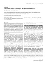
Table 1
Classification of shock
Type of shock
Parameter(s) Hypovolemic Cardiogenic Obstructive Distributive
Preload, filling pressures, end-diastolic volumes ↓↑
↑
↓
↓
Pump, cardiac output ↓↓ ↓↑
Afterload, systemic vascular resistance ↑↑ ↑↓
Systemic oxygen delivery ↓↓ ↓↑
Systemic oxygen consumption ↑↓
↑
↓
↑
↓
Systemic oxygen extraction ratio ↑↑ ↑↓
Global oxygen balance, Sv
O
2
or ScvO
2
↓↓ ↓↑
SvO
2
, mixed venous oxygen saturation; ScvO
2
, central venous oxygen saturation.
172
Critical Care April 2005 Vol 9 No 2 Rady
Invasive hemodynamic monitoring
Pulmonary artery catheterization (PAC) is the gold standard
technique for invasive hemodynamic monitoring during acute
resuscitation. Direct measurement of cardiac output, filling
pressures, and Sv
O
2
can guide therapy to optimize cardiac
function, normalize Sv
O
2
, and restore the balance between
systemic oxygen delivery and consumption. However, recent
controlled studies have raised questions regarding the utility
of PAC in the intensive care unit (ICU) setting because this
type of monitoring does not translate into a decrease in
mortality or morbidity as compared with conventional central
venous catheterization [11,12]. It is uncertain whether the
same conclusions can be drawn for the utility of PAC during
acute resuscitation in the ED. The technical expertise
required and demand imposed on nursing for this type of
monitoring has limited its use in the ED setting.
A modified form of central venous catheterization has been
developed to measure central venous pressure (CVP) and
central venous oxygen saturation (Scv
O
2
) simultaneously
during acute resuscitation in the ED [13,14]. Rivers and
coworkers [13] conducted a trial of early goal-directed
therapy, which included volume resuscitation with fluids to a
CVP of 12 mmHg or higher, vasopressor infusion to restore
mean arterial pressure to 65 mmHg or higher, followed by
transfusion of packed red blood cells and/or dobutamine
infusion to achieve a Scv
O
2
of 70% or greater. Early goal-
directed therapy restored systemic oxygen delivery with rapid
elimination of anaerobic metabolites and decreased mortality
from shock [13]. Therefore, ED resuscitation protocols that
attempt to normalize CVP and Scv
O
2
can improve global
oxygenation and result in better survival.
Cardiac output can be measured continuously using the
lithium dilution method and arterial waveform analysis
[15,16]. The lithium dilution method requires central or
peripheral intravenous infusion of lithium salt solution,
followed by arterial sampling to measure stroke volume and
cardiac output [17]. A small dose of lithium chloride is
injected as an intravenous bolus, and cardiac output is
derived from the dilution curve generated by a lithium-
sensitive electrode attached to the arterial line. Analysis of
arterial waveform energy provides a real-time calculation of
stroke volume and cardiac output. This method can also be
utilized with peripherally inserted central venous catheters in
upper extremities, eliminating the hazards associated with
central venous instrumentation. The lithium method has
limitations when assessing low cardiac output states (e.g.
hypovolemic or cardiogenic shock). However, in normal or
high cardiac output states it can provide reliable information
on stroke volume variation in real time, which can be difficult
to obtain using traditional thermodilution methods.
Noninvasive hemodynamic monitoring
Doppler echocardiography, in the form of transthoracic or
transesophageal echocardiography, permits intermittent or
continuous noninvasive evaluation of hemodynamic
parameters, including aortic blood flow, global and regional
ventricular wall motion, and valvular integrity [18]. Cardiac
output, preload, afterload, and contractility are measured or
derived from the esophageal Doppler waveform. This method
can yield valuable information regarding diastolic and systolic
functions of left and right ventricles, as well as stroke
volumes. However, the technology involved requires highly
experienced operators for accurate image acquisition and
interpretation in the ED. Cardiac output calculated from
Doppler flow measurements require certain assumptions
regarding the geometry and dimensions of cardiac chambers
and thoracic aorta, which are age dependent.
Other noninvasive technologies such as thoracic bio-
impedance for cardiac output determination are less operator
dependent and can be applied in the ED. Measurements of
stroke volume and cardiac output using the bioimpedance
method can be influenced by rapid changes in extravascular
and cellular fluid space content, especially during large
volume resuscitation.
Tissue oxygenation monitoring
Metabolic acidosis and lactic acidosis are byproducts of
anaerobic metabolism, and when they are measured in serum
they can be useful markers of persistent tissue hypoxia or
oxygen debt. Rapid bedside determination of blood lactate in
the ED has been made feasible with newly developed
enzymatic, substrate-specific electrodes [19]. A blood lactate
of 4 mmol/l or higher is a useful triage test for detecting
occult tissue hypoxia in the ED. Measurement of the
elimination rate of an elevated lactate is also a valuable
indicator of restoration of tissue oxygenation and relief from
regional ischemia [13]. Delayed elimination of elevated
lactate has been associated with subsequent development of
multiple organ dysfunction and high mortality [20].
Gastric mucosa or sublingual partial carbon dioxide tension
(P
CO
2
) can serve as a simple and noninvasive measurement
for the diagnosis and estimation of the severity of shock in the
ED. Gastric mucosal and sublingual P
CO
2
are measured
using tonometric catheters inserted into the stomach or
beneath the tongue, respectively [21,22]. Gastric and
sublingual P
CO
2
are measured using automated devices; the
device used to measure sublingual P
CO
2
is a hand-held,
portable device. A P
CO
2
above 70 mmHg is associated with
poor blood flow to the gastric or sublingual mucosa, and is
consistent with global tissue ischemia [23]. The delayed
response of mucosal P
CO
2
to therapy limits its use for real-
time monitoring of acute resuscitation.
Infrared and near infrared spectrometry, as is used in pulse
oximetry, has been employed to monitor the oxidation-reduction
state of hemoglobin and mitochondrial cytochrome in vivo. This
type of technology provides noninvasive means for assessing
cellular oxygenation and its recovery during acute resuscitation.
173
Transcutaneous oxygen and carbon dioxide electrodes have
been used experimentally for early detection of tissue hypoxia
and impending shock. However, the reproducibility of the
clinical data, the real-time response, and associated
background noise have been major obstacles to its wider
application in clinical practice and use in the ED [24].
Fluid therapy
The mainstay of cardiovascular resuscitation is administration
of intravenous fluids to increase circulating blood volume,
cardiac preload, cardiac output, and systemic oxygen
delivery. Current controversies remain focused on the type,
composition, and volumes of fluid used during resuscitation
[25]. Whether colloid or crystalloid should be used as the
fluid of first choice remains uncertain because there is no
difference in mortality between the two types of fluid [26]. A
recent large randomized clinical trial comparing saline versus
iso-oncotic human albumin solution for acute volume
resuscitation has indicated that clinical outcome is similar
with both fluid types [27].
Recently, renewed interest has focused on the use of small
fluid volumes for acute resuscitation in uncontrolled
hemorrhage and trauma, to avoid large increases in systolic
arterial pressure and dilution of coagulation factors [28,29].
Hyper-osmolar sodium chloride (7%) and/or hyper-oncotic
hydroxethyl starch (6%) have been utilized for small volume
resuscitation safely in acute hypovolemic shock [30,31].
These types of fluids can maximally augment cardiac output
at relatively small volumes and produce minimal hemodilution,
while augmenting systemic oxygen delivery [32]. The type
and volume of infused fluid can influence vascular endothelial
integrity and capillary permeability [33]. Intra-abdominal
compartment syndrome, intracranial hypertension, and
pulmonary extravascular water accumulation are frequently
associated with large fluid volume resuscitation. Compart-
ment syndromes have deleterious effects on respiratory
compliance, cardiovascular performance and splanchnic
perfusion, and can precipitate multiple organ dysfunction
[34,35]. Aggressive fluid resuscitation should focus on the
use of efficient plasma volume expanders such as colloids
and blood products in order to utilize the smallest volume of
fluid needed to restore sufficient global and tissue oxygen
delivery [36].
Blood transfusion
There has been growing concern regarding the relationship
between blood transfusion and the incidence of nosocomial
infections, organ dysfunction, and mortality in the critically
ill. Restrictive transfusion practices and tolerance of anemia
in a stable patient in the ICU was found more advantageous
than transfusion practice aimed at a higher hemoglobin
threshold in a randomized control trial [37,38]. Another
observational study [39] reported that blood transfusion
increased the risk for nosocomial infections and increased
length of stay for patients in the ICU after adjustment for
severity of illness at a single institution. However, no
randomized control trials to date justify a change in the
current transfusion practice of using either fresh or red cell
concentrate of short shelf-life for augmenting oxygen
delivery and avoiding the deleterious effects of high doses
of vasopressor and/or inotropic drugs for cardiovascular
support during resuscitation.
Cardiovascular support
Cardiovascular support during resuscitation may require
administration of pharmacologic vasoactive agents with
vasopressor and/or inotropic actions after blood volume is
restored [40]. Physiologic end-points of global and regional
oxygenation should be used for titration of vasoactive drugs
(Table 2) to avoid deleterious effects from their inappropriate
or excessive use [41]. Venous oxygen saturation (either Sv
O
2
or ScvO
2
), plasma or blood lactate clearance rate, sublingual
or gastric mucosal P
CO
2
, and urinary output are useful
parameters for assessing the effectiveness of pharmacologic
interventions. Temporary mechanical support with an intra-
aortic balloon pump may be necessary in cardiogenic shock
associated with acute coronary syndrome.
Definitive treatment
Although initial resuscitation will stabilize vital organs and
restore visceral perfusion and oxygenation, early definitive
treatment of the underlying cause of illness is required to
ensure survival. In penetrating and blunt trauma, certain
clinical indications require emergent surgical intervention.
Interventional revascularization in acute coronary syndrome
has been shown to improve survival from cardiogenic shock
secondary to acute coronary syndrome. Percutaneous
drainage, arterial or venous embolization, and insertion of
intraluminal stents or filters with the assistance of
interventional radiology can eliminate the need for surgical
intervention in certain situations [42,43].
Appropriate antimicrobial therapy and early intervention to
control sources of infection are the main pillars of definitive
treatment for sepsis [44]. Activated protein C can be given in
septic shock when cardiovascular dysfunction and/or
pulmonary dysfunction are established in order to reduce
mortality. Corticosteroid replacement for adrenal insufficiency
had been shown to improve cardiovascular stability and
perhaps decrease mortality in septic shock.
Outcomes of resuscitation
The success of resuscitation and treatment is commonly
measured in terms of 28-day or hospital survival.
Unfortunately, for many patients and families, survival is
unacceptable if it is associated with catastrophic morbidity,
such as care dependency or cognitive, psychological, and/or
physical disability [45]. A refocusing on patient-centered
outcomes that are meaningful to patients and families is
essential in measuring the success of resuscitation and
treatment [46]. Several factors are known to influence
Available online />174
resuscitation outcomes (Table 3). Adequacy and timing of
acute resuscitation, as well as definitive treatment of the
underlying illness, will influence short-term outcomes such as
incidence of multiple organ failure and hospital survival [47].
However, short-term outcomes do not always reflect
pertinent long-term outcomes such as physical, cognitive, or
psychological functioning, and subsequent life span. Long-
term consequences may be attributed to the precipitating
illness, resuscitation, or definitive treatment, or all of these
combined. Advanced age, debilitation, impaired pre-illness
functional status, chronic disease, genotype, and socio-
economic environment can also predispose to long-term
sequelae on survival.
Ethical considerations in resuscitation
A recent epidemiologic survey [48] indicated that intensive
care is used in one out of five deaths in the USA, raising
significant concerns regarding the appropriateness of the
type of care offered to terminally ill, hospitalized patients. That
survey emphasizes the importance of informed participation
of patients and surrogates in decision making regarding
resuscitation and life-sustaining therapy. Physicians are
obliged to preserve the principles of beneficence, non-
maleficence, and respect for patient autonomy under all
circumstances, including during acute resuscitation [49].
However, the urgency of acute resuscitation and the impaired
ability of the patient to make a reasonable autonomous
decision both conspire against adequate consideration of the
principles of medical ethics.
Health care providers often make initial resuscitation
decisions unilaterally. Under such circumstances, gaining
informed consent is not feasible and may lead to a common
misconception that consent is not required for resuscitation,
because it brings benefit and prevents harm, and the patient
is unable to give or withhold consent. Physicians who are
engaged in acute resuscitation have professional obligations
to address certain ethical issues. First, did the patient or their
surrogate participate in an informed decision making
process? Second, will treatment confer survival, restore
functional independence, and improve the patient’s quality of
life? Third, will treatment result in short-term or long-term
complications, disability, and/or care dependency? Finally,
will treatment represent efficient utilization of limited health
care resources?
Information gathered from advanced directives, living wills,
and family discussions that indicate patient-expressed
attitudes or wishes regarding such a situation must be
considered, along with the likely benefits and harms of the
resuscitation endeavor. The physician responsible for
initiating resuscitation or life-sustaining therapy must fulfill
that task [50]. Life-sustaining therapy that simply delays death
and prolongs suffering is unacceptable, and should be
carefully reconsidered. Medical futility of resuscitation
because of long-term permanent disability and poor quality of
life must be addressed in each clinical situation with
surrogate decision makers [51].
Conclusion
The modes and methods for monitoring acute resuscitation
have undergone significant changes over the past decade.
Patient-centered outcome measures and observance of
ethical principles are becoming integral to the everyday
practice of resuscitation.
Competing interests
The author has no affiliations or financial involvement with any
organization or entity with a direct financial interest in the
subject matter or materials discussed in the manuscript.
References
1. Orebaugh SL: Difficult airway management in the emergency
department. J Emerg Med 2002, 22:31-48.
2. Sigillito RJ, DeBlieux PM: Evaluation and initial management of
the patient in respiratory distress. Emerg Med Clin North Am
2003, 21:239-258.
3. Murray S: Bi-level positive airway pressure (BiPAP) and acute
cardiogenic pulmonary oedema (ACPO) in the emergency
department. Aust Crit Care 2002, 15:51-63.
Critical Care April 2005 Vol 9 No 2 Rady
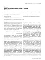
Table 2
Common vasoactive agents used for cardiovascular support
during resuscitation
Type of action Drugs
Inotropic Dobutamine
Milrinone
Dopexamine
Combined Dopamine
Epinephrine
Vasomotor Norepinephrine
Phenylephrine
Vasopressin
Table 3
Outcome of resuscitation
Outcome end-points Factors that impact on outcome
28-day survival Age
Hospital survival Pre-illness function and mobility
Long term care dependency Chronic end-stage organ disease
Return to independent function Etiology of acute illness
Quality of life-adjusted Timing of resuscitation and
survival definitive treatment
175
4. Pelosi P, Severgnini P, Aspesi M, Gamberoni C, Chiumello D,
Fachinetti C, Introzzi L, Antonelli M, Chiaranda M: Non-invasive
ventilation delivered by conventional interfaces and helmet in
the emergency department. Eur J Emerg Med 2003, 10:79-86.
5. Lightowler JV, Wedzicha JA, Elliott MW, Ram FS: Non-invasive
positive pressure ventilation to treat respiratory failure result-
ing from exacerbations of chronic obstructive pulmonary
disease: Cochrane systematic review and meta-analysis. BMJ
2003, 326:185.
6. L’Her E: Noninvasive mechanical ventilation in acute cardio-
genic pulmonary edema. Curr Opin Crit Care 2003, 9:67-71.
7. Gali B, Goyal DG: Positive pressure mechanical ventilation.
Emerg Med Clin North Am. 2003, 21:453-473.
8. Ram FS, Lightowler JV, Wedzicha JA: Non-invasive positive
pressure ventilation for treatment of respiratory failure due to
exacerbations of chronic obstructive pulmonary disease.
Cochrane Database Syst Rev 2003, 1:CD004104.
9. Sinuff T, Cook DJ, Randall J, Allen CJ: Evaluation of a practice
guideline for noninvasive positive-pressure ventilation for
acute respiratory failure. Chest 2003, 123:2062-2073.
10. Chaney JC, Derdak S: Minimally invasive hemodynamic moni-
toring for the intensivist: current and emerging technology.
Crit Care Med 2002, 30:2338-2345.
11. Rhodes A, Cusack RJ, Newman PJ, Grounds RM, Bennett ED: A
randomised, controlled trial of the pulmonary artery catheter
in critically ill patients. Intensive Care Med 2002, 28:256-264.
12. Richard C, Warszawski J, Anguel N, Deye N, Combes A, Barnoud
D, Boulain T, Lefort Y, Fartoukh M, Baud F, et al.: Early use of the
pulmonary artery catheter and outcomes in patients with
shock and acute respiratory distress syndrome: a randomized
controlled trial. JAMA 2003, 290:2713-2720.
13. Rivers E, Nguyen B, Havstad S, Ressler J, Muzzin A, Knoblich B,
Peterson E, Tomlanovich M; Early Goal-Directed Therapy Collabora-
tive Group: Early goal-directed therapy in the treatment of severe
sepsis and septic shock. N Engl J Med 2001, 345:1368-1377.
14. Ladakis C, Myrianthefs P, Karabinis A, Karatzas G, Dosios T, Fild-
issis G, Gogas J, Baltopoulos G: Central venous and mixed
venous oxygen saturation in critically ill patients. Respiration
2001, 68:279-285.
15. Hamilton TT, Huber LM, Jessen ME: PulseCO: a less-invasive
method to monitor cardiac output from arterial pressure after
cardiac surgery. Ann Thorac Surg 2002, 74:S1408-S1412.
16. Cottis R, Magee N, Higgins DJ: Haemodynamic monitoring with
pulse-induced contour cardiac output (PiCCO) in critical care.
Intensive Crit Care Nurs 2003, 19:301-307.
17. Jonas MM, Tanser SJ: Lithium dilution measurement of cardiac
output and arterial pulse waveform analysis: an indicator dilu-
tion calibrated beat-by-beat system for continuous estimation
of cardiac output. Curr Opin Crit Care 2002, 8:257-261.
18. Axler O, Megarbane B, Lentschener C, Fernandez H: Compari-
son of cardiac output measured with echocardiographic
volumes and aortic Doppler methods during mechanical ven-
tilation. Intensive Care Med 2003, 29:208-217.
19. Aduen J, Bernstein WK, Khastgir T, Miller J, Kerzner R, Bhatiani A,
Lustgarten J, Bassin AS, Davison L, Chernow B: The use and
clinical importance of a substrate-specific electrode for rapid
determination of blood lactate concentrations. JAMA 1994,
272:1678-1685.
20. Husain FA, Martin MJ, Mullenix PS, Steele SR, Elliott DC: Serum
lactate and base deficit as predictors of mortality and morbid-
ity. Am J Surg 2003, 185:485-491.
21. Rackow EC, O’Neil P, Astiz ME, Carpati CM: Sublingual cap-
nometry and indexes of tissue perfusion in patients with cir-
culatory failure. Chest 2001, 120:1633-1638.
22. Boswell SA, Scalea TM: Sublingual capnometry: an alternative
to gastric tonometry for the management of shock resuscita-
tion. AACN Clin Issues 2003, 14:176-184.
23. Weil MH, Nakagawa Y, Tang W, Sato Y, Ercoli F, Finegan R,
Grayman G, Bisera J: Sublingual capnometry: a new noninva-
sive measurement for diagnosis and quantitation of severity
of circulatory shock. Crit Care Med 1999,27:1225-1229.
24. Tatevossian RG, Wo CC, Velmahos GC, Demetriades D, Shoe-
maker WC: Transcutaneous oxygen and CO2 as early warning
of tissue hypoxia and hemodynamic shock in critically ill
emergency patients. Crit Care Med 2000, 28:2248-2253.
25. Bradley C: Crystalloid, colloid or small volume resuscitation?
Intensive Crit Care Nurs 2001, 17:304-306.
26. Rizoli SB: Crystalloids and colloids in trauma resuscitation: a
brief overview of the current debate. J Trauma 2003,
Suppl:S82-S88.
27. The SAFE Study Investigators: A comparison of albumin and
saline for fluid resuscitation in the intensive care unit. N Engl J
Med 2004, 350:2247-2256.
28. Dutton RP, Mackenzie CF, Scalea TM: Hypotensive resuscita-
tion during active hemorrhage: impact on in-hospital mortal-
ity. J Trauma 2002, 52:1141-1146.
29. Revell M, Greaves I, Porter K: Endpoints for fluid resuscitation
in hemorrhagic shock. J Trauma 2003, Suppl:S63-S67.
30. Oliveira RP, Velasco I, Soriano F, Friedman G: Clinical review:
Hypertonic saline resuscitation in sepsis. Crit Care 2002, 6:
418-423.
31. Freudenberg S, Palma P, Schuster K, Mkony C, Waschke K:
Small volume resuscitation with 7.5% hypertonic saline solu-
tion: treatment of haemorrhagic shock in the tropics. Trop
Doct. 2003, 33:165-166.
32. Mauritz W, Schimetta W, Oberreither S, Polz W: Are hypertonic
hyperoncotic solutions safe for prehospital small-volume
resuscitation? Results of a prospective observational study.
Eur J Emerg Med 2002, 9:315-319.
33. Jaeger K, Heine J, Ruschulte H, Juttner B, Scheinichen D, Kuse ER,
Piepenbrock S: Effects of colloidal resuscitation fluids on the
neutrophil respiratory burst. Transfusion 2001, 41:1064-1068.
34. Biffl WL, Moore EE, Burch JM, Offner PJ, Franciose RJ, Johnson
JL: Secondary abdominal compartment syndrome is a highly
lethal event. Am J Surg 2001, 182:645-648.
35. Balogh Z, McKinley BA, Cocanour CS, Kozar RA, Valdivia A,
Sailors RM, Moore FA: Supranormal trauma resuscitation
causes more cases of abdominal compartment syndrome.
Arch Surg 2003, 138:637-642; discussion 642-633.
36. Wu JJ, Huang MS, Tang GJ, Kao WF, Shih HC, Su CH, Lee CH:
Hemodynamic response of modified fluid gelatin compared
with lactated ringer’s solution for volume expansion in emer-
gency resuscitation of hypovolemic shock patients: prelimi-
nary report of a prospective, randomized trial. World J Surg
2001, 25:598-602.
37. Hebert PC, Wells G, Martin C, Tweeddale M, Marshall J, Blajch-
man M, Pagliarello G, Schweitzer I, Calder L: A Canadian survey
of transfusion practices in critically ill patients. Transfusion
Requirements in Critical Care Investigators and the Canadian
Critical Care Trials Group. Crit Care Med. 1998, 26:482-487.
38. Hebert PC, Yetisir E, Martin C, Blajchman MA, Wells G, Marshall
J, Tweeddale M, Pagliarello G, Schweitzer I; Transfusion Require-
ments in Critical Care Investigators for the Canadian Critical Care
Trials Group: Is a low transfusion threshold safe in critically ill
patients with cardiovascular diseases? Crit Care Med 2001,
29:227-234.
39. Taylor RW, Manganaro L, O’Brien J, Trottier SJ, Parkar N, Vere-
makis C: Impact of allogenic packed red blood cell transfusion
on nosocomial infection rates in the critically ill patient. Crit
Care Med 2002, 30:2249-2254.
40. Ruokonen E, Parviainen I, Uusaro A: Treatment of impaired per-
fusion in septic shock. Ann Med 2002, 34:590-597.
41. Trager K, DeBacker D, Radermacher P: Metabolic alterations in
sepsis and vasoactive drug-related metabolic effects. Curr
Opin Crit Care 2003, 9:271-278.
42. Ajani AE, Maruff P, Warren R, Eccleston D, Dick R, MacIsaac A,
Rowe M, Lefkovits J: Impact of early percutaneous coronary
intervention on short- and long-term outcomes in patients
with cardiogenic shock after acute myocardial infarction. Am J
Cardiol 2001, 87:633-635, A639-A610.
43. Kushimoto S, Arai M, Aiboshi J, Harada N, Tosaka N, Koido Y,
Yoshida R, Yamamoto Y, Kumazaki T: The role of interventional
radiology in patients requiring damage control laparotomy. J
Trauma 2003, 54:171-176.
44. Dellinger RP, Carlet JM, Masur H, Gerlach H, Calandra T, Cohen J,
Gea-Banacloche J, Keh D, Marshall JC, Parker MM, et al.: Surviv-
ing Sepsis Campaign guidelines for management of severe
sepsis and septic shock. Crit Care Med 2004, 32:858-873.
45. Buckley TA, Cheng AY, Gomersall CD: Quality of life in long-
term survivors of intensive care. Ann Acad Med Singapore
2001, 30:287-292.
46. Angus DC, Carlet J; 2002 Brussels Roundtable Participants: Sur-
viving intensive care: a report from the 2002 Brussels Round-
table. Intensive Care Med 2003, 29:368-377.
Available online />176
Critical Care April 2005 Vol 9 No 2 Rady
47. Gallagher SF, Williams B, Gomez C, DesJardins C, Swan S,
Durham RM, Flint LM: The role of cardiac morbidity in short-
and long-term mortality in injured older patients who survive
initial resuscitation. Am J Surg 2003, 185:131-134.
48. Angus DC, Barnato AE, Linde-Zwirble WT, Weissfeld LA, Watson
RS, Rickert T, Rubenfeld GD; Robert Wood Johnson Foundation
ICU End-Of-Life Peer Group: Use of intensive care at the end
of life in the United States: an epidemiologic study. Crit Care
Med 2004, 32:638-643.
49. Berger JT: Ethical challenges of partial do-not-resuscitate
(DNR) orders: placing DNR orders in the context of a life-
threatening conditions care plan. Arch Intern Med 2003, 163:
2270-2275.
50. Ardagh M: Resurrecting autonomy during resuscitation: the
concept of professional substituted judgment. J Med Ethics
1999, 25:375-378.
51. Hook CC, Koch KA: Ethics of resuscitation. Crit Care Clin
1996, 12:135-148.