Báo cáo khoa học: "High-frequency oscillatory ventilation in children: a single-center experience of 53 cases" pot
Bạn đang xem bản rút gọn của tài liệu. Xem và tải ngay bản đầy đủ của tài liệu tại đây (177.68 KB, 6 trang )
Open Access
Available online />R274
Vol 9 No 3
Research
High-frequency oscillatory ventilation in children: a single-center
experience of 53 cases
Fieke YAM Slee-Wijffels
1
, Klara RM van der Vaart
2
, Jos WR Twisk
3
, Dick G Markhorst
4
and
Frans B Plötz
5
1
Pediatrician, Department of Pediatric Intensive Care, VU Medical Center, Amsterdam, The Netherlands
2
PhD Student, Department of Pediatric Intensive Care, VU Medical Center, Amsterdam, The Netherlands
3
Epidemiologist, Department of Clinical Epidemiology and Biostatistics, VU Medical Center, Amsterdam, The Netherlands
4
Pediatric Intensivist, Department of Pediatric Intensive Care, VU Medical Center, Amsterdam, The Netherlands
5
Pediatric Intensivist, Department of Pediatric Intensive Care, VU Medical Center, Amsterdam, The Netherlands
Corresponding author: Frans B Plötz,
Received: 6 Feb 2005 Revisions requested: 2 Mar 2005 Revisions received: 4 Mar 2005 Accepted: 15 Mar 2005 Published: 8 Apr 2005
Critical Care 2005, 9:R274-R279 (DOI 10.1186/cc3520)
This article is online at: />© 2005 Slee-Wijffels et al, licensee BioMed Central Ltd.
This is an Open Access article distributed under the terms of the Creative Commons Attribution License ( />2.0), which permits unrestricted use, distribution, and reproduction in any medium, provided the original work is cited.
Abstract
Introduction The present article reports our experience with
high-frequency oscillatory ventilation (HFOV) in pediatric
patients who deteriorated on conventional mechanical
ventilation.
Methods The chart records of 53 consecutively HFOV-treated
patients from 1 January 1998 to 1 April 2004 were
retrospectively analyzed. The parameters of demographic data,
cause of respiratory insufficiency, Pediatric Index of Mortality
score, oxygenation index and PaCO
2
were recorded and
calculated at various time points before and after the start of
HFOV, along with patient outcome and cause of death.
Results The overall survival rate was 64%. We observed
remarkable differences in outcome depending on the cause of
respiratory insufficiency; survival was 56% in patients with
diffuse alveolar disease (DAD) and was 88% in patients with
small airway disease (SAD). The oxygenation index was
significantly higher before and during HFOV in DAD patients
than in SAD patients. The PaCO
2
prior to HFOV was higher in
SAD patients compared with DAD patients and returned to
normal values after the initiation of HFOV.
Conclusion HFOV rescue therapy was associated with a high
survival percentage in a selected group of children. Patients with
DAD primarily had oxygenation failure. Future studies are
necessary to evaluate whether the outcome in this group of
patients may be improved if HFOV is applied earlier in the
course of disease. Patients with SAD primarily had severe
hypercapnia and HFOV therapy was very effective in achieving
adequate ventilation.
Introduction
High-frequency oscillatory ventilation (HFOV) is, from a theo-
retical point of view, an ideal method of ventilation to minimize
ventilator-associated lung injury. HFOV avoids high peak
inspiratory pressures, thus preventing end-inspiratory overdis-
tension, and it avoids repetitive recruitment and de-recruitment
of the unstable lung alveoli, thus preventing end-expiratory col-
lapse [1-3]. Despite these factors, HFOV is primarily used as
a rescue therapy in pediatric patients with diffuse alveolar dis-
ease (DAD), and the reported survival varies between 18%
and 67% [4-15].
We have used HFOV as a rescue therapy in our pediatric
intensive care unit since 1995. In addition, in contrast to most
other centers, we also apply HFOV as a rescue therapy in chil-
dren with small airway disease (SAD). The purpose of the
present article is to report our HFOV experience with 53 con-
secutively treated pediatric patients who deteriorated on con-
ventional mechanical ventilation (CMV). In addition, we
considered whether the cause of respiratory insufficiency had
an effect on outcome.
CDP = continuous distending pressure; CMV = conventional mechanical ventilation; DAD = diffuse alveolar disease; HFOV = high-frequency oscil-
latory ventilation; OI = oxygenation index; SAD = small airway disease.
Critical Care Vol 9 No 3 Slee-Wijffels et al.
R275
Patients and methods
Our pediatric intensive care unit is a nine-bed combined med-
ical and surgical intensive care unit, staffed by trained pediatric
intensivists. The chart records of all HFOV-treated children
between 1 January 1998 and 1 April 2004 were retrospec-
tively analyzed. During this period a median of 356 patients
(range, 326–395 patients) were admitted per year. At the time
of the study, it was not institutional policy to require ethical
committee approval for a retrospective review of this nature.
The following demographic data were recorded: sex, age,
weight, cause of respiratory insufficiency, time on CMV prior to
HFOV, and Pediatric Index of Mortality score. The oxygenation
index (OI) was calculated 24, 12 and 6 hours before transition
to HFOV and at 1, 6, 12, 24 and 48 hours after the institution
of HFOV. The outcomes included survival at pediatric inten-
sive care unit discharge, the total number of ventilation days
(CMV and HFOV), and the change in the OI and PaCO
2
before and during HFOV. The OI was defined as: 100 × mean
airway pressure × (FiO
2
/ PaO
2
) [cmH
2
O/mmHg].
All patients with severe respiratory failure are initially managed
with CMV. We use an open lung ventilation strategy that is a
volume-targeted pressure-limited strategy, aimed at adequate
oxygenation and ventilation with limited pressures (plateau
pressures <30–35 cmH
2
O and tidal volumes of 8–10 ml/kg
bodyweight) with, when indicated, permissive hypercapnia
(pH >7.25) and optimal positive end-expiratory pressure to
achieve a goal of FiO
2
<0.6 with a minimum oxygen saturation
of 90% (PaO
2
>60 mmHg). We do not use exogenous sur-
factant to improve gas exchange in our pediatric intensive care
unit, and prone positioning is considered occasionally. In gen-
eral, we try to avoid the use of neuromuscular blockade agents
except in patients with small airway disease with refractory
acidosis.
The reason for converting to HFOV in these patients was per-
sistent oxygenation failure or ventilation failure, based on one
or both of the following criteria: intractable respiratory failure
with an OI >13 demonstrated by two consecutive blood gas
measurements over at least a 6-hour period, or a plateau pres-
sure exceeding 30 cmH
2
O despite the use of permissive
hypercapnia for at least 2 hours. However, this treatment was
not protocolized and the decision to start HFOV was, at times,
based on clinical discretion. Former prematurity with residual
bronchopulmonary dysplasia or obstructive airway disease
with clinical evidence of increased expiratory resistance or
hyperinflation on chest X-ray were not considered a contrain-
dication for HFOV. HFOV was performed using the Sensor-
Medics 3100A or 3100B (Yorba Linda, CA, USA).
Depending on the lung function and chest X-ray characteris-
tics during CMV, patients are classified either as having DAD
or SAD. DAD patients primarily had oxygenation disturbances
necessitating high plateau pressures and a chest X-ray with
bilateral diffuse whitening, whereas SAD patients primarily had
ventilation disturbances, with increased airway resistance and
prolonged time constants and a chest X-ray with hyperinfla-
tion. We use different HFOV strategies depending on the
underlying disease [6].
The 'high-volume' or 'open-lung' strategy for DAD
The initial continuous distending pressure (CDP) is set 4 cm
above the mean airway pressure used during CMV. Our oxy-
genation goal is to reach an adequate PaO
2
(>60 mmHg) with
FiO
2
<0.4. Thereafter, CDP is weaned once the patient
achieves FiO
2
<0.4. When hypoxemia persists with adequate
circulation and with no radiographic signs of lung overinflation,
CDP is increased further until the oxygenation targets are
reached and is subsequently rapidly weaned. The pressure
amplitude of oscillation is initially set to achieve chest wall
vibration to the level of the mid-thigh. The pressure amplitude
of oscillation and the frequency are sequentially adjusted to
achieve a PaCO
2
within the target range and to maintain a pH
>7.25. In children weighing <10 kg we used a frequency of 10
Hz, in children weighing >10 kg we used a frequency of 8 Hz.
The frequency is decreased with persistent respiratory acido-
sis despite maximization of the pressure amplitude of
oscillation.
The 'open-airway' strategy for SAD
In patients with SAD we used the same initial settings as
already described in the 'open-lung' strategy, but high CDP is
now used to open up the small airways, allowing oscillations
to move freely in and out of the alveolus. The CDP must be
applied carefully; if the airways are opened up, compliant alve-
oli can be faced with high pressures. Every incremental
change should be followed by PaCO
2
determination to see at
which CDP the airways are opened and the PaCO
2
decreases. When the airways are open, the lowest possible
CDP and pressure amplitude of oscillation are sought to mini-
mize the risk of overdistension. Overdistension is suspected if
the circulation becomes compromised and if this can be
restored by lowering the CDP. The degree of lung hyperinfla-
tion on chest X-ray is not used to modify CDP.
All patients are sedated during HFOV. Patients are either
weaned to continuous positive airway pressure or weaned to
CMV when CDP <20 cmH
2
O on FiO
2
<0.4 and endotracheal
suctioning is well tolerated.
Statistical analysis
Baseline characteristics for survivors and nonsurvivors were
compared with nonparametric Mann–Whitney tests for contin-
uous variables and with chi-square tests or Fisher exact tests
for dichotomous variables. The development over time in the
OI and PaCO
2
between groups of patients was analyzed with
generalized estimating equations [16].
Available online />R276
Generalized estimating equation analysis is an extended linear
regression analysis taking into account the fact that the same
patients are measured over time. The advantage of generalized
estimating equation analysis (for instance, compared with a
repeated-measures analysis of variance) is that each patient is
part of the analysis, irrespective of the number of repeated
measurements performed for that patient; that is, missing data
and an unequal number of measurements between patients
are allowed.
Time was added to the generalized estimating equation analy-
sis as a categorical variable (i.e. represented by dummy varia-
bles) in order to estimate the development over time as
accurately as possible. Five patients, after being switched
from HFOV to CMV, had another HFOV run (two nonsurvivors,
three survivors). This second run is not used in the analysis.
The significance level for all tests was set at P <0.05. All sta-
tistical analyses were performed with STATA (version 7; Stata
Corp LP, College Station, Texas, USA).
Results
During the study period 52 children were treated with HFOV
after failure on CMV. One patient was excluded from the anal-
ysis because differentiated HFOV and CMV for independent
lung ventilation was applied [17]. One patient underwent three
HFOV runs on different occasions. Thus 51 children (53
HFOV runs) composed the final study sample.
The overall survival rate was 32/53 (64%). The demographics
of the surviving and nonsurviving patients are presented in
Table 1. We observed that nine patients (47%) died during
HFOV rescue therapy. A remarkable difference in outcome
between DAD patients and SAD patients was observed; 18 of
32 (56%) DAD patients and 15 of 17 (88%) SAD patients sur-
vived. We therefore compared the course of the OI and
PaCO
2
between these two groups of patients.
The DAD patients had a significantly higher OI at the time of
transition than the SAD patients (Fig. 1). The observed rise in
the OI in the first hour after transition to HFOV in both groups
is due to the applied higher CDP when compared with the
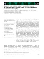
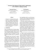
Figure 1
The oxygenation index (OI) before and during high-frequency oscillatory ventilation (HFOV) in patients with diffuse alveolar disease (DAD) (●) and in patients with small airway disease (SAD) (■)The oxygenation index (OI) before and during high-frequency oscillatory
ventilation (HFOV) in patients with diffuse alveolar disease (DAD) (●)
and in patients with small airway disease (SAD) (■). The OI became
significantly higher 6 hours prior to HFOV therapy and remained higher.
The observed rise in the OI in the first hour after transition to HFOV in
both groups is due to the applied higher CDP when compared with the
mean airway pressure during conventional mechanical ventilation. The
SAD patients had a higher, but not significant, PaCO
2
before transition
to HFOV than the DAD patients. PaCO
2
returned to normal values after
transition to HFOV. * P < 0.05.
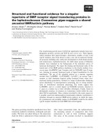
Figure 2
The oxygenation index (OI) was higher in the nonsurvivors (solid line) compared with the survivors (dash line) in the diffuse alveolar disease group before the start of high-frequency oscillatory ventilation (HFOV)The oxygenation index (OI) was higher in the nonsurvivors (solid line)
compared with the survivors (dash line) in the diffuse alveolar disease
group before the start of high-frequency oscillatory ventilation (HFOV).
The OI became significant after the start of HFOV. * P < 0.05.
Critical Care Vol 9 No 3 Slee-Wijffels et al.
R277
mean airway pressure during CMV. The OI was higher, but not
significantly, in the nonsurvivors in the DAD group before the
start of HFOV, and after the initiation of HFOV it became sig-
nificantly higher (Fig. 2). The SAD patients had a higher (66.9
± 27.9 mmHg), but not significant, PaCO
2
before transition to
HFOV than the DAD patients (55.2 ± 23.7 mmHg). The
PaCO
2
rapidly decreased after transition to HFOV (Fig. 1).
The mean PaCO
2
values 1 hour after the start of HFOV were
51.6 ± 15.5 mmHg in the SAD group and 55.4 ± 39.2 mmHg
in the DAD group, respectively.
Discussion
The overall survival rate was 64% in patients where adequate
oxygenation or ventilation could not be achieved with CMV.
We observed remarkable differences in outcome depending
on the cause of respiratory insufficiency, indicating that a dif-
ferent disease process carries a different prognosis and out-
come. In patients with DAD the survival rate was 56%, and this
rate was 88% in patients with SAD. The OI was significantly
higher in DAD patients than in SAD patients, whereas the
PaCO
2
prior to HFOV was higher in SAD patients than in DAD
patients.
Only one prospective study and a few retrospective observa-
tional studies report the outcome in pediatric patients treated
with HFOV [4-15]. Mortality rates vary between 18% and
67%. There are several reasons to explain this difference. First,
the numbers of patients included in the studies were very
small, ranging from four to 35 patients, so even the death of
one patient could substantially alter the mortality rate. Second,
mortality rates can be affected by the underlying cause of res-
piratory insufficiency. Most studies use HFOV as a rescue
therapy only in children showing signs of DAD. This in contrast
to our study, and we observed remarkable differences in out-
come depending on the cause of respiratory insufficiency.
Third, it is not evidently clear in the reports from the previous
studies whether all nonsurviving patients died of pulmonary
causes or because of other reasons. Finally, the experience
with HFOV differs between studies and hospitals, which could
have had an influence on the mortality rates reported. The
existence of a learning curve for new technologies, as for the
use of HFOV, has been widely acknowledged in the past.
Most rescue HFOV therapies are applied in patients with
DAD. It is suggested that an OI >13 may serve as an indication
for HFOV rescue therapy. When reviewing previous studies,
however, the actual OI at the time of transition varies widely
from 10 to 45.9 [4-6]. A large survey among 14 centers includ-
ing 232 pediatric patients also revealed a mean OI >27.1
before initiation of HFOV [18]. We started HFOV at a median
OI of 18 in the survivors and a median OI of 28 in the nonsur-
vivors (Fig. 1), suggesting that we may have started HFOV res-
cue therapy too late. However, the OI values 6 hours before
Table 1
Patient demographics
Parameter Survivors Nonsurvivors P value
Number of patients 34 19
Age (months)
a
9.5 (0–158) 14.0 (0–169) Not significant
Weight (kg)
a
6.7 (2.6–30.0) 10.0 (1.7–86.0) Not significant
Male 20 (58.8%) 7 (36.8%) Not significant
Pediatric Index of Mortality score (%)
a
2.8 (0.2–54.5) 3.9 (0.6–97.7) Not significant
Cause of respiratory insufficiency
Diffuse alveolar disease 18 (52.9%) 14 (73.7%)
Acute respiratory distree syndrome 10 (29.4%) 7 (36.8%)
Pneumonia 8 (23.6%) 6 (31.6%)
Aspiration 0 1 (5.3%)
Small airway disease 15 (44.2%) 2 (10.5%)
Bronchiolitis 15 (44.1%) 2 (10.5%)
Different 1 (2.9%) 3 (15.8%)
Duration of conventional mechanical ventilation before transition
(hours)
a
29.5 (0–690) 63.0 (2–473) Not significant
Duration of high-frequency oscillatory ventilation (hours)
a
214 (1–648) 177 (9–845) Not significant
Duration of conventional mechanical ventilation after high-frequency
oscillatory ventilation (hours)
a
66 (0–1218) 8 (0–427) Not significant
Number of pediatric intensive care unit days
a
23 (7–47) 22 (3–50) Not significant
a
Data presented as median (range).
Available online />R278
transition were comparable between survivors and nonsurvi-
vors (Fig. 2).
Most studies have focused on the OI as a predictor of mortality
after switching to HFOV. Sarnaik and colleagues proposed
that those patients with an initial OI >20 who did not have a
reduction of at least 20% in OI by 6 hours on HFOV can be
predicted to die [8]. We think it is more important to identify
early those patients who are at risk by prospectively recording
the OI at small time intervals. This may serve to switch these
patients to HFOV therapy before achieving OI >20 (Fig. 2). It
remains uncertain whether this will result in an improved sur-
vival. It is therefore necessary to perform a large prospective
multicenter trial to evaluate whether outcome in patients with
DAD may be improved if HFOV is applied earlier in the course
of the disease.
The use of HFOV in children with SAD is limited to a few case
reports and is usually avoided because of the assumption of
an associated increased risk of dynamic air trapping with this
condition [19,20]. The reason for converting to HFOV in
patients with SAD was primarily hypercapnia. HFOV therapy
was very effective in achieving rapid adequate ventilation,
resulting in an 88% survival. Our results suggest that HFOV is
safe but it remains very important to apply the adequate HFOV
strategy in this group of patients. HFOV is used to open up
and stent the small airways ('open airway' – a concept in anal-
ogy to the 'open lung' concept) to provide adequate ventila-
tion, which is in sharp contrast with the application of CDP to
provide optimal oxygenation. The airway diameter remains sta-
ble and oscillations can move freely in and out of the alveoli,
providing an adequate ventilation – particularly since expira-
tion during HFOV is active.
In conclusion, despite the retrospective nature of this study
creating several limitations, we observed that HFOV rescue
therapy was associated with a high survival percentage in a
selected group of children where CMV failed. Future studies
are necessary to evaluate whether the outcome in patients
with DAD may be improved if HFOV is applied earlier in the
course of disease. HFOV rescue therapy in patients with SAD
can be considered in refractory hypercapnia.
Competing interests
The author(s) declare that they have no competing interests.
Authors' contributions
FYAMS-W carried out the data collection and drafted the
manuscript. KRMvdV carried out the data collection and
drafted the manuscript. JWRT performed the statistical analy-
sis. DGM participated in the study design and helped to draft
the manuscript. FBP conceived of the study and participated
in its design and coordination, and helped to draft the manu-
script. All authors read and approved the final manuscript.
References
1. Froese AB, McCulloch PR, Sugiura M, Vaclavik S, Possmayer F,
Moller F: Optimizing alveolar expansion prolongs the effective-
ness of exogenous surfactant therapy in the adult rabbit. Am
Rev Respir Dis 1993, 148:569-577.
2. Froese AB: High-frequency oscillatory ventilation for adult res-
piratory distress syndrome: let's get it right this time! Crit Care
Med 1997, 25:906-908.
3. Venegas JG, Fredberg JJ: Understanding the pressure cost of
ventilation: why does high-frequency ventilation work? Crit
Care Med 1994, 22:S49-S57.
4. Arnold JH, Hanson JH, Toro-Figuero LO, Gutierrez J, Berens RJ,
Anglin DL: Prospective, randomized comparison of high-fre-
quency oscillatory ventilation and conventional mechanical
ventilation in pediatric respiratory failure. Crit Care Med 1994,
22:1530-1539.
5. Anton N, Joffe KM, Joffe AR: Inability to predict outcome of
acute respiratory distress syndrome in children when using
high frequency oscillation. Intensive Care Med 2003,
29:1763-1769.
6. Duval EL, Markhorst DG, Gemke RJ, van Vught AJ: High-fre-
quency oscillatory ventilation in pediatric patients. Neth J Med
2000, 56:177-185.
7. Tang JR, Yau KI, Shih HH: High-frequency oscillatory ventilation
for infants and children with adult respiratory distress syn-
drome. Zhonghua Min Guo Xiao Er Ke Yi Xue Hui Za Zhi 1997,
38:137-144.
8. Sarnaik AP, Meert KL, Pappas MD, Simpson PM, Lieh-lai MW, Hei-
demann SM: Predicting outcome in children with severe acute
respiratory failure treated with high-frequency ventilation. Crit
Care Med 1996, 24:1396-1402.
9. Fedora M, Klimovic M, Seda M, Dominik P, Nekvasil R: Effect of
early intervention of high-frequency oscillatory ventilation on
the outcome in pediatric acute respiratory distress syndrome.
Bratisl Lek Listy 2000, 101:8-13.
10. Rosenberg RB, Broner CW, Peters KJ, Anglin DL: High-fre-
quency ventilation for acute pediatric respiratory failure. Chest
1993, 104:1216-1221.
11. Lochindarat S, Srisan P, Jatanachai P: Factors effecting the out-
come of acute respiratory distress syndrome in pediatric
patients treated with high frequency oscillatory ventilation. J
Med Assoc Thai 2003, 86:S618-S627.
12. McDougall PN, Loughnan PM, Campbell NT, Hochmann M, Timms
BJ, Butt WW: High frequency oscillation in newborn infants
with respiratory failure. J Paediatr Child Health 1995,
31:292-296.
13. Watkins SJ, Peters MJ, Tasker RC: One hundred courses of high
frequence oscillatory ventilation: what have we learned? Eur J
Pediatr 2000, 159:134.
Key messages
- HFOV rescue therapy was associated with a high survival
percentage (64%) in a selected group of children.
- A remarkable difference in outcome was observed
depending on the cause of respiratory insufficiency,
indicating that a different disease process carries a dif-
ferent prognosis and outcome.
- In patients with diffuse alveolar disease the survival rate
was 56%, and this rate was 88% in patients with small
airway disease.
- The oxygenation index prior to HFOV was significantly
higher in diffuse alveolar disease patients than in small
airway disease patients.
HFOV rescue therapy in patients with small airway disease
can be considered in refractory hypercapnia.
Critical Care Vol 9 No 3 Slee-Wijffels et al.
R279
14. MacIntyre NR: High-frequency jet ventilation. Respir Care Clin N
Am 2001, 7:599-610.
15. Cox CE, Carson SS, Ely EW, Govert JA, Garrett JM, Brower RG,
Morris DG, Abraham E, Donnabella V, Spevetz A, Hall JB: Effec-
tiveness of medical resident education in mechanical
ventilation. Am J Respir Crit Care Med 2003, 167:32-38.
16. Twisk JWR: Applied Longitudinal Data Analysis for Epidemiology.
A Practical Guide Cambridge: Cambridge University Press; 2003.
17. Plötz FB, Hassing MB, Sibarani-Ponsen RD, Markhorst DG: Dif-
ferentiated HFO and CMV for independent lung ventilation in a
pediatric patient [Letter]. Intensive Care Med 2003, 29:1855.
18. Arnold JH, Anas NG, Luckett P, Cheifetz IM, Reyes G, Newth CJL,
Kocis KC, Heidemann SM, Hanson JH, Brogan TV, Bohn DJ:
High-frequency oscillatory ventilation in pediatric respiratory
failure: a multicenter experience. Crit Care Med 2000,
28:3913-3919.
19. Duval EL, Leroy PL, Gemke RJ, van Vught AJ: High-frequency
oscillatory ventilation in RSV bronchiolitis patients. Respir Med
1999, 93:435-440.
20. Duval EL, van Vught AJ: Status asthmaticus treated by high-fre-
quency oscillatory ventilation. Pediatr Pulmonol 2000,
30:350-353.