Báo cáo khoa học: "Clinical review: Role of triggering receptor expressed on myeloid cells-1 during sepsis" pptx
Bạn đang xem bản rút gọn của tài liệu. Xem và tải ngay bản đầy đủ của tài liệu tại đây (75.64 KB, 5 trang )
485
BAL = bronchoalveolar lavage; CDR = complementary determining region; CLP = caecal ligation and puncture; IL = interleukin; LPS = lipopoly-
saccharide; MFI = mean fluorescence intensity; NF-κB = nuclear factor-κB; TNF = tumour necrosis factor; sTREM = soluble TREM; TLR = Toll-like
receptor; TLT = TREM-like transcript; TREM = triggering receptor expressed on myeloid cells.
Available online />Abstract
Triggering receptor expressed on myeloid cells (TREM)-1 is a
recently identified molecule that is involved in monocytic activation
and in the inflammatory response. It belongs to a family related to
the natural killer cell receptors and is expressed on neutrophils,
mature monocytes and macrophages. The inflammatory response
mediated by Toll-like receptor-2 and -4 stimulation is amplified by
the engagement of TREM-1. The expression of membrane-bound
TREM-1 is greatly increased on monocytes during sepsis.
Moreover, infection induces the release of a soluble form of this
receptor, which can be measured in biological fluid and may be
useful as a diagnostic tool. Modulation of the TREM-1 signalling
pathway by the use of small synthetic peptides confers interesting
survival advantages during experimental septic shock in mice, even
when this teatment is administered late after the onset of sepsis.
Introduction
Sepsis is a complex clinical syndrome that results from a
harmful host response to infection. The initial line of defence
against invading pathogens is the immediate, innate host
immune response, which prevents proliferation of pathogens
until the more specialized adaptive response, provided by
specific T and B cells, can occur. The innate response
involves the coordinated action of effector cells such as
phagocytes and natural killer cells, which express numerous
membrane-bound receptors. Of these, the Toll-like receptors
(TLRs) detect microbial structures such as lipopoly-
saccharide (LPS), lipoteichoic acid, flagellin and bacterial
DNA, all of which are present in various micro-organisms
[1-3]. Innate effectors also express members of the
immunoglobulin and lectin-like superfamilies, which recognize
endogenous structures such as major histocompatibility
complex I molecules and CD47 [4]. These receptors contain
cytoplasmic immunoreceptor tyrosine-based inhibitory motifs
that recruit tyrosine phosphatases, which mediate inhibition.
Thus, in its basal state the innate immune system is subject to
constant inhibitory signalling. On detection of an infectious
agent, these inhibitory signals are overwhelmed by stimulatory
signals triggered by engagement of pathogen receptors.
The triggering receptor expressed on myeloid cells (TREM)
family is a member of the immunoglobulin superfamily and
includes at least two activating receptors, namely TREM-1
and TREM-2, as well as an inhibitory receptor called TREM-
like transcript (TLT)-1 [5,6]. TREM-1 and TREM-2 are trans-
membrane glycoproteins with a single extracellular immuno-
globulin-like domain, a transmembrane region with a charged
lysine residue, and a short intracellular region [5]. Engage-
ment of TREMs, after association with the adapter protein
DAP12 (which contains an immunoreceptor tyrosine-based
activation motif), triggers a signalling pathway involving ζ-
chain-associated protein 70 (ZAP70) and spleen tyrosine
kinase. This in turn leads to the recruitment and tyrosine
phosphorylation of adaptor molecules such as growth factor
receptor binding protein 2, and activation of phosphatidyl-
inositol 3-kinase, phospholipase C-γ, extracellular signal
regulated kinase-1 and -2, and p38 mitogen-associated
protein kinase [7]. Activation of these pathways leads to intra-
cellular calcium mobilization, actin cytoskeleton rearrange-
ment, and activation of transcription factors. TREM-1 has
been implicated in mounting the inflammatory response,
whereas TREM-2 regulates dendritic cells, osteoclasts and
microglia [6,8,9]. An alternative mRNA splice variant of
TREM-1 has also been detected, which encodes a putative
protein that lacks transmembrane and cytoplasmic domains
[10]. The TREM-1 gene cluster also includes a gene that
encodes an inhibitory receptor, namely TLT-1, that is found
exclusively in platelets and megakaryocytes [11-13]; its
expression is upregulated on platelet activation. TLT-1 does
Review
Clinical review: Role of triggering receptor expressed on myeloid
cells-1 during sepsis
Sébastien Gibot
Service de Réanimation Médicale, 29 Avenue du Maréchal de Lattre de Tassigny, Hôpital Central, Nancy, France
Corresponding author: Sébastien Gibot,
Published online: 3 June 2005 Critical Care 2005, 9:485-489 (DOI 10.1186/cc3732)
This article is online at />© 2005 BioMed Central Ltd
486
Critical Care October 2005 Vol 9 No 5 Gibot
not inhibit other members of the TREM family but it helps to
maintain vascular homeostasis and regulate coagulation at
sites of injury [12,13]. Murine counterparts of TREM-1 and
TREM-2 have also been described, along with a third cDNA
that encodes TREM-3 (a pseudogene in humans) [5,14-16].
TREM-1 as an amplifier of the inflammatory
response
TREM-1 is expressed by neutrophils, macrophages and
mature monocytes [5]. Its expression by effector cells is
dramatically increased in skin, biological fluids and tissues
infected by Gram-positive and Gram-negative bacteria and
fungi [17,18]. In contrast, TREM-1 is not upregulated in
samples from patients with noninfectious inflammatory
disorders such as psoriasis, ulcerative colitis, or vasculitis
caused by immune complexes [18]. In mice engagement of
TREM-1 with monoclonal agonist antibodies has been shown
to stimulate the production of proinflammatory cytokines and
chemokines such as IL-8, monocyte chemoattractant protein-
1 and -3, and macrophage inflammatory protein-1α [5,19], as
well as stimulating rapid neutrophil degranulation and
oxidative burst [20]. Activation of TREM-1 in the presence of
TLR-2 or TLR-4 ligands amplifies the production of pro-
inflammatory cytokines (tumour necrosis factor [TNF]-α,
IL-1β, and granulocyte–macrophage colony-stimulating factor)
while inhibiting the release of IL-10 [19]. In addition,
activation of these TLRs increases expression of TREM-1
[5,21] by activating a phosphatidylinositol-3-kinase-
dependent pathway [5,21].
Thus, TREM-1 and TLRs appear to cooperate to produce an
inflammatory response. Expression of TREM-1 may be under
the control of nuclear factor-κB (NF-κB; activated by the
TLRs), with engagement of TREM-1 possibly leading to
activation of several transcription complexes that synergize
with NF-κB in order to elicit transcription of proinflammatory
genes. The role of TREM-1 as an amplifier of the inflammatory
response has been confirmed in a mouse model of septic
shock in which blockade of TREM-1 signalling was able to
reduce mortality [18]. Moreover, transgenic mice that
overexpress DAP12 develop leucocytosis and pulmonary
macrophage infiltration, and are highly susceptible to LPS
[22].
Expression of TREM-1 in sepsis
Using experimental models of polymicrobial infection induced
by caecal ligation and puncture (CLP) in mice, we and others
[18,23] investigated whether sepsis alters membrane-bound
TREM-1 expression. In sham-operated animals, TREM-1 was
present at low levels on the surface of peripheral monocytes
and neutrophils, and peritoneal macrophages and neutro-
phils, as well as splenic macrophages. Sepsis induced a
marked (threefold to fivefold) increase in TREM-1 expression
on the surface of all cell types, with the most pronounced
increase observed on peritoneal macrophages. Conversely,
TREM-1 was undetectable on lymphocytes in both groups of
mice. Sepsis also induced the appearance of an
approximately 30-kDa protein in peritoneal lavage fluid
samples that was specifically recognized by a monoclonal
antibody directed against the extracellular domain of TREM-1
in Western blot analysis. The release of this soluble form of
TREM-1 (sTREM-1) was markedly increased in peritoneal
lavage fluid from septic animals but barely detectable in
sham-operated animals.
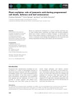
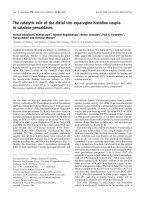
In healthy volunteers challenged with intravenous LPS,
granulocyte TREM-1 expression – initially high at baseline –
was immediately downregulated on LPS exposure, which
occurred together with an increase in sTREM-1 levels (Fig. 1).
In contrast, monocytes exhibited a progressive increase in
TREM-1 [21]. Interestingly, ligands for the predominantly
dendritic cell and B cell expressed TLRs (namely TLR-3, TLR-
7 and TLR-9) did not alter TREM-1 expression, and neither
did the surrounding concentrations of TNF-α [21]. This
pattern of monocytic TREM-1 expression found in healthy
volunteers was confirmed in septic shock patients [24].
Taken together, these data demonstrate that expression of
membrane-bound TREM-1 on neutrophils and monocytes/
macrophages is strongly altered during sepsis, as is the
release of its soluble form. Given that both cell surface
TREM-1 and sTREM-1 are upregulated during sepsis, this
protein may be useful in the diagnosis of infection.
TREM-1 as a diagnostic tool
The specific involvement of TREM-1 solely in cases of
infection led us to investigate the diagnostic value of a
plasma sTREM-1 assay in distinguishing sepsis from severe
systemic noninfectious inflammation among newly admitted
critically ill patients with suspected infection [25]. Baseline
plasma levels of C-reactive protein, procalcitonin and
sTREM-1 were higher among septic patients than in patients
with systemic inflammatory response syndrome only. Plasma
sTREM-1 levels appeared to be the most helpful parameter in
differentiating patients with sepsis from those with systemic
inflammatory response syndrome. Median plasma sTREM-1
levels at admission were 0 pg/ml (range 0–144 pg/ml) in
noninfected patients and 149 pg/ml (range 30–428 pg/ml) in
patients with sepsis (P < 0.001). Plasma sTREM-1 levels
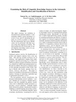
yielded the highest discriminative value (Table 1).
The diagnostic value of sTREM-1 has also been investigated
in the context of a more localized infectious process, namely
pneumonia, in a series of 148 consecutive mechanically
ventilated patients [26]. sTREM-1 levels were higher in
bronchoalveolar lavage (BAL) fluid from patients with
community-acquired and ventilator-associated pneumonia
than in BAL fluid from patients without pneumonia, but the
levels did not differ significantly between patients with
community-acquired pneumonia and those with ventilator-
associated pneumonia. The presence of elevated levels of
sTREM-1 in BAL fluid was the strongest predictor of
487
pneumonia (Table 1). Furthermore, Richeldi and coworkers
[27] recently studied TREM-1 expression levels in BAL
specimens from patients with community-acquired pneumonia,
tuberculosis (an intracellular infection that is unable to induce
upregulation of TREM-1 in vitro) and interstitial lung disease,
the latter being used as a model of noninfectious
inflammatory lung disease. TREM-1 expression was
significantly increased in lung neutrophils and in lung macro-
phages of patients with pneumonia (n = 7; 387.9 ± 61.4 MFI
[mean fluorescence intensity] and 660.5 ± 18.3 MFI,
respectively) in comparison with patients with pulmonary
tuberculosis (n = 7; 59.2 ± 13.1 MFI and 80.6 ± 291.2 MFI)
and patients with interstitial lung diseases (n = 10;
91.8 ± 23.3 MFI and 123.9 ± 22.8 MFI).
Hence, sTREM-1 appears to represent a reliable marker of
infection, particularly in plasma during sepsis and in BAL fluid
in cases of pneumonia.
TREM-1 as a follow-up marker
In a recent study [28] we sequentially measured plasma
sTREM-1 concentrations in 63 consecutive septic patients.
Soluble TREM-1 concentrations were significantly lower at
admission in nonsurviving patients than in surviving patients,
and an elevated baseline sTREM-1 level was found to be an
independent protective factor (an explanation for this intriguing
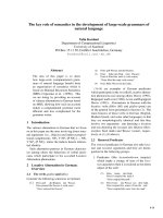
finding is given below). Moreover, sTREM-1 concentrations
remained stable or even increased in nonsurvivors whereas
they decreased in survivors (Fig. 2). A similar differential
pattern was found with regard to cell surface TREM-1
expression [24]. Although monocytic TREM-1 expression did
not differ on admission between septic survivors and
nonsurvivors, expression in these two groups diverged
significantly by day 3, with high and stable level in
nonsurvivors, but with levels in surviving patients rapidly
declining to those observed in healthy volunteers and
nonseptic patients. A progressive decline in plasma sTREM-1
or of its monocytic expression could therefore indicate a
favourable clinical evolution during the recovery phase of
sepsis.
The main cellular origin of sTREM-1 production is still unclear
(monocytes or neutrophils), and in view of the different
patterns of expression of TREM-1 between monocytes and
neutrophils [21], we require further clarification of the
relationship between soluble and membrane-bound forms of
TREM-1.
TREM-1 modulation as a therapeutic tool
Bouchon and coworkers [18] demonstrated that blockade of
TREM-1 with mTREM-1/IgG
1
(a murine TREM-1 extracellular
domain and human IgG
1
Fc fragment fusion protein)
protected mice against both LPS-induced shock and
microbial sepsis caused by administration of live Escherichia
coli or by CLP. We therefore designed a synthetic peptide
Available online />Figure 1
TREM-1 expression and release in healthy volunteers administered
lipopolysaccharide. (a) TREM-1 cell surface expression in healthy
volunteers administered 4 ng/kg lipopolysaccharide intravenously.
(b) Corresponding plasma concentrations of the soluble form of
TREM-1. Adapted with permission from Knapp and coworkers [21].
LPS, lipopolysaccharide; TREM, triggering receptor expressed on
myeloid cells.
0 2 4 6
–7
5
–5
0
–2
5
0
25
50
75
100
Monocytes
24
Neutrophils
% change of TREM-1 MFI
hours post LPS injection
0 2 4 6
0
2500
24
hours post LPS injection
Plasma sTREM-1 (pg/ml )
(a)
(b)
Table 1
Diagnostic accuracy of sTREM-1 determination in sepsis
sTREM-1 threshold Sensitivity Specificity Positive likelihood Area under the
Setting [ref.] (pg/ml) (% [95% CI]) (% [95% CI]) ratio ROC curve (95% CI)
Pneumonia [26] 5 98 (95–100) 90 (84–96) 10.4 0.93 (0.92–0.95)
Sepsis [25] 60 96 (92–100) 89 (82–95) 8.6 0.97 (0.94–1.00)
CI, confidence interval; ROC, receiver operating characteristic; sTREM, soluble triggering receptor expressed on myeloid cells.
488
(LP17) to mimic part of the extracellular domain of TREM-1
and examined its action both in vitro and in a mouse model of
endotoxaemia [29]. In monocytes cultured with LPS, LP17
reduced the production of TNF-α and IL-1β in a concen-
tration-dependent manner. In the mouse model, single
administration of LP17 60 min before a lethal dose of LPS
reduced mortality in a dose-dependent manner. Treatment
with LP17 after the onset of endotoxaemia also conferred
significant protection against a lethal dose of LPS, reducing
cytokine levels by 30% compared with controls. Similar
results were also obtained in a CLP model of polymicrobial
sepsis. The modulation of TREM-1 signalling reduced but did
not abolish NF-κB activation and cytokine production, and
protected septic animals from hyper-responsiveness and
death. Although crystallographic analyses [30,31] can predict
TREM-1 recognition by using antibody-equivalent comple-
mentary determining region (CDR) loops (such as T-cell
receptors, CD8 and cytotoxic T-lymphocyte associated
antigen-4), its natural ligand has yet to be identified.
Nevertheless, LP17 overlaps the CDR-3 and the ‘F’ β strand
of the extracellular domain of TREM-1, with the ‘F’ β strand
containing a tyrosine residue that mediates dimerization.
LP17 could therefore compete with the natural ligand of
TREM-1, thus acting as a decoy receptor, and/or it could
impair TREM-1 dimerization. Along similar lines, this hypothe-
Critical Care October 2005 Vol 9 No 5 Gibot
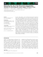
Figure 3
Overview of the role of TREM-1 in sepsis. DAG, diacylglycerol; ERK, extracellular signal regulated kinase; GRB, growth factor receptor binding
protein; MAPK, mitogen-activated protein kinase; MEK, mitogen-activated protein kinase kinase; PAMP, pathogen-associated molecular pattern;
PI3K, phosphatidylinositol 3-kinase; PKC, protein kinase C; PLC, phospholipase C; SOS, son of sevenless; TLR, Toll-like receptor; TREM,
triggering receptor expressed on myeloid cells; TREM-1L, TREM-1 ligand.
Monocyte/Macrophage
Decoy receptor for the TREM-1 ligand:
Inflammatory resp
onse modulation
SOS
Ras Raf
MEK1,2
GRB2
ERK1,2
Pi3K
PKC
PLC-
DAG
p
38 MAPK
Ca
PLC-γ
NFκB
Creb-1
AP-1
NFAT
Cytokines
Chemokines
DAP12
TREM-1
TREM-1L
sTREM-1
PAMP
TLR
trem-1
PI3K
Sheddin
g
?
Marker of
Infection
LP17
Figure 2
Time course of median plasma levels of sTREM-1 in septic patients.
Patients are subgrouped according to whether they survived (squares;
n = 42) or did not survive (triangles; n = 21). Adapted with permission
from Gibot and coworkers [29]. sTREM, soluble triggering receptor
expressed on myeloid cells.
0 7 14
0
100
200
300
P = 0.02
Time (days)
sTREM-1 (pg/ml)
P =0.02 P < 0.001
489
sis may also account for the protective effect of elevated
sTREM-1 concentrations observed in septic patients [28].
Conclusion
TREM-1 is a recently described cell surface molecule on
neutrophils and macrophages that acts as an amplifier of
inflammatory responses. During sepsis there is a significant
increase in both the expression of membrane-bound TREM-1
and in the release of its soluble form (Fig. 3). Although it
remains to be confirmed in larger and more heterogeneous
populations, the rapid assessment of sTREM-1 concentration
could prove to be a valuable tool for the diagnosis of
infection, particularly with regard to its plasma levels in sepsis
and BAL fluid levels in pneumonia. Although promising, the
therapeutic manipulation of the TREM-1 signalling pathway
still warrants further studies, particularly in assessing whether
such modulation does not bypass important steps in the
physiological reaction to pathogens.
Competing interests
Patent pending on sTREM-1 measurement.
References
1. Poltorak A, He X, Smirnova I, Liu MY, van Huffel C, Du X, Birdwell
D, Alejos E, Silva M, Galanos C, et al.: Defective LPS signaling
in C3H/HeJ and C57BL/10ScCr mice: Mutation in TLR4 gene.
Science 1998, 282:2085-2088.
2. Hofmann JA, Kafatos FC, Janeway CA, Ezekowitz RA: Phylogenic
perspective in innate immunity. Science 1999, 284:1313-1318.
3. Medzhitov R: Toll-like receptors and innate immunity. Nat Rev
Immunol 2001, 1:135-145.
4. Lanier LL: NK cell receptors. Annu Rev Immunol 1998, 16:359-
393.
5. Bouchon A, Dietrich J, Colonna M: Cutting edge: Inflammatory
responses can be triggered by TREM-1, a novel receptor
expressed on neutrophils and monocytes. J Immunol 2000,
164:4991-4995.
6. Colonna M: TREMs in the immune system and beyond. Nat
Rev Immunol 2003, 3:1-9.
7. McVicar DW, Taylor LS, Gosselin P, Willette-Brown J, Mikhael AI,
Geahlen RL, Nakamura MC, Linnemeyer P, Seaman WE, Ander-
son SK, et al.: DAP12-mediated signal transduction in natural
killer cells. A dominant role for the Syk protein-tyrosine
kinase. J Biol Chem 1998, 273:32934-32942.
8. Bouchon A, Hernandez-Munain C, Cella M, Colonna M: A
DAP12-mediated pathway regulates expression of CC
chemokine receptor 7 and maturation of human dendritic
cells. J Exp Med 2001, 194:1111-1122.
9. Paloneva J, Manninen T, Christman G, Hovanes K, Mandelin J,
Adolfsson R, Bianchin M, Bird T, Miranda R, Salmaggi A, et al.:
Mutations in two genes encoding different subunits of a
receptor signaling complex result in an identical disease phe-
notype. Am J Hum Genet 2002, 71:656-662.
10. Gingras MC, Lapillonne H, Margolin JF: TREM-1, MDL-1, and
DAP12 expression is associated with a mature stage of
myeloid development. Mol Immunol 2002, 38:817-824.
11. Washington AV, Quigley L, McVicar DW: Initial characterization
of TREM-like transcript (TLT)-1: A putative inhibitory receptor
within the TREM cluster. Blood 2002, 100:3822-3824.
12. Barrow AD, Astoul E, Floto A, Brooke G, Relou IA, Jennings NS,
Smith KG, Ouwehand W, Farndale RW, Alexander DR, et al.:
Cutting edge: TREM-like transcript, a platelet immunoreceptor
tyrosine-based inhibition motif encoding costimulatory
immunoreceptor that enhances, rather inhibits, calcium sig-
naling via SHP-2. J Immunol 2004, 172:5838-5842.
13. Washington AV, Schubert RL, Quigley L, Disipio T, Feltz R, Cho
EH, MacVicar DW: TREM family member, TLT-1, is found
exclusively in the
αα
-granules of megakaryocytes and
platelets. Blood 2004, 104:1042-1047.
14. Daws MR, Lanier LL, Seaman WE, Ryan JC: Cloning and charac-
terization of a novel mouse myeloid DAP12-associated recep-
tor family. Eur J Immunol 2001, 31:783-791.
15. Chung DH, Seaman WE, Daws MR: Characterization of TREM-
3, an activating receptor on mouse macrophages: definition of
a family of single Ig-domain receptors on mouse chromo-
some. Eur J Immunol 2002, 32:59-66.
16. Gibot S: TREM, new receptors mediating innate immunity.
Med Sci 2004, 20:503-505.
17. Colonna M, Fachetti F: TREM-1: a new player in acute inflam-
matory responses. J Infect Dis 2003, 187:S397-S401.
18. Bouchon A, Facchetti F, Weigand MA, Colonna M: TREM-1
amplifies inflammation and is a crucial mediator of septic
shock. Nature 2001, 410:1103-1107.
19. Bleharski JR, Kiessler V, Buonsanti C, Sieling PA, Stenger S,
Colonna M, Modlin RL: A role for triggering receptor expressed
on myeloid cells-1 in host defense during the early-induced
and adaptive phases of the immune response. J Immunol
2003, 170:3812-3818.
20. Radsak MP, Salih HR, Rammensee HG, Schild H: Triggering
receptor expressed on myeloid cells-1 in neutrophil inflam-
matory responses: differential regulation of activation and
survival. J Immunol 2004, 172:4956-4963.
21. Knapp S, Gibot S, de Vos A, Versteeg HH, Colonna M, van der
Poll T: Cutting edge: expression patterns of surface and
soluble triggering receptor expressed on myeloid cells-1 in
human endotoxinemia. J Immunol 2004, 173:7131-7134.
22. Lucas M, Daniel L, Tomasello E, Guia S, Horschowski N, Aoki N,
Figarella-Branger D, Gomez S, Vivier E: Massive inflammatory
syndrome and lymphocytic immunodeficiency in KARAP/
DAP12-transgenic mice. Eur J Immunol 2002, 32:2653-2663.
23. Gibot S, le Renard PE, Béné MC, Faure GC, Bollaert PE, Levy B:
Surface and soluble triggering receptor expressed on
myeloid cells-1: expression patterns in murine sepsis. Crit
Care Med 2005:in press.
24. Gibot S, le Renard PE, Bollaert PE, Kolopp-Sarda MN, Béné MC,
Faure GC, Levy B: Surface triggering receptor expressed on
myeloid cells 1 expression patterns in septic shock. Intensive
Care Med 2005, 31:594-597.
25. Gibot S, Kolopp-Sarda MN, Béné MC, Cravoisy A, Levy B, Faure
GC, Bollaert PE: Plasma level of a triggering receptor
expressed on myeloid cells-1: its diagnostic accuracy in
patients with suspected sepsis. Ann Intern Med 2004, 141:9-15.
26. Gibot S, Cravoisy A, Levy B, Béné MC, Faure GC, Bollaert PE:
Soluble triggering receptor expressed on myeloid cells and
the diagnosis of pneumonia. N Engl J Med 2004, 350:451-458.
27. Richeldi L, Mariani M, Losi M, Maselli F, Corbetta L, Buonsanti C,
Colonna M, Sinigaglia F, Panina-Bordignon P, Fabbri LM: Trigger-
ing receptor expressed on myeloid cells: role in the diagnosis
of lung infections. Eur Respir J 2004, 24:247-250.
28. Gibot S, Cravoisy A, Kolopp-Sarda MN, Béné MC, Faure GC,
Bollaert PE, Levy B: Time-course of soluble TREM-1, procalci-
tonine and C-reactive protein plasma concentrations during
sepsis. Crit Care Med 2005, 33:792-796.
29. Gibot S, Kolopp-Sarda MN, Béné MC, Bollaert PE, Lozniewski A,
Mory F, Levy B, Faure GC: A soluble form of the triggering recep-
tor expressed on myeloid cells-1 modulates the inflammatory
response in murine sepsis. J Exp Med 2004, 11:1419-1426.
30. Radaev S, Kattah M, Rostro B, Colonna M, Sun PD: Crystal
structure of the human myeloid cell activating receptor
TREM-1. Structure (Camb) 2004, 11:1527-1535.
31. Kelker MS, Debler EW, Wilson IA: Crystal structure of mouse
triggering receptor expressed on myeloid cells 1 (TREM-1) at
1.76 A. J Mol Biol 2004, 344:1175-1181.
Available online />