Báo cáo khoa học: "Clinical review: Mechanical ventilation in severe asthm" docx
Bạn đang xem bản rút gọn của tài liệu. Xem và tải ngay bản đầy đủ của tài liệu tại đây (275.46 KB, 7 trang )
581
NPPV = noninvasive positive pressure ventilation; PEEP = positive end-expiratory pressure; Pplat = plateau airway pressure; VEI = volume of gas at
end-inspiration above functional residual capacity.
Available online />Abstract
Respiratory failure from severe asthma is a potentially reversible,
life-threatening condition. Poor outcome in this setting is frequently
a result of the development of gas-trapping. This condition can
arise in any mechanically ventilated patient, but those with severe
airflow limitation have a predisposition. It is important that clinicians
managing these types of patients understand that the use of
mechanical ventilation can lead to or worsen gas-trapping. In this
review we discuss the development of this complication during
mechanical ventilation, techniques to measure it and strategies to
limit its severity. We hope that by understanding such concepts
clinicians will be able to reduce further the poor outcomes
occasionally related to severe asthma.
Introduction
Asthma continues to inflict significant morbidity and mortality
worldwide. Despite advances in therapy and in our under-
standing of its pathophysiology, the prevalence of asthma is
increasing [1-3], although there is significant age and
geographic variation [4]. While the prevalence of asthma has
increased, outcomes of severe asthma appear to be
improving, with lower complication rates and fewer in-hospital
deaths [3]. Nonetheless, it is estimated that about 10% of
individuals admitted to hospital for asthma go to the intensive
care unit, with 2% of all admitted patients being intubated [5].
Not surprisingly, admission to the intensive care unit and
need for mechanical ventilation are associated with mortality
[1,2]. When death does occur it is most commonly a result of
one of the complications of severe gas-trapping. These
complications include barotrauma, hypotension and refractory
respiratory acidosis. If the morbidity and mortality associated
with severe asthma is to continue to decrease, then it is
imperative that clinicians caring for such patients have a clear
understanding of how gas-trapping can occur and of how it
may be recognized/measured and limited.
This article reviews the principles of mechanical ventilation in
severe asthma, giving particular attention to the development
of gas-trapping as well as how to measure and limit it.
Specific details on pharmacological management and
prevention of future episodes of severe asthma are beyond
the scope of this review but can be found elsewhere [6,7].
Rationale for mechanical ventilation in severe
asthma
When a patient with severe asthma does not respond
adequately to medical therapy, prompt intervention in an effort
to provide adequate oxygenation and ventilation by means of
noninvasive positive pressure ventilation (NPPV) or invasive
positive pressure mechanical ventilation is frequently life
saving. Given that these patients have a propensity to develop
severe airflow limitation, making it difficult to exhale all of their
inspired gas, gas-trapping (which leads to dynamic
hyperinflation and is also referred to as intrinsic positive end-
expiratory pressure [PEEP] and auto-PEEP) frequently occurs.
As a result, one of the most important principles of mechanical
ventilation in this setting is to utilize a strategy aimed at
reducing the likelihood that this complication will occur.
Noninvasive positive pressure ventilation
It is possible that in some patients with severe asthma NPPV
may be preferential to intubation. However, to date only two
small, prospective, randomized trials have been completed that
evaluated the use of NPPV in patients with severe asthma: one
in children [8] and a pilot study in adults [9]. Both of those
studies suggested that, in selected patients with severe
asthma, NPPV could improve lung function and possibly
reduce the need for hospitalization. There are also some
observational studies, which yielded consistent results [10,11].
In chronic obstructive pulmonary disease – another condition
Review
Clinical review: Mechanical ventilation in severe asthma
David R Stather
1
and Thomas E Stewart
2
1
Fellow, InterDepartmental Division of Critical Care Medicine and Division of Respirology, Department of Medicine, Mount Sinai Hospital and University
Health Network, University of Toronto, Toronto, Canada
2
Associate Professor, Department of Medicine and Anaesthesia, and Administrative Director, Critical Care Medicine, Mount Sinai Hospital and
University Health Network, University of Toronto, Toronto, Canada
Corresponding author: Thomas E Stewart,
Published online: 8 September 2005 Critical Care 2005, 9:581-587 (DOI 10.1186/cc3733)
This article is online at />© 2005 BioMed Central Ltd
See related letter by Cole online [ />582
Critical Care December 2005 Vol 9 No 6 Stather and Stewart
frequently associated with severe airflow limitation – a number
of prospective randomized trials have shown that noninvasive
ventilation reduces the need for endotracheal intubation, length
of hospital stay and in-hospital mortality rate, and even that it
improves long-term survival [12-16]. The degree to which these
data can be applied to the asthmatic population is debatable.
Even though NPPV requires further investigation in severe
asthma, it is currently being used as an initial alternative to
mechanical ventilation in some centres. As is the case in
other conditions, the success of NPPV depends on a variety
of factors including clinician experience [17], patient
selection and interfaces [16], and that it is not used in
patients with any known contraindications [18,19]. It is
particularly important to be very cautious in using NPPV in
paediatric patients, in whom the margins of safety are narrow,
and a low threshold for intubation when required should be
maintained in these patients. The commonly accepted
contraindications to NPPV are as follows: cardiac/respiratory
arrest, severe encephalopathy, haemodynamic instability,
facial surgery/deformity, high risk for aspiration, non-
respiratory organ failure, severe upper gastrointestinal
bleeding, unstable arrhythmia, and upper airway obstruction.
The decision to intubate
The decision to intubate should be based mainly on clinical
judgement. Markers of deterioration include rising carbon
dioxide levels (including normalization in a previously hypo-
capnic patient), exhaustion, mental status depression, haemo-
dynamic instability and refractory hypoxaemia [20]. Clinical
judgement is crucial because many patients presenting with
hypercapnia do not require intubation [21], and thus the
decision should not be based solely on blood gases.
Development of gas-trapping
Severe airflow limitation is always associated with severe
asthma exacerbation and occurs as a result of broncho-
constriction, airway oedema and/or mucous plugging.
Consequently, the work of breathing is significantly
increased. Increased work occurs because the normally
passive process of expiration becomes active in an attempt
by the patient to force the inspired gas out of their lungs. In
addition, there is increased inspiratory work caused by high
airway resistance and hyperinflation. This hyperinflation
causes the lungs and chest wall to operate on a suboptimal
portion of their pressure–volume curves (i.e. they are
overstretched), resulting in increased work to stretch them
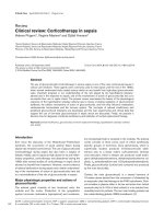
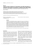
further in an attempt to ventilate adequately. Gas-trapping
occurs because the low expiratory flow rates mandate long
expiratory times if the entire inspired volume is to be exhaled.
If the next breath interrupts exhalation, then gas-trapping
results (Fig. 1). Because gas is trapped in the lungs there is
additional pressure at the end of expiration (auto-PEEP or
intrinsic PEEP) above applied PEEP, which leads to dynamic
hyperinflation. Auto-PEEP, intrinsic PEEP and dynamic hyper-
inflation are terms that are frequently used interchangeably.
Dynamic hyperinflation has been defined as failure of the lung
to return to its relaxed volume or functional residual capacity
at end-exhalation [22-24]. Of note, some refer to gas-
trapping as the component of hyperinflation that is due to
airway occlusion, and is therefore potentially less amenable to
ventilator manipulation (in some situations, the dominant
component of total hyperinflation in severe asthma [25]).
Hyperinflation can be adaptive in that with higher lung
volumes the increase in airway diameter and elastic recoil
pressure enhances expiratory flow; however, excessive
dynamic hyperinflation has been shown to predict the develop-
ment of hypotension and barotrauma during mechanical
ventilation of severe asthma [25]. These developments are
the usual causes of excess morbidity and mortality.
Measuring gas-trapping
Gas-trapping can be measured a variety of ways involving
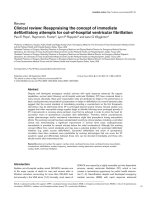
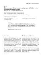
volume, pressure, or flow of gas. Estimating gas-trapping
using volume measures can be done by collecting the total
exhaled volume during 20–60 s of apnoea in a paralyzed
patient. Tuxen and coworkers [25,26] described this volume
as ‘VEI’, or the volume of gas at end-inspiration above
functional residual capacity (Fig. 2). Tuxen and Lane [25] also
showed that a VEI above 20 ml/kg predicted complications of
hypotension and barotrauma in mechanically ventilated
patients with severe asthma. Prospective studies involving
larger patient numbers are needed to validate the predictive
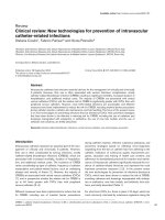
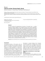
value of VEI. Another way to estimate gas-trapping is to
measure end-expiratory pressure in the lungs. If the expiratory
port of the ventilator is occluded at end-expiration, then the
proximal airway pressure will equilibrate with alveolar
pressure and permit measurement of auto-PEEP (end-
expiratory pressure above applied PEEP) at the airway
opening (Fig. 3). Expiratory muscle contraction can elevate
auto-PEEP without adding to dynamic hyperinflation, and
therefore for accurate measurement of auto-PEEP the patient
should be relaxed. Auto-PEEP measured in this manner has
not yet been shown to correlate with complications [27].
Another way to look for gas-trapping is to observe the flow
Figure 1
Mechanism of dynamic hyperinflation in the setting of severe airflow
obstruction. Reproduced with permission from Levy and coworkers [7].
583
versus time graphics on the ventilator. If inspiratory flow
begins before expiratory flow ends, then gas must be trapped
in the lungs.
Each of the measures of gas-trapping described thus far rely
on the assumption that the airways all remain in communi-
cation with the proximal airway throughout expiration because
pressure, flow, or gas volume cannot be measured from a
noncommunicating airway. Frequently, all of the airways may
not be in communication with the proximal airway in severe
asthma. For example, it has been noted (perhaps as a result
of complete airway closure) that there may at times be
‘unmeasured’ or ‘occult’ auto-PEEP [23]. This occult auto-
PEEP has all of the untoward effects of the measurable auto-
PEEP, but it cannot be quantified using the usual approaches
[23]. As a result, exercising good clinical judgement is
important. When assessing dynamic hyperinflation/gas-
trapping in mechanically ventilated patients with severe
asthma, clinicians should question low auto-PEEP measure-
ments in clinical situations that suggest otherwise.
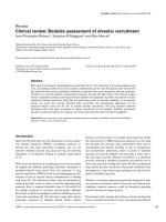
One such clinical situation would be increasing plateau
airway pressure (Pplat) unexplained by decreases in
respiratory system compliance during volume-cycled ventilation.
Pplat can be determined by stopping flow at end-inspiration
utilizing an end-inspiratory pause (typically 0.4 s). During this
pause, airway opening pressure falls from peak pressure (the
sum of static and resistive pressures) to Pplat (static
pressure alone) as resistive pressure falls to zero (Fig. 4).
Patients must be paralyzed or heavily sedated to obtain
reliable measurements. Because alveolar pressure increases
as lung volume increases, measurement of Pplat should
reflect gas-trapping (again assuming that there is no other
explanation, such as adjustments to the ventilator or changes
in respiratory system compliance). Some have pointed out
that if Pplat is kept at less than 30 cmH
2
O then complications
appear to be rare [28], although no studies have yet shown
Pplat to be a reliable predictor of complications. Similarly,
when using pressure cycled ventilation, decreasing tidal
volumes may indicate gas-trapping. Other situations in which
clinicians should suspect gas-trapping include increasing
chest wall girth, hyperinflation on chest imaging, reduced
efficiency of ventilation, increased patient effort, unexplained
patient agitation, development of barotrauma, haemodynamic
compromise and missed respiratory efforts (as patients attempt
to trigger the ventilator but cannot generate enough pressure
to overcome the auto-PEEP that has developed) [22].
Limiting gas-trapping
Because gas-trapping is potentially associated with
significant adverse events in severe asthma, clinicians must
be vigilant for its development and employ strategies to limit
it. Understanding how gas-trapping occurs is the first step in
developing such strategies. These strategies include controlled
hypoventilation (reduced tidal volumes [less gas to exhale]
and reduced respiratory rates [longer expiratory time]),
Available online />Figure 2
Measuring lung hyperinflation using VEI. VEI, volume of gas at end-inspiration above functional residual capacity. Reproduced with permission from
Tuxen [43].
Figure 3
Measurement of intrinsic positive end-expiratory pressure. Reproduced
with permission from The McGraw-Hill Companies [64].
584
relieving expiratory flow resistance (frequent airway suction-
ing if necessary, bronchodilators, steroids, large-bore endo-
tracheal tube), reducing inspiratory time by increasing the
inspiratory flow rate or incorporating nondistensible tubing,
and reducing the need for high minute ventilation by
decreasing carbon dioxide production (e.g. sedation/
paralysis, controlling fever/pain). The application of external
PEEP in severe asthma remains a controversial topic. It could
theoretically decrease the work of breathing and hence
carbon dioxide production, while limiting gas-trapping by
splinting the airways open [29,30]; however, in practice there
are situations in which the application of external PEEP may
increase total PEEP and worsen gas-trapping.
Assuming that appropriate medical therapy to alleviate airflow
obstruction has been administered (i.e. inhaled beta agonists,
inhaled ipratroprium bromide, steroids, with/without intra-
venous magnesium sulphate, etc.), by far the most effective
method of decreasing dynamic hyperinflation/gas-trapping is
to reduce the minute ventilation [31,32]. Reducing the minute
ventilation by adjusting the tidal volume, frequency, or set
pressure on the ventilator may result in carbon dioxide
retention. In this setting the controlled use of ‘permissive
hypercapnia’ is generally considered well tolerated [33,34].
Permissive hypercapnia that maintains a pH above 7.20 or an
arterial carbon dioxide tension below 90 mmHg has gained
widespread acceptance [27,34-36]. Permissive hypercapnia
has been used successfully in mechanically ventilated
patients with status asthmaticus [33].
Expiratory time can be lengthened by using higher inspiratory
flow settings (70–100 l/min) during volume cycled ventilation,
using a shorter inspiratory time fraction, reducing respiratory
rate, and eliminating any inspiratory pause. Prolongation of
expiratory time has been shown to decrease dynamic
hyperinflation in patients with severe asthma, as is evident by
decreased plateau pressures [37]. The magnitude of this
effect becomes relatively modest when the baseline minute
ventilation is 10 l/min or less and when the baseline
respiratory rate is low [37]. It should be emphasized that
while modifying the I/E ratio is important in fine tuning the
amount of gas-trapping, the single most effective way is by
reducing minute ventilation [6,7].
Applying adequate sedation and analgesia is a fundamental
step in lowering the production of carbon dioxide and
subsequently ventilatory requirements. Sedation and/or
paralysis may also allow the clinician to avoid patient–ventilator
dysynchrony and facilitate strategies to limit gas-trapping in the
most severe of cases. It is beyond the scope of this review to
recommend which agents or protocols are best for this. The
use of neuromuscular blocking agents should be limited to
short periods of time and only when absolutely necessary in
patients with severe asthma who are not achieving synchrony
with other agents. Although neuromuscular blocking agents
effectively promote synchrony, lower the risk for barotrauma,
reduce lactate accumulation [38] and reduce oxygen
consumption and carbon dioxide production, their prolonged
use, particularly when combined with steroids, can lead to
prolonged paralysis and/or myopathy [39,40].
The addition of extrinsic PEEP in the setting of auto-PEEP
may reduce work of breathing and possibly even prevent gas-
trapping by splinting the airways open [29]. In terms of
reducing the work of breathing, the addition of extrinsic PEEP
in patients with dynamic hyperinflation would theoretically
reduce the inspiratory muscle effort required to overcome
auto-PEEP and initiate an inspiration. It has been
demonstrated that in patients with chronic obstructive
pulmonary disease more than 40% of inspiratory muscle
effort can be expended to overcome auto-PEEP [41,42], and
that adding extrinsic PEEP can attenuate the inspiratory
muscle effort needed to trigger inspiration and improve
patient–ventilator interaction. In these patients extrinsic PEEP
must be titrated individually, with an average of 80% of the
auto-PEEP being tolerated before the plateau pressures and
total PEEP begin to increase. Such an approach is only
useful in those patients who are breathing spontaneously and
capable of triggering the ventilator. In addition, extrinsic PEEP
may prevent airway collapse (which could lead to occult auto-
PEEP) by splinting the airways open. If this is the case then
extrinsic PEEP would be most useful only in the most severe
of cases, including those patients who are not spontaneously
breathing. It should be noted that extrinsic PEEP has also
been shown to be effective at preventing ventilator-induced
lung injury in other forms of lung injury and hence may be of
added benefit in this situation. In practice, however, adding
extrinsic PEEP in some patients with severe asthma has been
shown to worsen auto-PEEP [43]. As mentioned above, it is
occasionally difficult to measure auto-PEEP reliably, and if the
extrinsic PEEP is greater than the auto-PEEP then gas-
trapping will likely worsen. This has led some to recommend
Critical Care December 2005 Vol 9 No 6 Stather and Stewart
Figure 4
Measurement of end-inspiratory plateau pressure, an estimate of
average end-inspiratory alveolar pressure. Reproduced with permission
from The McGraw-Hill Companies [64].
585
minimizing the use of extrinsic PEEP or not using it at all
[35,36] in the ventilation of patients with severe asthma. If
extrinsic PEEP is to be used, then careful bedside obser-
vation with a clear understanding of how the benefits
(reductions in auto-PEEP) and adverse effects (worsening
gas-trapping) would manifest is mandatory.
Considerations for initial ventilator settings
in patients with severe asthma
There have been a number of review articles recommending
initial ventilator settings and algorithmic approaches to
mechanical ventilation in severe asthma [6,7]. The fine details
of the ventilator settings are not as crucial as close attention
to the basic principles of ventilating patients with severe
asthma: employ low tidal volumes and respiratory rate;
prolong expiratory time as much as possible; shorten
inspiratory time as much as possible; and monitor for the
development of dynamic hyperinflation.
As a starting point for ventilating patients with severe asthma,
we recommend that the ventilator initially be used in pressure
control mode, setting the pressure to achieve a tidal volume
of 6–8 ml/kg, respiratory rate of 11–14 breaths/min and
PEEP at 0–5 cmH
2
O. We use these settings with a goal of
obtaining a pH, in general, above 7.2 and a Pplat under
30 cmH
2
O. If a Pplat under 30 cmH
2
O cannot be maintained,
then the patient must be evaluated for causes of decreased
respiratory system compliance (i.e. pneumothorax, misplaced
endotracheal tube, pulmonary oedema, etc.) beyond the
development of dynamic hyperinflation. If no such causes are
evident then efforts to limit gas-trapping further must be
considered. If permissive hypercapnia results in a pH below
7.2, then the same type of evaluation needs to occur,
including consideration of increased sedation/paralysis and
methods of decreasing carbon dioxide production (i.e.
reducing fever, preventing over-feeding, decreasing patient
effort, etc.). In addition to these examples, administration of
sodium bicarbonate to maintain a pH of 7.2 during controlled
hypoventilation has been investigated in patients with status
asthmaticus [44]; however, no studies have demonstrated
any benefit associated with bicarbonate infusion. Decisions
regarding ongoing ventilator management must be based on
the principles outlined in this review.
Adjuncts to mechanical ventilation
A large variety of unproven therapies that clinicians may need
to consider in an emergent situation have been proposed,
including intravenous magnesium sulphate, general anaes-
thesia, bronchoscopic lavage, heliox and extracorporeal
membrane oxygenation.
Intravenous magnesium sulphate has bronchodilating
properties and has been shown in limited studies to improve
pulmonary function in patients with severe asthma [45,46], at
least in the short term. Several inhalation anaesthetic agents
have intrinsic bronchodilator properties [47,48] and there are
reports of successful use of these agents in refractory status
asthmaticus [49,50]. The special equipment and personnel
needed for inhalation anaesthesia and the significant
haemodynamic complications associated with these agents
make their use problematic. Ketamine is an intravenous agent
that has analgesic and bronchodilating properties [51]. There
are limited clinical data available regarding the use of
ketamine in status asthmaticus [52,53], and its side effects of
tachycardia, hypertension, delirium and lowering the seizure
threshold should always be taken into account.
In patients with status asthmaticus and severe mucous
impaction, it has been suggested that bronchoscopic
examination of the airways and removal of secretions may be
beneficial [54]. As the presence of the bronchoscope may
worsen lung hyperinflation and increase the risk for
pneumothorax [55], we do not recommend this technique.
Heliox is a blend of helium and oxygen (usually at a 70 : 30
ratio), which is less dense than air, theoretically permitting
higher flow rates through a given airway segment for the
same driving pressure, thereby alleviating dynamic
hyperinflation. Several small studies have shown heliox to
reduce peak inspiratory pressure and arterial carbon dioxide
tension, and to improve oxygenation in mechanically
ventilated patients [56,57]. That heliox is expensive, has a
limited concentration of oxygen and has conflicting results in
the literature [58-61] make it a somewhat controversial
therapy, and at this time we cannot recommend it for routine
use in severe asthma.
Extracorporeal membrane oxygenation is another expensive
modality that has been successfully used in patients with
severe refractory asthma [62,63]. The use of these second-
line therapies should be on a case-by-case basis, carefully
weighing the risks and benefits.
Conclusion
Severe asthma exacerbation causing respiratory failure has
not yet been eliminated, and remains a potentially reversible,
life-threatening condition that imposes significant morbidity
and mortality. When mechanical ventilation is required in
severe asthma, it is important that clinicians managing these
patients understand why gas-trapping occurs, how to
measure it and how to limit its severity. We hope that by
understanding such concepts clinicians will be able to
reduce further the number of poor outcomes that are
occasionally associated with severe asthma.
Competing interests
The author(s) declare that they have no competing interests.
References
1. Richards GN, Kolbe J, Fenwick J, Rea HH: Demographic charac-
teristics of patients with severe life threatening asthma: com-
parison with asthma deaths. Thorax 1993, 48:1105-1109.
Available online />586
2. Turner MO, Noertjojo K, Vedal S, Bai T, Crump S, Fitzgerald JM:
Risk factors for near-fatal asthma. A case-control study in
hospitalized patients with asthma. Am J Respir Crit Care Med
1998, 157:1804-1809.
3. Kearney SE, Graham DR, Atherton ST: Acute severe asthma
treated by mechanical ventilation: a comparison of the chang-
ing characteristics over a 17 yr period. Respir Med 1998, 92:
716-721.
4. Manfreda J, Sears MR, Becklake MR, Chan-Yeung M, Dimich-Ward
H, Siersted HC, Ernst P, Sweet L, Van Til L, Bowie DM, et al.: Geo-
graphic and gender variability in the prevalence of bronchial
responsiveness in Canada. Chest 2004, 125:1657-1664.
5. Pendergraft TB, Stanford RH, Beasley R, Stempel DA, RobertsC,
McLaughlin T: Rates and characterisitics of intensive care unit
admissions and intubations among asthma-related hospital-
izations. Ann Allergy Asthma Immunol 2004, 93:29-35.
6. Corbridge TC, Hall JB: The assessment and management of
adults with status asthmaticus. Am J Respir Crit Care Med.
1995, 151:1296-1316.
7. Levy BD, Kitch B, Fanta CH: Medical and ventilatory manage-
ment of status asthmaticus. Intensive Care Med 1998, 24:105-
117.
8. Thill PJ, McGuire JK, Baden HP, Green TP, Checchia PA: Nonin-
vasive positive-pressure ventilation in children with lower
airway obstruction. Pediatr Crit Care Med 2004, 5:337-342.
9. Soroksky A, Stav D, Shpirer I: A pilot prospective, randomized,
placebo-controlled trial of bilevel positive airway pressure in
acute asthma attack. Chest 2003, 123:1018-1025.
10. Fernandez MM, Villagra A, Blanch L, Fernandez R: Non-invasive
mechanical ventilation in status asthmaticus. Intensive Care
Med 2001, 27:486-492.
11. Keenan SO, Gregor J, Sibbald WJ, Cook D, Gafni A: Noninva-
sive positive pressure ventilation in status asthmaticus. Chest
1996, 110:767-774.
12. Brochard L, Mancebo J, Wysocki M, Lofaso F, Conti G, Rauss A,
Simonneau G, Benito S, Gasparetto A, Lemaire F, et al.: Noninva-
sive ventilation for acute exacerbations of chronic obstructive
pulmonary disease. N Eng J Med 1995, 333:817-822.
13. Plant PK, Owen JL: Early use of non-invasive positive pressure
ventilation in the setting of severe, acute exacerbations of
chronic obstructive pulmonary disease on general respiratory
wards: a multicenter randomized controlled trial. Lancet 2000,
355:1941-1935.
14. Plant PK, Owen JL, Elliott MW: Non-invasive ventilation in acute
exacerbations of chronic obstructive lung disease: long term
survival and predictors of in-hospital outcome. Lancet 2001,
56:708-712.
15. Confalonieri M, Parigi P, Scartabellati A, Aiolfi S, Scorsetti S,
Nava S, Gandola L: Noninvasive mechanical ventilation
imporves the immediate and long-term outcome of COPD
patients with acute respiratory failure. Eur Respir J 1996, 9:
422-430.
16. Soo Hoo GW, Santiago S, Williams A: Nasal mechanical venti-
lation for hypercapnic respiratory failure in chronic obstructive
pulmonary disease: determinants of success and failure. Crit
Care Med 1994, 22:1253-1261.
17. Kramer N, Meyer TJ, Meharg J, Cece RD, Hill NS: Randomized,
prospective trial of noninvasive positive pressure ventilaiton
in acute respiratory failure. Am J Resp Crit Care Med 1995,
151:1799-1806.
18. Sinuff T, Keenan SP: Clinical practice guideline for the use of
noninvasive positive pressure ventilation in COPD patients
with acute respiratory failure. J Crit Care 2004, 19:82-91.
19. Evans TW, Albert RK, Angus DC, Bion JF, Chiche J-D, Epstein
SK, Fagon JY, Ranieri M, Sznajder JI, Torres A, Walley KR: Inter-
national consensus conferences in intensive care medicine:
noninvasive positive pressure ventilation in acute respiratory
failure. Am J Respir Crit Care Med 2001, 163:283-291.
20. National Heart, Lung and Blood Institute: Guidelines for the Diag-
nosis and Management of Asthma, Expert Panel Report 2. Publi-
cation number 97-4051. Bethesda: National Institutes of Health;
1997.
21. Mountain RD, Sahn S: Clinical features and outcomes in
patients with acute asthma presenting with hypercapnia. Am
Rev Respir Dis 1988, 138:535-539.
22. Stewart TE, Slutsky AS: Occult, occult auto-PEEP in status
asthmaticus. Crit Care Med 1996, 24:379-380.
23. Leatherman JW, Ravenscraft SA: Low measured auto-positive
end-expiratory pressure during mechanical ventilation of
patients with severe asthma: hidden auto-positive end-expira-
tory pressure. Crit Care Med 1996, 24:541-546.
24. Pepe PE, Marini JJ: Occult positive end-expiratory pressure in
mechanically ventilated paitents with airflow obstruction. The
auto-PEEP effect. Am Rev Resp Dis 1982, 126:166-170.
25. Tuxen DV, Lane S: The effects of ventilatory pattern on hyper-
inflation, airway pressures, and circulation in mechanical ven-
tilation of patients with severe air-flow obtruction. Am Rev
Respir Dis 1987, 136:872-879.
26. Tuxen DV, Williams TJ, Scheinkestel CD, Czarny D, Bowes G:
Use of a measurement of pulmonary hyperinflation to control
the level of mechanical ventilation in patients with acute
severe asthma. Am Rev Respir Dis 1992, 146:1136-1142.
27. Phipps P, Gerrard CS: The pulmonary physician in critical care:
acute severe asthma in the intensive care unit. Thorax 2003,
58:81-88.
28. Leatherman J: Life-threatening asthma. Clin Chest Med 1994,
15:453-479.
29. Kondili E, Alexopoulou C, Prinianakis G, Xirouchaki N, Geor-
gopoulos D: Pattern of lung emptying and expiratory resis-
tance in mechanically ventilated patients with chronic
obstructive pulmonary disease. Intensive Care Med 2004, 30:
1311-1318.
30. Kondili E, Prinianakis G, Athanasakis D, Georgopoulos D: Lung
emptying in patients with acute respiratory distress syn-
drome: effects of positive end-expiratory pressure. Eur Respir
J 2002, 19:811-819.
31. Williams TJ, Tuxen DV, Scheinkestel CD, Czarny D, Bowes G:
Risk factors for morbidity in mechanically ventilated patients
with acute severe asthma. Am Rev Respir Dis 1992, 146:607-
615.
32. Bellomo R, McLaughlin P, Tai E, Parkin G: Asthma requiring
mechanical ventilation. A low morbidity approach. Chest 1994,
105:891-896.
33. Darioli R, Perret C: Mechanical controlled hypoventilation in
status asthmaticus. Am Rev Respir Dis 1984, 129:385-387.
34. Bigatello LM, Patroniti N, Sangalli F: Permissive hypercapnia.
Curr Opin Crit Care 2001, 7:34-40.
35. Peigang Y, Marini JJ: Ventilation of patients with asthma and
chronic obstructive pulmonary disease. Curr Opin Crit Care
2002, 8:70-76.
36. Rodrigo GJ, Rodrigo C, Hall JB: Acute asthma in adults: a
review. Chest 2004, 125:1081-1102.
37. Leatherman JW, McArthur C, Shapiro RS: Effect of prolongation
of expiratory time on dynamic hyperinflation in mechanically
ventilated paitents with severe asthma. Crit Care Med 2004,
32:1542-1545.
38. Papiris S, Kotanidou A, Malagari K, Roussos C: Clinical review:
Severe asthma. Crit Care 2002, 6:30-44.
39. Leatherman JW, Fluegel WL, David WS, Davies SF, Iber C:
Muscle weakness in mechanically ventilated patients with
severe asthma. Am J Respir Crit Car Med 1996, 153:1686-
1690.
40. Behbehani NA, Al-Mane F, D’yachkova Y, Pare P, FitzGerald JM:
Myopathy following mechanical ventilation for acute severe
asthma: the role of muscle relaxants and corticosteroids.
Chest 1999, 115:1627-1631.
41. Appendini L, Purro A, Patessio A, Zanaboni S, Carone M, Spada
E, Donner CF, Rossi A: Partitioning of inspiratory muscle work-
load and pressure assistance in ventilator-dependent COPD
patients. Am J Respir Crit Care Med 1996, 154:1301-1309.
42. Guerin C, Milic-Emili J, Fournier G: Effect of PEEP on work of
breathing in mechanically ventilated COPD patients. Intensive
Care Med 2000, 26:1207-1214.
43. Tuxen DV: Detrimental effects of positive end-expiratory
pressure during controlled mechanical ventilation of patients
with severe airflow obstruction. Am Rev Respir Dis 1989,
140:5-10.
44. Menitove SM, Goldring RM: Combined ventilator and bicarbon-
ate strategy in the management of status asthmaticus. Am J
Med 1983, 74:898-901.
45. Rowe BH, Bretzlaff JA, Bourdon C, Bota GW, Camargo CA Jr:
Intravenous magnesium sulfate treatment for acute asthma in
the emergency department: a systematic review of the litera-
ture. Ann Emerg Med 2000, 36:181-190.
Critical Care December 2005 Vol 9 No 6 Stather and Stewart
587
46. Silverman RA, Osborn H, Runge J, Gallagher EJ, Chiang W,
Feldman J, Gaeta T, Freeman K, Levin B, Mancherje N, et al.: IV
magnesium sulfate in the treatment of acute severe asthma:
a multicenter randomized controlled trial. Chest 2002, 122:
489-497.
47. Rooke GA, Choi JH, Bishop M: The effect of isofluorane,
halothane, sevoflurane, and thiopental/nitrous oxide on respi-
ratory system resistance after tracheal intubation. Anesthesiol
1997, 86:1294-1299.
48. Maltais F, Sovilj M, Goldberg P, Gottfried SB: Respiratory
mechanics in status asthmaticus. Effects of inhalational anes-
thesia. Chest 1994, 116:296-300.
49. Otte RW, Fireman P: Isoflurane anesthesia for the treatment of
refractory status asthmaticus. Ann Allergy 1991, 66:305-309.
50. Bierman MI, Brown M, Muren O, Keenan RL, Glauser FL: Pro-
longed isoflurane anesthesia in status asthmaticus. Crit Care
Med 1986, 14:832-833.
51. Corseen G, Guitierrez J, Reves JG, Huber FC: Ketamine in the
anesthetic management of asthmatic patients. Anesth Analg
1972, 51:588-596.
52. Sarma V: Use of ketamine in acute severe asthma. Acta
Anaesthesiol Scand 1992, 36:106-107.
53. Hemmingsen C, Nielsen PK, Odorico J: Ketamine in the treat-
ment of bronchospasm during mechanical ventilation. Am J
Emerg Med 1994, 12:417-420.
54. Henke CA, Hertz M, Gustafson P: Combined bronchoscopy and
mucolytic therapy for patients with severe refractory status
asthmaticus on mechanical ventilation: a case report and
review of the literature. Crit Care Med 1994, 22:1880-1883.
55. Luksza AR, Smith P, Coakley J, Gordan IJ, Atherton ST: Acute
severe asthma treated by mechanical ventilation: 10 years’
experience from a district general hospital. Thorax 1986, 41:
459-463.
56. Gluck EH, Onorato DJ, Castriotta R: Helium-oxygen mixtures in
intubated patients with status asthmaticus and respiratory
acidosis. Chest 1990, 98:693-698.
57. Schaeffer EM, Pohlman A, Morgan S, Hall JB: Oxygenation in
status asthmaticus improves during ventilation with helium-
oxygen. Crit Care Med 1999, 27:2666-2670.
58. Kass JE, Terregino CA: The effect of heliox in acute severe
asthma: a randomized controlled trial. Chest 1999, 116:296-
300.
59. Tassaux D, Jolliet P, Roeseler J, Chevrolet JC: Effects of helium-
oxygen on intrinsic Positive end-expiratory pressure in intu-
bated and mechanically ventilated patients with severe
chronic obstructive pulmonary disease. Crit Care Med 2000,
28:2721-2728.
60. Carter LER, Webb CCR, Moffitt CER: Evaluation of heliox in
children hospitalized with acute severe asthma: a randomized
crossover trial. Chest 1996, 109:1256-1261.
61. Henderson SO, Acharya P, Kilaghbian T, Perez J, Korn CS, Chan
LS: Use of heliox-driven nebulizer therapy in the treatment of
acute asthma. Ann Emerg Med 1999, 33:141-146.
62. Kukita I, Okamoto K, Sato T, Shibata Y, Taki K, Kurose M, Terasaki
H, Kohrogi H, Ando M: Emergency extracorporeal life support
for patients with near-fatal status asthmaticus. Am J Emerg
Med 1997, 15:566-569.
63. Shapiro MB, Kleaveland AC, Bartlett RH: Extracorporeal life
support for status asthmaticus. Chest 1993, 103:1651-1654.
64. Corbridge TC, Hall JB: Status asthmaticus. In Principles of Criti-
cal Care. Edited by Hall JB, Schmidt GA, Wood LD. McGraw Hill;
1998:579-595.
Available online />