Báo cáo khoa học: "High frequency oscillatory ventilation compared with conventional mechanical ventilation in adult respiratory distress syndrome: a randomized controlled trial" potx
Bạn đang xem bản rút gọn của tài liệu. Xem và tải ngay bản đầy đủ của tài liệu tại đây (289.77 KB, 10 trang )
Open Access
Available online />R430
Vol 9 No 4
Research
High frequency oscillatory ventilation compared with conventional
mechanical ventilation in adult respiratory distress syndrome: a
randomized controlled trial [ISRCTN24242669]
Casper W Bollen
1
, Gijs Th J van Well
2
, Tony Sherry
3
, Richard J Beale
4
, Sanjoy Shah
5
,
George Findlay
5
, Mehran Monchi
6
, Jean-Daniel Chiche
6
, Norbert Weiler
7
, Cuno SPM Uiterwaal
8
and Adrianus J van Vught
9
1
Fellow, Intensive Care, University Medical Centre Utrecht, The Netherlands
2
Paediatrician, University Medical Centre Utrecht, The Netherlands
3
Intensivist, St Thomas Hospital, London, UK
4
Head, Intensive Care, St Thomas Hospital, London, UK
5
Intensivist, University Hospital of Wales, Cardiff, UK
6
Intensivist, Hopital Cochin, Paris, France
7
Intensivist, University Hospital Mainz, Germany
8
Clinical Epidemiologist, University Medical Centre Utrecht, The Netherlands
9
Head, Intensive Care University Medical Centre Utrecht, The Netherlands
Corresponding author: Adrianus J van Vught,
Received: 19 Dec 2004 Revisions requested: 17 Jan 2005 Revisions received: 22 Apr 2005 Accepted: 12 May 2005 Published: 21 Jun 2005
Critical Care 2005, 9:R430-R439 (DOI 10.1186/cc3737)
This article is online at: />© 2005 Bollen et al., licensee BioMed Central Ltd.
This is an Open Access article distributed under the terms of the Creative Commons Attribution License ( />2.0), which permits unrestricted use, distribution, and reproduction in any medium, provided the original work is cited.
Abstract
Introduction To compare the safety and efficacy of high
frequency oscillatory ventilation (HFOV) with conventional
mechanical ventilation (CV) for early intervention in adult
respiratory distress syndrome (ARDS), a multi-centre
randomized trial in four intensive care units was conducted.
Methods Patients with ARDS were randomized to receive either
HFOV or CV. In both treatment arms a priority was given to
maintain lung volume while minimizing peak pressures. CV
ventilation strategy was aimed at reducing tidal volumes. In the
HFOV group, an open lung strategy was used. Respiratory and
circulatory parameters were recorded and clinical outcome was
determined at 30 days of follow up.
Results The study was prematurely stopped. Thirty-seven
patients received HFOV and 24 patients CV (average APACHE
II score 21 and 20, oxygenation index 25 and 18 and duration of
mechanical ventilation prior to randomization 2.1 and 1.5 days,
respectively). There were no statistically significant differences
in survival without supplemental oxygen or on ventilator,
mortality, therapy failure, or crossover. Adjustment by a priori
defined baseline characteristics showed an odds ratio of 0.80
(95% CI 0.22–2.97) for survival without oxygen or on ventilator,
and an odds ratio for mortality of 1.15 (95% CI 0.43–3.10) for
HFOV compared with CV. The response of the oxygenation
index (OI) to treatment did not differentiate between survival and
death. In the HFOV group the OI response was significantly
higher than in the CV group between the first and the second
day. A post hoc analysis suggested that there was a relatively
better treatment effect of HFOV compared with CV in patients
with a higher baseline OI.
Conclusion No significant differences were observed, but this
trial only had power to detect major differences in survival
without oxygen or on ventilator. In patients with ARDS and
higher baseline OI, however, there might be a treatment benefit
of HFOV over CV. More research is needed to establish the
efficacy of HFOV in the treatment of ARDS. We suggest that
future studies are designed to allow for informative analysis in
patients with higher OI.
ARDS = adult respiratory distress syndrome; CDP = continuous distending pressure; CI = confidence interval; CV = conventional mechanical ven-
tilation; FiO2 = fraction of inspired oxygen; HFOV = high frequency oscillatory ventilation; MAP = mean airway pressure; OI = oxygenation index; OR
= odds ratio; paCO2 = pressure of arterial carbon dioxide; paO2 = pressure of arterial oxygen; PEEP = positive end-expiratory pressure; SaO2 =
arterial oxygen saturation.
Critical Care Vol 9 No 4 Bollen et al.
R431
Introduction
Mechanical ventilation of patients with adult respiratory dis-
tress syndrome (ARDS) may cause lung injury and, subse-
quently, multi-organ failure [1]. Multi-organ failure is a major
cause of death in ARDS [2]. In particular, repetitive opening
and closure of alveoli with significant shear forces exerted to
the alveolar walls and over-distension of alveoli and small air-
ways are thought to be main factors leading to ventilator
induced lung injury. Lung protective ventilation strategies with
low tidal volumes and high end-expiratory pressures are used
to prevent ventilator induced lung injury [3]. In high frequency
oscillatory ventilation (HFOV), extremely small tidal volumes
are combined with a high mean airway pressure to prevent
atelectasis and at the same time limit peak inspiratory pres-
sures. HFOV is suggested, by some, to be the theoretically
most optimal form of lung protective ventilation [4]. The role of
HFOV in ARDS, however, has to be established yet.
Most studies comparing HFOV with conventional mechanical
ventilation (CV) have been performed in premature neonatal
patients [5]. The routine use of HFOV as an elective treatment
in premature neonates with respiratory distress is equivocal. In
a recent paper we have argued that improvements in CV strat-
egies have diminished the relative benefit of HFOV [6]. There
is much less evidence in adult and paediatric patients. Three
non-randomized prospective trials and no more than two ran-
domized controlled trials in patients with ARDS have been
published to establish the safety and efficacy of HFOV [7-11].
In these trials, the oxygenation index (OI), a cost benefit ratio
of inspired oxygen times airway pressure divided by arterial
oxygen pressure (OI = FiO2 × MAP × 100)/paO2), was an
important predictor of mortality.
We performed a randomized controlled trial designed to test
the safety and efficacy of HFOV as a primary mode of ventila-
tion in ARDS patients compared with CV. This study was pre-
maturely terminated because of a low inclusion rate and the
completion of a similar trial [7]. We compared survival without
supplemental oxygen or on ventilator, mortality, therapy failure
and crossover.
Materials and methods
Between October 1997 and March 2001 61 patients were
enrolled in a randomized controlled trial comparing HFOV with
CV in patients with ARDS to detect differences in mortality,
therapy failure and ventilatory support at 30 days. This study
was conducted in intensive care units in London, Cardiff, Paris
and Mainz. Patients with ARDS and a bodyweight greater than
35 kg were randomized to receive either HFOV or CV. ARDS
was defined as the pressure of arterial oxygen divided by the
fraction of inspired oxygen (paO2/FiO2) < 200 mmHg, radio-
graphic evidence of bilateral infiltrates on chest X-ray and no
evidence of atrial hypertension. Patients with a non-pulmonary
terminal disease, severe chronic obstructive pulmonary dis-
ease or asthma and grade 3 or 4 air-leak were excluded.
Patients with FiO2 > 0.80 for 48 h or more than 10 days of
mechanical ventilation before meeting the entry criteria were
excluded as well. Randomization was by a sequentially num-
bered computerized randomization algorithm. The allocation to
treatment was concealed until study entry. This study was
approved by the ethical committee board of all participating
institutions and was in compliance with the Helsinki Declara-
tion. Informed consent was obtained from next of kin of
patients prior to study entry.
The general physiological targets for the two ventilator arms
were similar. The oxygenation goal was to maintain an O2 sat-
uration ≥ 88% or paO2 > 60 mmHg with a FiO2 < 0.6. The
ventilatory goal was to establish an arterial pH > 7.20 and a
HCO3 > 19 mmol/l while minimizing peak inspiratory pres-
sures irrespectively of arterial carbon dioxide pressure
(paCO2). The priority in both treatment arms was to maintain
lung volume by first weaning FiO2 to < 0.60 after which mean
airway pressure and FiO2 were given equal priority for reduc-
tion. Patients were crossed over to the alternative ventilator in
case of therapy failure: intractable hypotension despite maxi-
mum support (RR mean < 60 mmHg for > 4 h or < 50 mmHg
for > 1 h); intractable respiratory acidosis (pH 7.20 at HCO3
> 19 mmol/l for > 6 h); oxygenation failure (rising OI of more
than two times since study entry or OI > 42 after 48 h; OI =
(FiO2 × MAP × 100)/paO2)); and grade 4 air leak (air leak
with multiple recurrences (> 4); air leak requiring more than
two chest tubes per hemithorax; air leak continuing longer than
120 h; or pneumopericardium or pneumoperitoneum).
Patients could be withdrawn from the study treatment for the
following reasons: withdrawal of consent; weaned from
mechanical ventilation; death or treatment failure after
crossover.
In the CV treated group, patients were treated with time cycled
pressure controlled ventilation. Respiratory rate to achieve low
tidal volumes was free up to 60/minute. Maximum peak inspir-
atory pressure was limited to 40 cmH2O. To minimize the
inspiratory pressures, an arterial pH > 7.20 was acceptable
irrespectively of the level of paCO2. Positive end-expiratory
pressure was advocated up to 15 cmH2O. An inspira-
tory:expiratory ratio up to 2:1 could be used to achieve ade-
quate oxygenation. Otherwise, the patient was crossed over to
HFOV as indicated above. More detailed ventilation proce-
dures and methods of weaning were according to standard
protocols of the investigating centres.
Patients in the HFOV group were ventilated with the Sensor-
Medics 3100B ventilator (SensorMedics, Bilthoven, the Neth-
erlands). A high lung volume strategy was used as has been
previously described [12]. HFOV was started with continuous
distending pressure (CDP) at 5 cm H2O higher than mean air-
way pressure (MAP) on CV and then adjusted to achieve and
maintain optimal lung volume. Therefore, initially, CDP was
increased until an O2 saturation > 95% was achieved. CDP
Available online />R432
was not decreased until FiO2 < 0.60 was feasible applying
the general physiological targets mentioned earlier. Pulmonary
inflation was checked by chest X-rays if increasing CDP did
not result in O2 saturation > 88%. Frequency was initially set
at 5 Hz with an inspiratory time of 33%. Delta P was adjusted
according to paCO2 and chest wall vibrations. If ventilation
did not improve despite a maximum Delta P, the frequency
could be lowered. Weaning was instigated if paO2 > 60
mmHg at FiO2 < 0.40 and suction was well tolerated by
decreasing Delta P and CDP to continuous positive airway
pressure level. Ventilator weaning was continued on CV
according to the standard protocol of the unit.
Measurements
Assessment of the principal outcomes and repeated measure-
ments was not blinded. The principal outcomes consisted of:
cumulative survival without mechanical ventilation or oxygen
dependency at 30 days; mortality at 30 days; therapy failure;
crossover rate; and persisting pulmonary problems defined as
oxygen dependency or still being on a ventilator at 30 days.
Data collection began one hour following randomization for
the conventionally treated patients and at the initiation of
HFOV for the HFOV treated patients. The time period on CV
prior to the study, ET tube length and diameter, air leak score,
Acute Physiologic and Chronic Health Evaluation (APACHE)
II score at admission, arterial blood gases, ventilator settings
and cardiovascular measurements were recorded. Arterial
blood gases, ventilator settings, heart rate, blood pressure and
cardiac output, if available, were registered after study entry or
crossover and every eight hours for four days on the assigned
ventilator. Ventilator settings and blood gases were recorded
for every change of ventilator settings during the first three
days of treatment.
Statistical analysis
In analyses of primary outcomes, the intention to treat principle
was used. Based on a projected survival without mechanical
ventilation or oxygen dependency in the control group of 25%,
an increase to 51% in the HFOV group would be detectable
with 106 patients (alpha of 0.05, power of 0.80) [9]. Univariate
logistic regression analysis was used to calculate differences
in 30 day survival without mechanical ventilation or oxygen
dependency, mortality, crossover, therapy failure and inci-
dence of supplemental oxygen dependency or mechanical
ventilation at 30 days. Cox proportional hazard analysis was
conducted to detect differences in mortality. The proportional-
ity assumption was graphically tested using log minus log
plots. Multivariate logistic regression and Cox proportional
hazard analysis for mortality were used to adjust in case of
post-randomization differences in a priori defined pre-treat-
ment conditions (dummy variables for study site, OI, ventilatory
index (ventilatory index = (peak inspiratory pressure (mmHg) ×
respiratory rate × paCO2 (mmHg))/1000), APACHE II score,
age and weight). Furthermore, we looked at the relation
between the OI response and mortality. Average values and
standard errors of respiratory and circulatory parameters were
calculated for days 1, 2, 3, and 4 of the study. Significant dif-
ferences between treatment groups were tested by a general
linear mixed model analysis. P-values were calculated 2-sided.
All analyses were conducted using SPSS 12.0.1 for Windows
software (SPSS Inc., Chicago, Illinois, U.S.).
Results
The study was stopped prematurely after inclusion of 61
patients because of a low inclusion rate and the completion of
another trial comparing HFOV with CV in patients with ARDS
[7]. Of the 61 patients, 37 were randomized to receive HFOV
and 24 to receive CV. Follow up time to 30 days was incom-
plete in seven patients (five HFOV and two CV).
The baseline OI at study entry was higher in the HFOV group
than in the CV group, (25 versus 18; Table 1). Patients were
comparable for age and APACHE II score. The youngest
patient was 17 years and the oldest patient was 77 years. The
female:male ratio was lower in the HFOV group than in the CV
group (0.24 versus 0.42). The majority of patients (80%) were
diagnosed with sepsis or pneumonia. Prior to randomization,
patients were ventilated with an average tidal volume of 9.3 ml/
kg ideal bodyweight in the HFOV group and 8.4 ml/kg ideal
bodyweight in the CV group. (Ideal body weight was calcu-
lated as: males, weight = 50 + 0.91 × (height in centimetres
– 152.4); females, weight = 45 + 0.91 × (height in centime-
tres – 152.4)). Peak inspiratory pressures were comparable
for both treatment groups. In one case, the limitation of 40
mmHg for peak inspiratory pressures was violated in the CV
group. There were no major differences between treatment
groups in mean airway pressures or peak end-expiratory pres-
sures. Blood gas results prior to randomization showed a
lower arterial oxygen saturation and paO2 in the HFOV group
compared with the CV group.
The primary outcomes are presented in Table 2. There was no
difference in cumulative survival without oxygen dependency
or still on mechanical ventilation at 30 days between HFOV
and CV. Mortality at 30 days did not differ significantly
between HFOV and CV. An important cause of death was
withdrawal of treatment (10 cases in 24 deaths). None of the
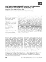
deaths were directly related to the assigned therapy. Figure 1
shows a nearly identical cumulative survival of the HFOV
group and the CV group corrected for the baseline covariates;
study site, OI, ventilatory index, APACHE II score, age and
weight. The survival curves of the duration of ventilation were
virtually identical for the HFOV group and the CV group (data
not shown). The median duration of ventilation was 20 days (±
6 SD) for HFOV and 18 days (± 5 SD) in the CV treatment
group.
Treatment failure occurred in 10 patients (27%) in the HFOV
group compared with five patients (21%) in the CV group.
Seven patients (19%) treated with HFOV crossed over to CV;
Critical Care Vol 9 No 4 Bollen et al.
R433
in the CV group four patients (17%) were switched to HFOV.
Of the four patients that crossed over in the CV group, two
patients died and one patient was on supplemental oxygen
therapy at 30 days. In the HFOV group, five patients that
crossed over died and two patients were still on ventilator or
needed extra oxygen. The occurrence of being on oxygen or
mechanical ventilation at 30 days in survivors was equal
between HFOV and CV.
Ventilatory settings and blood gas results at days 1, 2, 3 and
4 of the study are shown in Table 3. Patients with HFOV were
ventilated with higher mean airway pressures than patients on
Table 1
Patient characteristics at study entry
HFOV CV
N 37 24
Female:male ratio 9/28 (24%) 10/14 (42%)
Mean age (years) 81.0 ± 20.5 81.7 ± 12.5
Weight 50.7 ± 17.4 55.4 ± 12.8
APACHE II score 21.1 ± 7.6 20.1 ± 9.3
Diagnosis (%)
Trauma 1 (3) 2 (9)
Sepsis 25 (68) 13 (57)
Pneumonia 8 (22) 3 (13)
Other 3 (8) 5 (22)
Site (%)
United Kingdom 24 (65) 15 (63)
France 5 (21) 7 (19)
Germany 4 (17) 6 (16.2)
Ventilation time prior to study (days) 2.1 ± 2.6 1.5 ± 1.8
Oxygenation index 25.2 ± 13.0 18.0 ± 7.4
Ventilatory index 33.8 ± 20.4 30.3 ± 12.5
Respiratory rate (per min) 18.1 ± 4.1 17.8 ± 4.6
Tidal volume(ml) 618.4 ± 142.6 549.7 ± 130
Tidal volume per ideal bodyweight (ml/kg) 9.3 ± 2.2 8.4 ± 2.0
Peak inspiratory pressure (cmH2O) 33.1 ± 6.8 32.3 ± 5.4
Positive end-expiratory pressure (cmH2O) 13.9 ± 3.8 12.9 ± 3.2
Mean airway pressure (cmH2O) 21.5 ± 5.4 21.0 ± 5.1
FiO2 0.84 ± 0.19 0.76 ± 0.19
pH 7.3 ± 0.13 7.3 ± 0.11
paCO2 (mmHg) 53.5 ± 17.3 52.2 ± 11.9
paO2 (mmHg) 80.8 ± 24.1 93.3 ± 24.5
SaO2 (percentage) 90.8 ± 6.4 94.3 ± 3.1
Heart rate 109.8 ± 23.7 111.2 ± 29.5
Mean arterial pressure (cmH2O) 75.3 ± 13.1 72.2 ± 14.1
Central venous pressure (cmH2O) 13.5 ± 4.2 13.8 ± 4.9
Values are presented as means with standard deviations. APACHE II, Acute Physiologic and Chronic Health Evaluation II; CV, conventional
mechanical ventilation; FiO2, fraction of inspired oxygen; HFOV, high frequency oscillatory ventilation; OI, oxygenation index; paO2, pressure of
arterial oxygen, paCO2, pressure of arterial carbon dioxide; SaO2, arterial oxygen saturation.
Available online />R434
CV (p = 0.03). FiO2 was also higher in the HFOV group com-
pared with the CV group. This difference between the treat-
ment groups was not significant (p = 0.33). Results of blood
gases were comparable between the two treatment groups
including all patients. Patients that crossed over in the CMV
group had significantly lower pH than patients who did not
cross over in the CMV group (p = 0.02). This difference, how-
ever, was not found between patients who did and did not
cross over in the HFOV group (p = 0.56). The OI, on the other
hand, was higher in both patients that crossed over in the
CMV group and patients that crossed over in the HFOV group
compared with patients that did not cross over (p = 0.07 and
p = 0.05, respectively).
Systolic arterial blood pressure and mean arterial blood pres-
sure were higher in the HFOV treated patients compared with
CV treated patients (p = 0.06 versus p = 0.07). Cardiac out-
put was comparable between the two treatment groups (data
not shown).
Table 2
Primary outcomes
Unadjusted Adjusted
HFOV CV p-value OR 95% CI OR 95% CI
N3724
Survival without supplemental oxygen or on ventilator 12 (32%) 9 (38%) 0.79 0.80 0.27–2.53 0.80 0.22–2.97
Mortality 16 (43%) 8 (33%) 0.59 1.52 0.45–2.59 1.15 0.43–3.10
Circulatory failure 6 2
Cardiac arrhythmia 3 1
Brain death 0 2
Withdrawal of life support 7 3
Therapy failure 10 (27%) 5 (21%) 0.76 1.41 0.41–4.78 1.35 0.35–5.22
Hypotension 4 1
Acidosis 1 1
Oxygenation 4 2
Air leak 1 1
Cross-over 7 (19%) 4 (17%) 0.82 1.17 0.30–4.51 0.62 0.12–3.19
Supplemental oxygen or on ventilator at 30 days 9 (24%) 7 (29%) 0.96 0.96 0.26–3.58 0.67 0.12–3.84
Values between brackets are percentages of N (number of patients included in the analyses) except for CLD (Chronic Lung Disease) that has the
number of survivors in the denominator. CI, confidence interval; OR, odds ratio unadjusted and adjusted for study site, OI, ventilatory index,
APACHE II score, age and weight.
Figure 1
Cumulative mortality incidence for high frequency oscillatory ventilation (HFOV) versus conventional mechanical ventilation (CV)Cumulative mortality incidence for high frequency oscillatory ventilation
(HFOV) versus conventional mechanical ventilation (CV). Curves are
estimates of cumulative risk corrected for study site, baseline oxygena-
tion index and ventilatory index, APACHE II score, age and weight.
Critical Care Vol 9 No 4 Bollen et al.
R435
Table 3
Ventilatory conditions
HFOV CV
Cross-over No (30) Yes (7) No (20) Yes (4)
Day 1 N = 28 N = 7 (7 HFOV) N = 19 N = 4 (4 CV)
Peak inspiratory pressure (cmH2O) 32 ± 4.2 35 ± 6.9
Positive end-expiratory pressure (cmH2O) 14 ± 2.1 12 ± 4.5
Mean airway pressure (cmH2O) 30 ± 5.6
a
32 ± 6.3
a
22 ± 3.2 22 ± 6.1
Tidal volume per ideal bodyweight (ml/kg) 9 ± 1.7 8 ± 0.7
Frequency (HFOV, Hz; CV, breaths/min) 5 ± 0.5 5 ± 0.9 17.3 ± 3 17.3 ± 6
Delta P (cmH2O) 63 ± 14 70 ± 12.1
FiO2 0.78 ± 0.19 0.82 ± 0.12 0.68 ± 0.12 0.78 ± 0.21
pH 7.32 ± 0.08 7.31 ± 0.11 7.34 ± 0.08 7.22 ± 0.07
b
pCO2 (mmHg) 49 ± 11.3 57 ± 13 48 ± 9 52 ± 15.8
pO2 (mmHg) 126 ± 79.2 93 ± 37.1 98 ± 26.6 99 ± 25
SaO2 (percentage) 95 ± 3 90 ± 10.7 96 ± 2.4 94 ± 4.5
Oxygenation index 26 ± 16 31 ± 8.3
c
17 ± 7.5 19 ± 11.2
c
Day 2 N = 27 N = 7 (6 HFOV) N = 19 N = 4 (2 CV)
Peak inspiratory pressure (cmH2O) 25 ± 6.7 36 ± 7.2 31 ± 4.5 30 ± 2.6
Positive end-expiratory pressure (cmH2O) 11 ± 1.2 15 ± 1.9 14 ± 2.7 12 ± 4.7
Mean airway pressure (cmH2O) 28 ± 6.7
a
29 ± 4.3
a
21 ± 2.3 22 ± 9.1
Tidal volume per ideal bodyweight (ml/kg) 9 ± 1.6 10 ± 1.9 8 ± 1.6 8 ± 1
Frequency (HFOV, Hz; CV, breaths/min) 5.0 ± 0.4 4.8 ± 1.1 17.4 ± 2.6 17.2 ± 1.2
Delta P (cmH2O) 64 ± 14.5 73 ± 14.8 70 ± 13.8
FiO2 0.55 ± 0.17 0.57 ± 0.14 0.53 ± 0.12 0.76 ± 0.20
pH 7.36 ± 0.07 7.35 ± 0.04 7.38 ± 0.06 7.22 ± 0.08
b
pCO2 (mmHg) 45 ± 9 51 ± 8.9 46 ± 8.3 53 ± 8.5
pO2 (mmHg) 96 ± 21 83 ± 12.4 100 ± 27 87 ± 41.8
SaO2 (percentage) 95 ± 2.1 94 ± 1.9 96 ± 1.8 87 ± 16.1
Oxygenation index 17 ± 10.2 21 ± 8.2
c
12 ± 3.6 22 ± 10.5
c
Day 3 N = 23 N = 7 (4 HFOV) N = 19 N = 4 (2 CV)
Peak inspiratory pressure (cmH2O) 21 ± 3.1 32 ± 12 30 ± 4 27 ± 6
Positive end-expiratory pressure (cmH2O) 9 ± 3 10 ± 4.3 13 ± 2.8 11 ± 5.7
Mean airway pressure (cmH2O) 23 ± 7.1
a
25 ± 6.9
a
20 ± 2.8 24 ± 2.3
Tidal volume per ideal bodyweight (ml/kg) 9 ± 1.5 9 ± 3.5 9 ± 1.6 7 ± 1.6
Frequency (HFOV, Hz; CV, breaths/min) 5.0 ± 0.4 4.6 ± 0.5 18.8 ± 6.5 19.9 ± 5.8
Delta P (cmH2O) 66 ± 12.4 66 ± 19.1 67 ± 0.7
FiO2 0.46 ± 0.13 0.55 ± 0.15 0.46 ± 0.11 0.65 ± 0.26
pH 7.39 ± 0.06 7.37 ± 0.06 7.39 ± 0.06 7.33 ± 0.1
b
pCO2 (mmHg) 45 ± 10.4 47 ± 12.9 48 ± 9 47 ± 12.6
pO2 (mmHg) 89 ± 19.7 86 ± 46.2 91 ± 13.7 89 ± 22.4
Available online />R436
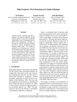
The OI response in all patients treated with either HFOV or CV
did not differ significantly between survivors and non-survivors
(Figure 2). The OI response from day 1 to day 2 was signifi-
cantly larger in HFOV than in CV treated patients (p < 0.01).
Within treatment groups there was a significant difference in
initial OI between survivors and non-survivors in CV treated
patients, but OI response to treatment did not differentiate
between survivors and non-survivors in CV treated patients. In
the HFOV treated patients there was no difference in the
baseline OI, nor was there a difference in OI response
between survivors and non-survivors.
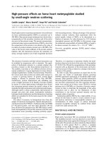
The results of a post hoc analysis are shown in Figure 3.
Adjusted odds ratios for mortality were calculated for samples
of the study population including patients with progressively
higher baseline OI prior to randomization. This suggested that,
in patients with a higher baseline OI, the effect of treatment
with HFOV was relatively better compared with CV. OI was
evaluated as an interaction term in a Cox Proportional Hazard
model with treatment, age and OI as explanatory variables. The
likelihood ratio test comparing the reduced (no-interaction)
with the full (interaction) model showed a p-value of 0.048.
Discussion
No significant differences between HFOV and CV were
observed, but this trial only had power to detect major differ-
ences in mortality or survival without oxygen dependency or on
ventilator. Furthermore, 11 of 61 patients were crossed over
to a different treatment arm; this also diminished the power to
detect potential treatment differences. A post hoc analysis,
however, suggested that in patients with a higher baseline OI,
HFOV may be more effective than CV.
This trial was stopped because of a low inclusion rate and the
completion of another similar trial [7]. The low inclusion rate
was not because of competing trials but probably due to the
limited number of investigators (four centres compared with
nine centres in the study by Derdak et al.). The number of
patients included in the two treatment arms differed consider-
ably. This misbalance was due to stopping the trial early. There
were no protocol violations. Furthermore, baseline OI at study
entry was higher in the HFOV group than in the CV group. The
OI has been recognized as an important prognostic determi-
nant of mortality [13].
HFOV was started early in the course of ARDS. Patients were
ventilated on HFOV according to the open lung concept. This
resulted in significantly higher mean airway pressures com-
pared with CV ventilated patients. This mainly determined the
higher OI in the HFOV group during the first days. FiO2 and
paO2 values were similar between HFOV and CV patients.
Potential theoretical risks of HFOV therapy, overdistension of
the pulmonary system leading to barotrauma or cardiovascular
compromise, packing of mucus leading to ineffective ventila-
SaO2 (percentage) 94 ± 6.7 89 ± 14.1 96 ± 1.9 95 ± 2.4
Oxygenation index 14 ± 7.2 19 ± 9.3
c
11 ± 3.7 20 ± 12.3
c
Day 4 N = 22 N = 7 (3 HFOV) N = 19 N = 2 (0 CV)
Peak inspiratory pressure (cmH2O) 25 ± 8 31 ± 6.9 28 ± 6.9
Positive end-expiratory pressure (cmH2O) 9 ± 4.6 11 ± 4.2 11 ± 3.2
Mean airway pressure (cmH2O) 22 ± 7.8
a
24 ± 6.2
a
17 ± 5.6 24 ± 3.2
Tidal volume per ideal bodyweight (ml/kg) 10 ± 2.4 7 ± 3.1 8 ± 2.2
Frequency (HFOV, Hz; CV, breaths/min) 5.0 ± 0.3 4.3 ± 0.6 17.9 ± 5.3
Delta P (cmH2O) 57 ± 11.4 70 ± 11.8 48 ± 14.8
FiO2 0.45 ± 0.11 0.57 ± 0.18 0.45 ± 0.11 0.51 ± 0.12
pH 7.42 ± 0.14 7.37 ± 0.1 7.43 ± 0.12 7.45 ± 0.06
b
pCO2 (mmHg) 43 ± 12.3 46 ± 7.5 41 ± 10.3 44 ± 11.1
pO2 (mmHg) 85 ± 22.3 84 ± 30.5 87 ± 27.4 74 ± 23.7
SaO2 (percentage) 89 ± 15.3 90 ± 14.1 89 ± 17.2 84 ± 20
Oxygenation index 12 ± 5.6 18 ± 7.9
c
10 ± 4.3 19 ± 9.5
c
The columns represent the treatment allocation. Measurements were made day 1, 2, 3 and 4 of the study. Peak inspiratory pressure, positive end-
expiratory pressure and tidal volume per ideal bodyweight were measured in high frequency oscillatory ventilation (HFOV) after crossover to
conventional mechanical ventilation (CV). Values are presented as means with standard deviations.
a
Higher mean airway pressures in HFOV
compared with CV (p = 0.03).
b
Significantly lower pH in patients that cross over in the CV group (p = 0.017).
c
Higher OI in patients that crossed
over compared with patients that did not cross over (p = 0.07 and p = 0.05, respectively). FiO2, fraction of inspired oxygen; paCO2, pressure of
arterial carbon dioxide; paO2, pressure of arterial oxygen; SaO2, arterial oxygen saturation.
Table 3 (Continued)
Ventilatory conditions
Critical Care Vol 9 No 4 Bollen et al.
R437
tion or blocking of the endotracheal tube were not encoun-
tered. None of the HFOV ventilated patients developed
necrotizing tracheobronchitis.
Patients in the CV group were ventilated following a lung pro-
tective strategy targeted to minimizing tidal volumes. The tidal
volumes per kg ideal bodyweight that were used in this study
were higher than tidal volumes used in studies of lung protec-
tive ventilation strategies [14]. On the other hand, tidal vol-
umes in our study were significantly lower than tidal volumes
that were found to be harmful in those studies. Peak inspira-
tory pressures were limited to 40 cmH2O in the CV group.
This restriction was violated in only one case. Nine patients
were ventilated with pressures above 35 cmH2O. Further-
more, the overall mortality and survival without mechanical
ventilation or oxygen dependency at 30 days did not suggest
that the ventilation treatment in the CV group was suboptimal.
The OI represents the pressure and oxygen cost for oxygena-
tion. It has been regarded as a marker of lung injury and prog-
nostic indicator of treatment success [15]. In CV treated
patients there was a significant difference in baseline OI
between survivors and non-survivors. Baseline OI did not,
however, differentiate between survivors and non-survivors in
HFOV treated patients. Although in some studies OI response
to treatment was a predictor of outcome [7,9], we could not
reproduce this relation. A possible explanation could be that
fewer numbers of patients were included in our analysis. Also,
we used a different time window; we compared OI on a daily
basis whereas in a study by Derdak et al. [7] OI was compared
every 4 h. In that study, OI response was maximally different at
16 h [7]. In our study, OI response only differed significantly
between HFOV and CV treated patients. This difference for
the most part could be explained by the higher mean airway
pressures used in the HFOV group.
A post hoc analysis suggested that baseline OI could be an
important effect modifier of the relative treatment effect of
HFOV compared with CV. We hypothesize that within the
pressure-ventilation curve there is a safe window between
under-inflation with atelectasis and shear stress and over-infla-
tion with barotrauma [4,16]. In patients with ARDS with higher
OI, this safe window possibly becomes too small for CV to
prevent ventilator induced lung injury. This concept is
supported by animal experiments where addition of positive
end-expiratory pressure (PEEP) resulted in additional over-
inflation contributing to ventilator-induced lung injury [17]. The
combination of high levels of PEEP and over-distension are
directly reflected in the OI. HFOV seemed to offer an advan-
tage over CV only in patients with a higher initial OI. This is in
Figure 2
Oxygenation index (OI) in survivors versus non-survivors and high frequency oscillatory ventilation (HFOV) versus conventional mechanical ventila-tion (CV)Oxygenation index (OI) in survivors versus non-survivors and high frequency oscillatory ventilation (HFOV) versus conventional mechanical ventila-
tion (CV). OIs are represented by diamonds as means with bars as 95% confidence intervals (CI). Reported p-values for baseline OI are corrected
for study site, ventilatory index, APACHE II score, age and weight. The baseline OI did not significantly predict mortality in all patients or in HFOV (p
= 0.06 and p = 0.41, respectively).
§
Baseline OI was significantly different between survivors and non-survivors in the CV group (p = 0.04). Signifi-
cant differences between OI responses were calculated by linear mixed model analyses.
#
Significant difference in OI response between HFOV and
CV (p = < 0.01). OI response did not differentiate between survivors and non-survivors in all patients or in CV and HFOV separately (p = 0.28, p =
0.12 and p = 0.95, respectively).
Available online />R438
accordance with observational studies that showed that better
survival rates in more severe ARDS with higher OI was asso-
ciated with HFOV treatment [11,18]. In fact, HFOV has been
recommended in patients who require high mean airway pres-
sure and FiO2 exceeding 60% corresponding to an OI > 20
when paO2 = 60 mmHg [12]. Because these findings result
from a post hoc analysis, however, they can only be regarded
as hypothesis generating still to be confirmed.
Previous trials did not show a significant difference in mortality
in patients with ARDS between HFOV and CV [19]. In our trial,
mortality in the HFOV group was similar to mortality reported
in the previous trials, but mortality in the CV group was consid-
erably less, in accordance with the imbalance in prognostic
indicators at baseline.
More evidence is needed to confirm a beneficial effect of
HFOV over CV in the treatment of ARDS. Our results and
those from previous trials seem promising but could depend
on other criteria to select patients with ARDS that benefit from
HFOV compared with CV. One of these criteria could be OI.
Therefore, we believe that in future research comparing HFOV
with CV as early treatment of ARDS, it is important to focus on
patients with higher levels of baseline OI. As treatment differ-
ences will be smaller than our prior estimate was, larger trials
are needed. We do not think that OI response can be used as
an alternative outcome measurement for treatment success or
failure.
Conclusion
In this study, we were not able to find significant differences in
efficacy or safety between HFOV and CV as early treatment of
ARDS. A post hoc analysis suggested that HFOV could pre-
vent mortality compared with CV in patients with a higher
baseline OI. Therefore, it is important in future studies to ena-
ble informative analysis of patients with higher baseline OI. To
achieve sufficient power to detect possible important treat-
ment differences in subgroups of patients with higher OI,
larger multi-centre trials are warranted.
Competing interests
Supported in part by SensorMedics Corporation, which also
provided use of the 3100B high-frequency oscillatory ventila-
tors. None of the study investigators have a financial interest in
SensorMedics Corporation. The authors declare that they
have no competing interests.
Authors' contributions
AJvV initiated the study, participated in its design and coordi-
nation and helped to draft the manuscript. CWB, CSPMU and
GTJvW performed the statistical analyses and wrote the man-
uscript. TS, RJB, SS, GF, MM, JC and NW participated in its
design and conducted the study. All authors read and
approved the final manuscript.
References
1. Frank JA, Matthay MA: Science review: mechanisms of ventila-
tor-induced injury. Crit Care 2003, 7:233-241.
2. Rubenfeld GD: Epidemiology of acute lung injury. Crit Care
Med 2003, 31:S276-S284.
3. Brower RG, Rubenfeld GD: Lung-protective ventilation strate-
gies in acute lung injury. Crit Care Med 2003, 31:S312-S316.
4. Froese AB: High-frequency oscillatory ventilation for adult res-
piratory distress syndrome: let's get it right this time! Crit Care
Med 1997, 25:906-908.
5. Henderson-Smart D, Bhuta T, Cools F, Offringa M: Elective high
frequency oscillatory ventilation versus conventional ventila-
tion for acute pulmonary dysfunction in preterm infants.
Cochrane Database Syst Rev 2003, 4:CD000104.
6. Bollen CW, Uiterwaal CS, van Vught AJ: Cumulative metaanaly-
sis of high-frequency versus conventional ventilation in pre-
mature neonates. Am J Respir Crit Care Med 2003,
168:1150-1155.
7. Derdak S, Mehta S, Stewart TE, Smith T, Rogers M, Buchman TG,
Carlin B, Lowson S, Granton J: High-frequency oscillatory venti-
lation for acute respiratory distress syndrome in adults: a ran-
domized, controlled trial. Am J Respir Crit Care Med 2002,
166:801-808.
Figure 3
Post hoc analysis of the treatment effect on mortality relative to baseline oxygenation index (OI)Post hoc analysis of the treatment effect on mortality relative to baseline
oxygenation index (OI). On the y-axis the odds ratio of mortality (OR)
adjusted for study site, OI, ventilatory index, APACHE II score, age and
weight is presented by diamonds and 95% confidence intervals by
bars. On the x-axis the different analyses are depicted including
patients with increasing levels of initial OI at study entry. N denotes the
number of patients in each subgroup. CI, confidence interval; CMV,
conventional mechanical ventilation; HFOV, high frequency oscillatory
ventilation.
Key messages
• This study was not powered to show significant differ-
ences in efficacy or safety between HFOV and CV as
early treatment of ARDS.
• However, a post hoc analysis suggested a better treat-
ment effect of HFOV compared with CV in patients with
higher baseline OI.
• Future studies should be designed to allow for informative
analysis in patients with higher OI.
Critical Care Vol 9 No 4 Bollen et al.
R439
8. David M, Weiler N, Heinrichs W, Neumann M, Joost T, Markstaller
K, Eberle B: High-frequency oscillatory ventilation in adult
acute respiratory distress syndrome. Intensive Care Med 2003,
29:1656-1665.
9. Arnold JH, Hanson JH, Toro-Figuero LO, Gutierrez J, Berens RJ,
Anglin DL: Prospective, randomized comparison of high-fre-
quency oscillatory ventilation and conventional mechanical
ventilation in pediatric respiratory failure. Crit Care Med 1994,
22:1530-1539.
10. Mehta S, Lapinsky SE, Hallett DC, Merker D, Groll RJ, Cooper AB,
MacDonald RJ, Stewart TE: Prospective trial of high-frequency
oscillation in adults with acute respiratory distress syndrome.
Crit Care Med 2001, 29:1360-1369.
11. Fort P, Farmer C, Westerman J, Johannigman J, Beninati W, Dolan
S, Derdak S: High-frequency oscillatory ventilation for adult
respiratory distress syndrome – a pilot study. Crit Care Med
1997, 25:937-947.
12. Derdak S: High-frequency oscillatory ventilation for acute res-
piratory distress syndrome in adult patients. Crit Care Med
2003, 31:S317-S323.
13. Sarnaik AP, Meert KL, Pappas MD, Simpson PM, Lieh-Lai MW,
Heidemann SM: Predicting outcome in children with severe
acute respiratory failure treated with high-frequency
ventilation. Crit Care Med 1996, 24:1396-1402.
14. Ventilation with lower tidal volumes as compared with tradi-
tional tidal volumes for acute lung injury and the acute respi-
ratory distress syndrome. The Acute Respiratory Distress
Syndrome Network. N Engl J Med 2000, 342:1301-1308.
15. van Genderingen HR, van Vught JA, Jansen JR, Duval EL,
Markhorst DG, Versprille A: Oxygenation index, an indicator of
optimal distending pressure during high-frequency oscillatory
ventilation? Intensive Care Med 2002, 28:1151-1156.
16. Lachmann B: Open up the lung and keep the lung open. Inten-
sive Care Med 1992, 18:319-321.
17. Ricard JD, Dreyfuss D, Saumon G: Ventilator-induced lung
injury. Eur Respir J Suppl 2003, 42:2s-9s.
18. Mehta S, Granton J, MacDonald RJ, Bowman D, Matte-Martyn A,
Bachman T, Smith T, Stewart TE: High-frequency oscillatory
ventilation in adults: the Toronto experience. Chest 2004,
126:518-527.
19. Wunsch H, Mapstone J: High-frequency ventilation versus con-
ventional ventilation for treatment of acute lung injury and
acute respiratory distress syndrome. Cochrane Database Syst
Rev 2004:CD004085.