Báo cáo khoa học: "Clinical review: The Israeli experience: conventional terrorism and critical care" docx
Bạn đang xem bản rút gọn của tài liệu. Xem và tải ngay bản đầy đủ của tài liệu tại đây (88.09 KB, 10 trang )
490
ARDS = acute respiratory distress syndrome; CT = computed tomography; ED = emergency department; GCS = Glasgow Coma Score; GICU =
general intensive care unit; HFJV = high-frequency jet ventilation; ICP = intracranial pressure; ICU = intensive care unit; ISS = injury severity score;
MCI = multiple-casualty incident; PACU = post-anesthesia care unit; PEEP = positive end-expiratory pressure; SIRS = systemic inflammatory
response syndrome.
Critical Care October 2005 Vol 9 No 5 Aschkenasy-Steuer et al.
Abstract
Over the past four years there have been 93 multiple-casualty
terrorist attacks in Israel, 33 of them in Jerusalem. The Hadassah-
Hebrew University Medical Center is the only Level I trauma center
in Jerusalem and has therefore gained important experience in
caring for critically injured patients. To do so we have developed a
highly flexible operational system for managing the general
intensive care unit (GICU). The focus of this review will be on the
organizational steps needed to provide operational flexibility,
emphasizing the importance of forward deployment of intensive
care unit personnel to the trauma bay and emergency room and the
existence of a chain of command to limit chaos. A retrospective
review of the hospital’s response to multiple-casualty terror
incidents occurring between 1 October 2000 and 1 September
2004 was performed. Information was assembled from the medical
center’s trauma registry and from GICU patient admission and
discharge records. Patients are described with regard to the
severity and type of injury. The organizational work within intensive
care is described. Finally, specific issues related to the diagnosis
and management of lung, brain, orthopedic and abdominal injuries,
caused by bomb blast events associated with shrapnel, are
described. This review emphasizes the importance of a
multidisciplinary team approach in caring for these patients.
Introduction
Every hospital should be able to respond to a multiple-
casualty terror attack as it can occur anywhere and anytime
[1]. Over the past four years there have been 93 multiple-
casualty terrorist attacks in Israel, 33 of them in Jerusalem.
The Hadassah-Hebrew University Medical Center is the only
Level I trauma center in Jerusalem and has therefore gained
important experience in caring for the critically injured
patients. Despite the violence, all surgical services continued
providing all routine services, including our general intensive
care unit (GICU). To do so we developed a highly flexible
operational system for managing the GICU. The focus of this
report will be on the organizational steps needed to provide
operational flexibility. In addition, issues related to the
Review
Clinical review: The Israeli experience: conventional terrorism
and critical care
Gabriella Aschkenasy-Steuer
1
, Micha Shamir
2
, Avraham Rivkind
3
, Rami Mosheiff
4
, Yigal Shushan
5
,
Guy Rosenthal
6
, Yoav Mintz
7
, Charles Weissman
8
, Charles L Sprung
9
and Yoram G Weiss
10
1
Resident in Anesthesiology, Department of Anesthesiology and Critical Care Medicine, Hadassah Hebrew University School of Medicine, Hadassah
Medical Organization, Jerusalem, Israel
2
Senior Anesthesiologist, Department of Anesthesiology and Critical Care Medicine, Hadassah Hebrew University School of Medicine, Hadassah
Medical Organization, Jerusalem, Israel
3
Associate Professor of Surgery, Department of Surgery, Hadassah Hebrew University School of Medicine, Hadassah Medical Organization,
Jerusalem, Israel
4
Associate Professor of Orthopedics, Department of Orthopedic Surgery, Hadassah Hebrew University School of Medicine, Hadassah Medical
Organization, Jerusalem, Israel
5
Senior Clinical Lecturer in Neurosurgery, Department of Neurosurgery, Hadassah Hebrew University School of Medicine, Hadassah Medical
Organization, Jerusalem, Israel
6
Senior Neurosurgeon, Department of Neurosurgery, Hadassah Hebrew University School of Medicine, Hadassah Medical Organization, Jerusalem,
Israel
7
Instructor in Surgery, Department of Surgery, Hadassah Hebrew University School of Medicine, Hadassah Medical Organization, Jerusalem, Israel
8
Professor of Medicine and Anesthesiology, Department of Anesthesiology and Critical Care Medicine, Hadassah Hebrew University School of
Medicine, Hadassah Medical Organization, Jerusalem, Israel
9
Professor of Medicine, Department of Anesthesiology and Critical Care Medicine, Hadassah Hebrew University School of Medicine, Hadassah
Medical Organization, Jerusalem, Israel
10
Senior Lecturer in Anesthesia and Critical Care Medicine, Hadassah Hebrew University Medical School, Jerusalem, Israel and Adjunct Assistant
Professor in Anesthesia and Critical Care Medicine, University of Pennsylvania Medical School, Philadelphia, PA, USA.
Corresponding author: Yoram G Weiss,
Published online: 29 June 2005 Critical Care 2005, 9:490-499 (DOI 10.1186/cc3762)
This article is online at />© 2005 BioMed Central Ltd
491
Available online />diagnosis and management of specific injuries associated
with terror events will be discussed.
Numbers and statistics
A retrospective review of the hospital’s response to multiple-
casualty terror incidents occurring between 1 October 2000
and 1 September 2004 was performed. Information was
assembled from the medical center’s trauma registry and
from GICU patient admission and discharge records. The
information we collected included the following: type of
attack, number of victims at the location, number of patients
treated and admitted to the intensive care unit (ICU), location
before admission to the ICU [operating rooms, imaging
department or emergency department (ED)], trauma injury
severity score (ISS), time for admission to the ED, time to
admission to the ICU, length of stay in the ICU and mortality
in the ICU.
ICU organization
The hospital intensive care facilities include 29 surgical ICU
beds (11 general, 6 pediatric, 6 neurosurgical, and 6 cardio-
thoracic). When these ICUs are full, patients are treated in
the 14-bed post-anesthesia care unit (PACU), which is
adjacent to the GICU. In addition, nine medical intensive care
beds are available. The GICU is part of the Department of
Anesthesiology and Critical Care Medicine. All ICU attending
professionals are board-certified anesthesiologists. All
anesthesiology residents have training in intensive care (6
months of an ICU rotation) and are routinely involved in the
daily care of patients in the GICU.
Patients
After 33 major terror attacks, 541 victims were admitted to
the ED of the Hadassah Hebrew University Medical Center,
of whom 208 were hospitalized. Twenty of these attacks
involved more than 10 wounded and therefore were defined
as a ‘multiple-casualty incident’ (MCI; Table 1). In preparation
for the admission of critically injured terror victims, 40
patients were transferred out of the GICU either to a regular
ward (75%) or to another ICU (25%). Additional patients
(postoperative) were discharged from the PACU if their
condition was deemed stable. This was done to increase the
number of GICU beds. One hundred and one (49%) patients
were admitted to an ICU (median 4 admissions per event;
range 0 to 9), 86 to the GICU, 8 to the neurosurgical ICU
and 7 to the pediatric ICU. The age distribution of terror
victims was skewed towards the younger generation (80%
aged 15 to 44 years, compared with 37% for other traumas).
During this period, a total of 2,647 patients were admitted to
the GICU, of whom 4% were victims of these terrorist
attacks. Twelve patients, who were victims of five different
attacks each associated with more than six admissions to the
GICU, were initially admitted to the PACU. Fifty-seven
patients were admitted from the operating rooms (56%), 11
from the angiography suite (10%) and the rest were admitted
directly from the ED (34%).
The average time from the initial trauma to admission to the
GICU was 5.5 ± 3.1 hours (mean ±SD) (range 1 to 13 hours).
Patients admitted directly from the ED or angiography suite
were admitted earlier than those from the operating rooms
(means 3.8, 3.7 and 6.3 hours, respectively). The severity of
injuries is demonstrated by the fact that 47 of 101 patients
(46%) had to be intubated in the ED in addition to those
already intubated at the scene. Out of all the 541 MCI terror
victims, 12 patients admitted to our center were intubated at
the scene and 16 patients were admitted as secondary
transfers after initial resuscitation from other hospitals.
Furthermore, 116 (56%) of the patients admitted to our Level
1 trauma center needed surgery within the first 8 hours after
the attack. Less severely injured patients were diverted by the
emergency medical services to other regional trauma centers
in the Jerusalem area [2]. The terror victims stayed in the
GICU for an average of 9 days (range 1 to 80 days; Table 2),
whereas the median length of stay for the entire GICU
population was 3 days. More than half of the patients
admitted to an ICU had an in-hospital length of stay of two
weeks or more (Table 3). The average overall mortality rate for
patients in the ICU was 8.5% in 2001 to 2004. Finally,
patients hospitalized after terror events had sustained more
severe injuries (74% versus 10% in other types of trauma
with an ISS of more than 16) and had double the mortality
(6.2% versus 3%) [3].
Type of injury
The hallmark of the injuries was a combination of blunt trauma
and penetrating injuries due to bolts. The injuries could be
divided into three categories. Blunt trauma was diagnosed in
51 patients, burns in 33 and penetrating injuries in 90 patients.
Blast injury was registered as blunt injury. Commonly victims
suffered injuries originating from more than one mechanism of
injury. Moreover, victims commonly had injuries to several
parts of the body, the most frequently injured region being the
head, neck and facial area (Tables 4 and 5). The ISS in the
101 patients requiring intensive care varied from 5 to 75 with
an average score of 24 (Table 6). Four patients with low ISS
scores (5 to 8) were admitted to the unit for a 24-hour
observation period: two patients for chest and neck burns,
one patient after neck exploration and one patient kept
intubated after a long surgical intervention. The Glasgow
Coma Score (GCS) on admission in these patients was as
follows: 29 (28.7%) injured had a GCS of 3 to 8; 6 (5.9%)
within the range 9 to 12; and 66 (65.3%) had a GCS of 13
to 15.
Exploring the sequence of events and
organizational issues
On 24 May 2001 the floor of Versailles Hall (located on the
third floor of a building in the center of Jerusalem) collapsed
during a wedding celebration. Over a 2-hour period more
than 200 victims were admitted to the Hadassah ED. On the
basis of our experience from this incident, we believe that the
response to a multiple-casualty terror attack does not differ
492
Critical Care October 2005 Vol 9 No 5 Aschkenasy-Steuer et al.
from the response to any other multiple-casualty trauma.
Hence, lessons learned during these events should be
implemented by others in preparation for catastrophes. A
previous publication discussed the in-hospital response to
the specific actions that were taken in response to the
various time periods of a multiple-casualty terror event:
assessment of incident size and severity; alerting of backup
personnel; initial casualty care; and definitive treatment [4].
To streamline the administration of a multiple-casualty event,
two important administrative concepts have been adopted:
first, peri-incident intensive care management (‘forward
Table 1
Data for 20 major bombing attacks with more than 10 wounded
Wounded ED Hospital ICU Acute In-hospital
on scene admissions admissions admissions Ventilated surgery deaths
Date Location (n)(n)(n)(n)(n)(n)(n)
Aug 2001 Restaurant 113 18 8 4 2 4 –
Dec 2001 City center 188 65 26 9 6 18 1
Jan 2002 City center 150 32 3 1 1 1 –
Mar 2002 Street 58 8 4 4 3 3 1
Mar 2002 Café 64 35 10 5 3 8 –
Mar 2002 Café 100 17 4 2 2 4 1
Mar 2002 Supermarket 50 33 3 1 1 2 –
Mar 2002 EMS station 10 7 2 2 2 2 –
Apr 2002 City center 66 23 6 4 3 4 –
Jun 2002 Bus 73 25 11 6 7 7 1
Jun 2002 Square 20 9 5 2 2 2 –
Jul 2002 University 93 25 15 8 8 13 2
Nov 2002 Bus 58 44 15 8 8 11 1
May 2003 Bus 27 5 4 4 4 3 –
Jun 2003 Bus 100 21 9 5 5 6 1
Aug 2003 Bus 154 33 16 9 9 7 2
Sep 2003 Café (missing) 26 6 3 2 5 –
Jan 2004 Bus 60 17 8 6 6 4 1
Feb 2004 Bus 69 26 14 3 2 6 –
Aug 2004 Checkpoint 22 8 7 6 4 3 –
Total 1,475 477 176 92 80 113 11
ED, emergency department; EMS, emergency medical services; ICU, intensive care unit.
Table 2
Length of stay in the intensive care unit for 101 terror victims
Length of stay (days) No. of patients (%)
0–3 44 (44)
4–7 23 (23)
8–14 20 (20)
15–21 5 (5)
>22 9 (9)
Table 3
Hospital length of stay in 101 terror victims primarily admitted
to the intensive care unit
Length of stay (days) No. of patients (%)
0–6 14 (14)
7–13 34 (34)
14–20 12 (12)
>21 41 (41)
493
deployment’) and second, maintaining a ‘chain of command’
[4]. Forward deployment of anesthesiology and surgical
personnel is the procedure used at Hadassah for responding
to all traumas. When severe injuries occur, an anesthesiology
resident with ICU training or a critical-care fellow continuously
cares for a severely injured patient from admission to the ED,
through imaging studies in the radiology department and
during surgery. Continuity of care is guaranteed and vital
information is collected by one dedicated team and the
complete medical picture of the specific patient is
maintained. This is especially important in severe multiple-
trauma patients for whom surgical teams often change during
several multi-disciplinary interventions.
A chain of command should be established by the institution
and the departments as soon as possible. This is essential to
control the chaos that will ensue as victims arrive en masse.
Command rests with the most senior personnel available on site
from general surgery, orthopedic surgery and anesthesiology/
critical care medicine and hospital administration. As events
evolve and more senior personnel arrive they will take charge.
A senior general surgeon performs triage at the door of the
ED. Another experienced general surgeon acts as the
‘surgical command officer’ who guides the trauma teams. The
command team maintains a log of the most severely injured
victims. They consult frequently in the ED as to the
disposition of these patients (operating room, radiology suite,
ICU or recovery room). The GICU attending professional is
present in the ED trauma area to evaluate those wounded
who may need intensive care. This early evaluation is of
utmost importance to help direct the placement of each of
these patients in the ICU or in a less intensive area such as
the PACU. Early knowledge of the type and magnitude of
injuries gives an immediate estimate of the number of ICU
beds required. In addition, it is helpful in planning the exact
ICU bed for each patient. It is important to prevent clustering
of the most complicated patients in one location, treated by
few nurses, while other areas are left out. Furthermore,
providing early information to the ICU team on the injuries
their patient has suffered allows time to organize special
equipment such as ventilators, rapid infusion and blood-
warming devices.
Sequence of events
Assessment of incident size and severity
After a terror attack there is a latent period, lasting at least
20 minutes, in which events are taking place outside the
hospital [4]. During this period, estimating the number of
victims and the possible severity of their injuries is crucial for
proportional ‘department wakeup’. Estimations depend on the
day of the week, time of day, location and nature of the
incident. It is important to realize that an explosion in a
confined space will result in a large number of severely
injured victims [5]. Early information may best be obtained
from the media, the Internet or emergency medical services’
radio communications. Estimates of casualties must be
updated frequently because information changes with time.
During this latent phase, lower-intensity care areas of the ED
should be cleared of patients. Patients in the ED should be
quickly triaged, admitted to a ward or discharged. The
attending professional in the ICU can use this period to
Available online />Table 5
Isolated versus combined injuries in 101 terror victims
admitted to the intensive care unit
Injured part of the body No. of patients
Isolated injury 15
Combined injuries 86
With head injury 40
Head, chest, abdomen 5
Head, chest 21
Head, abdomen 3
Without head injury 46
Chest, abdomen 11
Combined injuries including extremities 55
Table 4
Numbers of patients with injuries to specified parts of the
body out of 101 terror victims treated in the intensive care unit
Injured part of the body No. of patients
Face 56
Chest 51
Lower extremities 46
Head 44
Upper extremities 39
Abdomen 32
Neck 17
Spine 7
Note: several patients had more than one injury.
Table 6
Injury severity score (ISS) in the 101 terror victims admitted to
the intensive care unit
ISS No. of patients (%)
5–8 4 (4)
9–14 22 (22)
16–24 29 (29)
>25 46 (46)
494
quickly review the patients in the ICU, thus identifying the
potential vacant beds. A list of actions to be taken by the on-
call chief resident has been placed at the anesthesiology
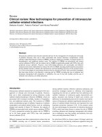
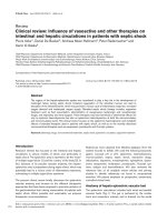
control office. Figure 1 summarizes these actions in context
with the time frame of events.
Meanwhile, lower-intensity care areas in the ED or PACU
should be quickly equipped to care for patients with major
injuries. Equipment available in all high-intensity care areas
should include oxygen, airway equipment (laryngoscope,
endotracheal tubes, bag/mask and suction devices),
intravenous supplies, drugs (ketamine, ethomidate, succinyl-
choline and a non-depolarizing muscle relaxant) and monitors.
Mobile ‘multiple-casualty carts’ containing these supplies can
save valuable time. It is of importance to check the availability
of rapid infusion devices and body heaters for these patients.
Backup
Recruiting additional staff is essential. We used an average of
16 anesthesiologists, attending and resident staff, per event
to manage all the department’s activities. Naturally, at the
beginning personnel are used for resuscitation of the injured
in the ED. An up-to-date list of all staff members, permanently
posted in a prominent place, is crucial for efficient personnel
recruitment. Staff are called according to residential distance
rather than professional status. On hearing of an event, the in-
house on-call ICU physician should call the at-home on-call
attending professional for the ICU, the department chair, and
a few other senior physicians with trauma expertise. Our
hospital is equipped with cellular phones that act as an
extension of the hospital telephone system (a virtual private
network). However, cellular networks tend to fail immediately
after an MCI and cannot be relied on, mandating the use of
Critical Care October 2005 Vol 9 No 5 Aschkenasy-Steuer et al.
Figure 1
Timeline of events after an incident and actions to be taken. ED, emergency department; ICU, intensive care unit; OR, operating room; PACU, post-
anesthesia care unit.
Time (min)
after
explosion
0
0–10
0–20
Event
Explosion
Assess
incident size
and severity
Backup and
preparation
of sites
Actions to be taken
Obtain information from emergency services and
media sources
Continue life-saving operations
Hold elective operations
Clear emergency department
Send patients from OR waiting area back to
departments
Consider taking out patients from OR if operation
not yet started
Notify all anesthesiologists present in the hospital
about the event
Prepare to call in additional personnel
Recruit additonal staff:
Head of ICU department, on-call senior physicians,
ICU head nurses. Other physicians and nurses as
needed
Triage patients to be discharged from the ICU and
recovery room
Send on-call most experienced physicians to the
trauma bay and emergency department
Prepare trauma bay, two operating rooms and ICU
beds (including PACU) for admission
Mean time of event (min)
0 30 50 100 150 200 250 300 350
Explosion
1st ambulance on scene
1st ambulance arrival to ED
Last ambulance arrival
1st surgery start
1st ICU admission from ED
1st ICU admission from angioplasty suite
1st ICU admission from OR
495
beepers and cable telephone systems [6]. There is also a
computerized call-in system that delivers a recorded message
using regular telephone lines. Cellular networks usually
resume normal function after some time and become
invaluable communication tools between physicians spread in
various locations throughout the hospital.
During our study period, a median of four patients per event
were admitted to the GICU, 5.5 ± 3.2 hours (mean ± SD)
after the event. Some patients, however, arrived in the ICU
soon after the event, either because they did not require
surgery or because they needed extensive stabilization before
surgery. The ICU must not be a limiting factor in clearing the
ED. One should also anticipate a second wave of wounded
referred from smaller hospitals. Finding vacant beds and
negotiating with the appropriate services should be done with
extensive help from nursing and hospital administrators. In
contrast to routine transfers, requests for patient transfer in
these circumstances are dealt with instantaneously and with
acceptance as part of the entire hospital’s response. The
transfer of a patient and preparation of the vacant bed for a
new admission consumes time and must therefore begin as
early as possible. The ICU attending professional present on
site decides which patients can be transferred to a ward and
which to another ICU and arranges their transfer. Shortly
thereafter, as additional personnel arrive, at least two to four
physicians are diverted to the ICU to help in the care of the ICU
patients and transfers. The ICU attending professional then
moves to help with triaging and managing patients in the ED.
Casualty care – chaotic phase
The arrival of the first ambulance, about 20 minutes after the
attack, signifies the beginning of the chaotic phase during
which the center of activity is the ED. There is a continual
flow of ambulances from the scene for about 30 minutes.
Patients can arrive via various transportation modes; mostly,
but not exclusively, by emergency medical service ambulances.
In addition, the Israeli emergency medical service has adopted
the ‘scoop and run’ approach [2]. This may explain the finding
that nearly 47 patients had to be intubated in the ED. Hence,
adequate pre-hospital triage is not guaranteed. However, this
may explain the survival to hospital admission of some
severely injured patients. Patients receive the same initial
evaluation as non-terror-related trauma victims. An important
task of the surgical command officer is to coordinate patient
evaluations according to injury severity. The victim’s initial
care requires the efforts of many health care professionals
and support staff, creating unavoidable, but ideally controlled,
chaos. Only surgeons and anesthesiologists care for major
trauma victims in our institution, whereas emergency
physicians treat minor injuries and medical patients.
At times the trauma admitting area was full and severely
injured patients had to be admitted to lower-intensity care
areas in the ED. At other times, patients were initially under-
triaged to lower-intensity care areas in the ED. Several of
these patients required intubation, mechanical ventilation or
urgent procedures (for example chest decompression, volume
resuscitation or surgery). Timely assessment of patients
admitted to such areas is important to identify deteriorating or
under-triaged victims. Anesthesiologists and ICU physicians
were therefore assigned to all areas of the ED to help assess
patients and perform timely intubations and resuscitations.
The observation that a median of 3.7 patients were initially
treated in the trauma admitting area (which has only three
bays) meant that patients were transferred from the trauma
admitting area rapidly enough to accommodate new patients.
This is in line with the expectation that a Level I trauma center
should rapidly prepare for new arrivals referred from
secondary trauma centers. We have adopted an approach of
unidirectional flow of patients. Patients who have gone for
radiological studies are not brought back to the ED.
Definitive treatment
During the definitive care phase, activities concentrate in and
around the ICU. The PACU was found to be an excellent
location for the care of unstable or ventilated patients
awaiting surgery or an ICU bed. Hence, sufficient staff should
be assigned to the additional high-intensity care areas. In
emergencies, additional staff were recruited to help the
PACU staff. Additional nurses may be recruited from other
ICUs or departments in the hospital. These nurses were well
acquainted with our ICU’s routines by having previously
worked additional hours in the recovery room. The staff may
also be expanded by nursing students, who have to undergo
strict selection. The students are carefully instructed in
advance regarding the tasks they are expected to perform.
The senior nursing staff are instructed how to manage these
inexperienced students during an unusually heavy workload.
Finally, volunteer workers, supervised by a senior nurse, may
help in preparing treatment carts and in undressing and
washing the victims. These volunteer workers may assist in
administrative activities: preparing forms and files, answering
telephone calls, and by being the contact person with the
families and the public.
Patient assessment is a detailed and lengthy process, as a
combination of many injury mechanisms (blunt, penetrating,
thermal and blast injuries) should be suspected [7]. Only
patients arriving in uncontrollable shock were operated on
immediately.
Despite meticulous preparations and previous experience, no
system is perfect and errors occur. For example, we have
learned that because of the large number of severely injured
patients the risk of missed injuries is high. We have
consequently called the surgeons for tertiary survey the day
after the event, to re-evaluate the patients.
Working in the ICU soon after a terror incident is difficult,
both emotionally and physically. It is therefore important to
Available online />496
provide relief after 8 to 12 hours of work. The activities
surrounding a multiple-casualty event have repercussions for
the ICU for up to 48 hours and even longer. It is important to
add both nurses and physicians to the subsequent shifts to
provide adequate care for a large number of severely and
sometimes unstable patients. This is highlighted by the relatively
large proportion of patients needing ICU admission, together
with their substantially longer ICU stays, again demonstrating
the severity of injuries in terror events. Debriefing as soon as
possible after the event, sometimes on the same day, proves
useful for improving procedures. Furthermore, it contributes to
inter-service communication and cooperation as well as to
identifying a lack of needed equipment.
Diagnosis and management of specific
injuries
Bombing injuries are caused by a combination of
mechanisms: blast (from changes in atmospheric pressure),
blunt (consequence of body displacement caused by
expanding gases), penetrating injuries (caused by shrapnel)
and burns [3]. The extent of injury will depend on several
factors, including the explosive power of the bomb, the
distance of the injured patient from the site of detonation, the
nature of the space in which the explosion occurred (closed
or open), and the nature of the shrapnel within the bomb. In
this section we will describe important issues for the
diagnosis and management of victims.
Acute lung injuries
Incidence and prevalence
Fifty-one (52%) of the injured in the bombings had some type
of acute lung injury. We and others have noted significantly
worse injuries after closed-space versus open-air explosions
[5]. The lung injuries observed after bombings include lung
contusion, penetrating injuries, barotrauma, hemorrhage,
acute lung injury, acute respiratory distress syndrome (ARDS)
and superimposed pneumonia. Several patients presented
with significant bronchopleural fistulae. Although our opinion
is not based on a review of the data, but as noted by others,
we believe that there is a correlation between the severity of
injuries by explosion in closed spaces and the distance of the
victim from the explosion’s epicenter [8].
Diagnosis
The diagnosis of acute lung injury is made by considering the
mechanism of injury and the patient’s oxygenation state. The
diagnosis is confirmed by chest X-ray or computed
tomography (CT). The chest CT scan is highly sensitive in
identifying acute lung injury and can help to predict the
severity and need for mechanical ventilation [9]. Others have
suggested that after lung trauma, hypoxemia and hypercarbia
are greatest over the first 72 hours after injury [9]. However,
as previously noted by us and others, patients with severe
blast injuries often develop symptoms compatible with acute
lung injury as early as several minutes to a few hours after the
injury [10].
Management and therapy
Respiratory support
The respiratory management of patients with severe blast
lung injury is challenging because of the combination of lung
contusion and extensive barotrauma, complicated by severe
secondary lung injury. In addition, these patients may present
with bronchopleural fistulae, penetrating injuries and burns.
Each of these entities may require somewhat contradictory
therapies. For example, managing acute lung injury may
require the application of high positive end-expiratory pressure
(PEEP) levels for lung recruitment, which may exacerbate the
leak from a bronchopleural fistula. Furthermore, management
of these patients may be complicated by the presence of
shock from hypovolemia, systemic inflammatory response
syndrome (SIRS) or sepsis as well as head injuries. We have
adopted a set of ventilatory guidelines, published in a
previous review [11].
A lung protective ventilatory strategy is started as soon as the
patient demonstrates the first signs of acute lung injury [12].
Hence, all patients admitted to our unit with blast injuries, or
with a combination of blast and penetrating injuries, are
ventilated with low tidal volumes (5 to 7 mL/kg) that keep peak
inspiratory pressures no higher than about 35 cmH
2
O and
plateau pressures of about 25 cmH
2
O, usually using pressure-
controlled ventilation, combined with a PEEP of 10 to
20 cmH
2
O [13,14]. The lowest F
i
O
2
(fraction of inspired
oxygen) to maintain an oxygen hemoglobin saturation of about
90% is used and, if necessary, permissive hypercapnia is
allowed [15-18]. The use of a low tidal volume for lung
protection has been accepted as mainstay therapy in patients
with ARDS, following publication of the article by the ARDS
Network group [19]. We preferentially use pressure-controlled
ventilation in patients with significant acute lung injury/ARDS.
To our knowledge, however, few well-designed studies have
compared the difference between using volume-controlled
ventilation and pressure-controlled ventilation in this setting.
Relative contraindications to the application of high levels of
PEEP are the presence, in addition to acute lung injury, of a
significant bronchopleural fistula, evidence of head injury
supported by CT findings, or measurement of an increased
intracranial pressure (ICP). A decision to apply higher PEEP
pressures (more than 10 mmHg) in a patient with even a
small bronchopleural leak may require the placement of
bilateral chest tubes to prevent the development of a tension
pneumothorax. Two of our patients with severe hypoxemia not
responsive to regular increments in PEEP were successfully
treated with recruitment manoeuvres: 40 cmH
2
O of
continuous positive airway pressure for 40 s [20]. When
using permissive hypercapnia, P
a
CO
2
(arterial CO
2
partial
pressure) was allowed to increase above normal values [18].
Permissive hypercapnia is relatively contraindicated in head-
injured patients; when used, it requires ICP monitoring.
Intermittent prone positioning was successfully applied in one
patient who was not responsive to any other therapy [21].
Critical Care October 2005 Vol 9 No 5 Aschkenasy-Steuer et al.
497
Additional therapies such as independent lung ventilation,
high-frequency jet ventilation (HFJV) [22] and nitric oxide
[23,24] are described in the literature as adjuncts for the
management of severe acute lung injury/ARDS. In those
patients with severe acute lung injury/ARDS we have used
nitric oxide to overcome severe hypoxemia, thereby reducing
the high oxygen concentrations and preventing secondary
lung injury. We have used HFJV only in one patient for a short
period. We do not use extracorporeal membrane oxygenation
on blast-injured patients because of the increased risk of
intra-pulmonary bleeding.
Bronchopleural air leaks present a major problem in the
ventilatory management of these patients. Although many
patients had bronchopleural leakage, few complications
related to this were noted in this group of patients, because
of adequate management. A high level of awareness is
required. Reducing plateau pressure and mean airway
pressure can be as important. This can be coupled with the
use of permissive hypercapnia. The use of the lowest PEEP
possible has been advocated. Finally, the placement of large
enough chest tubes to evacuate a pleural air leak is extremely
important in the prevention of tension pneumothoraces.
Several case reports have recommended the use of HFJV
and independent lung ventilation for ventilating patients with
severe bronchopleural fistulae.
The severely injured lung is prone to the development of
superimposed infections. Many of these patients develop
severe pneumonias within a few days, which may significantly
prolong their recovery.
Hemodynamic support
Patients with severe blast injuries can also present with
injuries to the abdominal cavity as well as to soft tissues due
to penetrating injuries by shrapnel. These patients frequently
develop shock as a result of hypovolemia, SIRS or sepsis
with significant hemodynamic perturbations and a propensity
to develop multiorgan failure. Shock therapy is primarily
adequate fluid resuscitation to maintain adequate cardiac
filling pressures and blood pressure.
Patients with SIRS or septic shock can develop significant
third spacing that requires massive fluid resuscitation, which,
in the presence of acute lung injury, can result in significant
respiratory deterioration. These patients therefore benefit
from invasive monitoring to optimize fluid management with
either central venous pressure (CVP) or pulmonary-artery
catheters and transthoracic or transesophageal echocardio-
graphy, or both. A CVP catheter is routinely placed in all
patients with life-threatening injuries. Pulmonary artery
catheters are placed only in those patients showing significant
hemodynamic instability. Hence, only 10% of injured patients
were monitored with a pulmonary artery catheter. Because of
increased peak inspiratory pressure transmitted through the
lung parenchyma, these patients can present with a relatively
high pulmonary artery occlusion pressure (PAOP) despite
being hypovolemic [25]. Therefore, after initial fluid
resuscitation to adequate filling pressures, we started
vasopressor therapy with, preferentially, norepinephrine
(noradrenaline) [26,27].
Brain injuries
Bomb blast injuries combine aspects of closed-head injury
due to blast effect with penetrating injuries from shrapnel
[28]. During our study period, 44 patients sustained head
injuries from bombs, of whom three died. The initial
presentation of the patients was widely varied. Three patients
presented with a GCS of 3 to 5, six with a GCS of 6 to 9, five
with a GCS of 10 to 12, and 30 with a GCS of 13 to 15.
Diagnosis
We consider CT to be the examination of choice. Three-
dimensional reconstruction CT of the skull is particularly
important if penetrating skull and brain injury is suspected. It
conveys a better understanding of the mechanism of injury
and tract definition, especially if surgery is considered. If the
patient is hemodynamically unstable and must be taken
urgently to the operating room by the trauma team without a
prior CT scan, the neurosurgeon’s clinical judgment and
experience must help to dictate further actions. X-rays of the
skull may help to define the projectile tract, the extent of bony
injury, and the presence of intracranial air. In these cases
placement of an ICP monitor is warranted until the patient is
stabilized to proceed to CT. In case of anisocoria, burr hole
placement or an exploratory craniotomy may be undertaken.
Intra-operative ultrasound is useful in localizing an
intracerebral clot in such cases.
Initial assessment and management
The patient’s neurological status at the scene should be
clearly defined in terms of GCS and lateralizing signs, and
should be communicated to the trauma team on arrival to the
trauma unit.
Monitoring of the ICP is an important part of the management
of patients with blast injury. The mean ICP at insertion in our
patients was 22.5 mmHg, and peak ICP ranged from 12 to
70 mmHg. Higher ICP values were seen in patients with
intraventricular blood, brain edema and large hematomas. We
believe that patients with penetrating brain injury presenting
with a GCS of 8 or less, intraventricular hemorrhage,
significant brain edema or significant intracerebral hematomas
require ICP monitoring. Ventriculostomy remains the method
of choice because this allows therapeutic drainage of
cerebrospinal fluid. When significant brain edema and small
ventricular size are present, parenchymal ICP monitor
placement is preferred. It is appropriate to administer a
loading dose of anticonvulsant medication intravenously to all
patients with penetrating brain injury [29]. Furthermore, in
these patients, initiation of prophylactic antibiotic therapy is
recommended. We use broad-spectrum (second to fourth
Available online />498
generation) cephalosporins with blood–brain barrier penetra-
tion. For combined cranial sinuses and brain injuries with
suspected skull-base defect an anti-anaerobic preparation
should be considered. We generally treat patients for 5 days
after injury, but vary our practice depending on the nature of
the wound [30].
Surgical management
In our series, two patients had documented migration of a
metallic fragment. Two patients developed a traumatic
intracranial aneurysm. In the two patients with documented
migration, as well as in two others in which a large metallic
fragment was accessible and considered to pose a potential
risk, we removed these with the aid of an image-guided
neurosurgical navigation system. In the four patients who
underwent this operation, outcome was excellent without new
neurological deficit or other complications.
Post-traumatic cerebrovascular lesions
In cases where the projectile crosses two dural compart-
ments, or involves the facial, orbital or pterional regions, a
higher rate of traumatic intracranial aneurysm has been
reported [31]. Today, we consider endovascular therapy an
excellent first-line therapeutic option.
Orthopedic injuries
Incidence
Gunshot wounds and multiple shrapnel injuries due to terror
attacks may differ in injury pattern and severity. The surge of
violence in our region has produced penetrating long bone
injuries with increased severity, often associated with multiple
trauma. During the review period, 85 patients suffering from
113 long bone fractures due to penetrating gunshot and
shrapnel injuries were treated. There were 36 femoral
fractures, 50 tibial fractures, 5 humeral fractures and 24
forearm fractures. Thirty-six percent of the patients had
multiple fractures. Forty-three percent of the patients suffered
from associated injuries, mainly vascular damage and/or
nerve injury to the fractured extremity. Fifty-eight percent of
these patients had an ISS in the range 9 to 14, and 21% had
an ISS of greater than 25. Seven (6.9%) patients had spinal
injuries (Petrov K, Weil Y, Mintz A, Peyser A, Mosheiff R,
Liebergall M, unpublished data).
Management
Controversy exists for protocols applied for the management
of these serious injuries. In the present experience, 77% of
the fractures were primarily fixated and 23% were splinted or
put in a cast. Limb amputation had to be performed in only
3%. A significant number of fractures needed arterial repair
(28%), nerve repair was required in 18%, and soft tissue
coverage procedures were necessary in 14% (Petrov K, Weil
Y, Mintz A, Peyser A, Mosheiff R, Liebergall M, unpublished
data). Many of these injuries became infected, requiring
repeated debridement and therapy with local and systemic
antibiotics.
When the injury consisted of an isolated fracture, the victim
could usually return to normal day-to-day life after treatment.
Patients with multiple limb injuries and/or multiple fractures
were in a more complicated situation, needing several
operative procedures and a long rehabilitation period.
In summary, the aggressive primary surgical approach, using
multidisciplinary teams, can result in favorable results in this
unique group of patients.
Abdominal blast injuries
Thirty-two (32%) of the trauma victims in our series suffered
abdominal injuries and required surgical workup and
intervention. Abdominal injuries may occur as a result of the
three phases of blast injury. In primary blast injury, gas-
containing organs are affected [32,33]. Bowel perforations
are the result of this mechanism and have been described in
up to 14% of all casualties suffering from primary blast injuries
[8]. It is not unusual to diagnose bowel perforations in these
casualties after a significant delay because of their multiple
injuries and minimal abdominal symptoms, partly owing to the
sedation provided to the ventilated patients [34]. It is believed
that these small perforations are due to hematomas in the
bowel wall, causing ischemia and delayed perforation, rather
than missed injuries. Indications for laparotomy include
hemodynamic instability, positive imaging studies and/or
peritoneal irritation. Because of the possibility of delayed
bowel perforation, these patients were closely followed for the
first 48 hours in anticipation of abdominal emergency.
Secondary blast injury entails the penetration of shrapnel into
the abdominal cavity, causing solid-organ, major vascular or
bowel-penetrating injuries. Most often these casualties have
multiple abdominal injuries including the stomach, small
bowel, colon, rectum, spleen and liver [32]. The presence of
penetrating torso injury or injury to four or more body regions
serves as an independent predictor of intra-abdominal injury.
The mechanism of tertiary blast injury is similar to blunt
abdominal trauma, which mostly affects the solid organs. The
probability of being injured as a result of each of these
mechanisms is determined by the distance of the casualty
from the epicenter of the explosion and whether it was in a
confined or an open space. The presence of penetrating
shrapnel injury signifies the proximity of the casualty to the
epicenter of the explosion. The finding of shrapnel injury to one
body region should alert the treating physicians to the
possibility of multiple body regions being injured. Penetrating
thoracic, abdominal and pelvic injuries frequently coincide and
one should also be aware that the trajectory of these
asymmetrical missiles is unpredictable. Therefore, a thorough
evaluation should be performed, mainly involving complete
exposure and liberal use of imaging studies such as CT scans.
The treating physicians should keep in mind that casualties
with blast abdominal injuries do not necessarily have external
Critical Care October 2005 Vol 9 No 5 Aschkenasy-Steuer et al.
499
signs of abdominal trauma. Hence, being injured in a
explosion in a confined space should by itself serve as a high
index of suspicion for abdominal blast injuries.
Conclusion
In this paper we have presented our approach to multiple-
casualty events. We have attempted to highlight the most
important issues relevant to patients with blunt and
penetrating injuries resulting from bombs containing shrapnel.
The paper emphasizes the importance of an aggressive
primary medical and surgical approach, using multi-
disciplinary teams, to care for this unique group of trauma
victims and resulting in a favorable outcome. We hope that
this information will not be needed in any other part of the
world. It is also our hope that our experience gained through
these events shall not be needed in the future.
Competing interests
The author(s) declare that they have no competing interests.
Acknowledgements
The authors would like to acknowledge the contributions of Mrs Iryna
Gertsenshtein for providing the trauma registry numbers, and Mrs Irit
Yagen for providing her insight on staff recruitment to the PACU.
Finally, the authors thank all teams – nurses, physiotherapists, nutri-
tional support personnel, social worker, pharmacists and physicians –
for their devotion in taking care of these patients.
References
1. Mallonee S, Shariat S, Stennies G, Waxweiler R, Hogan D, Jordan
F: Physical injuries and fatalities resulting from the Oklahoma
City bombing. JAMA 1996, 276:382-387.
2. Einav S, Feigenberg Z, Weissman C, Zaichik D, Caspi G, Kotler
D, Freund HR: Evacuation priorities in mass casualty terror-
related events: implications for contingency planning. Ann
Surg 2004, 239:304-310.
3. Kluger Y: Bomb explosions in acts of terrorism: detonation,
wound ballistics, triage and medical concerns. Isr Med Assoc J
2003, 5:235-240.
4. Shamir MY, Weiss YG, Willner D, Mintz Y, Bloom AL, Weiss Y,
Sprung CL, Weissman C: Multiple casualty terror events: the
anesthesiologist’s perspective. Anesth Analg 2004, 98:1746-
1752.
5. Leibovici D, Gofrit ON, Stein M, Shapira SC, Noga Y, Heruti RJ,
Shemer J: Blast injuries: bus versus open-air bombings – a
comparative study of injuries in survivors of open-air versus
confined-space explosions. J Trauma 1996, 41:1030-1035.
6. Feliciano DV, Anderson GV Jr, Rozycki GS, Ingram WL, Ansley JP,
Namias N, Salomone JP, Cantwell JD: Management of casual-
ties from the bombing at Centennial Olympics. Am J Surg
1998, 176:538-543.
7. Biancolini CA, Del Bosco CG, Jorge MA: Argentine Jewish com-
munity institution bomb explosion. J Trauma 1999, 47:728-732.
8. Katz E, Ofek B, Adler J, Abramowitz HB, Krausz MM: Primary
blast injury after a bomb explosion in a civilian bus. Ann Surg
1989, 209:484-488.
9. Cohn S: Pulmonary contusion: review of the clinical entity. J
Trauma 1997, 42:973-979.
10. Pizov R, Oppenheim-Eden A, Matot I, Weiss YG, Eidelman LA,
Rivkind AI, Sprung CL: Blast lung injury from an explosion on a
civilian bus. Chest 1999, 115:165-172.
11. Steuer G, Goodman S, Levin P, Einav S, Minz B, Sprung CL,
Rivkind AI, Weissman C, Weiss YG: Acute lung injuries among
survivors of suicide bomb attacks. In Terror and Medicine.
Medical Aspects of Biological, Chemical and Radiological Terror-
ism. Edited by Shemer J, Shoenfeld Y. Lengerich, Germany: Pabst
Science Publishers; 2003:420-432.
12. Amato MB, Barbas CS, Medeiros DM, Magaldi RB, Schettino GP,
Lorenzi-Filho G, Kairalla RA, Deheinzelin D, Munoz C, Oliveira R,
et al.: Effect of a protective-ventilation strategy on mortality in
the acute respiratory distress syndrome. N Engl J Med 1998,
338:347-354.
13. Gattinoni L, Pelosi P, Crotti S, Valenza F: Effects of positive end-
expiratory pressure on regional distribution of tidal volume
and recruitment in adult respiratory distress syndrome. Am J
Respir Crit Care Med 1995, 151:1807-1814.
14. Dreyfuss D, Saumon G: Barotrauma is volutrauma, but which
volume is the one responsible? Intensive Care Med 1992, 18:
139-141.
15. Sorkine P, Szold O, Kluger Y, Halpern P, Weinbroum AA, Fleishon
R, Silbiger A, Rudick V: Permissive hypercapnia ventilation in
patients with severe pulmonary blast injury. J Trauma 1998,
45:35-38.
16. Dries DJ: Permissive hypercapnia. J Trauma 1995, 39:984-989.
17. Gentilello LM, Anardi D, Mock C, Arreola-Risa C, Maier RV: Per-
missive hypercapnia in trauma patients. J Trauma 1995, 39:
846-852.
18. Slutsky AS: Consensus conference on mechanical ventilation.
Intensive Care Med 1993, 20:64-79.
19. The Acute Respiratory Distress Syndrome Network Investigators:
Ventilation with lower tidal volumes as compared with tradi-
tional tidal volumes for acute lung injury and the acute respi-
ratory distress syndrome. N Engl J Med 2000, 342:1301-1308.
20. Grasso S, Mascia L, Del Turco M, Malacarne P, Giunta F,
Brochard L, Slutsky AS, Marco Ranieri V: Effects of recruiting
maneuvers in patients with acute respiratory distress syn-
drome ventilated with protective ventilatory strategy. Anesthe-
siology 2002, 96:795-802.
21. Gattinoni L, Tognoni G, Pesenti A, Taccone P, Mascheroni D,
Labarta V, Malacrida R, Di Giulio P, Fumagalli R, Pelosi P, Brazzi
L, Latini R; Prone-Supine Study Group: Effect of prone position-
ing on the survival of patients with acute respiratory failure. N
Engl J Med 2001, 345:568-573.
22. Slutsky AS, Drazen JM: Ventilation with small tidal volumes. N
Engl J Med 2002, 347:630-631.
23. Dellinger RP, Zimmerman JL, Taylor RW, Straube RC, Hauser DL,
Criner GJ, Davis K Jr, Hyers TM, Papadakos P: Effects of inhaled
nitric oxide in patients with acute respiratory distress syn-
drome: results of a randomised phase II trial. Crit Care Med
1998, 26:15-23.
24. Beloucif S, Payen D: A European survey of the use of inhaled
nitric oxide in the ICU. Working Group on inhaled NO in the
ICU of the European Society of Intensive Care Medicine.
Intensive Care Med 1998, 24:864-877.
25. Weiss YG, Pollak A, Gilon D: Transesophageal echocardiography
in critical care medicine. Curr Opin Crit Care 1997, 3:232-237.
26. Matot I, Sprung CI: Definition of sepsis. Intensive Care Med
2001, 27(Suppl 1):S3-S9.
27. Reinhart K, Sakka SG, Meier-Hallman A: Hemodynamic man-
agement of a patient with septic shock. Eur J Anesthesiol
2000, 17:6-17.
28. Levi L, Borovich B, Guilburd JN, Grushkiewicz,I, Lemberger A,
Linn S, Schachter I, Zaaroor M, Braun J, Feinsod M: Wartime
neurosurgical experience in Lebanon, 1982-85. II: Closed
craniocerebral injuries. Isr J Med Sci 1990, 26: 555-558.
29. Temkin NR, Dikmen SS, Wilensky AJ, Keihm J, Chabal S, Winn
HR: A randomized, double-blind study of phenytoin for the
prevention of post-traumatic seizures. N Engl J Med 1990,
323:497-502.
30. Anonymous: Antibiotic prophylaxis for penetrating brain injury.
J Trauma 2001, 51:S34-S40.
31. Aarabi B: Management of traumatic aneurysms. Neurosurg
Clin N Am 1995, 6:775-797.
32. Almogy G, Luria T, Richter E, Pizov R, Bdolah-Abram T, Mintz Y,
Zamir G, Rivkind AI: Can external signs of trauma guide man-
agement? Lessons learned from suicide bombing attacks in
Israel. Arch Surg 2005, 140:390-393.
33. Almogy G, Rivkind AI: Suicide bombings: the General Sur-
geon’s view. In Terror and Medicine. Medical Aspects of Biologi-
cal, Chemical and Radiological Terrorism. Edited by Shemer J,
Shoenfeld Y. Lengerich, Germany: Pabst Science Publishers;
2003:409-419.
34. Paran H, Neufeld D, Shwartz I, Kidron D, Susmallian S, Mayo A,
Dayan K, Vider I, Sivak G, Freund U: Perforation of the terminal
ileum induced by blast injury: delayed diagnosis or delayed
perforation? J Trauma 1996, 40:472-475.
Available online />