Báo cáo khoa học: " Hospital-acquired sinusitis is a common cause of fever of unknown origin in orotracheally intubated critically ill patients" pptx
Bạn đang xem bản rút gọn của tài liệu. Xem và tải ngay bản đầy đủ của tài liệu tại đây (266.3 KB, 8 trang )
Open Access
Available online />R583
Vol 9 No 5
Research
Hospital-acquired sinusitis is a common cause of fever of
unknown origin in orotracheally intubated critically ill patients
Arthur RH van Zanten
1
, J Mark Dixon
2
, Martine D Nipshagen
3
, Remco de Bree
4
,
Armand RJ Girbes
5
and Kees H Polderman
6
1
Senior Consultant in Internal Medicine and Intensive Care, Department of Intensive Care, Gelderse Vallei Hospital, Ede, The Netherlands
2
Senior Consultant in Anaesthesiology and Intensive Care, Department of Anesthesiology and Intensive Care, Norfolk and Norwich University
Hospital, Norwich, UK
3
Resident in Plastic Surgery, Hospital Hilversum, Hilversum, The Netherlands
4
Professor of Intensive Care Medicine, Department of Intensive Care, VU University Medical Center, Amsterdam, The Netherlands
5
Senior Consultant in Otolaryngology, Department of Otolaryngology/Head and Neck Surgery, VU University Medical Center, Amsterdam, The
Netherlands
6
Senior Consultant in Intensive Care, Department of Intensive Care, VU University Medical Center, Amsterdam, The Netherlands
Corresponding author: Kees H Polderman,
Received: 21 Jun 2005 Revisions requested: 27 Jul 2005 Revisions received: 9 Aug 2005 Accepted: 12 Aug 2005 Published: 13 Sep 2005
Critical Care 2005, 9:R583-R590 (DOI 10.1186/cc3805)
This article is online at: />© 2005 van Zanten et al.; licensee BioMed Central Ltd.
This is an Open Access article distributed under the terms of the Creative Commons Attribution License ( />2.0), which permits unrestricted use, distribution, and reproduction in any medium, provided the original work is properly cited.
Abstract
Introduction Sinusitis is a well recognised but insufficiently
understood complication of critical illness. It has been linked to
nasotracheal intubation, but its occurrence after orotracheal
intubation is less clear. We studied the incidence of sinusitis in
patients with fever of unknown origin (FUO) in our intensive care
unit with the aim of establishing a protocol that would be
applicable in everyday clinical practice.
Methods Sinus X-rays (SXRs) were performed in all patients
with fever for which an initial screening (physical examination,
microbiological cultures and chest X-ray) revealed no obvious
cause. All patients were followed with a predefined protocol,
including antral drainage in all patients with abnormal or
equivocal results on their SXR.
Results Initial screening revealed probable causes of fever in
153 of 351 patients (43.6%). SXRs were taken in the other 198
patients (56.4%); 129 had obvious or equivocal abnormalities.
Sinus drainage revealed purulent material and positive cultures
(predominantly Pseudomonas and Klebsiella species) in 84
patients. Final diagnosis for the cause of fever in all 351 patients
based on X-ray results, microbiological cultures, and clinical
response to sinus drainage indicated sinusitis as the sole cause
of fever in 57 (16.2%) and as contributing factor in 48 (13.8%)
patients with FUO. This will underestimate the actual incidence
because SXR and drainage were not performed in all patients.
Conclusion Physicians treating critically ill patients should be
aware of the high risk of sinusitis and take appropriate
preventive measures, including the removal of nasogastric tubes
in patients requiring long-term mechanical ventilation. Routine
investigation of FUO should include computed tomography
scan, SXR or sinus ultrasonography, and drainage should be
performed if any abnormalities are found.
Introduction
A large proportion of patients admitted to the intensive care
unit (ICU) are likely to develop fever of unknown origin (FUO)
at some point of their stay there. Many of these episodes are
due to well-recognised hospital-acquired infections such as
ventilator-associated pneumonia (VAP) and central venous
catheter infections [1,2]. Various diagnostic strategies have
been developed to handle such infectious complications in the
ICU, many of which have been laid down in hospital or national
guidelines [3,4]. However, the potential role of sinusitis as a
source of hospital-acquired infections has been much less
well studied. It is well recognised that sinusitis can occur as a
complication of nasotracheal intubation; however, the inci-
dence of sinusitis in patients after orotracheal intubation is
unclear, and the data from the literature have been conflicting
[5-8]. We therefore decided to assess the role of sinusitis as
CT = computed tomography; ENT = ear, nose and throat; FUO = fever of unknown origin; ICU = intensive care unit; SXR = sinus X-ray; VAP = ven-
tilator-associated pneumonia.
Critical Care Vol 9 No 5 van Zanten et al.
R584
a hospital-acquired infection in mechanically ventilated and
orotracheally intubated patients admitted to our ICU, in a pro-
spective study using a rigorous protocol with predefined crite-
ria for suspecting sinusitis.
Our aim was not only to assess the incidence of hospital-
acquired sinusitis in patients with FUO but also to provide a
practical protocol for diagnostic work-up and treatment that
could be quickly implemented and easily applied in everyday
clinical practice. Diagnostic and therapeutic procedures were
therefore chosen in part on the basis of feasibility in daily clin-
ical practice in the care of critically ill patients.
The three main imaging techniques available to establish a
diagnosis of sinusitis are a standard sinus X-ray (SXR), ultra-
sound investigation, and computed tomography (CT) of the
sinuses. Of these, a CT scan of the sinus cavities is unques-
tionably the most accurate and reliable procedure to establish
the diagnosis of sinusitis. However, it would be highly imprac-
tical and costly to perform repeated CT scans on large num-
bers of ICU patients on a routine basis. In addition,
transporting critically ill patients from the ICU to the radiology
unit to perform a CT scan involves some risks [9-11]. A rela-
tively new and promising development is the use of ultrasound
as a diagnostic tool for sinusitis in the ICU setting, especially
for the detection of maxillary sinusitis [12-15]; however, the
reliability of this technique is strongly operator-dependent, and
its sensitivity, especially in detecting frontal sinusitis, and over-
all specificity are relatively low [15-20]. Varonen and associ-
ates performed a meta-analysis of studies comparing SXR and
ultrasound and reported that ultrasound was slightly less
accurate than radiography when compared with the gold
standard of sinus puncture [21]. Engels and associates [22]
also concluded that, in spite of some limitations, sinus radiog-
raphy rather than ultrasonography should still be viewed as the
most reliable initial screening procedure for sinusitis. The most
recent European Position Paper on Rhinosinusitis and Nasal
Polyps recommends a combination of SXR followed by sinus
puncture and aspiration as the diagnostically most accurate
procedure [23].
It should be pointed out that most of these studies were not
performed in mechanically ventilated ICU patients, and some
studies have suggested that ultrasound has a higher sensitivity
and specificity in the ICU setting. However, ultrasound has not
so far been widely adopted as a first-line diagnostic tool for
sinusitis, and most ICUs use plain SXRs as a first-line screen-
ing tool. We therefore chose SXR as our initial screening
technique.
Methods
Patients
The study was performed in accordance with guidelines laid
down by the hospital ethics committee. All mechanically venti-
lated adult patients admitted to the surgical wing of our inten-
sive care department during the 18-month study period who
spent more than 48 hours in the ICU and who developed fever
during their ICU stay were included in the study. Inclusion cri-
teria were as follows: age 18 to 80 years; core temperature
38.5°C (measured in oesophagus, bladder or rectum); not
admitted for infections or, if infection was the primary reason
for admission, infection treated and temperature normalised
for at least 72 hours before recurrence of FUO. At the time of
our study, gastric tubes were inserted nasally in most patients.
Sedation and analgesia were given in the context of a nurse-
driven sedation protocol using the Ramsey score to guide lev-
els of sedation. Exclusion criteria included severe head and
facial injuries, skull fractures and immunocompromised
patients.
FUO was defined as follows: the cause of fever not immedi-
ately clear; the patient was not admitted because of fever or
sepsis, or the patient had recovered from one or more previous
septic episodes or infections. This means that some patients
were admitted with, for example, abdominal sepsis, and devel-
oped sinusitis in the course of their admission. Such patients
were eligible for inclusion in our study.
Protocol
According to our protocol all patients who developed fever
first underwent routine analysis, which included physical
examination, drawing of blood cultures and analysis for white
blood cell count, and a chest X-ray. Central lines were
changed if they had been in place for 1 week or more, or if
there were any signs of local infection [2].
An SXR was taken if a cause of fever did not become clear
from the above mentioned analysis. An SXR was also taken if
a cause of fever was found on routine analysis but when fever
persisted for more than 48 hours in spite of antibiotic therapy
to exclude sinusitis as the primary cause of fever and/or a con-
tributing factor.
SXRs were taken in two directions, the straight anterior–pos-
terior view (Caldwell view) and the lateral view, using portable
devices in the ICU. Additional X-rays were taken if the first X-
rays were difficult to interpret, in accordance with our routine
for radiodiagnostic procedures [24]. Interpretations were
made by the attending physician and confirmed by an inde-
pendent radiologist. Three categories were used: abnormal
(clouded sinuses with fluid), equivocal and normal.
Patients with an abnormal SXR were treated by an ear, nose
and throat (ENT) surgeon with diagnostic and therapeutic
antral sinus tap and drainage [22,23]. The procedure had to
be performed as soon as possible, but a maximum interval of
12 hours was allowed if there was a need for correction of
coagulopathy. To prevent accidental contamination the nares
were swabbed with chlorhexidine before puncture. Macro-
scopic inspection of the aspirate was performed by the ENT
Available online />R585
surgeon using four categories: pus, purulent, bloody and clear.
In all cases samples were taken for both aerobic and anaero-
bic cultures. Cultures were performed using semi-quantitative
methods (no growth, 0 colonies; +, 1 to 10 colonies; ++, 10
to 100 colonies; +++, more than 100 colonies), with ++ or
+++ being regarded as positive and 0 or + as negative.
Repeated drainage could be scheduled at the discretion of the
attending ENT surgeon on clinical grounds. Patients with
equivocal and normal results on SXR were followed up. In
patients with equivocal results a repeat SXR was made 48
hours later unless the fever had resolved or another cause of
fever had been found. In patients with normal SXRs no repeat
was indicated except at the discretion of the attending
physician.
Final diagnosis for cause of fever in all 351 patients was based
on blood, sputum and sinus cultures as applicable, chest X-
rays and on clinical criteria (normalisation of temperature after
removal of the central line, or after sinus drainage, response to
antibiotic treatment, and so on).
Statistical analyses were performed with Student's t-test for
unpaired groups. Results are expressed as means ± SD. Sta-
tistical significance was accepted at P < 0.05.
Results
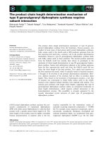
The results are summarised in Fig. 1.
During a period of 15 months a total of 351 patients met the
initial inclusion criteria. In 153 patients a probable cause of
fever was found on routine analysis. Therefore 198 patients
met the criteria for SXR. Patient data and the results of these
X-rays are shown in Table 1.
On the basis of the results of the SXR, sinus drainage was first
performed in those patients with evident abnormalities (n =
101). Drainage was performed in 98 of these 101 patients
within 12 hours (mean 2.05 ± 5.7 hours). Twenty-four patients
had been given platelets or fresh frozen plasma before the
procedure. In three patients the procedure was delayed for a
longer period because of the use of anticoagulants and/or
Figure 1
Flowchart depicting the organisation of our study in patients with fever of unknown origin (FUO), as well as the diagnostic work-up and culture resultsFlowchart depicting the organisation of our study in patients with fever of unknown origin (FUO), as well as the diagnostic work-up and culture
results.
Critical Care Vol 9 No 5 van Zanten et al.
R586
platelet aggregation inhibitors. In these patients the procedure
was performed within 48 hours.
Repeat drainage was performed in 41 patients after an aver-
age period of 52 ± 38 hours.
The initial (macroscopic) interpretation of the material
obtained during the draining procedure by the ENT surgeon
was pus in fluid from 17 of 101 patients (17%), purulent in 38
(38%), bloody in 13 (13%) and clear in 33 (33%). Culture
results of initial sinus drainage are shown in Table 2. Many
patients had more than one microorganism cultured from the
sinus fluid. A total of 140 microorganisms were cultured from
84 of these 101 sinus drainage fluids (84%). All cultures that
had been deemed as pus or purulent on macroscopic evalua-
tion turned out positive for pathogenic microorganisms. How-
ever, bacteria were also cultured from a substantial proportion
(18 of 33 (55%)) of the fluids that had been deemed clear on
microscopic inspection. The cultured pathogens are listed in
Table 2. The most predominant microorganisms in the sinus
fluids were Gram-negative pathogens such as Pseudomonas
and Klebsiella species.
Of the 28 patients with indeterminate or equivocal results on
the initial SXR, a repeat SXR was performed in 25 patients.
Ten (40%) now had obvious abnormalities, and drainage was
performed. Of these the diagnosis of sinusitis was confirmed
in 9 patients. Of the 69 patients with an initially normal SXR,
sinus drainage was nevertheless performed in the subsequent
72-hour period in 12 patients on clinical grounds (n = 2),
repeat SXR (n = 2) and following CT scan (n = 8). The diag-
nosis was confirmed by drainage and cultures in all 12 of
these patients. Thus, a total of 21 cases (22%) of microbio-
logical sinusitis were subsequently found in the group of 97
patients who initially had equivocal or normal findings on SXR.
On the basis of these clinical, radiological and microbiological
criteria we evaluated the final diagnoses in all 351 patients
with FUO initially included in our study. The results are shown
in Tables 3 and 4.
Discussion
The results of our study demonstrate that sinusitis is a fre-
quently occurring hospital-acquired infection in the ICU.
Sinusitis was initially diagnosed in 84 of 351 (24%) patients
with FUO, and in an additional 21 patients who had equivocal
or normal findings on initial SXR, giving a total of 105 of 351
patients (30%). This underestimates the true incidence
because SXRs were not taken in 153 patients who had obvi-
ous other causes of fever on initial screening, some of whom
might also have had sinusitis.
Sinusitis was the sole cause of fever in 57 patients (16%) and
one of several causes (for example sinusitis and purulent bron-
chitis) in 48 patients (14%). Pathogenic microorganisms were
cultured not only when material obtained by antral sinus punc-
ture was classified as 'purulent' but also in more than half of
the patients whose puncture material was less suspect on
macroscopic examination.
Previous studies on sinusitis in orotracheally intubated
patients have reported a lower incidence of sinusitis than was
observed in our study, ranging from 2% to 7.7% [5,7,25,26].
There are several possible reasons for this. First, the rate of
antibiotic resistance in the Netherlands is low, and antibiotics
are used relatively sparingly. This might have reduced the like-
lihood of undetected sinusitis being concomitantly treated
because patients were receiving antibiotics for other infec-
tions [25,27]. Second, our patients were more severely ill than
patients included in the previous studies, as demonstrated by
a high average severity of disease score (Acute Physiology
and Chronic Health Evaluation (APACHE)-II score of 21 ± 6.8
in our study, compared with Simplified Acute Physiology
Score (SAPS) II scores of 12 ± 4.5 [5] and 11.0 ± 3.5 [7];
other studies reported no severity scores). Third, risk factors
for sinusitis such as sedation and nasogastric tube feeding
were present more frequently in our patients, perhaps
because of the greater severity of disease.
Of the positive cultures in our patients, 77% contained Gram-
negative pathogens. This rate is higher than reported in previ-
ous studies, in which about 50% of cultured pathogens were
Table 1
Epidemiological data, results of SXR and macroscopic
evaluation of sinus fluids obtained by antral puncture
Parameter Value
Patient data (n = 351)
Sex (M:F) 193:158
Age (mean ± SD) 59 ± 21.2
APACHE II score (mean ± SD) 21 ± 6.8
ICU LOS (days) at diagnosis (mean ± SD) 5.9 ± 5.7
Results of sinus X-rays in patients with FUO (n = 198)
Sinus X-ray abnormal (two directions) 101 (51%)
Sinus X-ray equivocal 28 (14%)
Sinus X-ray normal 69 (35%)
Macroscopic evaluation of sinus fluid (n = 101)
Pus 17 (17%)
Purulent 38 (38%)
Bloody 13 (13%)
Clear 33 (33%)
APACHE, Acute Physiology and Chronic Health Evaluation; FUO,
fever of unknown origin; UCI LOS, length of stay in the intensive care
unit.
Available online />R587
Gram-positive [8,25,28,29]. This might be explained by differ-
ences in case mix, severity of illness and length of ICU stay, as
well as effects of previous antibiotic treatment on the patients'
microflora [25,28,29].
Our study has some limitations. The diagnosis was based on
abnormal findings on SXR and positive microbiological cul-
tures obtained after antral drainage. However, SXRs cannot
accurately distinguish purulent sinusitis from sterile fluids, so
abnormal SXRs may overestimate the incidence of sinusitis
[8,25]. Moreover, even positive microbiological cultures may
not prove clinically relevant sinusitis, because they may indi-
cate colonisation rather than actual infection. We tried to cir-
cumvent these problems by classifying only cultures with more
than 10 colonies of bacteria as positive and by basing our
diagnosis on a combination of radiological abnormalities, pos-
itive cultures, and clinical response to therapeutic measures
such as drainage and targeted antibiotic treatment. We are
therefore confident that our results accurately reflect the true
incidence of sinusitis.
Early detection and treatment is important because delays can
lead to the development of VAP, sepsis, and life-threatening
complications such meningitis, mastoiditis, intra-cranial
Table 2
Results of sinus fluid cultures of patients with gross abnormalities in their initial sinus X-ray
Bacterium Sinus fluid Same MO cultured from tracheal
aspirate
Same MO cultured from blood
Pseudomonas aeruginosa 32 22 8
Klebsiella oxytoca 531
Klebsiella pneumoniae 17 10 2
Enterococcus faecalis 10 2 1
Enterobacter cloacae 12 4 0
Escherichia coli 16 8 1
Staphylococcus aureus 831
Gram-positive mixture 11 - -
Gram-negative mixture 14 - -
Other
a
15 8 1
Total 140 60 15
There were 101 patients with gross abnormalities in their initial sinus X-ray. Positive cultures were obtained in 84 patients, with 140 different types
of microorganisms (MOs). Columns 3 and 4 show positive results of the same microorganisms (MOs) cultured from tracheal aspirate and blood,
cultured in the period between 24 hours before and 48 hours after sinus drainage.
a
Other pathogens included anaerobic bacteria (such as Bacteroides sp.) and fungi (Candida sp.).
Table 3
Initial diagnosis for fever of unknown origin in mechanically ventilated patients in intensive care unit
Cause of fever Sole cause One of multiple causes
Central line infection 43 1
Upper respiratory tract infection/pneumonia
a
93 42
Sinusitis 45 39
Abdominal focus 5 3
Other
b
21
Unknown 121
Total 188 86
Initial diagnosis was performed after initial screening with physical examination and chest X-ray in all 351 patients, sinus X-ray in 198 patients and
sinus drainage in 98 patients; cultures were not yet available. All patients had fever and leucocytosis.
a
Purulent tracheobronchial aspirate with
cultures positive for pathogenic microorganisms, combined with new or progressive pulmonary infiltrates on chest X-ray;
b
other causes of fever
included meningitis, phlebitis and deep venous thrombosis.
Critical Care Vol 9 No 5 van Zanten et al.
R588
abscesses and venous thrombosis of the sinus cavernosus
[24,30,31]. Early treatment of sinusitis may significantly
reduce the risk of VAP and perhaps also ICU mortality [8,32].
The results of our microbiological cultures underline the close
link between sinusitis and the development of VAP. Of 105
patients in whom positive sinus cultures were obtained, the
same microorganisms were cultured from bronchotracheal
aspirates in 40% of cases (n = 42). In some patients we were
able to demonstrate that positive sinus cultures preceded
positive cultures from the lungs, strongly suggesting that
sinusitis can lead to infections of the lower airway. Others
reported similar observations; for example, Holzapfel and co-
workers found that the early detection and treatment of hospi-
tal-acquired sinusitis could prevent the occurrence of VAP and
reduce mortality in nasotracheally intubated ICU patients [33].
Bacteraemia with the same microorganism as that cultured
from the sinus occurred in 12 patients; in five patients the
microorganism causing bacteraemia was cultured only from
the blood and the sinus, making sinusitis the most likely cause
of bacteraemia. However, no definite conclusions about cause
and effect can be drawn because bacteraemia can also lead
to sinusitis, with bacterial colonisation of sinus fluids following
bacteraemia [30,34].
Various mechanisms might explain the high incidence of
sinusitis in ICU patients. The first is anatomical. The paranasal
sinuses secrete mucus that flows to the natural ostia located
posteriorly towards the nasopharynx; this flow can be blocked
by infection, inflammation, anatomic abnormalities or the pres-
ence of foreign material such as nasotracheal intubation tubes.
Even tubes with smaller diameters (such as nasogastric feed-
ing and suction tubes) can cause significant obstruction in the
normal flow of sinus fluids, leading to an increased risk of bac-
terial colonisation and development of hospital-acquired
sinusitis [25,28]. The presence of nasogastric tubes has been
linked to a significant increase in the risk for sinusitis in
mechanically ventilated patients (odds ratio 14.1, 95% confi-
dence interval 1.7 to 117) [25]. Another important risk factor
is the use of sedatives (odds ratio 15.9, 95% confidence inter-
val 1.9 to 133.5) [25]. Underlying mechanisms may include
the suppression of normal cleansing mechanisms such as
coughing, sneezing and nose-blowing, because of sedation
and analgesia; in addition, immobility precludes positional
changes that improve mucous drainage under normal circum-
stances [24]. Remaining in a recumbent position can increase
nasal congestion and obstruction of the ostia of the maxillary
sinuses. This problem may be compounded by the positive
inspiratory and end-expiratory pressure in ventilated patients,
which also induces an increase in central venous pressure
[6,35]. In addition, critically ill patients recovering from earlier
episodes of sepsis may develop relative immune suppression,
so-called immunoparalysis [36].
ICU patients are often unable to communicate, and complaints
related to sinusitis may go unnoticed by the medical and nurs-
ing staff. Patients may have a 'runny nose', or discharge of
purulent material from the nasal cavity. However, this is seen
in only 27% of cases [37]. Thus elevations in white blood cell
count and/or FUO may be the first presenting symptoms [24].
In theory, the use of imaging modalities such as CT scans
[24,38,39] and B-mode ultrasound [12-14] could improve the
diagnostic yield. As discussed above, the CT scan should be
regarded as the gold standard for the diagnosis of sinusitis.
Unfortunately, CT scans are not easily performed in the ICU
setting, meaning that the patient has to be transported to the
department of radiology for this procedure. These in-hospital
transports can be risky [9,10,40,41]. The potential benefits in
establishing or confirming the diagnosis should therefore be
weighed against the risks of transport. The development of
mobile CT scans for use at bedside would significantly reduce
Table 4
Final diagnosis for FUO at ICU discharge, with final results of all cultures known
Cause of fever Sole cause One of multiple causes
a
Central line infection 44 11
Upper respiratory tract infection/pneumonia 132 58
Sinusitis 57 48
Abdominal focus 8 16
Other
b
12 28
Unknown 46
Total 253 161
See also Fig. 1.
a
Most patients with more than one cause of fever had sinusitis and bronchitis/pneumonia;
b
other causes of fever included
meningitis (not related to sinusitis), phlebitis and deep venous thrombosis.
Available online />R589
these problems; however, such devices are not yet available in
most hospitals.
Some authors have suggested that ultrasound may provide a
good or even better alternative to SXR for detecting sinusitis
at bedside in critically ill patients [12,13]. However, ultrasound
is not yet widely used for this purpose in routine clinical prac-
tice. Moreover, its diagnostic accuracy depends on the expe-
rience of the operator, and the costs are higher than for SXR.
In addition, the literature comparing diagnostic yields of ultra-
sound and SXR provides conflicting results [12,13,21,22,42].
Our study was not designed to compare the two techniques;
we based our choice mainly on the fact that ultrasound is not
yet widely used to detect sinusitis in the ICU setting, and on
our pre-existing clinical protocols. It seems unlikely that use of
ultrasound for initial screening would have significantly
affected our results; at best it could have increased our diag-
nostic yield, further strengthening our observation that sinusi-
tis is a frequent cause of FUO in ICU patients. In addition,
about 85% of the cases of hospital-acquired sinusitis associ-
ated with mechanical ventilation involve the maxillary sinuses
[6]. As conventional SXRs are most reliable in detecting max-
illary sinusitis (in comparison with frontal and ethmoidal sinusi-
tis) we feel that SXRs remain the most practical diagnostic
tool, with an acceptable sensitivity for detecting sinusitis in the
ICU setting. Hospitals favouring ultrasonography as initial
screening method could easily adapt our protocol, replacing
SXR by ultrasound. The CT scan remains the radiological gold
standard in the diagnosis of sinusitis.
On the basis of the results of our study we recommend that
hospital-acquired sinusitis be considered in all patients with
FUO in the ICU in whom a cause of fever is not immediately
apparent from initial examination and chest X-ray. SXR or ultra-
sound or (if possible) a CT scan should be included in the
diagnostic work-up, and sinus puncture with drainage should
be performed in case of abnormal or equivocal findings. In our
study all procedures were performed at thr bedside; 40% of
patients with confirmed sinusitis required repeat drainage, but
no patients required more than two procedures.
All nasal tubes should be removed if sinusitis is suspected;
antibiotics should be started empirically or based on Gram
staining, and adjusted for final culture results. In most patients
temperature normalises within 48 hours [37]; this was also
observed in our study. Radiological signs of sinusitis clear
more slowly but should disappear within ±1 week [43].
The results of our study have led to the implementation of sev-
eral measures to reduce the incidence of sinusitis. First,
nasogastric tubes are no longer used in intubated patients
unless it is expected that the endotracheal tube can be
removed within 24 hours. Gastric tubes in all other patients are
now inserted through the mouth. Second, patients intubated
for ≥24 hours now routinely receive topical administration of
saline 0.9% and/or decongestants such as xylometazoline
drops in the nasal cavities. Thirdly, the nursing staff keeps a far
more rigorous watch for signs of purulent nasal discharge in all
patients, and diagnostic procedures such as X-sinus are per-
formed if such discharge is observed. Finally, the routine diag-
nostic work-up in patients who develop fever in the ICU now
includes an SXR. Drainage (both as a diagnostic and thera-
peutic tool) takes place in all patients with clear or equivocal
signs of sinusitis. Topical decongestants are used to reduce
oedema and facilitate drainage. In patients with clear SXR in
whom no other diagnosis is established, SXR is repeated after
48 hours. These measures have led to a marked reduction in
the incidence of sinusitis in our ICU.
Conclusion
Hospital-acquired sinusitis is a frequent cause of FUO in oro-
tracheally intubated and mechanically ventilated critically ill
patients. ICU physicians should be aware of the numerous risk
factors for sinusitis simultaneously present in ICU patients and
take appropriate preventive measures. We recommend includ-
ing an SXR in the routine work-up for FUO in all ICU patients;
drainage should take place if SXR reveals clouding, and
should also be considered if the SXR is equivocal or difficult
to interpret. A normal SXR does not rule out sinusitis, and
when in doubt drainage or additional diagnostic procedures
such as CT scan should be performed.
Competing interests
The author(s) declare that they have no competing interests.
Authors' contributions
KHP, JMD and ARG designed and coordinated the study.
AvZ, RdB, JMD and KHP were involved in the collection, sta-
tistical analysis and interpretation of the data. MDN performed
literature analysis and assisted in the data collection. AvZ and
KHP drafted and revised the manuscript. All authors read and
approved the final manuscript.
Key messages
• Sinusitis is a frequent cause of FUO in the ICU (in this
study it was the sole cause in 16% and a contributing
factor in 13% of patients with FUO).
• Bacterial colonisation of the sinuses often precedes the
development of bronchitis and VAP; sinusitis may be a
frequent cause of hospital-acquired bronchitis and VAP.
• Diagnostic work-up of FUO should include an SXR,
ultrasound or CT scan; drainage should be performed if
any abnormalities are found.
• Physicians treating critically ill patients should be aware
of the high risk of sinusitis and take appropriate preven-
tive measures, including the removal of nasogastric
tubes in patients requiring long-term mechanical
ventilation.
Critical Care Vol 9 No 5 van Zanten et al.
R590
References
1. Chastre J, Fagon J: Ventilator-associated pneumonia. Am J
Respir Crit Care Med 2002, 165:867-903.
2. Polderman KH, Girbes AR: Central venous catheter use. Part 2:
Infectious complications. Intensive Care Med 2002, 28:18-28.
3. Anon: Hospital-acquired pneumonia in adults: diagnosis,
assessment of severity, initial antimicrobial therapy, and pre-
ventive strategies. A consensus statement. American Thoracic
Society, November 1995. Am J Respir Crit Care Med 1996,
153:1711-1725.
4. O'Grady NP, Alexander M, Dellinger EP, Gerberding JL, Heard
SO, Maki DG, Masur H, McCormick RD, Mermel LA, Pearson ML,
et al.: Guidelines for the prevention of intravascular catheter-
related infections. Centers for Disease Control and Prevention.
MMWR Recomm Rep 2002, 51:1-29.
5. Bach A, Boehrer H, Schmidt H, Geiss HK: Nosocomial sinusitis
in ventilated patients. Nasotracheal versus orotracheal
intubation. Anaesthesia 1992, 47:335-339.
6. Michelson A, Schuster B, Kamp HD: Paranasal sinusitis associ-
ated with nasotracheal and orotracheal long-term intubation.
Arch Otolaryngol Head Neck Surg 1992, 118:937-939.
7. Salord F, Gaussorgues P, Marti-Flich J, Sirodot M, Allimant C,
Lyonnet D, Robert D: Nosocomial maxillary sinusitis during
mechanical ventilation: a prospective comparison of orotra-
cheal versus the nasotracheal route for intubation. Intensive
Care Med 1990, 16:390-393.
8. Holzapfel L, Chastang C, Demingeon G, Bohe J, Piralla B, Coupry
A: A randomized study assessing the systematic search for
maxillary sinusitis in nasotracheally mechanically ventilated
patients. Influence of nosocomial maxillary sinusitis on the
occurrence of ventilator-associated pneumonia. Am J Respir
Crit Care Med 1999, 159:695-701.
9. Beckmann U, Gillies DM, Berenholtz SM, Wu AW, Pronovost P:
Incidents relating to the intra-hospital transfer of critically ill
patients. An analysis of the reports submitted to the Australian
Incident Monitoring Study in Intensive Care. Intensive Care
Med 2004, 30:1579-1585.
10. Smith I, Fleming S, Cernaianu A: Mishaps during transport from
the intensive care unit. Crit Care Med 1990, 18:278-281.
11. Waydhas C: Intrahospital transport of critically ill patients. Crit
Care 1999, 3:R83-R89.
12. Hilbert G, Vargas F, Valentino R, Gruson D, Chene G, Bebear C,
Gbikpi-Benissan G, Cardinaud JP: Comparison of B-mode ultra-
sound and computed tomography in the diagnosis of maxillary
sinusitis in mechanically ventilated patients. Crit Care Med
2001, 29:1337-1342.
13. Puidupin M, Guiavarch M, Paris A, Caroff P, Boutin JP, Le Bivic T,
Garcia JF: B-mode ultrasound in the diagnosis of maxillary
sinusitis in intensive care unit. Intensive Care Med 1997,
23:1174-1175.
14. Lichtenstein D, Biderman P, Meziere G, Gepner A: The 'sinuso-
gram', a real-time ultrasound sign of maxillary sinusitis. Inten-
sive Care Med 1998, 24:1057-1061.
15. Lucchin F, Minicuci N, Ravasi MA, Cordella L, Palu M, Cetoli M,
Borin P: Comparison of A-mode ultrasound and computed
tomography: detection of secretion in maxillary and frontal
sinuses in ventilated patients. Intensive Care Med 1996,
22:1265-1268.
16. Haapaniemi J, Laurikainen E: Ultrasound and antral lavage in the
examination of maxillary sinuses. Rhinology 2001, 39:39-42.
17. Pfleiderer AG, Drake-Lee AB, Lowe D: Ultrasound of the
sinuses: a worthwhile procedure? A comparison of ultrasound
and radiography in predicting the findings of proof puncture
on the maxillary sinuses. Clin Otolaryngol Allied Sci 1984,
9:335-339.
18. Jensen C, von Sydow C: Radiography and ultrasonography in
paranasal sinusitis. Acta Radiol 1987, 28:31-34.
19. Trigaux JP, Bertrand BM, Van Beers BE: Comparison of B-mode
ultrasound, radiography and sinuscopy in the diagnosis of
maxillary sinusitis. Report on 177 cases. Acta Otorhinolaryngol
Belg 1988, 42:670-679.
20. Shapiro GG, Furukawa CT, Pierson WE, Gilbertson E, Bierman
CW: Blinded comparison of maxillary sinus radiography and
ultrasound for diagnosis of sinusitis. J Allergy Clin Immunol
1986, 77:59-64.
21. Varonen H, Makela M, Savolainen S, Laara E, Hilden J: Compari-
son of ultrasound, radiography, and clinical examination in the
diagnosis of acute maxillary sinusitis: a systematic review. J
Clin Epidemiol 2000, 53:940-948.
22. Engels EA, Terrin N, Barza M, Lau J: Meta-analysis of diagnostic
tests for acute sinusitis. J Clin Epidemiol 2000, 53:852-862.
23. European Academy of Allergology and Clinical Immunology: Euro-
pean position paper on rhinosinusitis and nasal polyps. Rhinol
Suppl 2005, 18:1-87.
24. Seiden AM: Sinusitis in the critical care patient. New Horiz
1993, 1:261-270.
25. George DL, Falk PS, Umberto Meduri G, Leeper KV Jr, Wunderink
RG, Steere EL, Nunnally FK, Beckford N, Mayhall CG: Nosoco-
mial sinusitis in patients in the medical intensive care unit: a
prospective epidemiological study. Clin Infect Dis 1998,
27:463-470.
26. Borman KR, Brown PM, Mezera KK, Jhaveri H: Occult fever in sur-
gical intensive care unit patients is seldom caused by sinusitis.
Am J Surg 1992, 164:412-415.
27. Cars O, Molstad S, Melander A: Variation in antibiotic use in the
European Union. Lancet 2001, 357:1851-1853.
28. Vandenbussche T, De Moor S, Bachert C, Van Cauwenberge P:
Value of antral puncture in the intensive care patient with fever
of unknown origin. Laryngoscope 2000, 110:1702-1706.
29. Bert F, Lambert-Zechovsky N: Microbiology of nosocomial
sinusitis in intensive care unit patients. J Infect 1995, 31:5-8.
30. Bos AP, Tibboel D, Hazebroek FW, Hoeve H, Meradji M, Molenaar
JC: Sinusitis: hidden source of sepsis in postoperative pediat-
ric intensive care patients. Crit Care Med 1989, 17:886-888.
31. O'Reilly MJ, Reddick EJ, Black W, Carter PL, Erhardt J, Fill W,
Maughn D, Sado A, Klatt GR: Sepsis from sinusitis in nasotra-
cheally intubated patients. A diagnostic dilemma. Am J Surg
1984, 147:601-604.
32. Talmor M, Li P, Barie PS: Acute paranasal sinusitis in critically
ill patients: guidelines for prevention, diagnosis and treatment.
Clin Infect Dis 1997, 25:1441-1446.
33. Holzapfel L, Chevret S, Madinier G, Ohen F, Demingeon G, Cou-
pry A, Chaudet M: Influence of long-term oro- or nasotracheal
intubation on nosocomial maxillary sinusitis and pneumonia:
results of a prospective, randomized, clinical trial. Crit Care
Med 1993, 21:1132-1138.
34. Aebert H, Hunefeld C, Regel G: Paranasal sinusitis and sepsis
in ICU patients with nasotracheal intubation. Intensive Care
Med 1988, 15:27-30.
35. Aust R, Drettner B: The patency of the maxillary ostium in rela-
tion to body posture. Acta Otolaryngol 1975, 80:443-446.
36. Adrie C, Pinsky MR: The inflammatory balance in human
sepsis. Intensive Care Med 2000, 26:364-375.
37. Caplan ES, Hoyt NJ: Nosocomial sinusitis. JAMA 1982,
247:639-641.
38. Osguthorpe JD, Hadley JA: Rhinosinusitis. Current concepts in
evaluation and management. Med Clin North Am 1999,
83:27-41.
39. Zinreich SJ: Rhinosinusitis: radiologic diagnosis. Otolaryngol
Head Neck Surg 1997, 117:S27-S34.
40. Szem JW, Hydo LJ, Fischer E, Kapur S, Klemperer J, Barie PS:
High-risk intrahospital transport of critically ill patients: safety
and outcome of the necessary 'road trip'. Crit Care Med 1995,
23:1660-1666.
41. Evans A, Winslow EK: Oxygen saturation and hemodynamic
response in critically ill, mechanically ventilated adults during
intrahospital transport. Am J Crit Care 1995, 4:106-111.
42. de Bock GH, Houwing-Duistermaat JJ, Springer MP, Kievit J, van
Houwelingen JC: Sensitivity and specificity of diagnostic tests
in acute maxillary sinusitis determined by maximum likelihood
in the absence of an external standard. J Clin Epidemiol 1994,
47:1343-1352.
43. Deutschman CS, Wilton P, Sinow J, Dibbell D Jr, Konstantinides
FN, Cerra FB: Paranasal sinusitis associated with nasotracheal
intubation: a frequently unrecognized and treatable source of
sepsis. Crit Care Med 1986, 14:111-114.