Báo cáo khoa học: "Circulating anions usually associated with the Krebs cycle in patients with metabolic acidosis" potx
Bạn đang xem bản rút gọn của tài liệu. Xem và tải ngay bản đầy đủ của tài liệu tại đây (204.9 KB, 5 trang )
Open Access
Available online />R591
Vol 9 No 5
Research
Circulating anions usually associated with the Krebs cycle in
patients with metabolic acidosis
Lui G Forni
1
, William McKinnon
2
, Gwyn A Lord
3
, David F Treacher
4
, Jean-Marie R Peron
5
and
Philip J Hilton
6
1
Consultant Physician & Intensivist, Department of Critical Care, Worthing Hospital, Worthing, West Sussex, UK
2
Research Fellow, Renal Laboratory, St Thomas' Hospital, London, UK
3
MRC Scientist, MRC Toxicology Unit, Birkbeck College, London, UK
4
Consultant Physician & Intensivist, Renal Laboratory, St Thomas' Hospital, London, UK
5
Research Fellow, Department of Chemistry, Kingston University, Surrey, UK
6
Consultant Physician & Research Director, Renal Laboratory, St Thomas' Hospital, London, UK
Corresponding author: Lui G Forni,
Received: 29 Jun 2005 Revisions requested: 22 Jul 2005 Revisions received: 1 Aug 2005 Accepted: 12 Aug 2005 Published: 13 Sep 2005
Critical Care 2005, 9:R591-R595 (DOI 10.1186/cc3806)
This article is online at: />© 2005 Forni et al.; licensee BioMed Central Ltd.
This is an Open Access article distributed under the terms of the Creative Commons Attribution License ( />2.0), which permits unrestricted use, distribution, and reproduction in any medium, provided the original work is properly cited.
Abstract
Introduction Acute metabolic acidosis of non-renal origin is
usually a result of either lactic or ketoacidosis, both of which are
associated with a high anion gap. There is increasing
recognition, however, of a group of acidotic patients who have a
large anion gap that is not explained by either keto- or lactic
acidosis nor, in most cases, is inappropriate fluid resuscitation
or ingestion of exogenous agents the cause.
Methods Plasma ultrafiltrate from patients with diabetic
ketoacidosis, lactic acidosis, acidosis of unknown cause, normal
anion gap metabolic acidosis, or acidosis as a result of base
loss were examined enzymatically for the presence of low
molecular weight anions including citrate, isocitrate, α-
ketoglutarate, succinate, malate and d-lactate. The results
obtained from the study groups were compared with those
obtained from control plasma from normal volunteers.
Results In five patients with lactic acidosis, a significant
increase in isocitrate (0.71 ± 0.35 mEq l
-1
), α-ketoglutarate
(0.55 ± 0.35 mEq l
-1
), malate (0.59 ± 0.27 mEq l
-1
), and d-
lactate (0.40 ± 0.51 mEq l
-1
) was observed. In 13 patients with
diabetic ketoacidosis, significant increases in isocitrate (0.42 ±
0.35 mEq l
-1
), α-ketoglutarate (0.41 ± 0.16 mEq l
-1
), malate
(0.23 ± 0.18 mEq l
-1
) and d-lactate (0.16 ± 0.07 mEq l
-1
) were
seen. Neither citrate nor succinate levels were increased.
Similar findings were also observed in a further five patients with
high anion gap acidosis of unknown origin with increases in
isocitrate (0.95 ± 0.88 mEq l
-1
), α-ketoglutarate (0.65 ± 0.20
mEq l
-1
), succinate (0.34 ± 0.13 mEq l
-1
), malate (0.49 ± 0.19
mEq l
-1
) and d-lactate (0.18 ± 0.14 mEq l
-1
) being observed but
not in citrate concentration. In five patients with a normal anion
gap acidosis, no increases were observed except a modest rise
in d-lactate (0.17 ± 0.14 mEq l
-1
).
Conclusion The levels of certain low molecular weight anions
usually associated with intermediary metabolism were found to
be significantly elevated in the plasma ultrafiltrate obtained from
patients with metabolic acidosis. Our results suggest that these
hitherto unmeasured anions may significantly contribute to the
generation of the anion gap in patients with lactic acidosis and
acidosis of unknown aetiology and may be underestimated in
diabetic ketoacidosis. These anions are not significantly
elevated in patients with normal anion gap acidosis.
Introduction
Metabolic acidosis is a common presentation in acute medi-
cine and in only a minority of cases can renal failure alone be
considered the sole cause of the acidosis. Diabetic ketoacido-
sis (DKA) and 'classic' lactic acidosis (taken as a blood lactate
in excess of 5 mmol l
-1
) account for most of the remaining
cases [1,2]. Amongst clinicians there is an increasing aware-
ness that there is often an important discrepancy between the
measured acidosis and the associated base deficit, suggest-
ing that other anions must contribute to the generation of the
DKA = diabetic ketoacidosis.
Critical Care Vol 9 No 5 Forni et al.
R592
anion gap [3,4]. Further evidence to support the hypothesis of
additional 'missing anions' is supplied by observations on
patients receiving bicarbonate-buffered haemofiltration for
treatment of lactic acidosis where it is possible to calculate the
net rates of lactate removal and bicarbonate donation by the
haemofilter [5]. In patients whose blood pH is slowly falling
(thereby excluding any possible confounding effect of other
blood buffers), it is not uncommon for the rate of bicarbonate
donation to exceed the rate of lactate removal by as much as
twofold (PJ Hilton, unpublished observations). This implies
that not only lactic acid, but other 'unknown' acids are being
neutralised by the bicarbonate in the haemofiltration replace-
ment fluid.
Preliminary experiments performed in this laboratory with high
performance liquid chromatography coupled to mass spec-
trometry on plasma ultrafiltrate obtained from patients with
metabolic acidosis had suggested that anions principally
associated with the Krebs tricarboxylic acid cycle were signif-
icantly elevated in patients with unexplained metabolic acido-
sis. We undertook an enzymatic study of anions of the Krebs
cycle in patients with metabolic acidosis whose standard base
deficit was 8 mmol l
-1
or greater. The samples were obtained
from consecutive patients who fulfilled these requirements
except those with a normal anion gap, which were collected
subsequently. The results obtained from the patient samples
were compared to control values obtained from healthy
volunteers.
Methods
This study was approved by the Ethics Committee of Guy's
and St Thomas' National Health Service Trust (reference
number EC03/104). Prior to the sample being taken, informed
consent was obtained from the subject or, where this was not
possible, their next of kin. Patient studies were undertaken on
15 ml of arterial blood taken from arterial cannulae in patients
with metabolic acidosis whose standard base deficit was 8
mmol l
-1
or greater. The arterial blood was drawn into a non-
heparinised syringe before being rapidly transferred into SST
II (KODAK) Vaccutainers (BD Vaccutainer Systems Ltd, Ply-
mouth, UK). Control samples were obtained from venous
blood of laboratory workers and treated in the same manner as
that obtained from patients. Arterial blood gas levels were not
measured as it was felt that arterial puncture was
inappropriate.
Once obtained, the sample was chilled and rapidly trans-
ported to the laboratory. The plasma was isolated by centrifu-
gation of the Vaccutainers (1,500 g) at 4°C for 10 minutes.
The plasma was transferred to an Amicon 30,000 Da cutoff fil-
ter (Millipore, Watford, Herts, UK) where centrifugation at
1,560 g for 15 minutes produced ultrafiltrate. The generated
ultrafiltrate was either immediately analysed or stored at -20°C
for analysis within 24 h. Previous work had highlighted the
need for rapid assay of the samples due to an observed rapid
decrease in concentrations of the measured anions within
plasma.
The concentration of anions in the ultrafiltrate was determined
by enzymatic assay with reference to internal standards. The
plasma ultrafiltrate concentrations of citrate, succinate, malate,
d-lactate and l-lactate anions were estimated using commer-
cially available kits (Roche, Glasgow, UK). The levels of isoci-
trate and α-ketoglutarate were measured using enzyme assays
developed by ourselves using isocitrate dehydrogenase and
α-ketoglutarate dehydrogenase, respectively, and their associ-
ated co-factors (Sigma Chemicals, Poole, UK). All the enzy-
matic assays relied upon the interconversion of NAD
+
and
NADH or NADP
+
and NADPH and used the change in absorb-
ance due to the reduced co-enzyme at 340 nm. Oxaloacetate
concentrations could not be accurately measured as a result
of its short half-life (approximately 69 s) in aqueous systems at
near physiological pH [6]. All data are presented as mean ±
standard deviation. Unless stated otherwise, the data are nor-
mally distributed and statistical analysis was undertaken with
an unpaired t-test. Where the data was not normally distrib-
uted a Mann-Whitney non-parametric test was applied. In both
cases, significance was deemed to have been attained if p
was ≤0.05
Results
Samples were obtained from 28 patients in total and from 12
control subjects. Of the study patients, 13 had DKA at the time
of blood sampling and 5 had a lactic acidosis (defined as a
blood lactate concentration in excess of 5 mmol/l in the pres-
ence of acidosis). Five patients had a metabolic acidosis that
could not be ascribed to either lactic acidosis, ketoacidosis or
exogenous agents. A further five patients had an acidosis as a
result of gastrointestinal or renal ion losses. In all cases, the
patients were acidotic with an average pH of 7.18 (±0.11) and
a base deficit of 13.4 (±4.7) mmol l
-1
.
The mean results obtained for the plasma ultrafiltrate concen-
tration of citrate, succinate, malate, d-lactate, l-lactate, isoci-
trate and α-ketoglutarate are presented in Table 1. The mean
results obtained from the data together with the maximal value
for each anion measured in each type of acidosis are shown in
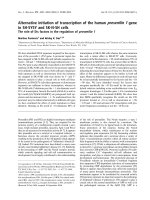
Fig. 1.
Patients with DKA showed significant increases relative to the
control values in isocitrate, α-ketoglutarate, malate and d-lac-
tate levels. The concentrations of both citrate and succinate
did not differ significantly from controls. Patients with lactic
acidosis showed significant increases relative to the control
values in citrate, isocitrate, α-ketoglutarate, succinate, malate
and d-lactate levels. Patients with acidosis of unknown origin
showed significant increases in the concentrations of isoci-
trate, α-ketoglutarate, succinate, malate and d-lactate. The
level of citrate did not differ significantly from that of the con-
trols. In those patients with a normal anion gap acidosis, the
Available online />R593
levels of citrate, isocitrate, α-ketoglutarate, succinate and
malate did not differ significantly from the control values. The
concentration of d-lactate was significantly raised compared
to control values in this patient group.
Discussion
The consequences of metabolic acidosis can be catastrophic
and a considerable body of literature highlights the poor out-
look in patients where lactic acid is the principal component of
the acidaemia [7-9]. This increase in blood lactate concentra-
tion reflects either increased lactate production, reduced lac-
tate metabolism or, more commonly, a combination of the two
[10]. In patients with DKA, 3-hydroxybutyric acid and, to a
lesser extent, acetoacetic acid play the major role in the gen-
eration of the anion gap. As outlined, however, in a third patient
group, neither lactate nor 3-hydroxybutyrate is responsible for
the elevated anion gap and the relevant anions responsible
remain unknown [11]. In the fourth patient group, acidosis is
generated as a result of uncontrolled electrolyte loss either
from the kidney (renal tubular acidosis) or the gut.
We have shown that the plasma concentrations of acids usu-
ally associated with the Krebs tricarboxylic acid cycle are sig-
nificantly increased in patients with lactic acidosis as well as
those with 'unexplained acidosis' with normal or near normal
blood lactate concentrations. In DKA, although the concentra-
tions of these acids are less strikingly elevated, they are still
abnormal in the majority of patients when compared to con-
trols. They are not, however, significantly elevated in patients
with normal anion gap acidosis secondary to excess base loss.
The accumulation of such acids may contribute significantly to
the production of the anion gap and account, in part, for the
'missing' anions in patients with certain forms of acidosis.
Recent studies, in keeping with previous work, have demon-
strated the predictive value of acid-base variables on outcome
in the critically ill [12]. Furthermore, the calculation of unmeas-
ured anions appears to be a better discriminator of outcome
than lactate or base deficit [13].
Table 1
Concentrations of measured anions in plasma (µEq l
-1
)
Acid Patient group
Diabetic ketoacidosis Lactic acidosis Unknown origin Normal anion gap Controls
Mean SD p Mean SD p Mean SD p Mean SD p Mean SD
Citrate 454.02 194.11 ns 1,453.13 513.95 <0.01 335.63 69.82 ns
a
239.1 105.1 ns 448.55 119.80
Isocitrate 421.93 352.56 0.02 704.6 347.6 <0.01 949.16 883.22 <0.01
a
84.48 69.6 ns
a
60.97 31.31
α-Ketoglutarate 413.41 158.48 <0.01
a
547.72 344.98 <0.01
a
651.51 203.06 <0.01
a
72.97 67.34 ns
a
79.17 106.74
Succinate 181.1 173.24 ns 358.27 112.49 <0.01
a
340.04 128.74 0.02
a
125.89 73.57 ns
a
90.29 49.97
Malate 229.81 181.87 <0.01
a
593.65 265.88 <0.01 485.16 189.67 <0.01
a
95.07 117.59 ns 59.82 32.94
d-Lactate 157.34 67.85 <0.01 397.69 511.15 <0.01
a
176.49 135.21 <0.01 69.27 55.39 <0.01 35.63 18.42
For each group, statistical analysis is presented relative to control value.
a
Mann-Whitney non-parametric test. ns, not significant; SD, standard
deviation.
Figure 1
The concentration of various weak acids grouped by their underlying aetiology (mean ± upper range)The concentration of various weak acids grouped by their underlying
aetiology (mean ± upper range). DKA, diabetic ketoacidosis; NAG, nor-
mal anion gap.
Critical Care Vol 9 No 5 Forni et al.
R594
With the partial exception of citrate and isocitrate (97% ion-
ised at pH 7.0) the anions examined in this study are effectively
fully ionised at the measured pH. Unlike lactate, not all the ani-
ons are monobasic, with tribasic acids (citric and isocitric)
contributing three protons, whilst the dibasic acids (α-ketogl-
utaric, malic and succinic) add two protons to the solution on
ionisation. Converting the concentrations of these observed
anions to mEq l
-1
(Table 1) shows that, on average, the contri-
bution to the observed anion gap due to such anions may be
in excess of 3 mEq l
-1
and in some cases may be over 5 mEq
l
-1
. Thus, the contribution of these anions to the generation of
the anion gap is of much greater significance than is apparent
from their molarity. The large standard deviation present in
these samples probably reflects their heterogeneous nature
and, in many ways, the range and maxima in each group are of
as much interest as the means, demonstrating the extreme
ranges that can be present in patients with metabolic acidosis
(Fig. 1).
The greatest deviations in the level of measured plasma ultra-
filtrate acids from that observed in the control group were seen
in the patients within the lactic acidosis and 'unexplained aci-
dosis' groups. In these two groups, the concentrations of most
of the acids studied were present in the plasma ultrafiltrate at
a concentration significantly higher than that observed in the
normal control population. In those with DKA, four of the six
acids measured were significantly elevated relative to the con-
trol values. Interestingly, in normal anion gap acidosis, only d-
lactate was significantly increased relative to the control
values.
It has been widely reported elsewhere that plasma citrate is
not elevated in acidosis [4,11] and we observed this in all the
groups of patients studied except those with lactic acidosis. In
this group, significantly elevated levels of citrate were
observed in comparison to the normal control values. This
result may be unreliable as four of the patients in this group
had received an infusion of heparin (containing sodium citrate
as an anticoagulant) prior to the blood sample being obtained.
Isocitrate concentrations were significantly elevated in
patients with ketoacidosis, lactic acidosis and those whose
acidosis was of unknown origin. Consequently, the citrate:iso-
citrate ratio is significantly reduced compared to control values
and we are unable to advance a simple explanation as to why
this ratio should be so low.
Although it seems reasonable to believe that the likely source
for the generation of these observed anions is the mitochon-
dria, we have no direct evidence for this and the results may
indeed reflect compartmentalised oxidative and glycolytic
energy production. Indeed, studies on mitochondria in isolated
rat skeletal muscle have demonstrated that lactic acidosis has
differential effects on actively phosphorylating and non-phos-
phorylating mitochondria, suggesting that the effect of acidae-
mia may depend on local physiological conditions [14]. The
rate of oxygen delivery to respiring tissue may also play a role
in generating intermediates of Krebs acids, with several
authors suggesting that hypoxia can cause an increase in
intermediates of the citric acid cycle [15-18], although in the
patients examined in this study none were significantly hypoxic
at the time the sample was taken. Furthermore, it seems
unlikely that the acidaemia per se is responsible for the
increased levels of Krebs intermediates within our patient pop-
ulation given the normal values found in the patients with nor-
mal anion gap acidosis and the lesser elevation seen in the
patients with DKA. An alternative explanation may be the gen-
eration of intermediates from anaplerotic pathways of metabo-
lism, which may reflect enhanced protein catabolism in these
patients
Other authors have proposed alternative hypotheses to
explain how the 'missing' ions in the elevated anion gap may
be generated. These include the choice of fluid used for resus-
citation, the effects of hypoproteinaemia or the presence of
other metabolites [19-21], in addition to various 'physical
chemical' approaches, including the calculation of the strong
anion difference [22]. This approach, which enjoys some pop-
ularity at the present time, represents an alternative way to
express the principles of acidosis. The 'strong ion difference'
model can be thought of as a restatement of the older concept
of buffer base and thus, predictably, must approach the cor-
rected anion gap, although some of its stronger proponents
suggest that it can explain the generation of acidosis per se.
We prefer to adhere to the concept that acidosis can best be
regarded as an excess of protons over those normally found in
physiological states, caused either from the loss of base or
excessive net proton production.
We have previously referred to the difficulties of measuring
plasma oxaloacetate in view of its extremely short half-life in
aqueous solution. Given the increases in plasma concentra-
tions of the other components of the Krebs cycle and the
known existence of cytosolic oxaloacetate, it seems likely that
some oxaloacetate does enter the plasma of these acidotic
patient groups along with the other acids described here.
There, it would spontaneously decarboxylate to pyruvate and
its presence might only be inferred as a small deviation of the
pyruvate:lactate ratio from that predicted on the basis of pH
alone.
The results obtained from this study suggest that the role of
anions principally associated with the Krebs cycle may be
greater than previously thought in the generation of the anion
gap in 'classic' lactic acidosis. In addition, these anions appear
to have a significant role in the generation of the anion gap in
patients with acidosis of unknown cause.
Conclusion
We have shown that the concentration of anions normally
associated with the Krebs tricarboxylic acid cycle are elevated
Available online />R595
in appreciable quantities in patients with a metabolic acidosis.
We propose that they may play a significant role in generating
the anion gap. Further work in this laboratory is currently
underway to explore the clinical implications of these findings.
Competing interests
This work was supported by The Special Trustees for St Tho-
mas' Hospital, London. The authors have no financial interests
relevant to the results of this research, nor are there any other
circumstances that could potentially provoke a conflict of
interest.
Authors' contributions
LGF conceived the study, participated in its design, collected
patient samples and drafted the manuscript. WM developed
and performed the enzyme assays, participated in the design
of the study, performed the statistical analysis and helped to
draft the manuscript. GAL participated in the design of the
study, performed the initial mass spectrometry and helped in
assay development. DFT collected patient samples and partic-
ipated in study design. JMP helped in assay development and
stability studies. PJH conceived the study, participated in its
design, collected patient samples, helped in assay develop-
ment and helped to draft the manuscript. All authors read and
approved the final manuscript.
References
1. Mizock BA, Falk JL: Lactic acidosis in critical illness. Crit Care
Med 1992, 20:80-93.
2. Cohen RD, Woods HF: Lactic acidosis revisited. Diabetes 1983,
32:181-191.
3. Van Lambalgen AA, Bronsveld W, Van Den Bos GC, Thijs LG:
Distribution of cardiac output, oxygen consumption and lac-
tate production in canine endotoxin shock. Cardiovasc Res
1984, 18:195-205.
4. Rackow EC, Mecher C, Astiz ME, Goldstein C, McKee D, Weil
MH: Unmeasured anion during severe sepsis with metabolic
acidosis. Circ Shock 1990, 30:107-115.
5. Wright DA, Forni LG, Carr P, Treacher DF, Hilton PJ: Use of con-
tinuous haemofiltration to assess the rate of lactate metabo-
lism in acute renal failure. Clin Sci (Lond) 1996, 90:507-510.
6. Tsai CS: Spontaneous decarboylation of oxalacetic acid.
Canadian J Chem 1967, 45:873-880.
7. Stacpoole PW, Wright EC, Baumgartner TG, Bersin RM, Bucha-
lter S, Curry SH, Duncan C, Harman EM, Henderson GN, Jenkin-
son S, et al.: Natural history and course of acquired lactic
acidosis in adults. The DCA-Lactic Acidosis Study Group . Am
J Med 1994, 97:47-54.
8. Weil MH, Afifi AH: Experimental and clinical studies on lactate
and pyruvate as indicators of the severity of acute circulatory
failure (shock). Circulation 1970, 41:989-1001.
9. Smith I, Kumar P, Molloy S, Rhodes A, Newman PJ, Grounds RM,
Bennett ED: Base excess and lactate as prognostic indicators
for patients admitted to intensive care. Intensive Care Med
2001, 27:74-83.
10. Hilton PJ, Taylor J, Forni LG, Treacher DF: Bicarbonate-based
haemofiltration in the management of acute renal failure with
lactic acidosis. QJM 1998, 91:279-283.
11. Mecher C, Rackow EC, Astiz ME, Weil MH: Unaccounted for
anion in metabolic acidosis during severe sepsis in humans.
Crit Care Med 1991, 19:705-711.
12. Haterwill M, Waggie Z, Purves L, Reynolds L, Argent A: Correc-
tion of the anion gap for albumin in order to detect occult tis-
sue anions in shock. Arch Dis Child 2002, 87:526-529.
13. Kaplan LJ, Kellum JA: Initial pH, base deficit, lactate, anion gap,
strong ion difference, and strong ion gap predict outcome
from major vascular injury. Crit Care Med 2004, 32:1120-1124.
14. Tonkonogi M, Sahlin K: Actively phosphorylating mitochondria
are more resistant to lactic acidosis than inactive
mitochondria. Am J Physiol 1999, 277:C288-C293.
15. Peuhkurinen KJ, Takala TES, Nuutinen EM, Hassinen IE: Tricarbo-
xylic acid cycle metabolites during ischemia in isolated per-
fused rat heart. Am J Physiol 1983, 244:H281-H288.
16. Folbergrova J, Ljunggren B, Norberg K, Siesjo BK: Influence of
complete ischemia on glycolytic metabolites citric acid cycle
intermediates and associated amino acids in the rat cerebral
cortex. Brain Res 1974, 80:265-279.
17. Hassel B, Ilebekk A, Tonnessen T: Cardiac accumulation of cit-
rate during brief myocardial ischaemia and reperfusion in the
pig in vivo. Acta Physiol Scand 1998, 164:53-59.
18. Taegtmeyer H: Metabolic responses to cardiac hypoxia.
Increased production of succinate by rabbit papillary muscles.
Circ Res 1978, 43:808-815.
19. Morgan TJ: The meaning of acid-base abnormalities in the
intensive care unit: part III - effects of fluid administration. Crit
Care 2005, 9:204-211.
20. Rossing TH, Maffeo N, Fencl V: Acid-base effects of altering
plasma protein concentration in human blood in vitro. J Appl
Physiol 1986, 61:2260-2265.
21. Kellum JA: Closing the gap on unmeasured anions. Crit Care
2003, 7:219-220.
22. Stewart PA: Modern quantitative acid-base chemistry. Can J
Physiol Pharmacol 1983, 61:1444-1461.
Key messages
• Low molecular weight anions usually associated with
intermediary metabolism are significantly elevated in the
plasma ultrafiltrate obtained from patients with meta-
bolic acidosis.
• These anions may contribute significantly to the ele-
vated anion gap observed in patients with metabolic aci-
dosis, in particular those of unknown aetiology.