Báo cáo khoa học: "Lactate as a marker of energy failure in critically ill patients: hypothes" potx
Bạn đang xem bản rút gọn của tài liệu. Xem và tải ngay bản đầy đủ của tài liệu tại đây (157.66 KB, 6 trang )
588
DO
2
= oxygen delivery; VO
2
= oxygen consumption.
Critical Care December 2005 Vol 9 No 6 Valenza et al.
Abstract
Lactate measurement in the critically ill has been traditionally used
to stratify patients with poor outcome. However, plasma lactate
levels are the result of a finely tuned interplay of factors that affect
the balance between its production and its clearance. When the
oxygen supply does not match its consumption, organisms such as
man who are forced to produce ATP for their integrity adapt in
many different ways up to the point when energy failure occurs.
Lactate, being part of the adaptive response, may then be used to
assess the severity of the supply/demand imbalance. In such a
scenario, the time to intervention becomes relevant: early and
effective treatment may allow the cell to revert to a normal state, as
long as the oxygen machinery (i.e. mithocondria) is intact.
Conversely, once the mithocondria are deranged, energy failure
occurs even in the presence of normoxia. The lactate increase in
critically ill patients may therefore be viewed as an early marker of a
potentially reversible state.
Lactate in critical illness
The normal reference values for lactate are traditionally
considered 1 ± 0.5 mmol/l in normal patients and <2 mmol/l
in critically ill patients [1]. Since 1975, values above 2 mmol/l
but lower than 5 mmol/l have been separated from values
above 5 mmol/l, associated with acidemia, as different clinical
entities – referring to hyperlactatemia states in the former
situation as opposed to lactic acidosis in the latter situation
[2]. A further stratification, initially proposed by Cohen in
1976 [3], has been subsequently used according to the
presence (type A) or absence (type B) of ‘evident’ causes of
tissue hypoxia to explain the underlying cause of increased
lactate. Over the years, however, more sophisticated means
of assessing regional and even local perfusion have changed
the aforementioned classification into a more perfusion-
oriented vision. In fact, our increased ability to assess tissue
oxygenation clearly implies that measured plasma lactate
concentration is only a small window of a much more
complicated scenario.
Lactate as a clinical marker of hypoxia
As will be described, lactate is one of the intermediate
products that increase as a consequence of the rearrange-
ment of metabolism during hypoxia. As such, lactate has been
widely considered a marker of tissue hypoxia. There are
several examples of the increase of lactate in hypoxic
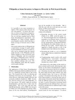
conditions [4,5]. Figure 1 shows our own experimental results.
The lactate increase is very fast, a matter of minutes, and is
proportional to the defect in the oxidative metabolism [6].
However, measured plasma lactate is the balance between
production and clearance. Liver failure does in fact influence
the kinetics of lactate increase [7].
It is also worth note that lactate is an intermediate compound
of normal metabolism. Erythrocytes, for instance, which are
equivalent to an organ weighing approximately 2500 g, are
obligatory anaerobes and ‘breathe’ via the lactate they
produce that is recycled from fatty acid oxidation in the liver.
Lactate, in this case, may be considered an energy shuttle
rather than a waste compound [8].
Outcome and stratification of severity
Lactate has been used as a marker in critically ill patients
since 1964 [9]. In 1970, Weil and Afifi clearly showed the
relationship between lactate concentration and outcome [10].
Several authors have subsequently confirmed those results
[11-15]. Interestingly, lactate measurements have also been
used to stratify patients. In fact, plasma lactate may be used as
Review
Lactate as a marker of energy failure in critically ill patients:
hypothesis
Franco Valenza, Gabriele Aletti, Tommaso Fossali, Giorgio Chevallard, Francesca Sacconi,
Manuela Irace and Luciano Gattinoni
Istituto di Anestesia e Rianimazione, Università degli Studi di Milano, Ospedale Maggiore Policlinico, Mangiagalli e Regina Elena – Fondazione IRCCS
di Natura Pubblica, Milan, Italy
Corresponding author: Franco Valenza,
Published online: 28 September 2005 Critical Care 2005, 9:588-593 (DOI 10.1186/cc3818)
This article is online at />© 2005 BioMed Central Ltd
See related commentary by Leverve in this issue, page 622 [ />589
Available online />a tool to discriminate patients with or without hemodynamic
failure, a process similar to the early definition of type B lactic
acidosis (see earlier). Many of the papers dealing with the
oxygen consumption/oxygen delivery (VO
2
/DO
2
) dependency
have included such an approach [16-24].
Therapeutic response
Even more important, however, is the use of lactate
measurement as a guide to therapy.
A lactate fall after a volume challenge may reveal a preload-
based energy failure. An increase of lactate following a
dobutamine challenge test may imply that the oxygen
machinery is unable to cope with the new workload [25]. The
use of lactate to assess the efficacy of therapy has been
recently shown by Rivers and colleagues in their frequently
quoted paper [26], and is confirmed by routine clinical use of
lactate in many clinical settings such as emergency
departments, operating rooms or intensive care units.
Despite the encouraging scenario, however, there are pitfalls
for lactate as a clinical marker. Specimen collection, the
stability of stored samples, the metabolic effects of blood
cells or even technical problems may affect the interpretation
of lactate concentrations [27]. Moreover, there are situations
in which plasma lactate does not increase despite its local
formation (exclusion of the territory from perfusion) or in
which the lactate increase does not correspond to energetic
failure (neoplastic cells, intoxications, etc.). Nevertheless, we
think the use of lactate may be of great value for clinical
practice, as long as it is ‘interpreted’ over time within other
signs, symptoms and biophysical measurements.
In the following we will propose a research hypothesis
according to which an increase of lactate in critically ill
patients may be considered positive when supply
dependency occurs. To describe this hypothesis we will first
briefly cover the basics of adaptation to tissue hypoxia in
order to define ‘energy failure’. We will then discuss the role
of lactate measurements in the critically ill and the meaning of
time to intervention following energy failure.
Adaptation to hypoxia and energy failure
Apart from the easy to understand differences between the
fresh water turtle, the hibernating frog and man, there is one
major difference: the first two species are able to ‘conform’ to
oxygen deprivation, whereas man definitively needs oxygen to
‘regulate’ his whole life [28].
The so-called oxygen conformers shut down energy
expenditure by arresting transmembrane ion traffic [29]. The
energy required for pumping ions across the membrane and
against the electrochemical gradient is in fact a great amount of
the resting energy expenditure. Ion channel arrest is therefore
valuable in decreasing oxygen requirements. Notably, ion
channel arrest does not interfere with cell integrity.
Oxygen regulators, such as man, lack this possibility to shut
down energy expenditure. They are forced to consume
energy irrespective of its supply. Such an imbalance of supply
and demand drives the cascade of events that eventually
leads the cell to death in the absence of oxygen (i.e. energy
substrate) [28].
Man does not always die even in critical hypoxic situations,
however, implying that a certain amount of adaptation to
hypoxia is possible. Some of the mechanisms of adaptation to
acute hypoxia are now described.
Flow redistribution
Blood flows to distal organs according to simple physical
laws. Hagen Pousille’s law clearly shows that flow depends
on the pressure gradient from the initial to the distal point of
Figure 1
Quick response to lactate production following exposure of laboratory
animals to hypoxia. The panel on the top represents changes in arterial
oxygenation (PO
2
) when inspiratory fraction of oxygen (FiO
2
) is
decreased to 8%. Bottom panel shows corresponding lactate changes.
590
Critical Care December 2005 Vol 9 No 6 Valenza et al.
flow and on the length and size of the vessels (i.e.
resistances). Given an initial pressure (i.e. heart function and
vessel tone), the flow distributes according to the least
pressure encountered downstream. Basically this is the
background for flow distribution to organs, given the local
neural control of vessel size that translates into resistance to
flow. The vasoconstriction and/or vasodilatation of different
vessels in critical conditions therefore allows blood flow to
redistribute to first-line organs, the function of which is
necessary to maintain integrity of the whole body to survive.
This simple yet sophisticated possibility is one of the
fundamentals of adaptation to hypoxia.
Flow redistribution, by increasing the number of capillaries
per tissue unit, is also one of the cornerstone mechanisms of
an oxygen extraction ratio increase.
Partial oxygen ‘shut down’
Years ago many authors and clinicians spent much time
dealing with the so-called VO
2
/DO
2
dependency [30]. This
dependency is defined as the critical point below which VO
2
depends on DO
2
. The VO
2
/DO
2
dependency may be viewed
as an adaptive mechanism: the organism, due to a lack of
energy, consumes less energy. Perhaps this state was for a
long time considered ‘bad’, but we may provocatively think of
it as ‘good’ (i.e. as a strategy to survive).
This phenomenon may be viewed as an adaptation, thus
assimilating man, at least in part, to oxygen conformers.
Metabolic rearrangement
Metabolic rearrangement is impressive. Our complicated
metabolic machinery, once the substrate for energy
production decreases or even ceases, shifts its pathways to
overcome the problem.
One of the most known and important metabolic rearrange-
ments is the Pasteur effect. Under a lack of oxygen, pyruvate
derived from the anaerobic conversion of glucose cannot enter
the Krebs’ cycle via acetyl-coenzyme A to produce energy. The
conversion of pyruvate to lactate, despite unfavorable
stechiometry, thus allows energy production without oxygen.
This is a major adaptive mechanism to survive hypoxia.
Meanwhile, a whole interplay of metabolic pathways favors
lactate utilization for gluconeogenesis in the liver [31],
decreases glucose oxidation via insulin resistance [32] and
even redirects the compartmentalization of glucose/lactate
metabolism between different types of cells within the same
organ [33].
Acid–base status
According to Stewart’s view, lactate production modifies the
strong ion difference thus influencing one of the determinants
of H
+
concentration. The resulting addition of protons drives
the CO
2
dissociation equation, CO
2
+ H
2
O ↔ H
2
CO
3
↔
HCO
3
–
+ H
+
, to the left causing CO
2
to dissolve (even if the
CO
2
content does not change). Both CO
2
and the protons
move the dissociation curve of hemoglobin to the right, thus
allowing a better transfer of oxygen to the tissue.
The influence of pH on the adaptive response, however, is
possibly even more important. Indeed, while extreme values
of acidemia (perhaps below 7.2) interfere with hemodynamic
stability, a pH value lower than normal is somewhat essential
for adaptation. In fact, while there are tissues whose
metabolic rearranged pathways need lactate as an
intermediate metabolite to survive [34], the production of
lactate itself depends on the pH level [35].
Interestingly, the inhibition of phosphofructokinase by low pH,
decreasing the utilization of glucose, may serve as a strategy
to spare metabolic fuel avoiding rapid consumption and
exhaustion of glucose [36]. Moreover, the pH value controls
the rate of lactate uptake from blood by hypoxic skeletal
muscles [37].
Gene regulation
Metabolic pathways are driven by dissociation constants and
are under tight enzymatic control. However, as we are
increasingly learning, several kinds of ‘regulations’ (adaptive
not excluded) are under gene control.
Interestingly, possibly via the amount of reactive oxygen
species or directly via the mitochondria [38], cells sense
hypoxia and trigger the upregulation of a powerful factor,
hypoxia inducible factor-1 [39]. This inducible factor, which is
constitutively expressed and rapidly degraded in normoxic
conditions, is accumulated during hypoxia. The increased
protein stability can activate many of the aforementioned
mechanisms of adaptation, including the Pasteur effect [40].
Table 1 summarizes some of the cellular and systemic
responses in which hypoxia inducible factor-1 is involved, all
of which are of importance in preventing energy failure by
rearranging more favorable fuel utilization and by increasing
local oxygen delivery.
It is of note that absolute oxygen deprivation at the
mitochondria site is the trigger for cell apoptosis [39]. This is
another key factor in the genetic regulation of adaptation to
hypoxia, allowing programmed cell death (apoptosis) as
opposed to the much more harmful cell necrosis.
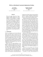
In conclusion, there are several mechanisms by which oxygen
regulators may initially adapt to an imbalance between energy
supply and energy demand. However, as shown in Fig. 2,
contrary to oxygen conformers, man is only able to cope with
an energy imbalance to a limited extent. Once the threshold
of adaptation is overcome, the obligate need for energy leads
to ‘energy failure’. This may be accounted for as an
unfavorable balance between ATP production and ATP
utilization (i.e. ATP turnover) according to the formula
591
describing energy charge: Energy charge = ([ATP] +
0.5[ADP]) / ([ATP] + [ADP] + [AMP]). A ratio between 0.8
and 0.95 is considered normal to survive, implying normal
supply for ATP synthesis and structurally intact machinery (i.e.
mithocondria).
Energy failure, lactate and time-course of illness
As already mentioned, a hypoxic insult may lead an oxygen-
regulated organism to death or to survival according to its
ability to adapt to hypoxia. However, even for a given genetic
trait, an individual may undergo adaptation or energy failure,
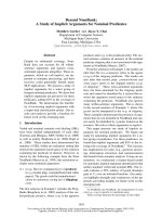
depending on time. In fact, if one expressed ATP turnover
over time there may be a scenario in which forced
hypometabolism rapidly decreases ATP turnover, eventually
leading to cell death. On the contrary, a slow decrease of
ATP turnover may allow a much longer survival (Fig. 3).
In the acute setting, oxygen conformance is unlikely because
a decrease of VO
2
is accompanied by an increase in lactate
levels. On the contrary, in the subacute settings, down-
regulation of some tissue metabolic activities is possible in
order to preserve more essential reactions [41]. There is also
evidence that oxygen conformance may exist in chronic
diseases in humans: Schumaker and colleagues reported
that VO
2
, determined indirectly by calorimetry, rapidly
increases after percutaneous valvuloplasty in patients with
severe aortic stenosis and cardiac cachexia [42].
When dealing with energy failure, time is the essence also
under a different perspective. In fact, there is a time window
in which the hypoxic cell, despite suffering, will revert to a
normal state if oxygen is supplied. After a time threshold,
however, an energy supply will be of no use. This time frame
is our therapeutic opportunity.
Let us consider hemorrhage as an example. Hemorrhage is
an acute preload hemodynamic impairment. Provided DO
2
is
the product of stroke volume and heart rate (cardiac output)
times the oxygen content (respiratory function – arterial
oxygen content), the energy failure during hemorrhage is due
to a stroke volume deficiency that eventually leads to cell
hypoxia. In the presence of normal adaptation processes,
however, the cell may cope with hypoxia for a while. If preload
is restored meanwhile, once oxygen is back the cell will be
able to utilize it because the oxygen machinery (i.e.
mitochondria) is still intact.
However, if the lack of oxygen persists, necrosis occurs and
organ failure emerges late during the course of illness. At that
time, even if an acid load due to cell lysis may persist, the
adaptive metabolic rearrangement that previously was
responsible for the increase of lactate (see earlier) my cease.
Plasma lactate may then decrease over time, provided
clearance is not impaired.
Similarly, an exaggerated aerobic glycolysis through Na
+
K
+
-
ATPase stimulation during septic shock may lead to
hyperlactatemia [43,44]. However, severe patients with poor
outcome have been shown to have mitochondrial dysfunction.
In fact, mediators such as nitric oxide may inhibit the
respiratory chain in the presence of normoxia or even
hyperoxia [45].
Available online />Table 1
Responses to acute and/or chronic hypoxia in which hypoxia
inducible factor-1 is involved
Pathway References
involved Gene controlled of interest
Glycolysis Aldolase A–C [46,47]
Enolase 1
Glucose transporter 1–3
Glyceraldehyde phosphate dehydrogenase
Hexokinase 1–2
Lactate dehydrogenase A
Phosphofructokinase L
Pyruvate kinase 1-M
Erythropoiesis Erythropoietin [48]
Angiogenesis Vascular endothelial growth factor [49]
Vascular tone Endothelin-1 [50-52]
Hemeoxygenase-1
Nitric oxide synthase 2
Figure 2
Different responses of oxygen conformers and oxygen regulators to
oxygen deprivation. Once a threshold of adaptation is reached, oxygen
regulators undergo an imbalance between energy supply and energy
consumption that leads to ‘energy failure’.
592
It thus seems that once oxygen machinery is out of order,
whether the initial insult was hemorrhage or sepsis, the
therapeutic opportunity is lost. This view may be applied to
the literature on the hemodynamic supranormal target. If we
consider the paper by Rivers and colleagues [26], it is clear
that their intervention was very early in the course of the
energy failure process and was very effective (see sections
on mixed venous saturation of oxygen and lactate changes
over time – few hours), as opposed to the late (third day in
the intensive care unit) and ineffective (see section on venous
saturation of oxygen) treatment by Gattinoni and colleagues
[25].
Conclusion
The metabolic fate of lactate in the body is under
sophisticated and finely tuned control. Many different
conditions may alter the balance between its production and
clearance. This per se is a limitation for the use of lactate as a
clinical marker in critically ill patients.
Nevertheless, when energy failure becomes relevant, lactate
measurement over time may be used as a metabolic marker
of energy failure. Contrary to what one may think, an increase
of lactate in critically ill patients in the presence of supply
dependency may be viewed as a positive feature, indicating
the presence of functioning adaptive metabolic pathways!
Increased lactate levels may be considered an early marker of
a potentially reversible state, possibly indicating that ‘there is
still room’ to boost fast intervention.
Competing interests
Franco Valenza received an honorarium of €500.00 in
December 2004 for his presentation on lactate and
mitochondria injury in an event organized by Instrumentation
Laboratories S.p.a. There are no other financial or non-
financial conflicts of interest.
References
1. Mizock BA: Lactic acidosis. Dis Mon 1989, 35:233-300.
2. Krebs H, Wood H, Alberti K: Hyperlactatemia and lactic acido-
sis. Essays Med Biochem 1975, 1:81-103.
3. Cohen R: Disorders of lactic acid metabolism. Clin Endocrinol
Metab 1976, 5:613-625.
4. Abu RS, Tannen RL: Amelioration of hypoxia-induced lactic
acidosis by superimposed hypercapnea or hydrochloric acid
infusion. Am J Physiol 1986, 250:F702-F709.
5. Pison CM, Chauvin C, Perrault H, Schwebel C, Lafond JL, Boujet
C, Leverve XM: In vivo hypoxic exposure impairs metabolic
adaptations to a 48 hour fast in rats. Eur Respir J 1998, 12:
658-665.
6. Mizock BA: Lactic acidosis in critical illness. Crit Care Med
1992, 20:80-93.
7. Chrusch C, Bands C, Bose D, Li X, Jacobs H, Duke K, Bautista E,
Eschun G, Light RB, Mink SN: Impaired hepatic extraction and
increased splanchnic production contribute to lactic acidosis in
canine sepsis. Am J Respir Crit Care Med 2000, 161:517-526.
8. Leverve X: Energy metabolism in critically ill patients: lactate is
a major oxidizable substrate. Curr Opin Clin Nutr Metab Care
1999, 2:165-169.
9. Broder G, Weil MH: Excess lactate: an index of reversibility of
shock in human patients. Science 1964, 143:1457-1459.
10. Weil MH, Afifi AA: Experimental and clinical studies on lactate
and pyruvate as indicators of the severity of acute circulatory
failure (shock). Circulation 1970, 41:989-1001.
11. Vitek V, Cowley RA: Blood lactate in the prognosis of various
forms of shock. Ann Surg 1971, 173:308-313.
12. Cady LDJ, Weil MH, Afifi AA, Michaels SF, Liu VY, Shubin H:
Quantitation of severity of critical illness with special refer-
ence to blood lactate. Crit Care Med 1973, 1:75-80.
13. Ronco JJ, Fenwick JC, Tweeddale MG, Wiggs BR, Phang PT,
Cooper DJ, Cunningham KF, Russell JA, Walley KR: Identifica-
tion of the critical oxygen delivery for anaerobic metabolism
in critically ill septic and nonseptic humans. JAMA 1993, 270:
1724-1730.
14. Levraut J, Ichai C, Petit I, Ciebiera JP, Perus O, Grimaud D: Low
exogenous lactate clearance as an early predictor of mortality
in normolactatemic critically ill septic patients. Crit Care Med
2003, 31:705-710.
15. Bakker J, Coffernils M, Leon M, Gris P, Vincent JL: Blood lactate
levels are superior to oxygen-derived variables in predicting
outcome in human septic shock. Chest 1991, 99:956-962.
16. Haupt MT, Gilbert EM, Carlson RW: Fluid loading increases
oxygen consumption in septic patients with lactic acidosis.
Am Rev Respir Dis 1985, 131:912-916.
17. Vincent JL, Roman A, De Backer D, Kahn RJ: Oxygen
uptake/supply dependency. Effects of short-term dobutamine
infusion. Am Rev Respir Dis 1990, 142:2-7.
18. Vincent JL, Roman A, Kahn RJ: Dobutamine administration in
septic shock: addition to a standard protocol. Crit Care Med
1990, 18:689-693.
19. Vallet B, Chopin C, Curtis SE, Dupuis BA, Fourrier F, Mehdaoui H,
LeRoy B, Rime A, Santre C, Herbecq P: Prognostic value of the
dobutamine test in patients with sepsis syndrome and normal
lactate values: a prospective, multicenter study. Crit Care Med
1993, 21:1868-1875.
20. De Backer D, Moraine JJ, Berre J, Kahn RJ, Vincent JL: Effects of
dobutamine on oxygen consumption in septic patients. Direct
versus indirect determinations. Am J Respir Crit Care Med
1994, 150:95-100.
21. Rhodes A, Lamb FJ, Malagon I, Newman PJ, Grounds RM,
Bennett ED: A prospective study of the use of a dobutamine
stress test to identify outcome in patients with sepsis, severe
sepsis, or septic shock. Crit Care Med 1999, 27:2361-2366.
22. Gilbert EM, Haupt MT, Mandanas RY, Huaringa AJ, Carlson RW:
The effect of fluid loading, blood transfusion, and cate-
cholamine infusion on oxygen delivery and consumption in
patients with sepsis. Am Rev Respir Dis 1986, 134:873-878.
23. Gattinoni L, Brazzi L, Pelosi P, Latini R, Tognoni G, Pesenti A,
Fumagalli R: A trial of goal-oriented hemodynamic therapy in
critically ill patients. SvO
2
Collaborative Group. N Engl J Med
1995, 333:1025-1032.
24. Hayes MA, Timmins AC, Yau EH, Palazzo M, Hinds CJ, Watson D:
Elevation of systemic oxygen delivery in the treatment of criti-
cally ill patients. N Engl J Med 1994, 330:1717-1722.
Critical Care December 2005 Vol 9 No 6 Valenza et al.
Figure 3
ATP turnover expressed over time. The fate of a cell exposed to a
decreased ATP turnover is shown. Time is essential to adaptation, the
lack of which brings the cell to death.
593
Available online />25. Gattinoni L, Valenza F, Carlesso E: ‘Adequate’ hemodynamics: a
question of time? In Functional Hemodynamic Monitoring.
Edited by Pinsky MR, Payen D. Berlin: Springer-Verlag; 2004:69-
86.
26. Rivers E, Nguyen B, Havstad S, Ressler J, Muzzin A, Knoblich B,
Peterson E, Tomlanovich M: Early goal-directed therapy in the
treatment of severe sepsis and septic shock. N Engl J Med
2001, 345:1368-1377.
27. Toffaletti JG: Blood lactate: biochemistry, laboratory methods,
and clinical interpretation. Crit Rev Clin Lab Sci 1991, 28:253-
268.
28. Boutilier RG: Mechanisms of cell survival in hypoxia and
hypothermia. J Exp Biol 2001, 204:3171-3181.
29. Hochachka PW: Defense strategies against hypoxia and
hypothermia. Science 1986, 231:234-241.
30. Vincent JL: DO
2
/VO
2
relationship. In Functional Hemodynamic
Monitoring. Edited by Pinsky MR, Payen D. Berlin: Springer-
Verlag; 2004:251-258.
31. Leverve XM, Mustafa I: Lactate: a key metabolite in the intercel-
lular metabolic interplay. Crit Care 2002, 6:284-285.
32. Vettor R, Lombardi A, Fabris R: Lactate infusion in anesthetised
rats produces insulin resistance in heart and skeletal
muscles. Metabolism 1997, 46:684-690.
33. Pellerin L, Magistretti P: How to balance brain energy budget
while spending glucose differently. J Physiol 2003, 546:325.
34. Schurr A, Rigor BM: Brain anaerobic lactate production: a
suicide note or a survival kit? Dev Neurosci 1998, 20:348-357.
35. Newsholme E, Leech A: Control of gluconeogenesis and gly-
colysis. In Biochemistry for Medical Sciences. Edited by New-
sholme E, Leech A. New York: Wyley; 1990:450-460.
36. Helperin M, Chhema-Dhadli S, Halperin F, Kamel K: Rationale for
the use of sodium bicarbonate in a patient with lactic acidosis
due to a poor cardiac output. Nephron 1994, 66:258-261.
37. Gutierrez LB, Hurtado FJ, Gutierrez AM, Fernandez E: Net uptake
of lacate by rabbit hindlimb during hypoxia. Am Rev Respir Dis
1993, 148:1204-1209.
38. Bunn HF, Poyton RO: Oxygen sensing and molecular adapta-
tion to hypoxia. Physiol Rev 1996, 76:839-885.
39. Brunelle J, Chandel N: Oxygen deprivation induced cell death:
an update. Apoptosis 2002, 7:475-482.
40. Seagroves TN, Ryan HE, Lu H, Wouters BG, Knapp M, Thibault P,
Laderoute K, Johnson RS: Transcription factor HIF-1 is a neces-
sary mediator of the pasteur effect in mammalian cells. Mol
Cell Biol 2001, 21:3436-3444.
41. Singer M, De Santi V, Vitale D, Jeffcoate W: Multiorgan failure is
an adaptive, endocrine-medaited, metabolic response to over-
whelming systemic inflammation. Lancet 2004, 364:545-548.
42. Shumacker PT, Soble JF, Feldman T: Oxygen delivery and
uptake relationships in patients with aortic stenosis. Am J
Respir Crit Care Med 1994, 149:1123-1131.
43. Howard JJ, Luchette FA, McCarter FD, Fisher JE: Lactate is an
unreliable indicator of tissue hypoxia in injury or sepsis.
Lancet 1999, 354:505-508.
44. Levy B, Gibot S, Frank P, Cravoisy A, Bollaert PE: Relation
between muscle Na
+
K
+
ATPase activity and rised lactate con-
centrations in septic shock: a prospective study. Lancet 2005,
365:871-875.
45. Brealey D, Brand M, Hargreaves I, Heales S, Land J, Smolenski R,
Davies NA, Cooper CE, Singer M: Association between mito-
chondrial dysfunction and severity and outcome of septic
shock. Lancet 2002, 360:219-223.
46. Iyer NV, Kotch LE, Agani F, Leung SW, Laughner E, Wenger RH,
Gassmann M, Gearhart JD, Lawler AM, Yu AY, Semenza GL: Cel-
lular and developmental control of O
2
homeostasis by
hypoxia-indicible factor 1
αα
. Genes Dev 1998, 12:149-162.
47. Semenza GL, Roth PH, Fang HM, Wang GL: Transcriptional
regulation of genes encoding glycolytic enzymes by hypoxia-
inducible factor 1. J Biol Chem 1994, 269:23757-23763.
48. Jiang B-H, Rue E, Wang GL, Roe R, Semenza GL: Dimerization,
DNA binding, and transactivation properties of hypoxia-
inducible factor 1
αα
. J Biol Chem 1996, 271:17771-17778.
49. Forsythe JA, Jiang B-H, Iyver NV, Agani F, Leung SW, Koos RD,
Semenza GL: Activation of vascular endothelial factor gene
transcription by hipoxia-inducible factor 1
αα
. Mol Cell Biol
1996, 16:4604-4613.
50. Hu J, Disher DJ, Bishopric NH, Webster KA: Hypoxia regulates
expression for the endothelin-1 gene through a proximal
hypoxia-inducible factor-1 binding on the antisense strand.
Biochem Biophys Res Commun 1998, 245:894-899.
51. Lee PJ, Jiang B-H, Chin BY, Iyer NV, Alam J, Semenza GL, Choi
AMK: Hypoxia-inducible factor 1 mediates heme oxygenase-1
gene in response to hypoxia. J Biol Chem 1997, 272:5375-
5381.
52. Palmer LA, Semenza GL, Stoler MH, Johns RA: Hypoxia induces
type II NOS gene expression in pulmonary artery endothelial
cells via HIF-1. Am J Physiol Lung Cell Mol Physiol 1998, 274:
L212-L219.