Báo cáo khoa học: "Bench-to-bedside review: Is there a place for epinephrine in septic shock" pptx
Bạn đang xem bản rút gọn của tài liệu. Xem và tải ngay bản đầy đủ của tài liệu tại đây (90.07 KB, 5 trang )
561
ICG = indocyanine green; IL = interleukin; L/P = lactate/pyruvate; MAP, mean arterial pressure; PCO
2
= partial CO
2
tension; SHO
2
= hepatic
venous oxygen saturation; SVO
2
= venous oxygen saturation.
Available online />Abstract
The use of epinephrine in septic shock remains controversial.
Nevertheless, epinephrine is widely used around the world and the
reported morbidity and mortality rates with it are no different from
those observed with other vasopressors. In volunteers, epinephrine
increases heart rate, mean arterial pressure and cardiac output.
Epinephrine also induces hyperglycemia and hyperlactatemia. In
hyperkinetic septic shock, epinephrine consistently increases
arterial pressure and cardiac output in a dose dependent manner.
Epinephrine transiently increases lactate levels through an increase
in aerobic glycolysis. Epinephrine has no effect on splanchnic
circulation in dopamine-sensitive septic shock. On the other hand,
in dopamine-resistant septic shock, epinephrine has no effect on
tonometric parameters but decreases fractional splanchnic blood
flow with an increase in the gradient of mixed venous oxygen
saturation (SVO
2
) and hepatic venous oxygen saturation (SHO
2
).
In conclusion, epinephrine has predictable effects on systemic
hemodynamics and is as efficient as norepinephrine in correcting
hemodynamic disturbances of septic shock. Moreover, epinephrine
is cheaper than other commonly used catecholamine regimens in
septic shock. The clinical impact of the transient hyperlactatemia
and of the splanchnic effects are not established.
Introduction
Early goal directed therapy [1] is now considered as a gold
standard in the early phase of septic shock. Fluid therapy and
vasoactive therapy may be immediately required in order to
maintain acceptable blood pressure levels. Invasive or non-
invasive assessment of hemodynamic status, although
essential to the rational management of septic shock, may
take time to establish. In this setting, there is good reason to
choose a broad spectrum catecholamine such as epinephrine
or dopamine rather than a pure α-adrenergic agonist, which
can cause substantial reductions in cardiac output, and as an
alternative to a pure β-agonist such as dobutamine, which
can exacerbate vasodilation and hypotension through its β
2
-
adrenergic action [2]. In contrast to norepinephrine-
dobutamine, epinephrine when used in septic shock
increases lactate level together with a slightly enhanced
lactate/pyruvate (L/P) ratio, decreases global splanchnic flow
and elevates the tonometric mucosal partial CO
2
tension
(PCO
2
) gap (tonometer PCO
2
minus arterial PCO
2
), a
surrogate marker of gastric mucosal metabolism and/or
perfusion. Based on these observations, The Task Force of
the American College of Critical Care Medicine and the
Society of Critical Care Medicine recommends the use of
epinephrine only in patients who fail to respond to traditional
therapies [3].
The aim of this paper is to provide an alternative point of view
regarding the somewhat dark side of epinephrine and to
moderate the interpretation of pharmacological data.
Epinephrine effects in volunteers
Hemodynamic effects
In volunteers [4,5], epinephrine increases heart rate as well as
mean arterial pressure (MAP), mainly as the result of a rise in
systolic blood pressure. Conversely, diastolic blood pressure
falls, irrespective of the dosage. Vasodilatation occurs in the
calf vascular bed while blood flow in skin capillaries and
arteriovenous anastomoses decreases. Concentration-
dependent increases in stroke volume and cardiac output
occur without any changes in end-diastolic volume, along with
decreases in vascular resistances of the systemic circulation,
calf and adipose tissue. Coronary blood flow, blood flow to
skeletal muscles as well as hepatic blood flow increase while
splanchnic vascular resistances decrease. Alternatively, renal
blood flow decreases with an increase in the filtration fraction
Metabolic effects
In healthy volunteers [4,5], epinephrine induces hyperglycemia
and hyperlactatemia. Because insulin secretion is suppressed
by alpha adrenergic stimulation, plasma concentration of
insulin remains low. Hyperglycemia is induced by an increase
Review
Bench-to-bedside review: Is there a place for epinephrine in
septic shock?
Bruno Levy
Service de Réanimation Médicale, Hôpital Central, 54000 Nancy, France
Corresponding author: Bruno Levy,
Published online: 4 November 2005 Critical Care 2005, 9:561-565 (DOI 10.1186/cc3901)
This article is online at />© 2005 BioMed Central Ltd
562
Critical Care December 2005 Vol 9 No 6 Levy
in glucose production caused by an increase in hepatic
glycogenolysis and an increase in gluconeogenesis. There is
also a marked increase in oxygen consumption (VO
2
). In
skeletal muscle, epinephrine increases glycolysis and glyco-
genolysis, inducing an upsurge in lactate. Muscular lactate
serves as a substrate for hepatic neoglucogenesis (Cori
cycle). Epinephrine also increases lipolysis and decreases
muscular proteolysis.
Clearly, epinephrine is the most potent natural β-agonist,
which explains the fact that in volunteers or in patients with
septic shock, epinephrine increased glucose and lactate
levels more than norepinephrine.
Epinephrine effects in septic shock
Epinephrine is effective in restoring global hemodynamics
In patients unresponsive to volume expansion or other cate-
cholamine infusions, epinephrine can increase MAP, primarily
by increasing cardiac index and stroke volume together with
more modest increases in systemic vascular resistance and
heart rate. This is an important advantage, especially in
patients with altered cardiac function. The effects of epi-
nephrine in hyperdynamic or normodynamic septic shock are
highly predictable, correlating an increase in MAP with an
increase in cardiac index [6]. Using epinephrine as a first line
agent, Moran et al. [7] reported a linear relationship between
epinephrine dosage and heart rate, MAP, cardiac index, left
ventricular stroke work index, and oxygen delivery and
consumption. Despite an increase in oxygen consumption, no
adverse cardiac side effects have been described in septic
shock. Electrocardiographic changes indicating ischemia or
arrhythmias have not been reported in septic patients. In
patients with right ventricular failure, epinephrine increases
right ventricular function by improving contractility [8].
Considering global hemodynamics, epinephrine is more
effective than dopamine and is just as efficient as nor-
epinephrine [9].
Epinephrine increases lactate concentration
In human septic shock, epinephrine increases lactate levels
and decreases arterial pH [10]. From the equation L/P =
K.NADH/NAD.[H
+
], where K is the dissociation constant, it
may be seen that a change in H
+
could result in a
proportional change in the L/P ratio. Thus, interpretation of
the L/P ratio should be done while accounting for arterial pH.
In the same study, we found that epinephrine increased
lactate level without any increase in the L/P ratio when the
latter was normalized to pH (H
+
= 10
–pH
). This rise in lactate
is transient, however, as levels return to baseline values after
12 hours [9]. The fact that β-receptor density is down-
regulated during sepsis [11] likely explains the transient
character of epinephrine increased lactate.
Epinephrine infusion is associated with an increase in lactate
concentration not only in septic conditions but also under fully
aerobic conditions, such as in healthy volunteers at rest and
during exercise. In a model of endotoxinic shock, we
demonstrated that the infusion of epinephrine was associated
with a significant increase in lactate without any change in L/P
ratio [12]. Moreover, epinephrine use was not associated with
a decrease in tissue ATP [12], demonstrating that epinephrine-
induced hyperlactatemia is probably related to direct effects of
epinephrine on carbohydrate metabolism and not to cellular
hypoxia. Indeed, elevated blood lactate concentrations during
shock states are often viewed as evidence of tissue hypoxia,
with lactate levels being proportional to the defect in oxidative
metabolism [1]. However, many tissues generate pyruvate and
lactate under aerobic conditions (so-called aerobic glycolysis)
in a process linking glycolytic ATP supply to activity of
membrane ion pumps such as Na
+
,K
+
-ATPase [13].
Stimulation of aerobic glycolysis (glycolysis not attributable to
oxygen deficiency or glycogenolysis) occurs not only in resting,
well-oxygenated skeletal muscles but also during experimental
hemorrhagic shock and experimental sepsis, and is closely
linked to stimulation of active sarcolemmal Na
+
,K
+
-ATPase
transport under epinephrine stimulation. Epinephrine
stimulates the release of lactate from skeletal muscle through
stimulation of Na
+
,K
+
-ATPase for oxidation purposes or
gluconeogenesis (Cori cycle). Thus, increased lactate
production is the result of aerobic glycolysis rather than the
result of anaerobic glycolysis. Although this is an ATP-
consuming process, the source of energy in the liver ultimately
comes from fatty acids. Thus, lactate provides glycolytic ATP
to several peripheral cells, this ATP being derived from energy-
producing lipid oxidation. This hypothesis was recently
demonstrated in human septic shock [14].
Epinephrine increases the PCO
2
gap
In a clinical setting of dopamine-resistant septic shock, we
compared the effects of norepinephrine-dobutamine versus
epinephrine alone on gastric tonometry using saline tonometry
[8]. Despite similar increases in arterial pressure and oxygen
delivery in both groups, the PCO
2
gap increased in
epinephrine-treated patients. This increase was transient,
however, as both groups had the same normal PCO
2
gap after
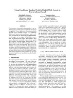
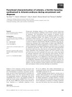
24 hours (Fig. 1). Moreover, the amplitude of the PCO
2
gap
increase was moderate and consistently below 18 mmHg [15].
This suggests one of two possibilities [16]. First, that
epinephrine increases splanchnic oxygen utilization and CO
2
production through a thermogenic effect, especially if gastric
blood flow does not increase to the same extent, inducing a
mismatch between splanchnic oxygen delivery and splanchnic
oxygen consumption. Second, that epinephrine decreases
mucosal blood flow with a decrease in CO
2
efflux, the net
result being an increase in CO
2
gap. The latter hypothesis is
not supported by Duranteau et al. [17], however, who
demonstrated, using laser Doppler flow, that epinephrine
induces higher gastric mucosal blood flow than norepinephrine
and dopamine without significant change in the PCO
2
gap.
Moreover, De Backer et al. [18] did not observe any variation
in the PCO
2
gap during epinephrine infusion using air
563
tonometry. Conversely, also using air tonometry, we have
frequently observed a decrease in the PCO
2
gap in the early
phase of septic shock when using epinephrine as a first line
agent (unpublished data). It is our hypothesis that the
improvement in arterial pressure and oxygen delivery induced
by epinephrine in severely hypotensive patients may offset the
putative deleterious effects on mucosal oxygen adequation.
Epinephrine decreases splanchnic blood flow and
increases the SVO
2
-SHO
2
gradient
Epinephrine decreases splanchnic blood flow, with transient
increases in arterial, splanchnic and hepatic venous lactate
concentrations. The reduction in splanchnic blood flow has
been associated with a decrease in oxygen delivery and a
reduction in oxygen consumption [19]. These effects may be
due to a reduction in splanchnic oxygen delivery to a level
that impairs nutrient blood flow, likely resulting in a reduction
in global tissue oxygenation, but may be potentially reversed
by the concomitant administration of dobutamine. The
addition of dobutamine to epinephrine-treated patients has
been shown to improve gastric mucosal perfusion, as
assessed by improvements in intramucosal pH, arterial
lactate concentration and the PCO
2
gap [20]. It is not clear
whether a transient decrease in hepatosplanchnic blood flow
in septic shock is deleterious [20]. The mucosa and the
submucosa are known to receive most of the splanchnic
blood flow. Indocyanine green (ICG) clearance explores both
splanchnic blood flow and liver function. De Backer and
colleagues [18] compared epinephrine, norepinephrine and
dopamine titrated for the same mean arterial pressure using
three different tools to evaluate splanchnic perfusion and
splanchnic metabolism. Splanchnic perfusion was assessed
using: ICG clearance as a reflection of global
hepatosplanchnic blood flow; hepatic venous saturation and
the gradient of mixed venous oxygen saturation (SVO
2
) and
hepatic venous oxygen saturation (SHO
2
) as a reflection of
the balance between splanchnic oxygen delivery and oxygen
consumption; and the gastric PCO
2
gap as a reflection of
gastric mucosa perfusion/metabolism adequacy. The authors
concluded that in patients who responded to dopamine, no
differences were found with regard to splanchnic effects. On
the other hand, in nine of ten cases of dopamine-resistant
septic shock, epinephrine, when associated with dobutamine,
decreased hepatosplanchnic blood flow, increased the
SVO
2
-SHO
2
gradient and increased arterial lactate and
hepatic lactate consumption without any net effect on the
PCO
2
gap, which may also indicate a constant blood flow in
the mucosa. Moreover, the absence of variation in the PCO
2
gap argues against a deleterious effect of epinephrine on
splanchnic circulation because gut mucosa is probably the
area of the body most sensitive to a decrease in blood flow. In
various animal models, a decrease in splanchnic blood flow is
associated with an increase in the PCO
2
gap. The more likely
explanation is that the energetic cost of metabolic processes
induced by epinephrine such as neoglucogenesis and lactate
consumption decreases the ability of the liver to metabolize
ICG. Nevertheless, metabolizing ICG is not a natural process.
Because epinephrine does not decrease liver lactate consump-
tion, liver energy equilibrium is likely to remain stable.
In contrast, Seguin et al. [21] demonstrated in patients with
septic shock that epinephrine at doses that induced the same
mean arterial pressure did not modify ICG clearance and
enhanced more gastric mucosal blood flow than the
combination of dobutamine at 5 µg/kg per minute and
norepinephrine.
Moreover, the effects of epinephrine may be different
according to the studied area. Duranteau et al. [10]
demonstrated using laser Doppler flow that epinephrine
induced higher gastric mucosal blood flow than nor-
epinephrine without any significant changes in intramucosal
pH. Thus, it is likely that despite a relative decrease in
splanchnic blood flow in the epinephrine-treated patient, gut
mucosa receives sufficient blood flow to meet its metabolic
needs. In fact, epinephrine exerts both sides of its β-2
properties: a redistribution of blood flow from the splanchnic
bed to the muscular bed, and a redistribution of splanchnic
flow towards the mucosa.
Limitation of splanchnic blood flow estimation
The clarification of the role of epinephrine in septic patients is
somewhat limited by the few techniques currently available
for estimating splanchnic tissue oxygenation, in addition to
each of these techniques having its own limitations. The ICG
method used by De Backer et al. [18] and other teams for
splanchnic blood flow determination actually measures liver
venous blood flow, which fails to distinguish supply from the
portal vein and the hepatic artery. Consequently, changes in
distribution of blood flow between the muscularis and the
mucosa of the gut are not detectable by this method. The
tonometric measurement raises the same types of concern
Available online />Figure 1
Evolution of the partial CO
2
tension (PCO
2
) gap (tonometer PCO
2
–
arterial PCO
2
) during infusion of epinephrine (open circles) or
norepinephrine-dobutamine (closed circles). Asterisks indicate
p < 0.01 versus baseline. (Reproduced from [8] with permission.)
564
because it only represents flow conditions in the gastric
region. It has been shown, at least in an animal model, that
changes in blood flow to the various organs in the splanchnic
region are quite variable following induction of sepsis. An
increase in SVO
2
-SHO
2
gradient signifies that the splanchnic
area consumes more O
2
than the rest of the body. It does not
mean that the splanchnic area is hypoxic.
Immunological and anticoagulant effects of
epinephrine during sepsis
An immunomodulatory effect of epinephrine has been
reported to supposedly be mediated via beta-adrenergic
receptors. In whole blood in vitro, Van Der Poll et al. [22]
demonstrated that epinephrine inhibits endotoxin-induced
IL-1β production through an inhibition of tumor necrosis
factor and an enhancement of IL-10. They concluded that
endogenous or exogenous epinephrine may attenuate
excessive activity of inflammatory cytokines during infection.
Oberbeck et al. [23] investigated in mice submitted to cecal
ligation and puncture the effects of epinephrine and/or beta-
adrenergic blockade on cellular immune functions. They
found that epinephrine infusion did not affect the lethality of
septic shock in mice but induced alterations in splenocyte
apoptosis, splenocyte proliferation and IL-2 release and was
associated with profound changes in circulating immune cell
subpopulations. Treatment with propranolol augmented the
epinephrine-induced increase of splenocyte apoptosis, did
not affect the decrease of splenocyte proliferation and IL-2
release, augmented the release of IL-6 and antagonized the
mobilization of natural killer cells observed in epinephrine-
treated animals. Furthermore, these immunological alterations
were accompanied by a significant increase of sepsis-
induced mortality. Co-administration of propranolol and
epinephrine augmented the propranolol-induced changes of
splenocyte apoptosis and IL-6 release and was associated
with the highest mortality of septic mice. These data clearly
indicate that adrenergic mechanisms modulate cellular
immune functions during sepsis, with these effects being
mediated via alpha- and beta-adrenergic pathways. The
conclusions on survival are not truly proven as epinephrine
and propranolol also act on hemodynamics. Therefore,
alterations in the serum concentrations of catecholamine may
affect the immunocompetence of the organism and may
thereby affect the clinical course of critically ill patients [24].
It is also interesting to note that epinephrine exerts anti-
thrombotic effects during endotoxemia by concurrent inhibition
of coagulation and stimulation of fibrinolysis. Thus, epinephrine,
whether endogenously produced or administered as a
component of treatment, may limit the development of dissemin-
ated intravascular coagulation during systemic infection [25].
In summary, although the clinical impact remains to be
demonstrated during septic conditions, epinephrine
modulates the inflammatory state and decreases the
hypercoagulation state.
Other properties of epinephrine
Unlike with norepinephrine, the hemodynamic effects of
epinephrine (MAP and cardiac index increase) were obtained
without the adjunction of dobutamine. This may prove to be
important from a practical standpoint in situations such as
transportation. Arrhythmia has not been described in the
setting of septic shock. Morever, epinephrine when used
alone is cheaper than vasopressin or the combination
norepinephrine-dobutamine.
Does the choice of catecholamine influence patient
evolution and prognosis?
Currently, there is no prospective randomized clinical study
indicating that one catecholamine is superior to the other
during septic shock. A recent meta-analysis by the Cochrane
group [26] failed to demonstrate any difference between
tested vasopressors. Furthermore, no study has demon-
strated a relationship between improvement in PCO
2
gap or
ICG clearance after pharmacological intervention and an
improvement in prognosis. Thus, all current data regarding
the splanchnic effects of catecholamine should be
considered as pharmacological investigations of a vasoactive
agent evaluated by a particular monitoring device. The
discrepancy observed between all of these measurements
further highlights the absence of clinical relevance.
Catecholamine use is not only limited to specialized
intensive care units
The initial choice of catecholamine in the intensive care unit is
relatively well standardized, at least for hyperkinetic septic
shock. Hemodynamic evaluation is easy and accessible (even
if the type of monitoring remains debatable), with the choice
of catecholamine based on rational evaluation. This is not the
case for many situations in other clinical settings. For
example, catecholamines are used on the ward, during
transportation, in the emergency room and even in patients’
homes. Physicians are often young and/or have little
experience in intensive care treatment. Diagnosis is not
always straightforward and, in some cases, it may be difficult
to distinguish between cardiogenic, hypovolemic or septic
shock. In these particular circumstances, it seems more
appropriate to use a catecholamine with predictable effects,
such as epinephrine, rather than a strong vasoconstrictor
such as norepinephrine.
Conclusion
Two opposite points of view are proposed. First, why should
we use epinephrine, a drug with such potential negative
effects, when there are other alternatives for the treatment of
septic patients. On the other hand, epinephrine is commonly
used worldwide and the reported morbidity and mortality
rates with it are no different from those observed with other
vasopressors. The French study comparing epinephrine and
norepinephrine-dobutamine has been presented only in an
abstract form [27]. These preliminary results seem to
demonstrate that there is no evidence for the superiority of
Critical Care December 2005 Vol 9 No 6 Levy
565
norepinephrine plus dobutamine over epinephrine alone for
the management of adults with septic shock. Thus, we have
to wait for the definitive publication to decide whether Dr
Jekyll or Mr Hyde is the true nature of epinephrine in the
treatment of septic shock [28].
Competing interests
The author(s) declare that they have no competing interests.
References
1. Rivers E, Nguyen B, Havstad S, Ressler J, Muzzin A, Knoblich B,
Peterson E, Tomlanovich M: Early goal-directed therapy in the
treatment of severe sepsis and septic shock. N Engl J Med
2001, 345:1368-1377.
2. Rudis MI, Basha MA, Zarowitz BJ: Is it time to reposition vaso-
pressors and inotropes in sepsis? Crit Care Med 1996, 24:
525-537.
3. Hollenberg SM, Ahrens TS, Annane D, Astiz ME, Chalfin DB,
Dasta JF, Heard SO, Martin C, Napolitano LM, Susla GM, et al.:
Practice parameters for hemodynamic support of sepsis in
adult patients: 2004 update. Crit Care Med 2004, 32:1928-
1948.
4. Bearn AG, Billing B, Sherlock S: The effect of adrenaline and
noradrenaline on hepatic blood flow and splanchnic carbohy-
drate metabolism in man. J Physiol 1951, 115:430-441.
5. Ensinger H, Weichel T, Lindner KH, Grunert A, Georgieff M: Are
the effects of noradrenaline, adrenaline and dopamine infu-
sions on VO2 and metabolism transient? Intensive Care Med
1995, 21:50-56.
6. Wilson W, Lipman J, Scribante J, Kobilski S, Lee C, Krause P,
Cooper J, Barr J: Septic shock: does adrenaline have a role as
a first-line inotropic agent? Anaesth Intensive Care 1992, 20:
470-474.
7. Moran JL, O’Fathartaigh MS, Peisach AR, Chapman MJ, Leppard
P: Epinephrine as an inotropic agent in septic shock: a dose-
profile analysis. Crit Care Med 1993, 21:70-77.
8. Le Tulzo Y, Seguin P, Gacouin A, Camus C, Suprin E, Jouannic I,
Thomas R: Effects of epinephrine on right ventricular function
in patients with severe septic shock and right ventricular
failure: a preliminary descriptive study. Intensive Care Med
1997, 23:664-670.
9. Levy B, Bollaert PE, Charpentier C, Nace L, Audibert G, Bauer P,
Nabet P, Larcan A: Comparison of norepinephrine and dobuta-
mine to epinephrine for hemodynamics, lactate metabolism,
and gastric tonometric variables in septic shock: a prospec-
tive, randomized study. Intensive Care Med 1997, 23:282-287.
10. Day NP, Phu NH, Bethell DP, Mai NT, Chau TT, Hien TT, White
NJ: The effects of dopamine and adrenaline infusions on acid-
base balance and systemic haemodynamics in severe infec-
tion. Lancet 1996, 348:219-223.
11. Silverman HJ, Penaranda R, Orens JB, Lee NH: Impaired beta-
adrenergic receptor stimulation of cyclic adenosine
monophosphate in human septic shock: association with
myocardial hyporesponsiveness to catecholamines. Crit Care
Med 1993, 21:31-39.
12. Levy B, Mansart A, Bollaert PE, Franck P, Mallie JP: Effects of
epinephrine and norepinephrine on hemodynamics, oxidative
metabolism, and organ energetics in endotoxemic rats. Inten-
sive Care Med 2003, 29:292-300.
13. James JH, Luchette FA, McCarter FD, Fischer JE: Lactate is an
unreliable indicator of tissue hypoxia in injury or sepsis.
Lancet 1999, 354:505-508.
14. Levy B, Gibot S, Franck P, Cravoisy A, Bollaert PE: Relation
between muscle Na+K+ ATPase activity and raised lactate
concentrations in septic shock: a prospective study. Lancet
2005, 365:871-875.
15. Levy B, Gawalkiewicz P, Vallet B, Briancon S, Nace L, Bollaert
PE: Gastric capnometry with air-automated tonometry pre-
dicts outcome in critically ill patients. Crit Care Med 2003, 31:
474-480.
16. Chapman MV, Mythen MG, Webb AR, Vincent JL: Report from
the meeting: Gastrointestinal Tonometry: State of the Art.
22nd-23rd May 1998, London, UK. Intensive Care Med 2000,
26:613-622.
17. Duranteau J, Sitbon P, Teboul JL, Vicaut E, Anguel N, Richard C,
Samii K: Effects of epinephrine, norepinephrine, or the combi-
nation of norepinephrine and dobutamine on gastric mucosa
in septic shock. Crit Care Med 1999, 27:893-900.
18. De Backer D, Creteur J, Silva E, Vincent JL: Effects of dopamine,
norepinephrine, and epinephrine on the splanchnic circulation
in septic shock: which is best? Crit Care Med 2003, 31:1659-
1667.
19. Meier-Hellmann A, Reinhart K, Bredle DL, Specht M, Spies CD,
Hannemann L: Epinephrine impairs splanchnic perfusion in
septic shock. Crit Care Med 1997, 25:399-404.
20. Levy B, Bollaert PE, Lucchelli JP, Sadoune LO, Nace L, Larcan A:
Dobutamine improves the adequacy of gastric mucosal perfu-
sion in epinephrine-treated septic shock. Crit Care Med 1997,
25:1649-1654.
21. Seguin P, Bellissant E, Le Tulzo Y, Laviolle B, Lessard Y, Thomas
R, Malledant Y: Effects of epinephrine compared with the com-
bination of dobutamine and norepinephrine on gastric perfu-
sion in septic shock. Clin Pharmacol Ther 2002, 71:381-388.
22. Van der Poll T, Lowry SF: Epinephrine inhibits endotoxin-
induced IL-1 beta production: roles of tumor necrosis factor-
alpha and IL-10. Am J Physiol 1997, 273:R1885-1890.
23. Oberbeck R, Schmitz D, Wilsenack K, Schuler M, Pehle B,
Schedlowski M, Exton MS: Adrenergic modulation of survival
and cellular immune functions during polymicrobial sepsis.
Neuroimmunomodulation 2004, 11:214-223.
24. Oberbeck R: Therapeutic implications of immune-endocrine
interactions in the critically ill patients. Curr Drug Targets
Immune Endocr Metabol Disord 2004, 4:129-139.
25. van der Poll T, Levi M, Dentener M, Jansen PM, Coyle SM, Braxton
CC, Buurman WA, Hack CE, ten Cate JW, Lowry SF: Epineph-
rine exerts anticoagulant effects during human endotoxemia.
J Exp Med 1997, 185:1143-1148.
26. Mullner M, Urbanek B, Havel C, Losert H, Waechter F, Gamper
G: Vasopressors for shock. Cochrane Database Syst Rev
2004, CD003709.
27. Annane D, Vignon P, Bollaert PE, Charpentier C, Martin C, Troche
G, Ricard JD, Nitenberg G, Bellissant E, for the CATS study
group: Norepinephrine plus dobutamine versus epinephrine
alone for the management of septic shock. Intensive Care Med
2005, 31:S1-S18.
28. Levy B: Epinephrine in septic shock: Dr. Jekyll or Mr. Hyde?
Crit Care Med 2003, 31:1866-1867.
Available online />