Retrovirology Research BioMed Central Open Access Analysis of transcribed human endogenous ppsx
Bạn đang xem bản rút gọn của tài liệu. Xem và tải ngay bản đầy đủ của tài liệu tại đây (452.53 KB, 17 trang )
BioMed Central
Page 1 of 17
(page number not for citation purposes)
Retrovirology
Open Access
Research
Analysis of transcribed human endogenous retrovirus W env loci
clarifies the origin of multiple sclerosis-associated retrovirus env
sequences
Georg Laufer
1
, Jens Mayer
2
, Benedikt F Mueller
1
, Nikolaus Mueller-Lantzsch
1
and Klemens Ruprecht*
1,3
Address:
1
Institute of Virology, Saarland University Hospital, Homburg, Germany,
2
Department of Human Genetics, Saarland University Hospital,
Homburg, Germany and
3
Department of Neurology, Saarland University Hospital, Homburg, Germany
Email: Georg Laufer - ; Jens Mayer - ; Benedikt F Mueller - ;
Nikolaus Mueller-Lantzsch - ; Klemens Ruprecht* -
* Corresponding author
Abstract
Background: Multiple sclerosis-associated retrovirus (MSRV) RNA sequences have been
detected in patients with multiple sclerosis (MS) and are related to the multi-copy human
endogenous retrovirus family type W (HERV-W). Only one HERV-W locus (ERVWE1) codes for
a complete HERV-W Env protein (Syncytin-1). Syncytin-1 and the putative MSRV Env protein have
been involved in the pathogenesis of MS. The origin of MSRV and its precise relation to HERV-W
were hitherto unknown.
Results: By mapping HERV-W env cDNA sequences (n = 332) from peripheral blood mononuclear
cells of patients with MS and healthy controls onto individual genomic HERV-W env elements, we
identified seven transcribed HERV-W env loci in these cells, including ERVWE1. Transcriptional
activity of individual HERV-W env elements did not significantly differ between patients with MS and
controls. Remarkably, almost 30% of HERV-W env cDNAs were recombined sequences that most
likely arose in vitro between transcripts from different HERV-W env elements. Re-analysis of
published MSRV env sequences revealed that all of them can be explained as originating from
genomic HERV-W env loci or recombinations among them. In particular, a MSRV env clone
previously used for the generation of monoclonal antibody 6A2B2, detecting an antigen in MS brain
lesions, appears to be derived from a HERV-W env locus on chromosome Xq22.3. This locus
harbors a long open reading frame for an N-terminally truncated HERV-W Env protein.
Conclusion: Our data clarify the origin of MSRV env sequences, have important implications for
the status of MSRV, and open the possibility that a protein encoded by a HERV-W env element on
chromosome Xq22.3 may be expressed in MS brain lesions.
Background
Multiple sclerosis (MS) is a chronic inflammatory demy-
elinating disease of the central nervous system thought to
result from an as yet incompletely understood complex
interplay of genetic and environmental factors [1]. Stimu-
lated by the finding that human T cell leukemia virus type
Published: 15 April 2009
Retrovirology 2009, 6:37 doi:10.1186/1742-4690-6-37
Received: 14 January 2009
Accepted: 15 April 2009
This article is available from: />© 2009 Laufer et al; licensee BioMed Central Ltd.
This is an Open Access article distributed under the terms of the Creative Commons Attribution License ( />),
which permits unrestricted use, distribution, and reproduction in any medium, provided the original work is properly cited.
Retrovirology 2009, 6:37 />Page 2 of 17
(page number not for citation purposes)
1 (HTLV-1), a human exogenous retrovirus, causes a neu-
rological disease (HTLV-1 associated myelopathy/tropical
spastic paraparesis) with similarities to MS [2], retrovi-
ruses have also been searched for in MS. This led to the
detection of retrovirus-like particles containing reverse
transcriptase activity in the supernatants from cultured
cell of patients with MS [3-5]. After molecular characteri-
zation of retroviral RNA sequences within such particles,
this novel retroviral element was named MS-associated
retrovirus (MSRV) [6,7]. Subsequent probing of human
genomic DNA with MSRV sequences revealed endog-
enous retroviral sequences closely related to MSRV, the
human endogenous retrovirus (HERV) family type W
(HERV-W) [8]. HERVs are remnants of ancestral germ line
infections by active retroviruses, which have thereafter
been transmitted in a Mendelian manner. In humans,
HERVs comprise approximately 8% of the genome (for
review see [9,10]). They typically consist of an internal
region containing gag, pro, pol, and env genes, flanked by
two long terminal repeats (LTR). HERV-W is a multicopy
family consisting of ~650 elements dispersed in the
human genome [11]. About 280 of those elements con-
tain internal sequences. The remaining elements lack
internal regions because of recombinational deletion
between the two LTRs, leaving a solo LTR [11]. Like
almost all HERV families, HERV-W is highly defective due
to acquisition of stop-codons, frameshift mutations, and
deletions. In addition, many members of the HERV-W
family represent processed pseudogenes that were gener-
ated through retrotransposition by long interspersed ele-
ments (LINE) [11-13]. Notably, no replication-competent
HERV-W provirus could be identified so far [8,14]. Yet, a
single HERV-W env locus (ERVWE1, chromosomal loca-
tion 7q21.2) retained a complete open reading frame
(ORF) for a functional envelope (Env) protein, termed
Syncytin-1 [15]. Syncytin-1 is highly expressed in the pla-
centa where it likely participates in the fusion of cytotro-
phoblast cells into the syncytiotrophoblast layer [16]. The
ERVWE1 locus therefore appears to have been diverted
into a bona fide human gene [17].
Intriguingly, in addition to the physiological function of
Syncytin-1 in placental morphogenesis, several studies
have provided evidence in support of a possible patho-
genic role of HERV-W Env/Syncytin-1 in MS. HERV-W
env/Syncytin-1 RNA levels were found to be higher in
autopsied brain tissue from patients with MS than in
brain tissue from controls [18-21]. Neuropathological
investigations reported increased expression of HERV-W
Env/Syncytin-1 protein in astrocytes and microglia in
actively demyelinating brain lesions from patients with
MS [18,21,22]. A potential pathogenic significance of
HERV-W Env/Syncytin-1 expression in MS can be inferred
from data showing that Syncytin-1 has indirect cytotoxic
effects on oligodendrocytes in vitro, and that expression of
Syncytin-1 in murine models results in demyelination in
vivo [18,22].
A putative MSRV Env protein was previously reported
[7,23]. The surface (SU) domain of MSRV Env
(AF331500), the amino acid sequence of which is 87%
identical to Syncytin-1, has been shown to stimulate pro-
duction of proinflammatory cytokines in human mono-
cytes via engagement of CD14 and toll-like receptor 4. It
also triggers maturation of human dendritic cells and con-
fers on them the potential to polarize naive T-cells into
Th-1-like effector T-lymphocytes [24]. Production of IFN-
γ, IL-6, and IL-12p40 following stimulation with MSRV
Env SU of peripheral blood mononuclear cells (PBMC)
from patients with MS is significantly higher compared to
PBMC from healthy controls [25]. In addition, MSRV Env
induces a polyclonal activation of T-cells bearing specific
Vβ chains, reminiscent of immunopathogenic effects trig-
gered by superantigens [23]. Altogether, those proinflam-
matory properties of MSRV Env appear compatible with a
potential relevance of MSRV Env in the context of MS, too.
Despite the possible role of MSRV and HERV-W (Syncy-
tin-1) in MS, the exact origin of MSRV and the precise rela-
tionship between MSRV and HERV-W have been hitherto
unclear. MSRV has been defined by different overlapping
cDNA clones that were generated from particle-associated
RNA from plasma or supernatants of cultured cells from
patients with MS [6,7,23]. Sequences of those cloned
cDNAs were reported to be similar to HERV-W, but indi-
vidual HERV-W proviruses from which those sequences
may have originated could not be identified so far. To
date, there exists no molecular clone containing a com-
plete infectious MSRV genome, and the very nature of
MSRV has thus remained uncertain [26,27]. This has fur-
ther stimulated a discussion over the relative contribu-
tions of MSRV and HERV-W to the pathogenesis of MS
[20,28-30].
By analogy to some well-characterized animal retroviruses
(e.g. Jaagsiekte sheep retrovirus), it has been speculated
that MSRV could be an exogenous member of an endog-
enous retrovirus family which consequently might be able
to form novel proviral insertions in human genomic DNA
[6,30-32]. If this was the case, it may be assumed that RNA
transcripts from such proviral MSRV insertions are specif-
ically detectable in individuals infected with MSRV. How-
ever, complicating the identification of such MSRV RNA
transcripts, many of the HERV-W elements present in
human genomic DNA, which all are very similar to MSRV,
may produce RNA transcripts as well. We therefore asked
which HERV-W loci are transcriptionally active in human
cells, and whether transcripts corresponding to previously
published MSRV sequences might be specifically detecta-
ble in patients with MS. Given the potential pathogenic
Retrovirology 2009, 6:37 />Page 3 of 17
(page number not for citation purposes)
role of MSRV/HERV-W Env proteins in MS, we focused
our analysis on MSRV/HERV-W env sequences. Published
MSRV env sequences were obtained from plasma of
patients with MS [7,23]. Since MSRV sequences found in
plasma are likely to originate from PBMC and since
MSRV/HERV-W env sequences have previously been
detected in human PBMC [18-21] we chose to analyze
PBMC in this work.
Herein, we identify transcriptionally active HERV-W env
loci in PBMC from patients with MS and healthy controls.
We also demonstrate that analysis of transcribed HERV-W
env elements is complicated by frequent recombinations
that are most likely generated in vitro. Based on these
results we show that the published MSRV env and gag
sequences can be explained as originating from endog-
enous HERV-W loci or recombinations among them. By
clarifying the origin of MSRV sequences, our data help to
settle a longstanding debate, and have important implica-
tions for the status of MSRV as well as for the potential
role of MSRV/HERV-W Env in the pathogenesis of MS.
Results
Transcriptionally active HERV-W env loci in human PBMC
We previously described an experimental strategy to iden-
tify distinct transcriptionally active HERV loci in human
tissues and cells, which we applied to detect transcribed
proviral loci for the HERV-K(HML-2) family [33-35].
Using this strategy, we aimed at identifying transcribed
HERV-W env loci and/or MSRV env sequences in PBMC
from patients with MS and healthy controls. To this end,
we performed RT-PCR on total RNA isolated from PBMC
of 4 patients with MS and 4 healthy controls. An extensive
DNAse digestion protocol assured the absence of contam-
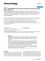
inating genomic DNA in all samples studied (Figure 1).
We employed a pair of HERV-W env-specific PCR primers
located in the region of HERV-W env coding for the SU
domain and generating a PCR product of about 640 bp.
The HERV-W env-specific PCR primers were designed to
amplify the previously reported MSRV env sequence
AF331500 and the HERV-W env locus on chromosome
7q21.2 (ERVWE1). In addition, they potentially recognize
at least eight other HERV-W env loci in the human
genome, as determined by BLAT PCR analyses http://
genome.brc.mcw.edu/cgi-bin/hgPcr. However, according
to more detailed comparisons with HERV-W env
sequences retrieved from the human genome sequence,
the actual number of HERV-W env loci possibly amplified
by the HERV-W env primers is probably even higher, since
further loci with few mismatches to the primers are very
likely to be amplified as well. PCR-products were subse-
quently cloned, and individual cDNA clones were
sequenced. HERV-W env cDNA sequences were assigned
to specific HERV-W env loci in the human genome, based
on characteristic nucleotide differences between HERV-W
env loci (see also Figure 2).
From each individual, we generated a median of 42 (range
40–44) HERV-W env cDNA clones, resulting in a total of
332 cDNA clones, the sequences of which are provided in
additional file 1. To map HERV-W env cDNA sequences
onto individual genomic HERV-W env loci, all 332
sequences were analyzed using human BLAT searches at
the UCSC Human Genome Browser [36]. We thereby
identified, in total, 7 transcribed HERV-W env loci in
human PBMC. A list of those HERV-W env loci and their
main characteristics are provided in Table 1[37]. In partic-
ular, the previously well characterized HERV-W env locus
on chromosome 7q21.2 (ERVWE1), that is, the gene
encoding Syncytin-1, was found to be transcribed in
human PBMC. The 7q21.2 locus contains a full-length
HERV-W proviral copy, flanked by two complete HERV-W
LTRs. As for the structure of the other 6 transcriptionally
active HERV-W env loci, all of them display incomplete
3'LTRs ending just downstream from the poly-A signal,
the expected 3' end of the LTR R-region. In addition, two
of those 6 elements (located on chromosome 6q21, and
15q21.3) show a deletion of the 5' LTR's first 255 nucle-
otides, corresponding to the expected LTR U3 region. The
four remaining elements (5q11.2, 14q21.3, 17q12, and
Xq22.3) are severely truncated at the 5' end, lacking the
5'LTR, the gag region, and varying portions of the 5' pol
region. Structures of transcribed HERV-W env loci are pro-
vided in additional file 2. In summary, except for the
7q21.2 locus, all HERV-W env loci found to be transcrip-
tionally active in human PBMC show characteristic fea-
tures of HERV-W pseudogenes that have been generated
by LINE machinery [11]. In keeping with results obtained
by others [38,39], our data therefore indicate that despite
Expression of HERV-W env in human PBMCFigure 1
Expression of HERV-W env in human PBMC. RT-PCR
using HERV-W env-specific primers was carried out on total
RNA isolated from human PBMC which was subjected (+) or
not (-) to reverse transcription. The expected size of the
amplified HERV-W env fragment is ~640 bp. M, DNA size
marker; H
2
O, PCR negative control.
P
B
M
C
M H
2
O + -
650 bp
500 bp
Retrovirology 2009, 6:37 />Page 4 of 17
(page number not for citation purposes)
having truncated or completely missing 5'LTRs HERV-W
pseudogenes can be transcribed. This implies that as yet
unidentified promotors located upstream of those HERV-
W pseudogenes drive their transcription.
In accordance with previous analyses of the coding capac-
ity of the HERV-W family [14,15,40], except for the
7q21.2 HERV-W env locus, none of the transcribed HERV-
W env loci disclosed ORFs for full-length Env proteins.
Still, a transcriptionally active HERV-W env locus on chro-
mosome Xq22.3 contains an almost complete env ORF,
only interrupted by a single premature stop codon in its 5'
region (codon 39) followed by several in-frame ATGs. If
the longest possible env ORF from this transcribed locus
Examples of recombined HERV-W env cDNA sequencesFigure 2
Examples of recombined HERV-W env cDNA sequences. A multiple alignment of the genomic DNA sequences (March
2006 human genome assembly) of the seven HERV-W env loci identified as transcriptionally active in human PBMC in this study
is shown. HERV-W env loci are designated according to their chromosomal location. The 7q21.2 HERV-W env locus
(ERVWE1) serves as reference sequence. Note that the 7 HERV-W env loci can be distiguished by unique nucleotides and/or
indels. Two of the cloned HERV-W env cDNA sequences, MS-III-K11 (from a patient with MS) and KO-IV-K6 (from a healthy
control) are shown as examples of recombined cDNAs. The proviral origin of cDNA sequence portions is indicated by a color
code. Gray shaded areas represent regions in which recombination events have taken place. Sequences of the primers used in
this study are underlined.
Herv-W_Chr7q21.2 1 TTCACTG-CCCACACCCATATGCCCCGCAACTGCTATCACTCTGCCACTCTTTGCATGCATGCAAATACTCATTATTGGACAGGAAAAATGATTAATCCTAGTTGTCCTGGAGGACTTGG
Herv-W_Chr17q12 1 A A C C
Herv-W_Chr15q21.3 1 C A A G
Herv-W_Chr6q21 1 A A G A
Herv-W_ChrXq22.3 1 A G
Herv-W_Chr5q11.2 1 T T A G T G
Herv-W_Chr14q21.3 1 C T G.A G TT G
MS-III-K11 1 A A C C
KO-IV-K6 1 A A C C
Herv-W_Chr7q21.2 120 AGTCACTGTCTGTTGGACTTACTTCACCCAAACTGGTATGTCTGATGGGGGTGGAGTTCAAGATCAGGCAAGAGAAAAACATGTAAAAGAAGTAATCTCCCAACTCACCCGGGTACATGG
Herv-W_Chr17q12 120 C T T CA C CA G G T A.
Herv-W_Chr15q21.3 120 C G T A C G G T.A A.
Herv-W_Chr6q21 120 C C T G G C G A.
Herv-W_ChrXq22.3 120 C T CA A G A G C G G A.
Herv-W_Chr5q11.2 120 .AC G G T.T T C G G A.
Herv-W_Chr14q21.3 121 C T CA C G CA G G A.
MS-III-K11 120 C T T CA C CA G G T A.
KO-IV-K6 120 C T T CA C CA G G T A.
Herv-W_Chr7q21.2 240 CACCTCTAGCCCCTACAAAGGACTAGATCTCTCAAAACTACATGAAACCCTCCGTACCCATACTCGCCTGGTAAGCCTATTTAATACCACCCTCACTGGGCTCCATGAGGTCTCGGCCCA
Herv-W_Chr17q12 227 C G A G G
Herv-W_Chr15q21.3 240 GC T.T
Herv-W_Chr6q21 240 C T A T
Herv-W_ChrXq22.3 240 C T G C A
Herv-W_Chr5q11.2 238 C A T A
Herv-W_Chr14q21.3 241 C A A A A C T
MS-III-K11 227 C G T.T
KO-IV-K6 227 C G T.T
Herv-W_Chr7q21.2 360 AAACCCTACTAACTGTTGGATATGCCTCCCCCTGAACTTCAGGCCATATGTTTCAATCCCTGTACCTGAACAATGGAACAACTTCAGCACAGAAATAAACACCACTTCCGTTTTAGTAGG
Herv-W_Chr17q12 347 G C T C.CA A T
Herv-W_Chr15q21.3 360 G T C A CA G A
Herv-W_Chr6q21 360 G T.T GCA A
Herv-W_ChrXq22.3 360 G C CA T
Herv-W_Chr5q11.2 358 G C C.CA G.C T T
Herv-W_Chr14q21.3 361 A G C A C A A
MS-III-K11 347 G T C A CA G A
KO-IV-K6 347
Herv-W_Chr7q21.2 480 ACCTCTTGTTTCCAATCTGGAAATAACCCATACCTCAAACCTCACCTGTGTAAAATTTAGCAATACTACATACACAACCAACTCCCAATGCATCAGGTGGGTAACTCCTCCCACACAAAT
Herv-W_Chr17q12 467 T T.G G
Herv-W_Chr15q21.3 476 G.T.G C G
Herv-W_Chr6q21 480 GT.G G
Herv-W_ChrXq22.3 480 T.G G A G
Herv-W_Chr5q11.2 474 C T.G T G
Herv-W_Chr14q21.3 481 C T.G G A
MS-III-K11 463 G.T.G C G
KO-IV-K6 467
Herv-W_Chr7q21.2 600 AGTCTGCCTACCCTCAGGAATATTTTTTGTCTGTGGTACCTC
Herv-W_Chr17q12 584
Herv-W_Chr15q21.3 595
Herv-W_Chr6q21 600 T
Herv-W_ChrXq22.3 600
Herv-W_Chr5q11.2 594 A
Herv-W_Chr14q21.3 601
MS-III-K11 582
KO-IV-K6 587
Retrovirology 2009, 6:37 />Page 5 of 17
(page number not for citation purposes)
were translated, starting at an in-frame ATG at codon 68,
the Xq22.3 HERV-W env locus could give rise to an N-ter-
minally truncated 475 amino acid HERV-W Env protein.
A close inspection of HERV-W env cDNAs reveals a high
number of recombined sequences
Ideally, a HERV-W env cDNA sequence is expected to dis-
play no nucleotide mismatches to the genomic HERV-W
env locus that it originated from. About one third of
HERV-W env cDNAs analyzed in this work indeed per-
fectly matched with genomic DNA sequences. However,
the remaining two thirds of HERV-W env cDNAs con-
tained between 1 and 24 nucleotide differences compared
to the best matching genomic HERV-W env locus.
Although minor nucleotide differences may well be
explained by the inaccuracy of Taq polymerase, sequenc-
ing errors, or sequence variations (SNPs) in genomic
HERV-W env loci, those possibilities seem unlikely to
account for the relatively high numbers of nucleotide mis-
matches observed in some of the cDNA sequences. It has
recently been shown that analyses of transcribed HERV
sequences are complicated by recombinations between
individual HERV transcripts, which most likely arise in
vitro during reverse transcription because of template
switches of reverse transcriptase and/or through PCR-
mediated recombinations [41]. To investigate whether
similar recombinations also occurred in the present study,
we generated multiple sequence alignments of the 7 tran-
scribed HERV-W env loci and the 332 HERV-W env cDNA
sequences. A close inspection of multiple alignments
unambiguously demonstrated that a high number of
HERV-W env cDNAs, that is, 99 out of 332 (29.8%), rep-
resented recombinations between transcripts from differ-
ent HERV-W env loci. Notably, the alleged breakpoints of
recombined sequences appeared to be randomly distrib-
uted. Typical examples of recombined sequences are
shown in Figure 2.
When assuming recombinations, the number of nucle-
otide differences between HERV-W env cDNAs and the
best matching genomic HERV-W env loci was strongly
reduced compared to the number of nucleotide mis-
matches when recombinations were not assumed (Figure
3). Within the ~640 bp sequence analyzed, the average
number of nucleotide mismatches between HERV-W env
cDNAs and the best matching genomic HERV-W env loci
was 3.69 per 640 bp (= 5.77/kb) when no recombinations
were asssumed, as opposed to 0.98 per 640 bp (= 1.53/kb)
when recombinations were assumed. The majority of
recombined cDNAs (67%) resulted from one recombina-
tion event involving transcripts from two different HERV-
W env loci. As for the other sequences, we were able to
detect up to 4 recombination events involving up to five
different HERV-W env loci (Table 2).
Differential transcriptional activity of HERV-W env loci in
human PBMC
Supposing that the relative cloning frequencies (the
number of cDNA clones from a given individual HERV-W
env locus relative to all cDNA clones analyzed) roughly
reflect the relative abundance of RNA transcripts from
individual HERV-W env loci in the total pool of HERV-W
env RNAs in PBMCs, it is possible to estimate the relative
transcriptional activity of individual HERV-W env loci in
PBMC [34,35]. In a first analysis of pooled data from all 8
individuals studied, we used the 233 non-recombined
Table 1: Characteristics of HERV-W env loci identified in this study as transcribed in human PBMC
HERV-W env locus strand location of amplicon in genome 5' LTR 3' LTR processed pseudogene full-length Env ORF
5q11.2 - 56852791 56853425 absent Δ325–780 + -
6q21 + 106788519 106789159 Δ1–255 Δ327–780 + -
7q21.2 (ERVWE1) - 91936808 91937448 complete complete - + (538 aa)
14q21.3 - 44559628 44559996 absent Δ327–780 + -
15q21.3 - 53385554 53386189 Δ1–255 Δ327–780 + -
17q12 + 32765922 32766546 absent Δ327–780 + -
Xq22.3 - 106183197 106183837 absent Δ327–780 + -*
The chromosomal location of transcriptionally active HERV-W env loci is indicated in the first column. Nucleotide positions of amplicons on the
respective chromosomes (human genome sequence March 2006 assembly) are indicated. Structures of the 5' and 3' LTRs of transcribed HERV-W
env loci are given with respect to the 780-bp HERV-W LTR consensus sequence (LTR17) obtained from Repbase />[37]. Missing nucleotides (Δ) in the LTRs as compared to the consensus LTR sequence are indicated. All but one locus represent processed
pseudogenes. Presence of full length ORFs for HERV-W Env proteins is given in the final column.
* The Xq22.3 locus contains an ORF for a hypothetical N-terminally truncated 475 amino acid HERV-W Env protein.
Retrovirology 2009, 6:37 />Page 6 of 17
(page number not for citation purposes)
cDNA sequences to calculate relative cloning frequencies.
As shown in Figure 4, this demonstrated a differential
transcriptional activity of expressed HERV-W env loci in
human PBMC, with transcripts from a HERV-W env locus
on chromosome 15q21.3 being most abundant (111 out
of 233 sequences [47.6%]). In contrast, cDNAs from the
14q21.3 and 5q11.2 HERV-W env loci were only very
rarely cloned (1 sequence from each locus out of 233
sequences [0.4%]).
To compare the transcriptional activity of different HERV-
W env loci the PCR efficiencies with which these loci are
amplified should be similar. PCR-efficiency mostly relies
on the binding of primers to their target sequences. As can
be seen in Figure 2, three of the seven amplified loci
(7q21.2, 17q12, Xq22.3) perfectly matched to the primers
and are thus expected to be amplified with similar effi-
ciencies. The binding regions of three further loci (5q11.2,
6q21, 15q21.3) contained a single nucleotide mismatch
in the 5' end of either the forward or the reverse primer.
Because the 15q21.3 and the 6q21 loci were the most fre-
quently cloned loci in our study, the single nucleotide
mismatches in these two loci appear unlikely to have had
a significant negative impact on amplification. In the case
of the 5q11.2 locus this possibility cannot be excluded,
but seems unlikely given the results for the 15q21.3 and
6q21 loci. One locus (14q21.3) displayed one additional
nucleotide and one nucleotide mismatch in the binding
region of the forward primer, and it seems possible that
those mismatches may have negatively affected its ampli-
fication.
Since the finding of recombined HERV-W env cDNA
sequences implies that the different HERV-W env loci that
contributed to the recombined sequences must have been
transcribed, we also estimated relative cloning frequencies
based on the 99 recombined sequences, counting individ-
ually all HERV-W env loci that were present in the recom-
bined sequences (see also Table 2). The relative cloning
frequencies obtained in this evaluation were overall com-
parable to those of the non-recombined sequences (Figure
4), suggesting that the likelihood of taking part in recom-
binations correlates with transcript abundance.
Finally, not all transcriptionally active loci were detected
as cDNA in every individual. Regarding non-recombined
and recombined sequences together, transcripts from the
5q11.2 locus were detected in one, transcripts from the
14q21.3 in three, and transcripts from the 7q21.2 locus in
seven of the eight individuals studied. The remaining
HERV-W env loci (6q21, 15q21.3, 17q12, Xq22.3) were
found to be transcriptionally active in the PBMC of every
individual studied.
Similar transcriptional activity of individual HERV-W env
loci in PBMC from patients with MS and healthy controls
We next evaluated whether the relative cloning frequen-
cies and thus transcriptional activities of individual
HERV-W env loci differ between patients with MS and
healthy controls. Since the general pattern of transcrip-
tional activity of individual HERV-W env loci was essen-
tially the same regardless of whether recombined
sequences were included in the evaluation or not (Figure
Table 2: Recombined HERV-W env cDNA sequences generated in this study
Recombination events Number of involved loci Number of cDNA sequences Sum of involved HERV-W env transcripts
01 233 233
12 66 132
2 2 11 33
2 3 16 48
33 2 6
34 1 4
45 3 15
Σ = 332 Σ = 471
Recombinations were detected by inspection of multiple alignments of cloned HERV-W env cDNA sequences and transcribed genomic HERV-W
env loci identified in this work (for details, see Methods). Altogether, 99 out of 332 (29.8%) cDNA sequences analyzed represented recombined
sequences. The sum of RNA transcripts from which recombined cDNA orignated is given in the right column. In the case that multiply recombined
sequences involved the same locus more than once, it was assumed that sequences from the same locus originated from different transcripts from
that locus.
Retrovirology 2009, 6:37 />Page 7 of 17
(page number not for citation purposes)
4), we analyzed data from all (non-recombined and
recombined) sequences (see also Table 2). Figure 5 shows
that the variability of the transcriptional activity of the dif-
ferent HERV-W env loci among the different individuals
studied was overall quite high, suggesting inter-individual
differences in the transcriptional activity of HERV-W env
loci. However, there were no significant differences in the
relative cloning frequencies of the different HERV-W env
loci when the group of patients with MS was compared
with the group of healthy controls (p > 0.05; two-tailed
Fisher's exact test).
MSRV sequences can be explained as originating from
distinct HERV-W loci and recombinations among them
Having identified transcriptionally active HERV-W env
loci in human PBMC, we were interested to know how
previously published MSRV env sequences are related to
those transcribed HERV-W env loci. Given the high fre-
quency of in vitro recombinations between HERV-W env
transcripts, we also wondered whether recombinations
may be detectable in MSRV env sequences. To this end, we
retrieved published MSRV sequences comprising parts of
or the entire env region (n = 5) [7,23] from the NCBI data-
base and analyzed them by BLAT searches (see Methods
for details). Since all published MSRV env sequences are
heterogeneous, it is a priori unlikely that all those
sequences are derived from a single proviral insertion.
Quite strikingly, three of the MSRV env containing
sequences (AF127227, AF127228, AF123882) could each
be assigned to a distinct genomic HERV-W locus, namely,
the HERV-W elements on chromosome 3q23, Xq22.3,
and 15q21.3 (Table 3). The other two MSRV env
sequences (AF127229 and AF331500) could be explained
as recombinations between different HERV-W loci.
AF127229 represents a recombination between two
HERV-W loci located on chromsome 3p12.3 and
18q21.32. Likewise, AF331500 represents a recombina-
tion between two HERV-W elements on Xq22.3 and 5p12.
Similar to the data shown in Figure 3, the number of
nucleotide mismatches between AF127229 or AF331500
and the best matching genomic HERV-W loci was strongly
reduced when recombinations were assumed (Table 3).
Alignments of MSRV sequences with the respective best
matching HERV-W loci are provided in additional file 3.
Notably, two HERV-W elements (15q21.3 and Xq22.3)
Nucleotide mismatches between HERV-W env cDNAs and best matching genomic HERV-W env lociFigure 3
Nucleotide mismatches between HERV-W env cDNAs and best matching genomic HERV-W env loci. White
bars represent the number of nucleotide mismatches between HERV-W env cDNAs (n = 332) and their best matching genomic
HERV-W env locus without assuming the presence of recombined HERV-W env sequences among those cDNAs. Black bars
indicate the number of nucleotide mismatches between HERV-W env cDNAs and their best matching genomic HERV-W env
loci when the presence of recombination events in 99 out of 332 HERV-W env cDNAs (see Table 2) was taken into account.
0
20
40
60
80
100
120
140
160
180
0123456789101112131415161718192021222324
number of nucleotide mismatches to best matching HERV-W loci
number of sequences
non-recombined
recombined
Retrovirology 2009, 6:37 />Page 8 of 17
(page number not for citation purposes)
from which MSRV env sequences originated were found to
be transcribed in human PBMC in the present work. The
four other HERV-W elements (3q23, 3p12.3, 18q21.32,
and 5p12) from which MSRV env sequences are derived
were not identified as transcriptionally active in human
PBMC in our investigation. However, due to various dele-
tions the binding sites for one or both of the HERV-W env
primers employed in our work are missing in those four
HERV-W loci, indicating that corresponding cDNAs could
not be amplified (data not shown). Therefore, it remains
possible that those four HERV-W loci are transcriptionally
active, too.
We could also assign the formerly published MSRV gag
sequence (AF123881) to a distinct HERV-W element on
chromosome 3q26.32. This HERV-W locus has formerly
been identified as transcriptionally active in human
PBMC [39] and is identical to a HERV-W gag gene on
chromosome 3 (AF156961), previously characterized by
Voisset et al. [14]. Although the 3q26.32 HERV-W gag
gene is incomplete, it contains the largest HERV-W gag
ORF in the human genome, with a putative coding capac-
ity for a 45 kDa HERV-W Gag protein, consisting of a com-
plete matrix domain and a C-terminally truncated capsid,
but lacking nucleocapsid [14].
Notably, the average number of nucleotide mismatches
between MSRV env and gag sequences and the respective
best matching genomic HERV-W loci (1.97/kb; see Table
3) were in the same range as that observed in our study of
transcribed HERV-W env loci in human PBMC (1.53/kb).
In summary, our analyses suggest that previously pub-
Relative cloning frequencies of transcriptionally active HERV-W env loci in human PBMCFigure 4
Relative cloning frequencies of transcriptionally active HERV-W env loci in human PBMC. The relative cloning fre-
quencies are given as the number of cDNA clones from a particular HERV-W env locus relative to the number of all cDNA
clones analyzed. Frequencies were calculated separately for all non-recombined clones (n = 233 sequences; white bars), all
recombined clones (n = 99 sequences, originating from 238 transcripts [see text and Table 2]; black bars), and for non-recom-
bined and recombined clones together (n = 332; originating from 471 transcripts [see text and Table 2]; gray bars).
0
10
20
30
40
50
60
15q21.3 6q21 Xq22.3 7q21.2 17q12 14q21.3 5q11.2
HERV-W
env
locus
relative cloning frequency (%)
non-recombined
recombined
all
Retrovirology 2009, 6:37 />Page 9 of 17
(page number not for citation purposes)
lished MSRV sequences originated from genomic HERV-
W loci, or recombinations among them.
Relationship between Xq22.3 HERV-W env and MSRV env
The MSRV env clone AF127228 and the SU and N-termi-
nal TM regions of the MSRV env clone AF331500 corre-
spond to the HERV-W env element on chromosome
Xq22.3 which harbors a long ORF for a putative 475
amino acid HERV-W Env protein (Table 3). Accordingly,
the amino acid sequence of a recombinant MSRV Env SU
protein, which has been shown to have proinflammatory
effects in various assays [24], and which was generated
using the AF331500 MSRV env clone, is identical to the
amino acid sequence of the HERV-W Env protein puta-
tively encoded by Xq22.3 HERV-W env (Figure 6) [42].
However, in contrast to Xq22.3 HERV-W Env which is N-
terminally truncated due to a stop codon (TGA) at posi-
tion 39, this stop codon is a tryptophan residue (TGG) in
the AF331500 MSRV env clone. Remarkably, the elimina-
tion of the stop codon at position 39 of HERV-W env
Xq22.3 results in an uninterrupted full-length HERV-W
env ORF, which could encode a complete HERV-W Env
protein that contains a signal peptide (Figure 6). The ori-
gin of the stop codon mutation in the MSRV env
AF331500 clone is unknown. Since several genomic
HERV-W env elements display a TGG at the particular
position, it is conceivable that a recombination event
involving transcripts from the Xq22.3 locus and a short
Relative cloning frequencies of transcriptionally active HERV-W env loci in human PBMC from patients with MS and healthy controlsFigure 5
Relative cloning frequencies of transcriptionally active HERV-W env loci in human PBMC from patients with
MS and healthy controls. Relative cloning frequencies were calculated for recombined and non-recombined clones together
(n = 332 sequences, originating from 471 transcripts [see text and Table 2]). The box represents the mean, and the whiskers
represent the minimum and maximum of the relative cloning frequencies of cDNAs from individual HERV-W env elements for
the groups of patients with MS (n = 4) and healthy controls (n = 4). There were no statistically significant differences between
patients and controls (p > 0.05; two-tailed Fisher's exact test).
HERV-W env locus
relative cloning frequency (%)
healthy controls
patients with MS
Retrovirology 2009, 6:37 />Page 10 of 17
(page number not for citation purposes)
sequence stretch from one of the TGG containing HERV-
W env loci might account for the reversal of the stop codon
(data not shown).
In contrast to AF331500, the MSRV env clone AF127228,
which likewise originates from the Xq22.3 HERV-W env
locus, displays the stop codon at position 39. A DNA frag-
ment comprising amino acids 68 to 446 of the HERV-W
env ORF encoded by AF127228 has previously been used
to generate the monoclonal anti HERV-W Env antibody
6A2B2 [16]. As shown in Figure 6, except for two amino
acid exchanges in its C-terminus, the AF127228 amino
acids 68 to 446 sequence is identical to the amino acid
sequence of Xq22.3 HERV-W Env, but displays 38 mis-
matches to the Syncytin-1 amino acid sequence. Never-
theless, the 6A2B2 antibody may cross react with
Syncytin-1 [16,43] and all previous neuropathological
studies that reported a dysregulated expression of Syncy-
tin-1 in MS lesions relied on the 6A2B2 antibody
[18,21,22]. Still, assuming that HERV-W Xq22.3 env may
have the potential to code for a HERV-W Env protein, our
findings open the intriguing possibility that the protein
detected by 6A2B2 in MS lesions could instead have orig-
inated from the HERV-W env locus on chromosome
Xq22.3.
Discussion
When studying HERV RNA expression in human diseases,
it seems important to clearly dissect from which genomic
HERV loci the detected HERV RNA transcripts originate
[44]. Consistent with previous findings suggesting that
expression of HERV transcripts is a ubiquitious phenome-
non occurring in every human tissue [34,45,46], we
herein show that at least seven HERV-W env loci are tran-
scribed in PBMC from patients with MS and healthy con-
trols. Since the primers used in this investigation only
amplify a limited number of genomic HERV-W env ele-
ments our study is not exhaustive, and it seems rather
likely that more than seven HERV-W env elements are
transcriptionally active in human PBMC. Additionally,
HERV-W env loci that are transcribed at very low rates
could be missed in the cloning procedure unless much
higher numbers of clones are generated. Three of the tran-
scribed HERV-W env elements (15q21.3, Xq22.3, 17q12)
identified in this study were previously found to be
expressed in human PBMC by a cloning and sequencing
approach [38,39]. Assignment of cDNAs to genomic
HERV-W env loci in the former investigations was based
on rather short sequences (30 bp, excluding primers),
containing only few informative nucleotides, that is,
nucleotides that are exclusively present in a single
genomic HERV-W env locus and thereby allow unambig-
uous assignment of cDNAs. Usage of a ~600 bp sequence
(excluding primers) in the present work resulted in a
higher number of informative nucleotides and thus
strengthened the accuracy of the assignment. Our finding
of ERVWE1 transcripts in human PBMC is consistent with
previous observations [19,20] and corroborates that
Table 3: Origin of previously described MSRV sequences
MSRV sequences HERV-W source locus/loci Number of nucleotide mismatches
env no recombinations recombinations
AF127227 (544 bp) 3q23 1 (1.84/kb) n.a.
AF127228 (1932 bp) Xq22.3 4 (2.07/kb) n.a.
AF127229 (2004 bp) 3p12.3/18q21.32 94 (46.91/kb) 3 (1.5/kb)
AF123882 (2477 bp) 15q21.3 5 (2.02/kb) n.a.
AF331500 (1629 bp) Xq22.3/5p12 31 (19.03/kb) 5 (3.07/kb)
gag
AF123881 (1511 bp) 3q26.32 2 (1.32/kb) n.a.
Previously published MSRV sequences were assigned by BLAT searches to corresponding HERV-W elements in the human genome. The accession
number and length (base pairs, bp) of published MSRV clones are provided in the left column. The best matching HERV-W locus or loci (in case of
recombined sequences) are indicated in column 2, and the number of nucleotide mismatches between MSRV sequences and the best matching
genomic HERV-W elements in column 3. Note that for the recombined sequences, the number of nucleotide mismatches after assuming
recombinations is markedly reduced. The average number of nucleotide differences of the 6 analyzed MSRV sequences to their best matching
genomic HERV-W locus/loci was 1.97/kb. n.a., not applicable
Retrovirology 2009, 6:37 />Page 11 of 17
(page number not for citation purposes)
Figure 6 (see legend on next page)
Xq22.3 1 MALPYHIFLFTVLLPPFALTAPPPCCCTTSSSPYQEFL·RTRLPGNIDAPSYRSLSKGNS
AF127228 1 T ·
AF331500 1 T W
syncytin-1 1 S.T R.M W.MQR TP
Xq22.3 61 TFTAHTHMPRNCYNSATLCMHANTHYWTGKMINPSCPGGLGATVCWTYFTHTSMSDGGGI
AF127228 61
AF331500 61
syncytin-1 61 H V Q.G V
Xq22.3 121 QGQAREKQVKEAISQLTRGHSTPSPYKGLVLSKLHETLRTHTRLVSLFNTTLTRLHEVSA
AF127228 121
AF331500 121
syncytin-1 121 .D H V V.G.S D G
Xq22.3 181 QNPTNCWMCLPLHFRPYISIPVPEQWNNFSTEINTTSVLVGPLVSNLEITHTSNLTCVKF
AF127228 181
AF331500 181
syncytin-1 181 I N V
Xq22.3 241 SNTIDTTSSQCIRWVTPPTRIVCLPSGIFFVCGTSAYHCLNGSSESMCFLSFLVPPMTIY
AF127228 241
AF331500 241
syncytin-1 241 TY N Q R
SU TM
Xq22.3 301 TEQDLYNHVVPKPHNKRVPILPFVIRAGVLGRLGTGIGSITTSTQFYYKLSQEINGDMEQ
AF127228 301
AF331500 301
syncytin-1 301 SY.IS R G A G L R
Xq22.3 361 VTDSLVTLQDQLNSLAAVVLQNRRALDLLTAKRGGTCLFLGEECCYYVNQSRIVTEKVKE
AF127228 361 R
AF331500 361 R
5p12 1 N E N G.I
syncytin-1 361 .A E G
Xq22.3 421 IRDRIQCRAEELQNTEHWGLLSQWMPWVLPFLGPLAALILLLLFGPCIFNLLVKFVSSRI
AF127228 421 R
AF331500 421 R T I.F F
5p12 61 .DR I D AP T I.F F
syncytin-1 421 R R GP I I N
Xq22.3 481 EAVKLQMVLQMEPQMQSMTKIYHGPLDQPASPCSDVNDIKGTPPEEISTAQPLPCPISAG
AF127228 481
AF331500 481 I R R RL EV LHSN.V.
5p12 121 I R R RL EV LHSN.V.
syncytin-1 481 K K RR R R A LR.N
Xq22.3 541 SR
AF127228
AF331500 541 .S
5p12 181 .S
syncytin-1 537 .S
Retrovirology 2009, 6:37 />Page 12 of 17
(page number not for citation purposes)
although ERVWE1 expression is most abundant in pla-
centa this locus is transcribed in non-placental tissues as
well [15].
Several studies have analyzed expression of HERV-W env
RNA in PBMC or brain tissue from patients with MS [18-
21]. Lack of systematic cloning, sequencing, and assign-
ment of cDNA sequences to genomic HERV-W env loci
have impaired the exact identification of transcriptionally
active genomic HERV-W env loci responsible for the
observed HERV-W env RNA expression in these investiga-
tions. Whereas in the present detailed analysis we could
identify distinct transcriptionally active HERV-W env loci,
we did not observe significant differences in the transcrip-
tional activity of those loci in PBMC from patients with
MS versus healthy controls. Although the number of indi-
viduals studied was rather small, these data argue against
a dysregulated transcription pattern of HERV-W env in
PBMC from patients with MS. In contrast, a consistent
finding of former investigations was a significantly higher
global HERV-W env RNA expression in brain tissue from
patients with MS as compared to brain tissue from
patients with other neurological diseases or normal brain
tissue [18-21]. Using the methodological approach of the
present work, it will therefore be interesting to identify the
HERV-W env elements underlying upregulated HERV-W
env RNA expression in MS brain tissue.
Antony and coworkers addressed this question by design-
ing primers that specifically amplify HERV-W env 7q21.2
(ERVWE1) and the MSRV env clone AF123882 [20],
which, as shown by our analyses, corresponds to a HERV-
W env element on chromosome 15q21.3. These authors
also employed a pair of degenerate (HERV-W
deg
) env
primers that were based on the MSRV env clone
AF331500, which, again as shown in this work, corre-
sponds to a recombined cDNA originating from HERV-W
env loci on Xq22.3 and 5p12. According to the Antony et
al. study, elevated HERV-W env RNA expression in MS
brain tissue originates mainly from the HERV-W env ele-
ments amplified by the HERV-W
deg
env primers and
(somewhat less) from HERV-W env 7q21.2, while HERV-
W env 15q21.3 expression was similar in patients and con-
trols [20]. A BLAT-PCR search showed that the HERV-W
deg
env primer pair potentially amplifies at least three
genomic HERV-W env loci, among them HERV-W env
Xq22.3. It is thus tempting to speculate that HERV-W env
Xq22.3 may significantly contribute to increased HERV-W
env RNA expression in MS brain tissue. Again, using the
methods described herein, this issue could be resolved in
a straightforward manner.
Remarkably, we observed a high number (29.8%) of
recombined sequences among the analyzed HERV-W env
cDNAs. As detailed in a previous study on transcribed
HERV-K(HML-2) sequences [41], those recombinant
cDNA sequences very likely resulted from in vitro recom-
binations that were due to template switches of reverse
transcriptase during cDNA synthesis and/or PCR-medi-
ated recombinations. Both of these mechanisms are well-
recognized and have been proven experimentally to pro-
duce chimeric sequences [41,47-53]. The percentage of
recombined sequences detected in the present study was
higher than that in the study on HERV-K(HML-2) in
which ~5% of recombined clones were observed [41].
This is most likely explained by the fact that in the HERV-
K(HML-2) study only cDNA sequences with more than 17
nucleotide mismatches to the best matching locus were
analyzed for recombinations, whereas in the present work
all cDNA sequences were scrutinized for recombinations.
Altogether, our data indicate that during experimental
studies of repetitive elements by RT-PCR, in vitro recombi-
nations are relatively common and almost inevitable
complications.
An important result of this investigation is that previously
published MSRV env and gag sequences appear to either
be derived from transcripts of specific genomic HERV-W
elements or to result from recombinations among such
transcripts (Table 3). Given the high frequency of in vitro
recombinations between transcripts from different HERV
loci observed in this and the study by Flockerzi et al. [41],
Relationship between Xq22.3 HERV-W Env, MSRV Env, and Syncytin-1Figure 6 (see previous page)
Relationship between Xq22.3 HERV-W Env, MSRV Env, and Syncytin-1. An amino acid sequence alignment of
Xq22.3 HERV-W Env, MSRV Env (clones AF127228 and AF331500), and Syncytin-1 is shown. The sequence of a HERV-W ele-
ment on chromosome 5p12 from which the C-terminal region of the MSRV env clone AF331500 is derived (see also Table 3
and Additional file 2) is also shown. For the sake of simplicity, only the C-terminal region of the 5p12 element is included. The
region of MSRV Env (AF331500) originating from HERV-W 5p12 is highlighted in yellow. Predicted signal peptides (according
to SignalP 3.0, />) are shaded in gray. The stop codon at position 39 of Xq22.3 HERV-W
Env and AF127228 is indicated by a dot (·). The consensus C-X-X-C motif conserved among C-type and D-type retroviral Env
proteins [42] is shown in boldface. The border between the SU and TM regions is indicated by arrows. The proteolytic cleav-
age site (consensus R/K-X-R/K-R) between SU and TM is highlighted in red letters. The sequences of the MSRV Env SU protein
(generated using the MSRV env clone AF331500) studied by Rolland et al. [24] is marked in red. The fragment of the MSRV env
clone AF127228 used for generation of the anti HERV-W Env monoclonal antibody 6A2B2 [16] is shown in green.
Retrovirology 2009, 6:37 />Page 13 of 17
(page number not for citation purposes)
and given that MSRV clones were generated by methodo-
logically similar approaches, it seems possible that the
recombined MSRV env sequences (AF127229, AF331500)
have resulted from in vitro recombinations as well.
An alternative explanation is that the recombined MSRV
env sequences, and the recombined HERV-W env
sequences isolated in this study, originated from novel,
recombined, genomic HERV-W insertions. Hypotheti-
cally, such insertions could have formed in vivo after
recombination of RNA transcripts from different HERV-W
env loci through template switches during reverse tran-
scription. Although we cannot formally exclude this pos-
sibility, a number of points argue against it. First, all
known HERV-W elements are defective and replication-
incompetent [8,14]. Therefore, HERV-W is a priori rather
unlikely to have the capacity to form new insertions in
human DNA. Second, if there were novel recombined
HERV-W loci in human DNA, one would expect to repeat-
edly observe defined recombined sequences originating
from such insertions. However, this is neither the case
with the 99 recombined HERV-W env cDNA sequences
analyzed in this study nor with the published MSRV env
clones. Third, given that about 30% of HERV-W env and
33% (2 of 6) of the investigated MSRV sequences repre-
sent recombinants, if all these recombinant MSRV/HERV-
W env sequences were derived from novel proviral inser-
tions, formation of such novel insertions would be an
astonishingly frequent event. It seems very unlikely that as
many recombined HERV-W loci should have been over-
looked in previous genome sequencing projects.
Collectively, the most plausible and simplest explanation
for the origin of MSRV env and gag sequences seems to be
that those sequences originate from RNA transcripts from
various endogenous HERV-W loci, or from in vitro recom-
binations among them. All of the HERV-W loci from
which MSRV sequences are derived are defective and
except for the 5p12 HERV-W env element, all of those loci
resemble processed HERV-W pseudogenes. The human
genome sequence was not yet available when MSRV was
described, which hampered the identification of the pre-
cise origin of MSRV sequences at that time. It was, how-
ever, noted that those sequences cannot be attributed to a
single replication-competent genome [7]. Nevertheless,
the nature of MSRV was subsequently controversial, and it
has been speculated that MSRV could be an exogenous,
replication-competent retrovirus [6,30-32]. In contrast,
our present data clearly suggest that the published MSRV
env and gag RNA sequences are not derived from the
genome of a currently replication-competent exogenous
retrovirus. In the light of these results and previous obser-
vations of an increased prevalence of MSRV pol transcripts
in plasma from patients with MS as compared to healthy
controls [54,55], it may similarly be interesting to analyze
which HERV-W pol elements those MSRV pol transcripts
could be derived from.
Although our findings argue against MSRV being an
autonomous retroviral entity, they do by no means rule
out that individual HERV-W env loci that correspond to
MSRV sequences, or the Syncytin-1 (ERVWE1) gene, could
be of relevance in MS. Indeed, we show that two MSRV env
clones (AF331500, AF127228), which have been instru-
mental for the characterization of proinflammatory
effects of MSRV Env [24] and the generation of a mono-
clonal anti-MSRV/HERV-W Env antibody (6A2B2) [16],
are derived from a HERV-W env locus on chromosome
Xq22.3. This locus is highly remarkable as it is interrupted
by only a single premature stop at codon position 39 and
otherwise harbors a long ORF for a N-terminally trun-
cated 475 amino acid HERV-W Env protein (Figure 6).
Bonnaud and colleagues described frameshift insertions/
deletions (indels), that is, indels whose length is not a
multiple of three, in 33 out of 36 analyzed genomic
HERV-W env loci. Interestingly, among the three loci with-
out frameshift indels were the ERVWE1 gene and the
Xq22.3 HERV-W env element [43]. We further note that
the 475 amino acid Xq22.3 HERV-W env ORF is also
present in the orthologous locus in chimpanzees (data
not shown). These data may be taken as hints that selec-
tive pressure could act on the Xq22.3 HERV-W env locus,
raising the possibility that Xq22.3 HERV-W env could
exert a biological function. Our finding that the Xq22.3
HERV-W env locus is transcriptionally active in human
cells indicates that it fulfills at least one essential prerequi-
site for a protein expression capacity in vivo.
Neuropathological studies revealed that the 6A2B2 anti-
HERV-W Env antibody reacts with an antigen that is
strongly expressed by glial cells in MS brain lesions, but
not in normal control brain tissue [18,21,22]. Because
Syncytin-1 has been thought to be the only HERV-W env
locus capable of producing a HERV-W Env protein, and
because 6A2B2 may crossreact with Syncytin-1 [16,43],
the antigen detected by 6A2B2 in MS brain lesions was
considered to be Syncytin-1. However, our analyses show
that the protein against which the 6A2B2 antibody was
raised is practically identical to the Xq22.3 HERV-W Env
protein (Figure 6) [16]. We meanwhile cloned Xq22.3
HERV-W env into a eukaryotic expression vector. Transient
transfection of HeLa cells with this clone showed that the
Xq22.3 HERV-W env has retained a coding capacity and
can produce a HERV-W Env protein in vitro which is
detected by the 6A2B2 antibody in immunocytochemistry
and immunoblots (C. Crusius, S. Wahl, K. Ruprecht, man-
uscript in preparation). These data suggest that the anti-
gen recognized by 6A2B2 in MS lesions could likewise
originate from the Xq22.3 HERV-W env locus, provided
that this locus has a protein expression capacity in vivo.
Retrovirology 2009, 6:37 />Page 14 of 17
(page number not for citation purposes)
More elaborate studies will be required to clarify the exact
nature of the HERV-W Env protein detected in MS lesions.
Further characterization of the putative Xq22.3-encoded
HERV-W Env protein, especially in comparison to Syncy-
tin-1, will be necessary for such clarification.
Conclusion
In conclusion, we demonstrate that several HERV-W env
loci are transcribed in human PBMC, and that analysis of
such transcribed HERV-W env elements is complicated by
frequent recombinations, which are most likely generated
in vitro. Based on these findings, we show that previously
reported MSRV env and gag sequences can be explained as
originating from (in some instances recombined) tran-
scripts of defective HERV-W elements, arguing against
MSRV sequences being derived from an infectious exoge-
nous retrovirus. Our results should help to settle the issue
of the nature of MSRV and contribute to the clarification
of the roles of MSRV versus HERV-W Env (Syncytin-1) in
MS. Indeed, our findings raise the intriguing possibility
that a protein encoded by a HERV-W env element on chro-
mosome Xq22.3 could be expressed in MS brain lesions.
Methods
Patients with MS and healthy controls
Four patients with MS (3 female, 1 male) and 4 healthy
controls (2 female, 2 male) were included in this study.
The median age of patients was 34 (range 29–39) and of
controls 34.5 (29–41) years. Clinical data of patients with
MS were obtained by review of the medical records. All
patients with MS had a diagnosis of definite MS according
to Poser's criteria [56]. Three patients had relapsing-remit-
ting MS, and one patient had secondary progressive MS.
The median expanded disability status scale score of
patients with MS was 3.25 (1.5–6.5). One patient was
treated with interferon-beta 1a, and two patients were
treated with glatiramer acetate by the time of blood collec-
tion. None of the patients had been treated with glucocor-
ticosteroids for at least 6 months before blood collection.
Participants provided written informed consent, and the
study was approved by the ethics committee of the faculty
of medicine, Julius-Maximilians University, Würzburg.
PBMC samples used in this work were collected and puri-
fied with Lymphoprep (Axis Shield, Oslo, Norway) gradi-
ent centrifugation as described before [57]. Samples were
stored at -80°C prior to the present analysis.
RT-PCR
PBMC were thawed and cultured overnight in RPMI 1640
(BioWhittaker) supplemented with 10% FCS and penicil-
lin (100 U/ml) and streptomycin (100 μg/ml) at 37°C in
a humidified 5% (v/v) CO
2
atmosphere. Total RNA was
extracted from PBMC using the RNeasy Mini kit (Qiagen)
and eluted in 60 μl of distilled water. RNA concentration
and purity were assessed spectrophotometrically. Con-
taminating DNA was removed using the TURBO DNA-
free Kit (Ambion Inc.) following the protocol for rigorous
DNAse treatment. In brief, 2 units of TURBO DNase were
added to a 50-μl reaction containing 10 μg RNA and incu-
bated for 30 minutes at 37°C. Another 2 units of TURBO
DNase were added and the incubation was continued for
30 minutes at 37°C. DNAse was removed using 10 μl of
the provided DNAse inactivation reagent. Subsequently,
0.3–0.5 μg of DNase digested cellular RNA was reverse
transcribed in a 20-μl reaction using Superscript II (Invit-
rogen) and 25 μM random hexamer primers (MWG-Bio-
tech AG) according to the protocol of the manufacturer.
Negative controls were generated in parallel for each sam-
ple by omission of Superscript II from the reaction. PCR
primer sequences for amplification of HERV-W env were
as follows: forward primer 5'-TTCACTGCCCACACCCAT-
3'; reverse primer 5'-GAGGTACCACAGACAAAAAATAT-
TCCT-3'. Conventional PCR was performed in a 50-μl
reaction containing 1 μl of cDNA, 0.5 μM of each primer,
200 μM of each dNTP, reaction buffer (10 mM Tris-HCl,
50 mM KCl, 1.5 mM MgCl
2
), and 0.05 units/μl of Taq
DNA Polymerase (D1806, Sigma). Cycling parameters
were as follows: 3 minutes at 95°C; 40 cycles of 50 sec at
95°C, 50 sec at 58°C, and 1 minute at 72°C; and 10 min-
utes at 72°C.
Cloning of HERV-W env transcripts and assignment to
proviral HERV-W loci
PCR products were excised from agarose gels, purified
(NucleoSpin Extract II, Macherey-Nagel), and ligated into
the pGEM-T vector (Promega). Plasmid DNA from ran-
domly selected insert-containing clones was purified with
the QIAprep Miniprep kit (Qiagen) and sequenced on an
Applied Biosystems 3730x Capillary Sequencer using vec-
tor-specific primers (Institut für Immunologie und Gene-
tik, Kaiserlautern, Germany). The quality of
chromatograms was assessed by visual inspection. Poor-
quality reads (< 0.1% of all sequences) were excluded
from the analysis.
Assignment of cDNA sequences to corresponding HERV-
W env loci is based on random and thus characteristic
nucleotide differences between the various genomic
HERV-W env loci. The proviral HERV-W env locus with no
or very few nucleotide mismatches to a HERV-W env
cDNA sequence can be assumed to represent the origin of
this cDNA, if all other alternative loci displayed more
nucleotide differences. A detailed discussion of the
sequence assignment strategy has recently been provided
[34].
To assign HERV-W env cDNA clones to specific HERV-W
env loci in the human genome, HERV-W env cDNA
sequences were first analyzed by BLAT searches (http://
genome.ucsc.edu/cgi-bin/hgBlat; March 2006 human
Retrovirology 2009, 6:37 />Page 15 of 17
(page number not for citation purposes)
genome assembly). To further study recombinations
between different HERV-W env loci in HERV-W env cDNA
sequences, sequences of the seven transcribed HERV-W
env loci were retrieved from the human genome sequence
(March 2006 assembly) at the Human Genome Browser
and multiply aligned with HERV-W env cDNA sequences
using Muscle 3.6 [58]. Candidate HERV-W env cDNA
sequences were then inspected for recombination events.
Analysis of MSRV sequences
Previously published MSRV env and gag sequences were
retrieved from GenBank and analyzed by BLAT searches to
identify endogenous HERV-W loci with similarities to
MSRV sequences. Alignments of the MSRV sequences with
the best matching HERV-W locus were manually
inspected for evidence of recombination events. In recom-
bined sequence portions, nucleotide mismatches between
MSRV sequences and the best matching HERV-W
sequence usually clustered in defined subregions. Presum-
ably recombined subregions were used as probe
sequences for another BLAT search to detect their best
matching HERV-W locus. Sequences of thus identified
HERV-W loci were again retrieved from the Human
Genome Browser and aligned with the corresponding
MSRV sequences.
Competing interests
The authors declare that they have no competing interests.
Authors' contributions
JM, KR, and NML conceived of the study, participated in
its design, and provided funding. GL, BFM, and KR carried
out the molecular genetic studies. GL, KR, and JM ana-
lyzed the data. KR drafted the manuscript. All authors read
and approved the final manuscript.
Additional material
Acknowledgements
This study was supported by grants from HOMFOR to KR and JM. JM and
NML are furthermore supported by grants from the Deutsche Forschungs-
gemeinschaft (DFG).
References
1. Noseworthy JH: Progress in determining the causes and treat-
ment of multiple sclerosis. Nature 1999, 399:A40-A47.
2. Gessain A, Barin F, Vernant JC, Gout O, Maurs L, Calendar A, de The
G: Antibodies to HTLV-1 in patients with tropical spastic
paraparesis. The Lancet 1985, 2:407-410.
3. Perron H, Geny C, Laurent A, Mouriquand C, Pellat J, Perret J, Sei-
gneurin JM: Leptomeningeal cell line from multiple sclerosis
with reverse transcriptase activity and viral particles.
Research in Virology 1989, 140:551-561.
4. Perron H, Lalande B, Gratacap B, Laurent A, Genoulaz O, Geny C,
Mallaret M, Schuller E, Stoebner P, Seigneurin JM: Isolation of ret-
rovirus from patients with multiple sclerosis. Lancet 1991,
337:862-863.
5. Haahr S, Sommerlund M, Moller-Larsen A, Nielson R, Hansen HJ: Just
another dubious virus in cells from a patient with multiple
sclerosis? Lancet 1991, 337:863-864.
6. Perron H, Garson JA, Bedin F, Beseme F, Paranhos-Baccala G, Komu-
rian-Pradel F, Mallet F, Tuke PW, Voisset C, Blond JL, et al.: Molecu-
lar identification of a novel retrovirus repeatedly isolated
from patients with multiple sclerosis. Proc Natl Acad Sci USA
1997, 94:7583-7588.
7. Komurian-Pradel F, Paranhos-Baccala G, Bedin F, Ounanian-Paraz A,
Sodoyer M, Ott C, Rajoharison A, Garcia E, Mallet F, Mandrand B, et
al.: Molecular cloning and characterization of MSRV-related
sequences associated with retrovirus-like particles. Virology
1999, 260:1-9.
8. Blond J-L, Beseme F, Duret L, Bouton O, Bedin F, Perron H, Man-
drand B, Mallet F: Molecular characterization and placental
expression of HERV-W, a new human endogenous retrovi-
rus family. J Virol 1999, 73:1175-1185.
9. Bannert N, Kurth R: Retroelements and the human genome:
new perspectives on an old relation. Proc Natl Acad Sci USA 2004,
101(Suppl 2):14572-14579.
10. de Parseval N, Heidmann T: Human endogenous retroviruses:
from infectious elements to human genes.
Cytogenet Genome
Res 2005, 110:318-332.
11. Pavlicek A, Paces J, Elleder D, Hejnar J: Processed pseudogenes of
human endogenous retroviruses generated by LINEs: their
integration, stability, and distribution. Genome Res 2002,
12:391-399.
12. Belshaw R, Katzourakis A, Paces J, Burt A, Tristem M: High copy
number in human endogenous retrovirus families is associ-
ated with copying mechanisms in addition to reinfection. Mol
Biol Evol 2005, 22(4):814-817.
13. Costas J: Characterization of the intragenomic spread of the
human endogenous retrovirus family HERV-W. Mol Biol Evol
2002, 19(4):526-533.
14. Voisset C, Bouton O, Bedin F, Duret L, Mandrand B, Mallet F, Para-
nhos-Baccala G: Chromosomal distribution and coding capac-
ity of the human endogenous retrovirus HERV-W family.
AIDS Res Hum Retroviruses 2000, 16:731-740.
15. de Parseval N, Lazar V, Casella JF, Benit L, Heidmann T: Survey of
human genes of retroviral origin: identification and tran-
Additional file 1
Sequences of the 332 HERV-W env cDNAs analyzed in this study. This
file contains raw sequence data of the 332 HERV-W env cDNAs analyzed
in this work.
Click here for file
[ />4690-6-37-S1.doc]
Additional file 2
Pustell matrix comparisons of the seven HERV-W env loci identified
as transcriptionally active in human PBMC in this study. This file con-
tains Pustell matrix comparisons of the Repbase />update/ HERV-W reference sequence with the seven HERV-W env loci
identified as transcriptionally active in human PBMC in this work.
Click here for file
[ />4690-6-37-S2.ppt]
Additional file 3
Alignments of previously published MSRV env and gag sequences with
their corresponding genomic HERV-W elements. This file contains
annotated alignments of previously published MSRV env and gag
sequences with the genomic HERV-W elements from which the respective
MSRV sequences are most likely derived.
Click here for file
[ />4690-6-37-S3.doc]
Retrovirology 2009, 6:37 />Page 16 of 17
(page number not for citation purposes)
scriptome of the genes with coding capacity for complete
envelope proteins. J Virol 2003, 77(19):10414-10422.
16. Blond JL, Lavillette D, Cheynet V, Bouton O, Oriol G, Chapel-Fern-
andes S, Mandrand B, Mallet F, Cosset FL: An envelope glycopro-
tein of the human endogenous retrovirus HERV-W is
expressed in the human placenta and fuses cells expressing
the type D mammalian retrovirus receptor. J Virol 2000,
74:3321-3329.
17. Mallet F, Bouton O, Prudhomme S, Cheynet V, Oriol G, Bonnaud B,
Lucotte G, Duret L, Mandrand B: The endogenous retroviral
locus ERVWE1 is a bona fide gene involved in hominoid pla-
cental physiology. Proc Natl Acad Sci USA 2004, 101(6):1731-1736.
18. Antony JM, van Marle G, Opii W, Butterfield DA, Mallet F, Yong VW,
Wallace JL, Deacon RM, Warren K, Power C: Human endogenous
retrovirus glycoprotein-mediated induction of redox reac-
tants causes oligodendrocyte death and demyelination. Nat
Neurosci 2004, 7(10):1088-1095.
19. Antony JM, Izad M, Bar-Or A, Warren KG, Vodjgani M, Mallet F,
Power C: Quantitative analysis of human endogenous retro-
virus-W env in neuroinflammatory diseases. AIDS Res Hum
Retroviruses 2006, 22(12):1253-1259.
20. Antony JM, Zhu Y, Izad M, Warren KG, Vodjgani M, Mallet F, Power
C: Comparative expression of human endogenous retrovi-
rus-W genes in multiple sclerosis. AIDS Res Hum Retroviruses
2007, 23(10):1251-1256.
21. Mameli G, Astone V, Arru G, Marconi S, Lovato L, Serra C, Sotgiu S,
Bonetti B, Dolei A: Brains and peripheral blood mononuclear
cells of multiple sclerosis (MS) patients hyperexpress MS-
associated retrovirus/HERV-W endogenous retrovirus, but
not human herpesvirus 6. J Gen Virol 2007, 88:264-274.
22. Antony JM, Ellestad KK, Hammond R, Imaizumi K, Mallet F, Warren
KG, Power C: The human endogenous retrovirus envelope
glycoprotein, syncytin-1, regulates neuroinflammation and
its receptor expression in multiple sclerosis: a role for endo-
plasmic reticulum chaperones in astrocytes. J Immunol 2007,
179(2):1210-1224.
23. Perron H, Jouvin-Marche E, Michel M, Ounanian-Paraz A, Camelo S,
Dumon A, Jolivet-Reynaud C, Marcel F, Souillet Y, Borel E, et al.: Mul-
tiple sclerosis retrovirus particles and recombinant envelope
trigger an abnormal immune response in vitro, by inducing
polyclonal Vbeta16 T-lymphocyte activation. Virology 2001,
287(2):321-332.
24. Rolland A, Jouvin-Marche E, Viret C, Faure M, Perron H, Marche PN:
The envelope protein of a human endogenous retrovirus-W
family activates innate immunity through CD14/TLR4 and
promotes Th1-like responses. J Immunol 2006,
176(12):7636-7644.
25. Rolland A, Jouvin-Marche E, Saresella M, Ferrante P, Cavaretta R,
Creange A, Marche P, Perron H: Correlation between disease
severity and in vitro cytokine production mediated by MSRV
(Multiple Sclerosis associated RetroViral element) envelope
protein in patients with multiple sclerosis. J Neuroimmunol
2005, 160(1–2):195-203.
26. Blomberg J, Ushameckis D, Jern P: Evolutionary aspects of human
endogenous retroviral sequences (HERVs) and disease. In
Retroviruses and primate genome evolution Edited by: Sverdlov ED.
Eurekah.com; 2005:204-238.
27. Voisset C, Weiss RA, Griffiths DJ: Human RNA "rumor" viruses:
the search for novel human retroviruses in chronic disease.
Microbiol Mol Biol Rev 2008, 72(1):157-196.
28. Garson J, Creange A, Dolei A, Ferrante P, Jouvin-Marche E, Marche
PN, Rieger F, Ruprecht K, Saresella M, Sotgiu S, et al.: MSRV, Syn-
cytin and the role of endogenous retroviral proteins in demy-
elination. Mult Scler 2005, 11(2):249-250.
29. Dolei A: MSRV/HERV-W/syncytin and its linkage to multiple
sclerosis: the usability and the hazard of a human endog-
enous retrovirus. J Neurovirol 2005, 11(2):232-235.
30. Dolei A, Perron H: The multiple sclerosis-associated retrovirus
and its HERV-W endogenous family: a biological interface
between virology, genetics, and immunology in human phys-
iology and disease. J Neurovirol 2009, 15(1):4-13.
31. Serra C, Mameli G, Arru G, Sotgiu S, Rosati G, Dolei A: In vitro
modulation of the multiple sclerosis (MS)-associated retrovi-
rus by cytokines: implications for MS pathogenesis. J Neurovi-
rol 2003, 9(6):637-643.
32. Perron H, Perin J-P, Rieger F, Alliel PM: Particle-associated retro-
viral RNA and tandem RGH/HERV-W copies on human
chromosome 7q: possible components of a 'chain-reaction'
triggered by infectious agents in multiple sclerosis? J Neurovi-
rol 2000, 6:
S67-S75.
33. Mayer J, Ehlhardt S, Seifert M, Sauter M, Muller-Lantzsch N, Mehraein
Y, Zang KD, Meese E: Human endogenous retrovirus HERV-
K(HML-2) proviruses with Rec protein coding capacity and
transcriptional activity. Virology 2004, 322(1):190-198.
34. Flockerzi A, Ruggieri A, Frank O, Sauter M, Maldener E, Kopper B,
Wullich B, Seifarth W, Muller-Lantzsch N, Leib-Mosch C, et al.:
Expression patterns of transcribed human endogenous ret-
rovirus HERV-K(HML-2) loci in human tissues and the need
for a HERV Transcriptome Project. BMC Genomics 2008, 9:354.
35. Ruprecht K, Ferreira H, Flockerzi A, Wahl S, Sauter M, Mayer J, Muel-
ler-Lantzsch N: Human endogenous retrovirus family HERV-
K(HML-2) RNA transcripts are selectively packaged into ret-
roviral particles produced by the human germ cell tumor
line Tera-1 and originate mainly from a provirus on chromo-
some 22q11.21. J Virol 2008, 82(20):10008-10016.
36. Kent WJ: BLAT – the BLAST-like alignment tool. Genome Res
2002, 12(4):656-664.
37. Jurka J, Kapitonov VV, Pavlicek A, Klonowski P, Kohany O, Walichie-
wicz J: Repbase Update, a database of eukaryotic repetitive
elements. Cytogenet Genome Res 2005, 110(1–4):462-467.
38. Nellaker C, Yao Y, Jones-Brando L, Mallet F, Yolken RH, Karlsson H:
Transactivation of elements in the human endogenous ret-
rovirus W family by viral infection. Retrovirology 2006, 3:44.
39. Yao Y, Schroder J, Nellaker C, Bottmer C, Bachmann S, Yolken RH,
Karlsson H: Elevated levels of human endogenous retrovirus-
W transcripts in blood cells from patients with first episode
schizophrenia. Genes Brain Behav 2007, 7(1):103-112.
40. Villesen P, Aagaard L, Wiuf C, Pedersen FS: Identification of
endogenous retroviral reading frames in the human
genome. Retrovirology 2004, 1:32.
41. Flockerzi A, Maydt J, Frank O, Ruggieri A, Maldener E, Seifarth W,
Medstrand P, Lengauer T, Meyerhans A, Leib-Mosch C, et al.: Expres-
sion pattern analysis of transcribed HERV sequences is com-
plicated by ex vivo recombination. Retrovirology 2007, 4:
39.
42. Pinter A, Kopelman R, Li Z, Kayman SC, Sanders DA: Localization
of the labile disulfide bond between SU and TM of the
murine leukemia virus envelope protein complex to a highly
conserved CWLC motif in SU that resembles the active-site
sequence of thiol-disulfide exchange enzymes. J Virol 1997,
71(10):8073-8077.
43. Bonnaud B, Bouton O, Oriol G, Cheynet V, Duret L, Mallet F: Evi-
dence of selection on the domesticated ERVWE1 env retro-
viral element involved in placentation. Mol Biol Evol 2004,
21(10):1895-1901.
44. Ruprecht K, Mayer J, Sauter M, Roemer K, Mueller-Lantzsch N:
Endogenous retroviruses and cancer. Cell Mol Life Sci 2008,
65:3366-3382.
45. Seifarth W, Frank O, Zeilfelder U, Spiess B, Greenwood AD, Hehl-
mann R, Leib-Mosch C: Comprehensive analysis of human
endogenous retrovirus transcriptional activity in human tis-
sues with a retrovirus-specific microarray. J Virol 2005,
79(1):341-352.
46. Stauffer Y, Theiler G, Sperisen P, Lebedev Y, Jongeneel CV: Digital
expression profiles of human endogenous retroviral families
in normal and cancerous tissues. Cancer Immun 2004, 4:2.
47. Fagan JB, Pastan I, de Crombrugghe B: Sequence rearrangement
and duplication of double stranded fibronectin cDNA proba-
bly occurring during cDNA synthesis by AMV reverse tran-
scriptase and Escherichia coli DNA polymerase I. Nucleic Acids
Res 1980, 8(13):3055-3064.
48. Bowman RR, Hu WS, Pathak VK: Relative rates of retroviral
reverse transcriptase template switching during RNA- and
DNA-dependent DNA synthesis. J Virol 1998, 72(6):5198-5206.
49. Luo GX, Taylor J: Template switching by reverse transcriptase
during DNA synthesis. J Virol 1990, 64(9):4321-4328.
50. Ouhammouch M, Brody EN: Temperature-dependent template
switching during in vitro cDNA synthesis by the AMV-
reverse transcriptase. Nucleic Acids Res 1992, 20(20):5443-5450.
51. Svarovskaia ES, Delviks KA, Hwang CK, Pathak VK: Structural
determinants of murine leukemia virus reverse tran-
Publish with BioMed Central and every
scientist can read your work free of charge
"BioMed Central will be the most significant development for
disseminating the results of biomedical research in our lifetime."
Sir Paul Nurse, Cancer Research UK
Your research papers will be:
available free of charge to the entire biomedical community
peer reviewed and published immediately upon acceptance
cited in PubMed and archived on PubMed Central
yours — you keep the copyright
Submit your manuscript here:
/>BioMedcentral
Retrovirology 2009, 6:37 />Page 17 of 17
(page number not for citation purposes)
scriptase that affect the frequency of template switching. J
Virol 2000, 74(15):7171-7178.
52. Judo MS, Wedel AB, Wilson C: Stimulation and suppression of
PCR-mediated recombination. Nucleic Acids Res 1998,
26(7):1819-1825.
53. Meyerhans A, Vartanian JP, Wain-Hobson S: DNA recombination
during PCR. Nucleic Acids Res 1990, 18(7):1687-1691.
54. Garson JA, Tuke PW, Giraud G, Paranhos-Baccala G, Perron H:
Detection of virion-associated MSRV-RNA in serum of
patients with multiple sclerosis. Lancet 1998, 351:33.
55. Dolei A, Serra C, Mameli G, Pugliatti M, Sechi G, Cirotto MC, Rosati
G, Sotgiu S: Multiple sclerosis-associated retrovirus (MSRV) in
Sardinian MS patients. Neurology 2002, 58(3):471-473.
56. Poser CM, Paty DW, Scheinberg L: New diagnostic criteria for
multiple sclerosis: guidelines for research protocols. Ann Neu-
rol 1983, 13:227-231.
57. Gronen F, Ruprecht K, Weissbrich B, Klinker E, Kroner A, Hofstetter
H, Rieckmann P: Frequency analysis of HLA-B7-restricted
Epstein-Barr virus-specific cytotoxic T lymphocytes in
patients with multiple sclerosis and healthy controls. J Neu-
roimmunol 2006, 180:185-192.
58. Edgar RC: MUSCLE: multiple sequence alignment with high
accuracy and high throughput. Nucleic Acids Res 2004,
32(5):1792-1797.