Báo cáo y học: " Proteasome-independent degradation of HIV-1 in naturally non-permissive human placental trophoblast cells" pptx
Bạn đang xem bản rút gọn của tài liệu. Xem và tải ngay bản đầy đủ của tài liệu tại đây (553.39 KB, 9 trang )
BioMed Central
Page 1 of 9
(page number not for citation purposes)
Retrovirology
Open Access
Research
Proteasome-independent degradation of HIV-1 in naturally
non-permissive human placental trophoblast cells
Anna Laura Ross, Claude Cannou, Françoise Barré-Sinoussi and
Elisabeth Menu*
Address: Institut Pasteur, Unit of Regulation of Retroviral Infections, Department of Virology, 25 rue du Docteur Roux, Paris, France
Email: Anna Laura Ross - ; Claude Cannou - ; Françoise Barré-Sinoussi - francoise.barre-
; Elisabeth Menu* -
* Corresponding author
Abstract
Background: The human placenta-derived cell line BeWo has been demonstrated to be
restrictive to cell-free HIV-1 infection. BeWo cells are however permissive to infection by VSV-G
pseudotyped HIV-1, which enters cells by a receptor-independent mechanism, and to infection by
HIV-1 via a cell-to-cell route.
Results: Here we analysed viral entry in wild type BeWo (CCR5
+
, CXCR4
+
) and BeWo-CD4
+
(CD4
+
, CCR5
+
, CXCR4
+
) cells. We report that HIV-1 internalisation is not restricted in either cell
line. Levels of internalised p24 antigen between VSV-G HIV-1 pseudotypes and R5 or X4 virions
were comparable. We next analysed the fate of internalised virions; X4 and R5 HIV-1 virions were
less stable over time in BeWo cells than VSV-G HIV-1 pseudotypes. We then investigated the role
of the proteasome in restricting cell-free HIV-1 infection in BeWo cells using proteasome
inhibitors. We observed an increase in the levels of VSV-G pseudotyped HIV-1 infection in
proteasome-inhibitor treated cells, but the infection by R5-Env or X4-Env pseudotyped virions
remains restricted.
Conclusion: Collectively these results suggest that cell-free HIV-1 infection encounters a surface
block leading to a non-productive entry route, which either actively targets incoming virions for
non-proteasomal degradation, and impedes their release into the cytoplasm, or causes the
inactivation of mechanisms essential for viral replication.
Background
The human immunodeficiency virus type 1 (HIV-1) must
overcome and counteract a number of cellular factors to
productively infect human cells. HIV-1 has evolved the
ability to hijack several host molecules and mechanisms,
thus using cellular factors as accomplices for viral infec-
tion. Although HIV-1 is able to most efficiently infect CD4
expressing T lymphocytes, other cell types can also be
infected [1-3].
Conversely, many cell populations do not sustain HIV-1
replication either because they lack molecules essential for
viral infection or they have restriction factors which can
actively inhibit infection. A number of cellular proteins
Published: 15 May 2009
Retrovirology 2009, 6:46 doi:10.1186/1742-4690-6-46
Received: 6 March 2009
Accepted: 15 May 2009
This article is available from: />© 2009 Ross et al; licensee BioMed Central Ltd.
This is an Open Access article distributed under the terms of the Creative Commons Attribution License ( />),
which permits unrestricted use, distribution, and reproduction in any medium, provided the original work is properly cited.
Retrovirology 2009, 6:46 />Page 2 of 9
(page number not for citation purposes)
(notably APOBEC3G and TRIM5α) have been demon-
strated to possess specific anti-viral properties and to be
involved in restricting HIV-1 infection in non-permissive
cells [4,5]. The cellular mechanism of proteasomal degra-
dation has also been shown to play a role in restricting
HIV-1 infection [6-8]. Several reports provide evidence
supporting the role of proteasomal degradation, either in
directly targeting incoming virions for degradation [6] or
by modulating the cell cycle or cellular factors which are
involved in viral infection [9]. The proteasome has been
demonstrated to play a role in the TRIM5α-mediated
restriction of HIV-1 in rhesus macaque monkey cells.
Indeed, the treatment of rhesus cells with proteasome
inhibitors resulted in an increase in the levels of HIV-1
reverse transcription. This did not, however, result in
increased infectivity, leading to the hypothesis that there
is a further restriction block in the viral cycle [6,10].
A number of reports suggested that human placental tro-
phoblast cells may lack one or several essential host fac-
tors or be characterised by an active restriction factor
which hinders HIV-1 infection in these cells. Human tro-
phoblast cells form the outer barrier of the chorionic villi
of the placenta, separating the maternal and fetal blood
systems, and are therefore the first cells of the placenta to
be exposed to cell-free or cell-associated virus in HIV
+
pregnant women [11]. The rate of early in uterotransmis-
sion of HIV-1 is relatively low indicating that the placenta
acts as a physical barrier to the virus [12]. Transmission
through the placenta could occur by direct infection, by
transcytosis of the virus through placental cells [13], or via
the passage of virions in physical breaks of the placental
tissue [14]. Whereas it is clear that the placental tissue may
be infected (either in utero or in vitro) by HIV-1, and that
trophoblast cells are susceptible to infection by direct con-
tact with infected PBMCs, the permissivity of trophoblast
cells to cell-free virion infection remains a topic of much
debate [15-18].
Various in vitro experimental models, such as the human
choriocarcinoma BeWo cell line, have been used to study
the mechanisms involved in trophoblast cell infection by
HIV-1. Our studies and those from other groups have
demonstrated that contact with HIV-1 infected cells leads
to the infection of polarised monolayers of trophoblast
cells [19,20]. However, in vitro models show that trophob-
last cells are restricted for infection by cell-free HIV-1 [15].
Indeed, following exposure of trophoblast-derived BeWo
cells to cell-free HIV-1 virions, no productive infection is
detected. Even when trophoblast cell lines are exposed to
cell-free virions in the presence of pro-inflammatory
cytokines (such as TNF-α and IL-1, known to increase
HIV-1 replication), a highly permissive reporter cell line
was needed to detect the extremely low levels of viral pro-
duction [17]. Following exposure to cell-free HIV-1 viri-
ons, the transformed BeWo trophoblast cell line does not
contain any detectable integrated forms of HIV-1, indicat-
ing that the restriction to infection occurs at a viral step
prior to integration [15]. BeWo cells are, however, capable
of sustaining HIV-1 replication when transduced with the
HIV-1 viral genome, or when infected with an HIV-1 pseu-
dotyped virus bearing the G protein of the Vesicular Sto-
matitis Virus (VSV) [21,22], which does not require fusion
for cell entry [23].
Primary trophoblast cells are known to express CCR5 and
CXCR4, the two main co-receptors for HIV-1 entry
[24,25]. Conversely, contradictory evidence exists con-
cerning the expression of the main CD4 receptor on the
surface of trophoblast cells, suggesting that CD4 may also
be expressed, albeit at low levels; and, as for the two main
co-receptors, CD4 expression may be regulated during
pregnancy [25,26]. Published reports focusing on the uti-
lisation of the CD4 receptor and the CCR5 and CXCR4 co-
receptors for HIV-1 infection in trophoblast cells are con-
tradictory: some evidence suggests that the virus makes
use of the receptors for membrane fusion [27], while
other reports suggest that HIV-1 entry in trophoblast cells
occurs independently of the CD4 receptor [28] or via an,
as yet undefined, endocytic pathway [27,29,30]. The
BeWo cell line endogenously expresses the CCR5 and
CXCR4 co-receptors, but does not express the CD4 mole-
cule on the cell surface. For this reason, our group previ-
ously derived a CD4 expressing BeWo cell line to study
HIV-1 infection. Despite the presence of the CD4 mole-
cule as well as CCR5 and CXCR4, no cell-free HIV-1 infec-
tion was detected in this cell line [15]. However, we
recently Recently our group has demonstrated that the
CCR5 and CXCR4 co-receptors are implicated in cell-to-
cell infection of trophoblast cells, as treatment with recep-
tor antagonists drastically reduces HIV-1 infection [31].
The experimental evidence, therefore, indicates that tro-
phoblast cell lines are restricted to HIV-1 cell-free infec-
tion and that this restriction likely affects the early steps of
the viral life cycle.
The objective of the work described in this report is to gain
insight into the mechanism of restriction of cell-free HIV-
1 infection in the BeWo trophoblast cell line by investigat-
ing viral entry in both wild-type and CD4
+
cells as well as
the potential role of proteasomal degradation.
Results
Cell-free HIV-1 infection is restricted in BeWo cells despite
the presence of a functional CD4 receptor and CCR5 and
CXCR4 co-receptors
As previously reported, cell-free HIV-1 infection remains
restricted in the BeWo-CD4
+
cell line [15]. To ensure that
the restriction in this stably transfected cell line is not due
to a nonfunctional or incorrectly folded CD4 receptor, we
Retrovirology 2009, 6:46 />Page 3 of 9
(page number not for citation purposes)
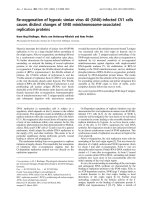
analysed the binding capacity of the exogenously
expressed CD4 receptor in the BeWo transfected cell line.
To verify this functional aspect of the CD4 receptor in this
transfected cell line, the cells were incubated with a CD4
antibody which specifically recognises the gp120 binding
site of the receptor and the cells were then analysed by
flow cytometry. The flow cytometry analyses indicated
that approximately 40% of the BeWo-CD4
+
population
expressed the CD4 surface molecule (Figure 1A). In order
to ascertain the complete binding capacity of the exoge-
nously expressed CD4 molecule, the BeWo-CD4
+
cells
were used in a CD4-gp120 binding assay. Following incu-
bation with recombinant gp120 (from HIV-1 BaL virus),
the BeWo-CD4
+
cells showed a dose-dependent increase
of fluorescently labeled cells (Figure 1B) compared to
background levels in BeWo wild type cells (data not
shown), indicating that the CD4 molecule in BeWo-CD4
+
cells is not only correctly expressed on the cell surface, but
also capable of binding the HIV-1 gp120 envelope pro-
tein. However, despite a functional CD4 receptor and the
endogenously expressed CCR5 and CXCR4 co-receptors,
cell-free HIV-1 infection was still restricted in BeWo-CD4
+
cells (data not shown)[15].
Viral internalisation in BeWo cells is envelope-independent
As cell-free infection by X4-Env and R5-Env pseudotyped
HIV-1 is restricted in BeWo trophoblast cells despite the
presence of functional CD4 receptor and co-receptors, we
wished to verify that viral entry was not affected or dimin-
ished in these cells. The BeWo-CD4
+
were exposed to VSV-
G pseudotyped HIV-1 as well as to X4 and R5 HIV-1 pseu-
dotypes. An envelope deficient HIV-1 "virus" was also
used as control. To determine the background level of vir-
ions attached to the cell surface, the experiments were per-
formed in parallel at 37°C and at 4°C. As shown in figure
2A, the level of internalised HIV-1 (as determined by p24
antigen quantification) was significantly higher in the
cells treated at 37°C than in the cells treated at 4°C. This
result suggested that the small quantity of p24 antigen
detected in the cell lysates exposed to the HIV-1 pseudo-
types at 4°C was residual amounts of virions attached to
the cell surface following pronase treatment and washes.
The amount of intracellular p24 antigen in VSV-G infected
BeWo-CD4
+
cells was slightly higher than the amount of
intracellular p24 antigen following exposure to X4 or R5
viral pseudotypes. However, the amount of intracellular
p24 antigen for X4 and R5 viruses was consistently sub-
stantially higher than the 4°C control (10–100 fold
increase), indicating that the p24 antigen detected in the
cell lysates was not simply bound to the cell surface of the
BeWo-CD4
+
cells. Interestingly, even the cells exposed to
an envelope-deficient HIV-1 pseudotype showed an inter-
nalisation of p24 antigen (figure 2A); this result suggested
that the viral entry route most likely occurred via an endo-
cytic pathway and not via membrane fusion. Having
determined that viral entry occurs in the BeWo-CD4
+
cells,
we performed the same experiments in the BeWo wild
type cells. As shown in figure 2B, intracellular p24 antigen
was also detected in wild type trophoblast cells (despite
the absence of the CD4 receptor), and at even higher lev-
els than those observed in the transfected BeWo-CD4
+
cell
line.
The BeWo-CD4
+
cell line expresses CD4 capable of binding HIV-1 gp120 recombinant proteinFigure 1
The BeWo-CD4
+
cell line expresses CD4 capable of
binding HIV-1 gp120 recombinant protein. A) Expres-
sion of the CD4 surface molecule on the cell membrane of a
selected BeWo cell line. The CD4 antibody used recognises
the gp120 binding domain of CD4. The left panel shows wild
type BeWo cells stained with anti-CD4 antibody; the right
panel shows staining of the CD4
+
BeWo cell line. B) gp120-
CD4 binding assays on the CD4
+
BeWo cell line. The CD4
+
cell line was incubated with increasing concentrations of
recombinant gp120 protein and subsequently stained with an
anti-gp120 antibody, followed by a FITC-conjugated second-
ary antibody; cells were analysed by flow cytometry. The
gp120 recombinant protein is derived from a BaL HIV-1
strain.
A
319 164
7.9%
44.8%
CD4-PC5
B
gp120-FITC
104
95
48
44
control + 76 nM gp120
+ 765 nM gp120 + 1.5 μM gp120
6.2% 7.6%
32.2%
44.1%
# Cells# Cells# Cells
Retrovirology 2009, 6:46 />Page 4 of 9
(page number not for citation purposes)
X4 and R5 HIV-1 viruses are less stable in trophoblast cells
than infectious VSV-G HIV-1 pseudotypes
Although HIV-1 particles are able to gain intracellular
access to trophoblast cells, these cells are unable to sustain
HIV-1 replication; we therefore analysed the fate of the
virions once inside the BeWo cells. To follow the fate of
intracellular p24 antigen, BeWo-CD4
+
cells were exposed
to the different enveloped HIV-1 pseudotypes and
infected as previously described. The intracellular levels of
p24 antigen of the VSV-G pseudotype decrease, in the
early time points, remained relatively stable in the BeWo
cells (remaining at approximately 60% of total p24 anti-
gen post-viral exposure) (Figure 3A). Conversely, the
amount of intracellular p24 antigen for both the X4
(HXB2) and R5 (BaL) HIV-1 pseudotypes decreased more
rapidly; and at 6 hours post-exposure, the detected level of
remaining p24 antigen was less than 10% of the initial
p24 antigen quantity (Figure 3A). The envelope-deficient
viral genome (ΔEnv) was also tested in this assay, and its
profile was found to be very similar to that of the envel-
oped HIV-1 pseudotypes. The fate of intracellular p24
antigen in BeWo wild type cells was also investigated; as
previously determined, the absolute amount of p24 anti-
HIV-1 pseudotypes, including envelope-deficient pseudotypes enter BeWo cellsFigure 2
HIV-1 pseudotypes, including envelope-deficient
pseudotypes enter BeWo cells. A) BeWo-CD4
+
cells
were exposed to 20 ng/ml of p24 antigen of the HIV-1 pseu-
dotypes at 37°C or 4°C by one hour spinoculation and one
hour incubation at according temperature; the cells were
washed, treated with pronase to eliminate residual virions on
the cell surface, and the cell pellet was lysed in preparation
for p24 antigen detection in lysates. Experiments were
repeated at least three times. Graph shows a representative
experiment. B) Wild type BeWo cells and CD4 expressing
BeWo cells were exposed to HIV-1 pseudotypes. Viral entry
was determined by p24 antigen quantification in cell lysates.
The bars indicate the mean of at least three independent
experiments; the error bars indicate SEM.
A
B
0
1
2
3
4
5
6
wt
CD4+
VSV-G
VSV-G
BaL
BaL
HXB2
HXB2
ΔEnv
ΔEnv
p24 ng/mlp24 ng/ml
5
4
3
6
2
1
0
1
2
3
0
37 ºC
4 ºC
37 ºC
4 ºC
VSV-G pseudotyped HIV-1 is more stable over time in BeWo wild type cells (A) and in BeWo-CD4
+
cells (B) compared to BaL, HXB2 and envelope-deficient HIV-1 virionsFigure 3
VSV-G pseudotyped HIV-1 is more stable over time
in BeWo wild type cells (A) and in BeWo-CD4
+
cells
(B) compared to BaL, HXB2 and envelope-deficient
HIV-1 virions. Following viral exposure, the viral suspen-
sion was removed, and replaced with fresh medium; cell sam-
ples were collected at different time points. The amount of
intracellular p24 antigen present in cells lysed immediately
after viral exposure (time 0) was considered to be 100%, and
all subsequent time points were normalised to time 0. The
results are the mean of at least three independent experi-
ments. Error bars indicate SEM.
0
10
20
30
40
50
60
70
80
90
100
A
B
p24 (%)p24 (%)
0
10
20
30
40
50
60
70
80
90
100
02 4 6
0246
Time p.i. (hours)
Time p.i. (hours)
VSV-G
∆Env
HXB2
BaL
100
90
80
70
60
50
40
30
20
10
0
100
90
80
70
60
50
40
30
20
10
0
Retrovirology 2009, 6:46 />Page 5 of 9
(page number not for citation purposes)
gen was consistently higher in the wild type cells than in
the BeWo-CD4
+
cells. However, the kinetics for the
decrease of intracellular p24 antigen in BeWo wild type
cells were very similar to those seen in the CD4 transfected
cell line (Figure 3B).
Intracellular p24 antigen levels of HIV-1 pseudotypes
accumulated in proteasome-inhibitor treated BeWo cells
To determine whether the internalised virions are subject
to degradation by the proteasome, the BeWo cells were
treated with the proteasome inhibitor lactacystin. The lac-
tacystin-treated BeWo cells were tested for optimal viabil-
ity and proteasome inhibition levels; and consequently
the cells were exposed to the HIV-1 pseudotypes as before,
and the intracellular levels of p24 antigen were measured
over-time (Figure 4). The data showed that the levels of
p24 antigen in lactacystin-treated cells were higher in the
early time points following exposure to both the HIV-1
enveloped pseudotypes as well as the VSV-G pseudotyped
HIV-1. However, 3 hours post viral exposure, treatment
with proteasome-inhibitors did not show any effect on
the R5 and X4 HIV-1 pseudotypes, as their p24 antigen
levels were similar in both treated and untreated cells.
Conversely, the levels of intracellular p24 antigen for VSV-
G HIV-1 exposed BeWo cells remained consistently higher
over time (p = 0.0312) in lactacystin-treated cells com-
pared to untreated cells.
Proteasome inhibition in BeWo trophoblast cells did not
overcome HIV-1 restriction
To determine whether the accumulation of intracellular
p24 antigen levels could constitute an escape mechanism
for the restriction of viral replication in BeWo cells, we
examined the effect of proteasome inhibition on viral rep-
lication. The level of luciferase activity detected in BeWo
cells (both wild type and CD4
+
) infected with VSV-G HIV-
1 was significantly higher (p = 0.002) in the cells which
had been previously treated with the proteasome inhibi-
tor (Figure 5). As expected, the untreated cells exposed to
the X4 and R5 HIV-1 pseudotypes showed only back-
ground levels of luciferase activity; this baseline level of
luciferase activity remained unaltered in the proteasome-
inhibited cells (Figure 5). The experiments were repeated
using two other proteasome inhibitors: epoxomicin and
MG132. Similar to the results with lactacystin, VSV-G
HIV-1 infection levels were increased, but no infection
was detected for HXB2 and BaL enveloped HIV-1 pseudo-
types (data not shown), suggesting that inhibition of pro-
teasomal degradation is not sufficient to relieve HIV-1
restriction in BeWo cells.
Discussion
The BeWo cell line is a standard in vitro model for the
study of placental trophoblast cells. BeWo cells do not
naturally express the CD4 molecule necessary for fusion
entry of HIV-1 viral particles. Hence, to determine
whether the restriction of cell-free HIV-1 infection is sim-
ply due to the lack of the main CD4 receptor, we previ-
ously transfected BeWo cells to obtain a CD4 expressing
cell line which, however, also fails to sustain viral infec-
tion. Although the CD4 receptor was clearly expressed on
the cell membrane, it was deemed important to verify that
the receptor was capable of binding the gp120 protein of
the HIV-1 envelope. We report that the CD4 molecule
present on the surface of the BeWo trophoblast cells is
capable of binding a recombinant HIV-1 BaL gp120 pro-
tein. Despite the gp120 binding capacity of CD4 and the
functional expression of co-receptors, the trophoblast
cells do not show any viral infection when exposed to the
X4 and R5 HIV-1 pseudotypes. It has been suggested that
the restriction of HIV infection in trophoblast cells is due
to the lack of the appropriate receptors on the cell surface;
indeed the levels of CD4 and the two co-receptors vary
during pregnancy and several reports have confirmed the
variable expression of CD4, CCR5, CXCR4 and other
chemokine receptors [25,26]. However, while it is not
possible to completely exclude a role of the CD4 receptor,
the data reported here strengthen the hypothesis of a
The intracellular levels of p24 antigen of VSV-G, HXB2 and BaL enveloped HIV-1 pseudotypes accumulate in early time points in lactacystin-treated BeWo cellsFigure 4
The intracellular levels of p24 antigen of VSV-G,
HXB2 and BaL enveloped HIV-1 pseudotypes accu-
mulate in early time points in lactacystin-treated
BeWo cells. Intracellular p24 antigen levels for VSV-G (A),
HXB2 (B), BaL (C) and envelope deficient (D) pseudotypes
normalised against p24 antigen level at time point 0 (100%).
BeWo cells were treated for 2 hours with 50 μM lactacystin
prior to viral exposure, which greatly reduces proteasomal
activity (as determined by the diminished release of the fluor-
ogenic substrate Suc-LLVY-AMC; data not shown) without
affecting cell viability (as determined by Wst-1 cell viability
assays; data not shown). Bars indicate the mean of at least
three independent experiments; error bars indicate SEM.
A -
VSV-G
D -
Env
C -
BaL
B -
HXB2
Lactacystin treated
Untreated
Time (hours)
p24 (%)
0,0 0,5 1,0 1,5 2,0 2,5 3,0
0
100
200
300
0,0 0,5 1,0 1,5 2,0 2,5 3,0
0
100
200
300
0,0 0,5 1,0 1,5 2,0 2,5 3,0
0
100
200
300
0.0 0.5 1.0 1.5 2.0 2.5 3.0
0
100
200
300
Retrovirology 2009, 6:46 />Page 6 of 9
(page number not for citation purposes)
restriction in trophoblast cells which goes beyond the
simple lack of receptor and co-receptor expression on the
cell surface.
Given the controversial data on viral infection in trophob-
last cells, it was important to determine whether viral
entry is hindered in the BeWo cell line by comparing viral
entry between the different HIV-1 pseudotypes and deter-
mine entry levels in wild type versus CD4
+
cells. Several
studies have shown the significant role of an undefined
endocytic pathway in virion internalisation in trophoblast
cells [17,30]. However, despite virion internalisation, pro-
ductive HIV-1 infection is restricted. Although virion
internalisation by endocytosis pathways is generally asso-
ciated with non-productive entry, there is evidence to
show that HIV-1 entry via different endocytic processes
can result in productive infection [32,33]. Our analyses
show that HIV-1 virions gain intracellular access to both
wild-type and CD4
+
BeWo cells. Interestingly our results
also show that the levels of intracellular virus are similar,
independent of the HIV-1 pseudotype used in the experi-
ments (including an envelope deficient virus), and inde-
pendent of whether the viral exposure leads to infection
or not (VSV-G HIV-1 pseudotype compared to the HXB2
and BaL enveloped pseudotypes). The data indicate that
the expression of the CD4 receptor is not necessary for
viral entry, which likely occurs through an endocytic path-
way, as suggested by others for different trophoblastic cell
lines [27,29,30]. Given that all pseudotypes tested enter
the BeWo cells at comparable levels, yet only the VSV-G
bearing HIV-1 is capable of sustaining an infection, it is
likely that the entry mechanisms or the very early post-
entry events involved are different; and as a result, the
intracellular compartmentalisation of the restricted pseu-
dotypes versus VSV-G HIV-1 pseudotypes might be differ-
ent. Intriguingly, the data described in this report
demonstrate that the level of viral entry is higher in wild
type BeWo cells (which do not naturally express the CD4
receptor) than in the CD4
+
transfected cell line. This
observation could be explained by the fact that the CD4
receptor on BeWo cells has been demonstrated to bind the
gp120 protein of HIV-1 (as shown in the protein binding
assays), and therefore the exogenously expressed receptor
may capture and sequester a portion of the HIV-1 virions,
which are consequently no longer able to enter via a
fusion-independent mechanism. Another potential expla-
nation could be that the exogenous expression of the CD4
molecule on the cell surface could alter the natural char-
acteristics of the cell membrane, reducing the levels of
endocytosis, which in turn results in a decreased amount
of internalised virus.
The data reported here indicate that all the HIV-1 pseudo-
types tested enter the BeWo cells via an envelope-inde-
pendent, non-fusion route at similar levels and kinetics,
yet only the VSV-G bearing HIV-1 pseudotype can result in
an infection. Following internalisation, all pseudotyped
virus levels decrease to some extent, although the p24
antigen levels of VSV-G HIV-1 pseudotype are more stable
than those of BaL, HXB2 or envelope-deficient HIV-1
pseudotypes. We hypothesized several non-mutually
exclusive mechanisms which could explain the decrease of
the p24 antigen levels of BaL and HXB2 enveloped HIV-1
pseudotypes. Once internalised by endocytosis, the
restricted HIV-1 pseudotypes may be recycled and
released from the cell by an exocytic pathway. Internalised
virions may also be degraded by the proteasome or by lys-
osomes or they could be recognised by a restriction factor
which impedes release into the cytoplasm preventing sub-
sequent uncoating and reverse transcription.
The amount of p24 antigen detected in the culture super-
natants following viral exposure increased slightly over-
time (data not shown). This observation is consistent with
the hypothesis of a portion of internalised virions being
released by exocytosis. However, the low levels of p24
antigen detected in the supernatants more likely resulted
from the detachment of membrane-bound particles. Thus
the intracellular instability of the viral p24 antigen levels
might not exclusively be due to release by exocytosis. The
p24 antigen quantification experiments performed in the
The reduction of proteasomal degradation increases VSV-G pseudotyped HIV-1 replication, but not replication of HIV-1 enveloped pseudotypesFigure 5
The reduction of proteasomal degradation increases
VSV-G pseudotyped HIV-1 replication, but not repli-
cation of HIV-1 enveloped pseudotypes. VSV-G HIV-1
viral infection in lactacystin treated BeWo cells increases
compared to untreated cells. The cells were exposed to the
pseudotyped HIV-1 viruses (X4 and R5 pseudotypes, as well
as the VSV-G pseudotype) and the level of infection in the
cells was determined by the amount of luciferase activity
detected in the cell lysates 96 hours post-infection. Results
are expressed as the fold induction of luciferase activity as
compared to uninfected cells. The results show mean of
three independent experiments. Error bars indicate SEM.
Statistical analyses were performed using Mann Whitney test.
VSV-G HXB2 BaL
0
5
10
15
45
70
95
Fold induction (RLU/sec)
p=0.0012
untreated
+50 μM lactacystin
Retrovirology 2009, 6:46 />Page 7 of 9
(page number not for citation purposes)
presence of the proteasome inhibitors demonstrated that
a portion of the incoming virus was degraded by the pro-
teasome. However the increased amount of internal p24
antigen in proteasome-inhibited cells for the R5 and X4
HIV-1 pseudotypes was only seen at the first time point
measurements following infection. By 3 hours post viral
exposure, the difference in p24 antigen levels of R5 and X4
HIV-1 viruses between proteasome-inhibitor treated cells
and untreated cells was no longer detected. These data
suggest that although the proteasome may contribute to
the degradation of incoming virions immediately after
viral exposure, the instability of internalised viral particles
can also be due to other mechanism(s). On the contrary,
when the proteasome-inhibited BeWo cells were exposed
to the VSV-G HIV-1 pseudotype, the higher level of intra-
cellular p24 antigen appears to be stable over-time. This
increased level of internal p24 antigen of VSV-G HIV-1
seen in the lysates of proteasome-inhibited cells translates
into an increase in the level of viral replication, as deter-
mined by the increase of luciferase activity following
infection. Despite an initial increase of internal virus in
BeWo cells exposed to HXB2 or BaL enveloped HIV-1
pseudotypes, the inhibition of proteasomal activity does
not equate with an escape from the restriction of viral rep-
lication.
Conclusion
In conclusion, although both VSV-G and HIV-1 pseudo-
typed virions all gained intracellular access to trophoblast
cells with similar kinetics and comparable quantities, only
VSV-G pseudotyped virions can productively infect these
cells. The data presented here suggest that HIV-1 virions
are engulfed by endocytosis, but in contrast with VSV-G
enveloped HIV-1 virions, they are not released from the
endocytic vesicles into the cytoplasm. A portion of inter-
nalised HIV-1 virions is subject to degradation by the pro-
teasome, as demonstrated by the temporary increase of
p24 antigen levels in proteasome-inhibited cell lysates.
However, this transient increase is not sufficient to over-
come HIV-1 restriction, and the virions are likely targeted
for degradation by alternative mechanisms, such as the
detrimental effect of the vacuolar pathway. Crucially, cell-
to-cell HIV-1 infection is not restricted in trophoblast
cells, bolstering the notion that the entry mechanism and
early post-entry viral steps are critical in determining the
restriction of cell-free HIV-1 virions. Direct contact with
infected cells entrains separate entry mechanisms which
allow HIV-1 virions to circumvent restriction in trophob-
last cells. The notion that the transmission mode of HIV-
1 may be key in determining restriction is highlighted by
recent data on the ability of rhTRIM5α to restrict cell-free
but not cell-associated HIV-1 transmission [34]. The
results described in this report support the notion that tro-
phoblast cells may recognise HIV-1 virions during the
binding of the virus to the cell surface, internalise them in
an endocytic, receptor-independent manner, and either
actively target HIV-1 for degradation (which is principally
proteasomal independent), or impede the release of the
virions from the endosomes into the cytoplasm.
The elucidation of the mechanisms involved in HIV-1
infection and restriction of placental trophoblast cells
remains of utmost importance to the understanding of the
natural mechanisms of control of in uteroHIV-1 mother-
to-child infection. This understanding holds potential
implications for the identification of new therapeutic drug
targets.
Methods
Cell lines
The human choriocarcinoma trophoblast cell line, BeWo
[35] was obtained from the American Type Culture Col-
lection (ATCC#CCL98, Rockville, Maryland, USA). A
BeWo cell line stably transfected for the CD4 receptor was
additionally used (BeWo-CD4
+
) [15]. BeWo cells were
cultured in Dulbecco's Modified Eagle's Medium (DMEM,
Gibco BRL), supplemented with 20% fetal calf serum
(FCS). The titration of the viral pseudotype stocks was per-
formed on HeLa P4P cells (CD4
+
, CCR5
+
, CXCR4
+
) cul-
tured in DMEM supplemented with 10% FCS, containing
500 μg/ml G418 and 1 μg/ml puromycin. The 293T
Human Embryonic Kidney cells (HEK) were used in tran-
sient transfections to obtain HIV-1 pseudotypes. The 293T
HEK cells were cultured in DMEM, supplemented with
10% FCS.
HIV-1 pseudotype infections
The luciferase expressing viral pseudotypes were based on
the NL4-3 HIV-1 and VSV-G, BaL (R5) or HXB2 (X4) env
plasmids [36-38]. The viral pseudotypes were generated
by transfecting 293T cells with the corresponding cDNA
plasmid (pNL4-3ΔEnvLuc+ plus the appropriate env
cDNA) using the transfection reagent SuperFect (Qiagen)
following the manufacturer's protocol. The resulting
supernatants were collected 48–72 hours following trans-
fection. The viral pseudotypes in the supernatants were
quantified by p24 antigen ELISA (Zeptometrix) and
titrated on HeLa P4P cells. The rate of infection of the viral
pseudotypes was determined by luciferase expression
using Luciferase Reagent (Promega) and a Glomax lumi-
nometer.
Viral infections were performed by treating 2 × 10
5
cells
with 20 ng/ml p24 antigen of HIV-1 pseudotypes, fol-
lowed by 1 hour spinoculation and 1 hour incubation at
37°C. Cells were subsequently incubated at 37°C for 72–
96 hours, then washed in phosphate buffered saline (PBS)
and lysed with 100 μl of Cell Lysis Buffer (Promega). 10
μl of cell lysates were used to determine levels of luciferase
activity using the Luciferase Reagent (Promega) and read-
Retrovirology 2009, 6:46 />Page 8 of 9
(page number not for citation purposes)
ing was performed using Glomax Luminoter. Results are
expressed as RLU/sec/100 μl lysate.
CD4-gp120 binding assays
The gp120 binding assays were performed as previously
described [39]. Both BeWo wild-type and BeWo-CD4
+
cells were incubated with different concentrations (76
nM, 765 nM, 1.5 μM) of recombinant HIV-1 BaL gp120
protein (NIH AIDS Research and Reference Reagent Pro-
gram) for 4 hours at 4°C. Cells were washed twice in
buffer (PBS, 1% FCS) and the cell pellet was resuspended
in 50 μl of 2 μg/ml of anti-gp120 antibody (Aalto Biorea-
gents Ltd). After incubation for 1 hour at 4°C, the cells
were washed and resuspended in 50 μl of anti-sheep IgG-
FITC antibody (Sigma) and incubated for 1 hour at 4°C.
Cells were washed and analysed by flow cytometry. All
cell flow cytometry analysis was performed using Beck-
man Coulter DC500; results were analysed by CXP and
FlowJo software.
Viral entry assays
Cells were infected with viral pseudotypes as described
above. BeWo cells were infected with R5 (BaL) or X4
(HXB2) viral pseudotypes or with envelope-deficient
virus. Pseudotypes expressing the G protein of the Vesicu-
lar Stomatitis Virus (VSV), for which BeWo cells are per-
missive, were used as positive control. Following
spinoculation and incubation, cells were washed twice
with PBS and fresh medium added. Cells were then incu-
bated at 37°C until cell lysis at the different time points.
Cell lysates were prepared by washing cells in PBS, treat-
ment with 1 mg/ml pronase for 3 minutes at room tem-
perature to eliminate residual virus followed by two
further washes in PBS. The cell pellet was resuspended in
100 μl of lysis buffer (Zeptometrix) and HIV-1 p24 anti-
gen detection was performed using an ELISA assay (Zep-
tometrix).
p24 antigen ELISA assays
The quantification of p24 antigen in cell lysates or super-
natants was performed by p24 antigen ELISA (Zeptome-
trix), according to the manufacturer. The optical density of
the samples was measured at 405 nm using Revel ELISA
plate reader and software.
Proteasome Inhibition
The proteasome inhibitor assays were performed using
lactacystin (Sigma), epoxomicin (Sigma) and MG132
(Sigma). The effect of the proteasome inhibitors was
determined by analysing the capability of the treated cells
to release the fluorogenic substrate Suc-LLVY-AMC (Bio-
mol International) compared to untreated cells. The cells
were treated for 2 hours at 37°C with the appropriate con-
centration of proteasome inhibitor. After a 2 hour incuba-
tion period, the supernatant was removed, the cells were
treated with lysis buffer and Suc-LLVY-AMC was added to
the lysate. The fluorescence of the cells was measured
using a Victor plate reader (Wallac). In parallel, the viabil-
ity of the cells was tested by the Wst-1 assay (Boehringer
Mannheim) to determine the toxicity of the proteasome
inhibitors on the cells.
Competing interests
The authors declare that they have no competing interests.
Authors' contributions
ALR performed all experiments, participated in the design
of the study and drafted the manuscript. CC provided
technical assistance for experimental work. FBS partici-
pated in the conception and design of the project. EM par-
ticipated in the design of the study, the conception of the
project and helped draft the manuscript. All authors read
and approved the final manuscript.
Acknowledgements
This work was supported by a grant from the National French Agency for
AIDS Research (ANRS) and by the Institut Pasteur and INSERM, France.
ALR was awarded a Sidaction fellowship and was subsequently the recipient
of a Marie Curie Intra-European Fellowship (European Commission). The
following reagent was obtained through the NIH AIDS Research and Ref-
erence Reagent Program, Division of AIDS, NIAID, NIH: HIV-1
BaL
gp120
from DAIDS, NIAID.
We thank Désirée Kunkel for help with flow cytometry analysis.
References
1. Clapham PR, Weber JN, Whitby D, McIntosh K, Dalgleish AG, Mad-
don PJ, Deen KC, Sweet RW, Weiss RA: Soluble CD4 blocks the
infectivity of diverse strains of HIV and SIV for T cells and
monocytes but not for brain and muscle cells. Nature 1989,
337:368-370.
2. Yahi N, Baghdiguian S, Moreau H, Fantini J: Galactosyl ceramide
(or a closely related molecule) is the receptor for human
immunodeficiency virus type 1 on human colon epithelial
HT29 cells. Journal of Virology 1992, 66:4848-4854.
3. Schneider-Schaulies J, Schneider-Schaulies S, Brinkmann R, Tas P, Hal-
brügge M, Walter U, Holmes HC, Ter Meulen V: HIV-1 gp120
receptor on CD4-negative brain cells activates a tyrosine
kinase. Virology 1992, 191:765-772.
4. Sheehy AM, Gaddis NC, Choi JD, Malim MH: Isolation of a human
gene that inhibits HIV-1 infection and is suppressed by the
viral Vif protein. Nature 2002, 418:646-650.
5. Stremlau M, Owens C, Perron M, Kiessling M, Autissier P, Sodroski J:
The cytoplasmic body component TRIM5α restricts HIV-1
infection in Old World monkeys. Nature 2004, 427:848-853.
6. Anderson JL, Campbell EM, Wu X, Vandegraaff N, Engelman A, Hope
TJ: Proteasome inhibition reveals that a functional preinte-
gration complex intermediate can be generated during
restriction by diverse TRIM5 proteins. Journal of Virology 2006,
80:9754-9760.
7. Chatterji U, Bobardt MD, Gaskill P, Sheeter D, Fox H, Gallay PA:
Trim5alpha accelerates degradation of cytosolic capsid asso-
ciated with productive HIV-1 entry. J Biol Chem 2006,
281:37025-37033.
8. Sakuma R, Noser JA, Ohmine S, Ikeda Y: Inhibition of HIV-1 rep-
lication by simian restriction factors, TRIM5 alpha and
APOBEC3G. 2007, 14:185-189.
9. Dueck M, Guatelli J: Evidence against a direct antiviral activity
of the proteasome during the early steps of HIV-1 replica-
tion. Virology 2007, 361:1-8.
Publish with BioMed Central and every
scientist can read your work free of charge
"BioMed Central will be the most significant development for
disseminating the results of biomedical research in our lifetime."
Sir Paul Nurse, Cancer Research UK
Your research papers will be:
available free of charge to the entire biomedical community
peer reviewed and published immediately upon acceptance
cited in PubMed and archived on PubMed Central
yours — you keep the copyright
Submit your manuscript here:
/>BioMedcentral
Retrovirology 2009, 6:46 />Page 9 of 9
(page number not for citation purposes)
10. Campbell EM, Perez O, Anderson JL, Hope TJ: Visualization of a
proteasome-independent intermediate during restriction of
HIV-1 by rhesus TRIM5alpha. J Cell Biol 2008, 180:549-561.
11. Burton GJ, Watson AL: The Structure of the Human Placenta:
Implications for Initiating and Defending Against Virus Infec-
tions. Rev Med Virol 1997, 7:219-228.
12. Chouquet C, Burgard M, Richardson S, Rouzioux C, Costagliola D:
Timing of mother-to-child HIV-1 transmission and diagnosis
of infection based on polymerase chain reaction in the neo-
natal period by a non-parametric method. AIDS 1997,
11:1183-1184.
13. Lagaye S, Derrien M, Menu E, Coïto C, Tresoldi E, Mauclère P, Scar-
latti G, Chaouat G, Barré-Sinoussi F, Bomsel M: HIV-1 ENft-
SoIUTo: Cell-to-cell contact results in a selective
translocation of maternal human immunodeficiency virus
type 1 quasispecies across a trophoblastic barrier by both
transcytosis and infection. Journal of Virology 2001, 75:4780-4791.
14. Burton GJ, O'Shea S, Rostron T, Mullen JE, Aiyer S, Skepper JN, Smith
R, Banatvala J: Physical breaks in the placental trophoblastic
surface: significance in vertical transmission of HIV. AIDS
1996, 10:1294-1296.
15. Dolcini G, Derrien M, Chaouat G, Barré-Sinoussi F, Menu E: Cell-
free HIV type 1 infection is restricted in the human trophob-
last choriocarcinoma BeWo cell line, even with expression
of CD4, CXCR4 and CCR5. AIDS Res Hum Retroviruses 2003,
19:857-864.
16. Muñoz LD, Serramía MJ, Fresno M, Muñoz-Fernández MA: Proges-
terone inhibits HIV-1 replication in human trophoblast cells
through inhibition of autocrine tumor necrosis factor secre-
tion. J Infect Dis 2007, 195:1294-1302.
17. Vidricaire G, Tardif MR, Tremblay MJ: The low viral production in
trophoblastic cells is due to a high endocytic internalization
of the human immunodeficiency virus type 1 and can be
overcome by the pro-inflammatory cytokines tumor necro-
sis factor-alpha and interleukin-1. J Biol Chem 2003,
278:15832-15841.
18. Bourinbaiar AS, Nagorny R: Human immunodeficiency virus
type 1 infection of choriocarcinoma-derived trophoblasts.
Acta Virol
1993, 37:21-28.
19. Derrien M, Faye A, Dolcini G, Chaouat G, Barre-Sinoussi F, Menu E:
Impact of the placental ctokine-chemokine balance on regu-
lation of cell-cell contact-induced human immunodeficiency
virus type 1 translocation across a trophoblastic barrier in
vitro. Journal Of Virology 2005, 79:12304-12310.
20. Vidricaire G, Tremblay MJ: Rab5 and Rab7, but not ARF6, gov-
ern the early events of HIV-1 infection in polarized human
placental cells. J Immunol 2005, 175:6517-6530.
21. Kilani RT, Chang LJ, Garcia-Lloret MI, Hemmings D, Winkler-Lowen
B, Guilbert LJ: Placental trophoblasts resist infection by multi-
ple human immunodeficiency virus (HIV) type 1 variants
even with cytomegalovirus coinfection but support HIV rep-
lication after provirus transfection. Journal of Virology 1997,
71:6359-6372.
22. Zachar V, Spire B, Hirsch I, Chermann JC, Ebbesen P: Human trans-
formed trophoblast-derived cells lacking CD4 receptor
exhibit restricted permissiveness for human immunodefi-
ciency virus type 1. Journal of Virology 1991, 65:2102-2107.
23. Roberts PC, Kipperman T, Compans RW: Vesicular stomatitis
virus G protein acquires pH-independent fusion activity dur-
ing transport in a polarized endometrial cell line. Journal of
Virology 1999, 73:10447-10457.
24. Kumar A, Kumar S, Dinda AK, Luthra K: Differential expression
of CXCR4 receptor in early and term human placenta. Pla-
centa 2004, 25:347-351.
25. Mognetti B, Moussa M, Croitoru J, Menu E, Dormont D, Roques P,
Chaouat G: HIV-1 co-receptor expression on trophoblastic
cells from early placentas and permissivity to infection by
several HIV-1 primary isolates. Clinical And Experimental Immunol-
ogy 2000, 119:486-492.
26. David FJ, Autran B, Tran HC, Menu E, Raphael M, Debre P, Hsi BL,
Wegman TG, Barre-Sinoussi F, Chaouat G: Human trophoblast
cells express CD4 and are permissive for productive infec-
tion with HIV-1. Clin Exp Immunol 1992, 88(1):10-16.
27. David FJ, Tran HC, Serpente N, Autran B, Vaquero C, Djian V, Menu
E, Barré-Sinoussi F, Chaouat G: HIV infection of choriocarci-
noma cell lines derived from human placenta: the role of
membrane CD4 and Fc-Rs into HIV entry. Virology 1995,
208:
784-788.
28. Zachar V, Nørskov-Lauritsen N, Juhl C, Spire B, Chermann JC,
Ebbesen P: Susceptibility of cultured human trophoblasts to
infection with human immunodeficiency virus type 1. J Gen
Virol 1991, 72(Pt 6):1253-1260.
29. Vidricaire G, Imbeault M, Tremblay MJ: Endocytic host cell
machinery plays a dominant role in intracellular trafficking
of incoming human immunodeficiency virus type 1 in human
placental trophoblasts. Journal of Virology 2004, 78:11904-11915.
30. Vidricaire G, Tremblay MJ: A clathrin, caveolae, and dynamin-
independent endocytic pathway requiring free membrane
cholesterol drives HIV-1 internalization and infection in
polarized trophoblastic cells. Journal of Molecular Biology 2007,
368:1267-1283.
31. Ayouba A, Cannou C, Nugeyre MT, Barre-Sinoussi F, Menu E: Dis-
tinct efficacy of HIV-1 entry inhibitors to prevent cell-to-cell
transfer of R5 and X4 viruses across a human placental tro-
phoblast barrier in a reconstitution model in vitro. Retrovirol-
ogy 2008, 5:31.
32. Daecke J, Fackler OT, Dittmar MT, Krausslich HG: Involvement of
clathrin-mediated endocytosis in human immunodeficiency
virus type 1 entry. J Virol 2005, 79:1581-1594.
33. Maréchal V, Prevost MC, Petit C, Perret E, Heard JM, Schwartz O:
Human immunodeficiency virus type 1 entry into macro-
phages mediated by macropinocytosis. Journal of Virology 2001,
75:11166-11177.
34. Richardson MW, Carroll RG, Stremlau M, Korokhov N, Humeau LM,
Silvestri G, Sodroski J, Riley JL: Mode of Transmission Affects the
Sensitivity of HIV-1 to Restriction by Rhesus TRIM5{alpha}.
J Virol 2008, 82:11117-28.
35. Pattillo RA, Gey GO: The establishment of a cell line of human
hormone-synthesizing trophoblastic cells in vitro. Cancer Res
1968, 28:1231-1236.
36. Akkina RK, Walton RM, Chen ML, Li QX, Planelles V, Chen IS: High-
efficiency gene transfer into CD34+ cells with a human
immunodeficiency virus type 1-based retroviral vector pseu-
dotyped with vesicular stomatitis virus envelope glycopro-
tein G. Journal of Virology 1996, 70:2581-2585.
37. Connor RI, Chen BK, Choe S, Landau NR: Vpr is required for effi-
cient replication of human immunodeficiency virus type-1 in
mononuclear phagocytes. Virology 1995, 206:935-944.
38. Landau NR, Page KA, Littman DR: Pseudotyping with human T-
cell leukemia virus type I broadens the human immunodefi-
ciency virus host range. Journal of Virology 1991, 65:162-169.
39. Mondor I, Ugolini S, Sattentau QJ: Human immunodeficiency
virus type 1 attachment to HeLa CD4 cells is CD4 independ-
ent and gp120 dependent and requires cell surface heparans.
J Virol 1998, 72:3623-3634.