Báo cáo khoa học: "Upregulated but insufficient generation of activated protein C is associated with development of multiorgan failure in severe acute pancreatitis" pot
Bạn đang xem bản rút gọn của tài liệu. Xem và tải ngay bản đầy đủ của tài liệu tại đây (345.49 KB, 9 trang )
Open Access
Available online />Page 1 of 9
(page number not for citation purposes)
Vol 10 No 1
Research
Upregulated but insufficient generation of activated protein C is
associated with development of multiorgan failure in severe acute
pancreatitis
Outi Lindstrom
1
, Leena Kylanpaa
1
, Panu Mentula
1
, Pauli Puolakkainen
1
, Esko Kemppainen
1
,
Reijo Haapiainen
1
, Jose A Fernandez
2
, John H Griffin
2
, Heikki Repo
3,4
and Jari Petaja
5
1
Second Department of Surgery, Helsinki University Central Hospital and University of Helsinki, Finland
2
Department of Molecular and Experimental Medicine, The Scripps Research Institute, La Jolla, CA, USA
3
Department of Medicine, University of Helsinki, Finland
4
Department to Bacteriology and Immunology, Haartman Institute, University of Helsinki, Finland
5
Department of Pediatrics, Jorvi Hospital, Espoo, Finland
Corresponding author: Leena Kylanpaa,
Received: 19 Aug 2005 Revisions requested: 26 Sep 2005 Revisions received: 15 Oct 2005 Accepted: 12 Dec 2005 Published: 13 Jan 2006
Critical Care 2006, 10:R16 (doi:10.1186/cc3966)
This article is online at: />© 2006 Lindstrom et al.; licensee BioMed Central Ltd.
This is an open access article distributed under the terms of the Creative Commons Attribution License ( />),
which permits unrestricted use, distribution, and reproduction in any medium, provided the original work is properly cited.
Abstract
Introduction Disturbed protein C (PC) pathway homeostasis
might contribute to the development of multiple organ failure
(MOF) in acute pancreatitis (AP). We therefore evaluated
circulating levels of PC and activated protein C (APC),
evaluated monocyte deactivation in AP patients, and determined
the relationship of these parameters to MOF.
Patients and methods Thirty-one patients in the intensive care
unit were categorized as cases (n = 13, severe AP with MOF)
or controls (n = 18, severe AP without MOF). Blood samples
were drawn every second day to determine the platelet count,
the levels of APC, PC, and D-dimer, and the monocyte HLA-DR
expression using flow cytometry. The APC/PC ratio was used to
evaluate turnover of PC to APC.
Results During the initial two weeks of hospitalization, low PC
levels (<70% of the adult mean) occurred in 92% of cases and
44% of controls (P = 0.008). The minimum APC level was lower
in cases than in controls (median 85% versus 97%, P = 0.009).
Using 87% as the cut-off value, 8/13 (62%) cases and 3/18
(17%) controls showed reduced APC levels (P = 0.021). A total
of 92% of cases and 50% of controls had APC/PC ratios
exceeding the upper normal limit (P = 0.013). Plasma samples
drawn before MOF showed low PC levels and high APC/PC
ratios. HLA-DR-positive monocytes correlated with PC levels (r
= 0.38, P < 0.001) and APC levels (r = 0.27, P < 0.001),
indicating that the PC pathway was associated with systemic
inflammation-triggered immune suppression.
Conclusion PC deficiency and decreased APC generation in
severe AP probably contributed to a compromised
anticoagulant and anti-inflammatory defence. The PC pathway
defects were associated with the development of MOF. The
data support feasibility of testing the use of APC or PC to
improve the clinical outcome in AP.
Introduction
Acute pancreatitis (AP) is a common disease with widely vari-
able clinical outcome. Twenty-five per cent of patients suffer
from the severe form of the disease with local and/or systemic
complications, resulting in a mortality rate ranging from 2 to
10% [1]. Increased morbidity and mortality are associated
with organ failure in 50% of severe AP cases [2].
Systemic inflammatory reaction and the development of organ
failure in AP share similarities with complicated courses of
sepsis, major trauma, and burns [3]. In systemic inflammation,
excessive proinflammatory burst is rapidly followed by an anti-
inflammatory reaction that may result in immune suppression
[4]. Likewise, rapid activation of coagulation may turn into glo-
bal or selected exhaustion of physiological anticoagulant sys-
tems. In sepsis, for example, coagulation, inflammation, and
AP = acute pancreatitis; APC = activated protein C; HLA = human leukocyte antigen; IL = interleukin; mAb = monoclonal antibody; MOF = multiple
organ failure; PC = protein C; PT = prothrombin time.
Critical Care Vol 10 No 1 Lindstrom et al.
Page 2 of 9
(page number not for citation purposes)
apoptosis all contribute to organ dysfunction and permanent
damage. The interactions between coagulation and inflamma-
tory pathways are essential in the pathogenesis of dissemi-
nated intravascular coagulation. For example, the
proinflammatory cytokines tumour necrosis factor alpha, IL-1,
and IL-6 upregulate thrombin formation and downregulate
physiological antithrombotic defence mechanisms, especially
the protein C (PC) pathway [5].
The PC pathway is both a major physiological anticoagulant
system and a central link between inflammation and coagula-
tion. The zymogen PC is converted to an active serine pro-
tease activated protein C (APC) by thrombin bound to
thrombomodulin on the endothelial surface [6]. This effect is
enhanced by the endothelial PC receptor [7]. APC conveys its
anticoagulant function mainly by proteolytic inactivation of
coagulation activated factor V and activated factor VIII. APC
also exhibits distinct anti-inflammatory and anti-apoptotic
properties [8-11]. While the underlying mechanism remains
incompletely understood, recombinant APC decreased the
levels of IL-6 and D-dimer and reduced mortality in severe sep-
sis patients [12].
Few studies have explored systematically haemostatic distur-
bances during AP [13-15]. An increase in plasma-soluble
thrombomodulin predicted a lethal course of AP [16]. No data
on APC in AP patients have been published. Based on the
central role of the PC pathway in the acute systemic inflamma-
tory response and the availability of two therapeutic
approaches, zymogen PC concentrate [17] and recombinant
APC [12], we decided to evaluate how the PC pathway
evolves during the course of severe AP. In the current study
we tested the hypothesis that a failure of the PC pathway
homeostasis might be involved in the development of organ
failure in AP patients.
Patients and methods
Patients
The study population consists of 31 patients with AP treated
in the intensive care unit at Helsinki University Central Hospital
between April 2001 and February 2003. The study protocol
was approved by the local ethics committee and informed con-
sent was obtained from each patient. Diagnosis of AP was
based on elevated serum amylase concentration (at least two-
fold higher than the upper reference limit) and/or a typical
appearance of AP on computed tomography associated with
typical clinical findings, including acute abdominal pain. The
patients had severe AP according to the Atlanta classification
[18], in which severe AP is associated with organ failure and/
or local complications such as necrosis, abscess, or pseudo-
cyst. Computed tomography was performed on all patients.
Organ failure was defined as the development of respiratory
failure necessitating mechanical ventilation and/or renal failure
necessitating haemodialysis. The criteria for initiating mechan-
ical ventilation were tachypnea (respiratory rate >35/min) and/
or the need for an inspiratory oxygen fraction >0.6 in order to
maintain an arterial partial pressure of oxygen >8 kPa. Haemo-
dialysis was started in patients with significantly impaired renal
function as indicated by increased concentrations of serum
Table 1
Characteristics of patients with acute pancreatitis either with (cases, n = 13) or without (controls, n = 18) organ failure
Variable Cases Controls
Age 47 (31–83) 44 (24–75)
Gender (male/female) 12/1 11/7
Aetiology
Alcohol 11 15
Biliary 2 3
Length of hospitalization (days) 35 (4–130) 13 (2–90)
Length of the stay in the intensive care unit (days) 26 (2–129) 6 (2–20)
Mechanical ventilation 12
Length of mechanical ventilation (days) 10 (4–129)
Haemodialysis 5
Need for inotropes 10
Died of acute pancreatitis 3
Data presented as median (range) or number of patients.
Available online />Page 3 of 9
(page number not for citation purposes)
creatinine (>300 µmol/l) and serum urea (>40 mmol/l) and
progressive metabolic acidosis (pH < 7.28) in serial measure-
ments regardless of urine output.
Organ failure developed in 13 patients (case group). The
remaining 18 patients, all of whom met the criteria of severe
AP but did not develop vital organ dysfunction, served as con-
trols (control group). The characteristics of the two patient
groups are presented in Table 1. Values for routine coagula-
tion parameters (prothrombin time [PT], D-dimer, and platelet
count) were recorded from the hospital records. Blood sam-
ples were taken for the study purpose every second day during
the patient's stay in the intensive care unit.
Protein C and activated protein C
Plasma levels of PC and APC were determined by enzyme
capture assay, as previously described [19]. Briefly, a mono-
clonal antibody against PC and APC was immobilized in
microplates. Plasma samples supplemented with benzami-
dine, a reversible inhibitor of thrombin, APC, and trypsin-like
proteases, were incubated in the wells for the capture of APC
and PC. The plates were then washed to remove sample con-
stituents and benzamidine. The amidolytic activity of the cap-
tured APC was measured with chromogenic substrate S-
2366 (Chromogenix AB, Mölndal, Sweden). Assays were run
in duplicate, and a noncommercial plasma pool containing
benzamidine was used as the standard. The sensitivity of the
assay is five pmol/l, corresponding to 13% of the normal mean
plasma level of APC in healthy resting adults [19].
The total PC was measured by activating the bound PC in the
wells by Protac (American Diagnostica, Greenwich, CT, USA)
and then measuring the amidolytic activity with the chromoge-
nic substrate S-2366 (Chromogenix AB). The amidolytic activ-
ity observed after Protac activation is essentially equal to the
total PC. Assays were run in duplicate. The results of APC and
total PC are expressed as a percentage relative to the plasma
pool from healthy adults, defined as 100%. The reference
range (± 2 standard deviations) for APC is 44–200% and that
for APC/PC is 0.64–1.48 [20]. For PC, 70% of the normal
adult mean was used as the lower limit of normal.
Flow cytometry
APC acts in vitro as an anti-inflammatory agent largely through
modulating monocyte activation during inflammation [9-11].
Monocyte HLA-DR expression reflects the recent history of
activation/functional suppression of the circulating monocyte
population [21]. HLA-DR was determined using whole blood
flow cytometry, as described previously [4,21]. The mAbs
were phycoerythrin-conjugated anti-HLA-DR mAb (IgG2a,
clone L243), phycoerythrin-conjugated irrelevant mAb (IgG2a,
clone X39), and fluorescein isothiocyanate-conjugated anti-
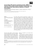
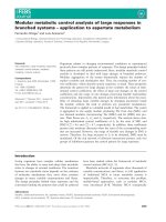
Figure 1
Levels of (a) protein C (PC), (b) activated protein C (APC), and (c) APC/PC ratio during hospital stay in patients with acute pancreatitisLevels of (a) protein C (PC), (b) activated protein C (APC), and (c) APC/PC ratio during hospital stay in patients with acute pancreatitis. Control,
patients with acute pancreatitis without multiple organ failure. The dotted lines indicate reference values for healthy individuals. Day 1 indicates the
day of admission to the research hospital
Critical Care Vol 10 No 1 Lindstrom et al.
Page 4 of 9
(page number not for citation purposes)
CD14 mAb (IgG2b, clone MFP9) (Becton Dickinson, San
Jose, CA, USA). HLA-DR expression is defined as the propor-
tion of positively fluorescing monocytes (HLA-DR%).
Statistics
All data are expressed as medians and ranges. Comparisons
of marker levels between the two groups were performed by
the Mann-Whitney U test. Fisher's exact test or chi-square
tests were used to compare the proportions of patients
between the two groups when appropriate. Spearman's rank
correlation was used for assessing correlations. Receiver
operating characteristics analysis was used for determination
of an optimal cut-off level for APC in differentiating cases from
controls. Comparisons of the follow-up samples were per-
formed with Friedman's test, followed by Dunn's test for post-
hoc comparisons when appropriate. SPSS 12.0.1 for Win-
dows (ACITS; The University of Texas at Austin, Austin, TX,
USA) statistical software was used. P < 0.05 was considered
statistically significant.
Results
PC pathway during hospital care
During the observation period all patients showed evidence for
activated haemostatic system. The levels of D-dimer were ele-
vated by 2.2-fold to 95-fold compared with the normal upper
limit of 0.5 mg/l. All values of PC levels, APC levels, and the
APC/PC ratio during the hospital stay are shown in Figure 1a–
c.
Decreased PC values (defined as <70% of the normal plasma
pool) were a frequent finding; found in 43% of all samples
(68% of all patients) at various stages of the disease (Figure
1a). The intrapatient variation in PC levels from day to day was
large. However, no association between the time of sampling
and the PC levels was found. Administration of blood products
did not readily explain the intrapatient variation of PC levels
(data not shown).
The levels of APC showed much less variation than did the PC
levels. All but one of the APC values fell within the range
observed in healthy resting adults (Figure 1b). As for PC, no
general dependency of APC levels with respect to the timing
of the samples was observed.
The rate of conversion of PC to APC was estimated by calcu-
lating the APC/PC ratios, for which we previously defined a
preliminary normal range [20] (Figure 1c). The APC/PC ratio
did not fall below the lower limit of normal in any sample but
exceeded the upper normal limit in 40% of the samples. At
least one APC/PC ratio value was above normal in 74% of all
patients.
Associations between general coagulation screening parame-
ters and PC pathway components were analysed. APC levels
correlated positively with the PT (r = 0.28, P = 0.01) and with
the platelet count (r = 0.32, P = 0.01), as did the PC concen-
tration (r = 0.48 for PT and r = 0.32 for platelet count, respec-
tively; both P = 0.01). The APC/PC ratio correlated negatively
with the PT (r = -0.43, P = 0.01). The D-dimer levels did not
correlate with APC levels (r = 0.15, P = 0.08), PC levels (r =
0.14, P = 0.10), or the APC/PC ratio (r = -0.06, P = 0.48).
Cases versus controls
Samples on admission
Samples on admission (defined as sampling within 36 hours
of actual admission) were available from 11 cases and 15 con-
trol patients. The APC concentration was significantly lower in
cases than in controls (median APC 86% versus 105%, P =
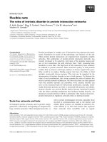
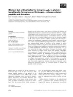
Figure 2
The lowest (a) protein C (PC) and (b) activated protein PC (APC) values during the first two weeks of hospital care in acute pancreatitis patients with multiple organ failure (cases) and in those without organ failure (controls)The lowest (a) protein C (PC) and (b) activated protein PC (APC) values during the first two weeks of hospital care in acute pancreatitis patients
with multiple organ failure (cases) and in those without organ failure (controls). P = 0.055 and P = 0.022 for PC and APC, respectively; Mann-Whit-
ney U test.
Available online />Page 5 of 9
(page number not for citation purposes)
0.027), whereas the PC level and the APC/PC ratio did not
differ significantly between the two groups. However, 89% of
cases and 43% of controls showed an abnormally high APC/
PC ratio (P = 0.04).
Follow-up samples
Multiple organ failure (MOF) developed from -2 to 14 days
(median, 1 day) after admission to the research hospital. We
compared the levels of the two PC pathway components
between cases and controls during this time period (Figure 2).
A decreased PC level was observed in 92% of cases and in
44% of controls (P = 0.008). The minimum (for instance the
lowest observed value) PC level was lower in cases than in
controls but the difference was not statistically significant (P =
0.055). The minimum observed APC level was lower in cases
than in controls (median 51% versus 73%, P = 0.022; Figure
2b). Utilizing receiver operating characteristics analysis, the
optimal cut-off value for APC to differentiate cases from con-
trols was found to be 87%. Eight out of 13 (62%) patients with
MOF showed a minimum APC level below this limit, while the
same was true for 3/18 (17%) of controls (P = 0.021). During
the first 2 weeks of hospitalization, 92% of cases and 50% of
controls had APC/PC ratios exceeding the upper normal limit
(P = 0.020).
The results of coagulation parameters during the stay in the
intensive care unit are presented in Table 2. There were no dif-
ferences in D-dimer level, the PT, or the platelet count
between cases and controls during the hospitalization. When
only the samples from the first 14 days were analysed, how-
ever, the maximum D-dimer level was higher (median 11.7 mg/
l versus 6.3 mg/l, P = 0.01) and minimum platelet count was
lower (105 × 10
9
/l versus 171 × 10
9
/l, P = 0.001) in cases
than in controls, respectively. The minimum PT was equal in
the two groups during the first 14 days.
Because MOF was diagnosed at various time points relative to
hospital admission, samples preceding the diagnosis of organ
failure were available from only five patients. The PC level was
below 70%, the APC level was within the normal range, and
the APC/PC ratio was above normal in all these patients.
When the pre-MOF samples of these five patients were com-
pared with control samples, the median APC value was equal
(P = 0.56), the median PC value was lower (P = 0.025), and
the APC/PC ratio was higher (P = 0.02).
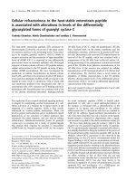
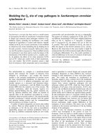
We also evaluated whether the time spent in hospital care
could affect the measured parameters in the subgroup of
patients with MOF. For this purpose, only patients with a suffi-
cient number of follow-up samples were analysed. Follow-up
samples with a maximum 2-day interval for 10 days were avail-
able from nine cases. During this 10-day period both the APC
and the PC levels tended to increase (Figure 3a,b). The
median APC/PC ratio was the lowest on day 7. The apparent
mechanism for the decreasing APC/PC ratio during hospitali-
zation was a gradual improvement of PC levels without a con-
comitant increase in APC values (Figure 3a–c). There was
thus a trend of gradual improvement of early PC pathway dis-
turbances during the course of MOF.
There were three deaths among the 13 patients in the case
group. In these three nonsurvivors the median level of APC
was 105% (85–188), that of PC was 74% (8–165), and that
of the APC/PC ratio was 1.45 (1.13–13.2). In the 10 survivors
of the case group, the median level of APC was 98% (76–
109), that of PC was 71% (35–165), and that of the APC/PC
ratio was 1.32 (0.87–2.6).
HLA-DR% and the protein C pathway
There was no difference in HLA-DR% between cases and
controls during their stay in the intensive care unit (Table 2).
Table 2
Coagulation parameters and the proportion of positively fluorescing monocytes (HLA-DR%) in patients with organ failure (cases)
and control patients during the stay in the intensive care unit
Variable Cases Controls P value
Lowest activated protein C level (%) 85 (76–102) 97 (73–136) 0.009
Lowest protein C level (%) 52 (8–70) 70 (18–113) 0.03
Lowest activated protein C/protein C ratio 1.1 (0.87–13.2) 1.3 (1.0–3.8) 0.02
Highest activated protein C/protein C ratio 1.8 (1.5–13.2) 1.6 (1.2–4.1) 0.03
D-dimer (mg/l) 5.5 (1.1–40.1) 4.7 (1.4–47.3) 0.22
Prothrombin time (%) 77 (17–147) 77 (13–138) 0.10
Platelet count (× 10
9
/l) 185 (48–738) 208 (40–530) 0.38
HLA-DR% 46 (8–82) 44 (11–84) 0.80
Data presented as median (range).
Critical Care Vol 10 No 1 Lindstrom et al.
Page 6 of 9
(page number not for citation purposes)
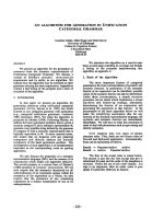
During the first 14 days in the intensive care unit, however, the
lowest HLA-DR% tended to be lower in cases (26%) than in
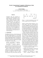
controls (35%) (P = 0.095). Positive correlations between
HLA-DR% and the PC concentration (r = 0.38, P < 0.001),
the APC concentration (r = 0.27, P < 0.001), and the platelet
count (r = 0.39, P < 0.001) were observed. HLA-DR% did not
correlate with the PT (r = 0.12) or the D-dimer level (r = -0.03)
(Figure 4a–d).
Discussion
The current observational study of the course of PC and APC
levels during AP in 31 patients with a 42% incidence of MOF
was conducted to address how often and to what extent the
PC pathway would be disturbed and whether such perturba-
tions would be associated with the development of MOF. The
study setting inevitably resulted in limitations in interpreting the
results in terms of causality. For example, the patients entered
the university hospital at various stages of AP, some having
MOF developed prior to the admission. Also, despite our con-
tinuous efforts during the two years of patient recruitment, the
sampling schedule in the original protocol was not completely
fulfilled. The patient series was rather uniform in disease sever-
ity and the incidence of MOF approached 50%, however, giv-
ing us the opportunity to test the hypothesis of whether a
defective PC pathway would be detrimental to patients with
AP.
The minimum levels of APC in MOF patients were significantly
lower than in controls. The PC deficiency was therefore logi-
cally associated with a decrease of APC levels in patients with
MOF. A more significant finding, however, may be that the
APC levels seemed not to be grossly elevated. In fact, only one
sample in one patient showed an absolute APC level that
exceeded the reported upper limit of normality (200%) [20].
Thrombin is the APC activator, and APC is a feedback inhibitor
for thrombin generation [22,23]. The complicated balance
Figure 3
Follow-up concentrations of (a) protein C (PC), (b) activated protein C (APC), and (c) APC/PC ratio during the stay in hospital in nine patients with organ failureFollow-up concentrations of (a) protein C (PC), (b) activated protein C (APC), and (c) APC/PC ratio during the stay in hospital in nine patients with
organ failure. There was no significant influence of time of sampling on any parameter (Friedman's test P > 0.05).
Available online />Page 7 of 9
(page number not for citation purposes)
between thrombin, PC, and APC varies depending on the cir-
cumstances. In resting healthy adults, the PC level rather than
the thrombin level may be the major determinant of the circu-
lating APC level [20,24,25]. During cardiopulmonary bypass,
upon rapid thrombin generation during reperfusion, the APC/
PC correlation was lost and a significant positive correlation
between fibrinopetide A, a thrombin marker, and APC devel-
oped [24].
The upper limit for APC formation may be estimated from APC
determinations in various clinical settings where coagulation is
known to be activated. In our previous studies the most pro-
nounced and fast enhancements from normal resting APC lev-
els to levels typically ranging from 250% to over 800% of the
normal mean were observed during the first minutes of reper-
fusion in liver transplantation [26] and during an infusion of
antithymocyte globulin, a strong proinflammatory stimulus, in
renal transplantation [27]. Liaw and colleagues recently
reported that in acute sepsis patients, despite ongoing activa-
tion of coagulation, 25% of patients failed to increase their
APC levels above 250% (while 75% had levels ranging from
approximately 250 to 800%) [28]. They concluded that the
septic patients varied markedly in their ability to generate APC
in response to the physiological thrombin stimulus, and attrib-
uted the phenomenon to endothelial dysfunction [28]. In chil-
dren with meningococceal sepsis, on the other hand, patients'
ability to generate APC in response to thrombin did not seem
to be grossly affected as infusion of PC resulted in elevated
levels of APC, and significant correlations were observed
between APC and thrombin markers at baseline [17].
Even though D-dimer is not a direct thrombin marker, the
highly elevated D-dimer levels in the current study indicated
that ample thrombin formation occurred throughout the obser-
vation period. Therefore, in good accordance with the study by
Liaw and colleagues in adult septic patients [28], we assume
that the lack of elevated free APC levels in the presence of
activated coagulation in patients with AP in most cases
reflected dysfunctional PC activation on the endothelium, or
possibly enhanced inhibition of APC by plasma protease inhib-
itors. This suggestion, however, does not exclude the possibil-
ity that significant PC deficiency could also be rate limiting for
APC formation in patients with severe AP, as seems to be the
case in septic children [17].
The PC levels were subnormal in 92% of patients with MOF
and in 44% of controls. Low PC was found to precede MOF,
and early APC/PC ratios were frequently high in MOF
Figure 4
Correlations between percentage of HLA-DR-positive monocytes and (a) protein C (PC), (b) activated PC (APC), (c) platelet count, and (d) D-dimer concentrationsCorrelations between percentage of HLA-DR-positive monocytes and (a) protein C (PC), (b) activated PC (APC), (c) platelet count, and (d) D-
dimer concentrations. Correlation coefficients are presented for combined data of organ failure and control patients. E9, × 10
9
.
Critical Care Vol 10 No 1 Lindstrom et al.
Page 8 of 9
(page number not for citation purposes)
patients. In an animal model of AP, the PC level was found to
decline already one hour after initiation of AP [29]. The PC lev-
els often fall rapidly in sepsis patients [30,31], and this may
associate with the development of organ failure [32]. Thus, in
logical accordance with animal AP data and human sepsis
studies, early PC deficiency was a most frequent finding dur-
ing severe AP. While the causal relationship between low PC
levels and MOF development cannot be proven in an observa-
tional study setting, one probable bias could be excluded.
Namely, there was no indication that PC deficiency would
associate with MOF after its diagnosis and, further, there was
a significant trend for improvement of PC homeostasis during
the course of MOF. It thus seems that, in patients developing
MOF, the PC activation system reached its limits prior to the
diagnosis of MOF, resulting in secondary deficiency of PC
concomitantly with a failure to maintain steady APC levels sim-
ilar to those observed in AP patients without MOF.
APC and PC correlated significantly with monocyte HLA-DR
expression but failed to associate with the level of D-dimer.
The existence of anti-inflammatory and anti-apoptotic proper-
ties of APC is currently widely accepted [22,33]. Compared
with the accumulating in vitro and animal data, however,
observational human clinical studies provide scarce data on
the specific relationship between APC and inflammation. The
obvious limitation is the fact that thrombin formation is also
enhanced whenever APC generation is enhanced, and the
question remains whether APC or other components of the
activated coagulation pathways might be involved.
The concomitant decrease in D-dimer and IL-6 during APC
infusion in the PROWESS trial has been taken as circumstan-
tial evidence of specific anti-inflammatory action of APC [12].
The remaining human studies finding associations between
APC and inflammatory phenomena have involved only hypera-
cute proinflammatory/procoagulant situations such as reper-
fusion after cardiopulmonary bypass [34], liver transplantation
[26], and renal transplantation [27]. We chose to measure
HLA-DR%, which reflects the recent history of activation/func-
tional suppression of the circulating monocyte population
[21]. APC acts as an anti-inflammatory agent in vitro, largely
through modulating monocyte activation during inflammation
[9-11]. The current correlations between HLA-DR expression
and the PC and APC levels may therefore support the concept
of interaction between inflammation and the PC pathway. In
severe AP, levels of PC and APC were moderately associated
with monocyte activation status.
The current study suggests that testing the therapeutic use of
APC or PC to improve patient outcome might be feasible in
AP. The concurrent activation of inflammation and coagulation
during AP, the frequent occurrence of PC pathway defects in
AP patients, their association with a significant clinical end-
point (MOF), and the timing of major PC defects to the early
phase of the disease all lend support to attempt APC or PC
infusion in severe AP.
Conclusion
In summary, the current study demonstrated significant PC
pathway pathology in severe AP. The PC pathway defects
were more frequent in patients developing MOF.
Competing interests
The authors declare that they have no competing interests.
Authors' contributions
OL and LK executed the study and drafted the manuscript. LK,
JAF, JHG, PP, HR, and JP participated in the original design
and coordination of the study, and in writing the original proto-
col. JAF and JHG carried out the PC and APC assays. LK, PM,
HR, and JP analysed the data. PM, EK, PP, HR, and JP
assisted in drafting the manuscript. RH assisted in the original
design and drafting of the final manuscript. All authors read
and approved the final manuscript.
Acknowledgements
This study was supported by grants from the Helsinki University Central
Hospital Research Funds (EVO) and the Foundation for Pediatric
Research (JP)
References
1. Gronroos J, Nylamo E: Mortality in acute pancreatitis in Turku
University Central Hospital 1971–1995. Hepatogastroenterol-
ogy 1999, 46:2572-2574.
2. Tenner S, Sica G, Hughes M, Noordhoek E, Feng S, Zinner M,
Banks B: Relationship of necrosis to organ failure in severe
acute pancreatitis. Gastroenterology 1997, 113:899-903.
3. Deitch E: Multiple organ failure. Ann Surg 1992, 216:117-134.
4. Mentula P, Kylanpaa-Back M-L, Kemppainen E, Takala A, Jansson
S-E, Kautiainen H, Puolakkainen P, Haapiainen R, Repo H:
Decreased HLA (human leucocyte antigen)-DR expression on
peripheral blood monocytes predicts the development of
organ failure in patients with acute pancreatitis. Clin Sci 2003,
105:409-417.
5. Levi M, ten Cate H: Disseminated intravascular coagulation. N
Engl J Med 1999, 341:586-592.
6. Shen L, Dahlback B: Factor V and protein S as synergistic
cofactors to activated protein C in degradation of factor VIIIa.
J Biol Chem 1994, 269:18735-18738.
7. Taylor FB Jr, Peer GT, Lockhart MS, Ferrell G, Esmon CT:
Endothelial cell protein C receptor plays an important role in
protein C activation in vivo. Blood 2001, 97:1685-1688.
8. Mosnier L, Griffin JH: Inhibition of staurosporine-induced apop-
tosis of endothelial cells by activated protein C requires pro-
Key messages
• Severe AP patients suffer from significant PC pathway
pathology, which is associated with the development of
organ failure.
• Monocyte HLA-DR% correlated with PC and APC lev-
els, indicating interaction between inflammation and the
PC pathway in severe AP.
• Modulating the PC pathway might improve the outcome
of severe AP patients.
Available online />Page 9 of 9
(page number not for citation purposes)
tease-activated receptor-1 and endothelial cell protein C
receptor. Biochem J 2003, 373:65-70.
9. Grey S, Tcuchida A, Hau H, Orthner C, Salem H, Hancock W:
Selective inhibitory effects of the anticoagulant activated pro-
tein C on the responses of human mononuclear phagocytes to
LPS, IFN-gamma, or phorbol ester. J Immunol 1994,
153:3664-3672.
10. White B, Schmidt M, Murphy C, Livingstone W, O'Toole D, Lawler
M, O'Neill L, Kelleher D, Schwarz H, Smith O: Activated protein C
inhibits lipoplysaccharide-induced nuclear translocation of
nuclear factor κB (NF-κB) and tumour necrosis factor α (TNF-
α) production in the THP-1 monocytic cell line. Br J Haematol
2000, 110:130-134.
11. Yuksel M, Okajima K, Uchiba M, Horiuchi S, Okabe H: Activated
protein C inhibits lipopolysaccharide-induced tumor necrosis
factor-alpha production by inhibiting activation of both nuclear
factor-kappa B and activator protein-1 in human monocytes.
Thromb Haemost 2002, 88:267-273.
12. Bernard G, Vincent J, Laterre P, LaRosa S, Dhainaut J-F, Lopez-
Rodriguez A, Steingrub J, Garber G, Helterbrand J, Ely W, et al.:
Efficacy and safety of recombinant human activated protein C
for severe sepsis. N Engl J Med 2001, 344:699-709.
13. Lasson A, Ohlsson K: Consumptive coagulopathy, fibrinolysis
and protease-antiprotease interactions during acute human
pancreatitis. Thromb Res 1986, 41:167-183.
14. Radenkovic D, Bajec D, Karamarkovic A, Stefanovic B, Milic N, Ign-
jatovic S, Gregoric P, Milicevic M: Disorders of hemostasis dur-
ing the surgical management of severe necrotizing
pancreatitis. Pancreas 2004, 29:152-156.
15. Salomone T, Tosi P, Palareti G, Tomassetti P, Migliori M, Guariento
A, Saieva C, Raiti C, Romboli M, Gullo L: Coagulative disorders
in human acute pancreatitis: role for the D-dimer. Pancreas
2003, 26:111-116.
16. Mantke R, Pross M, Kunz D, Ebert M, Kahl S, Peters B, Malfer-
theiner P, Lippert H, Schulz H-U: Soluble thrombomodulin
plasma levels are an early indication of a lethal course in
human acute pancreatitis. Surgery 2002, 131:424-432.
17. de Kleijn E, de Groot R, Hack C, Mulder P, Engl W, Moritz B,
Joosten K, Hazelzet J: Activation of protein C following infusion
of protein C concentrate in children with severe meningococ-
cal sepsis and purpura fulminans: a randomized, double-
blinded, placebo-controlled, dose-finding study. Crit Care Med
2003, 31:1839-1847.
18. Bradley E III: A clinically based classification system for acute
pancreatitis. Summary of the international symposium on
acute pancreatitis, Atlanta, Ga, September 11–13, 1992. Arch
Surg 1993, 128:586-90.
19. Gruber A, Griffin JH: Direct detection of activated protein C in
blood from human subjects. Blood 1992, 79:2340-2348.
20. Petaja J, Hakala L, Rasi V, Griffin JH: Circulating activated protein
C in subjects with heterozygous Gln506-factor V. Haemostasis
1998, 28:31-36.
21. Kylanpaa-Back M, Takala A, Kemppainen E, Puolakkainen P, Kau-
tiainen H, Jansson S-E, Haapiainen R, Repo H: Cellular markers
of systemic inflammation and immune suppression in patients
with organ failure due to severe acute pancreatitis. Scand J
Gastroenterol 2001, 36:1100-1107.
22. Esmon CT: Protein C anticoagulant pathway and its role in con-
trolling microvascular thrombosis and inflammation. Crit Care
Med 2001:48-51.
23. Griffin JH: The thrombin paradox. Nature 1995, 378:337-338.
24. Petaja J, Pesonen E, Fernández JA, Vento A, Rämö OJ, Griffin JH:
Cardiopulmonary bypass and activation of antithrombotic
plasma protein C. J Thorac Cardiov Surg 1999, 118:422-431.
25. Fernández JA, Petaja J, Gruber A, Griffin JH: Activated protein C
correlates inversely with thrombin levels in resting healthy
individuals. Am J Hematol 1997, 56:29-31.
26. Ilmakunnas M, Petaja J, Fernandez JA, Griffin JH, Repo H, Höcker-
stedt K, Mäkisalo H, Pesonen E: Activation of antithrombotic and
anti-inflammatory protein C during reperfusion in clinical liver
transplantation. Transplantation 2003, 75:467-472.
27. Turunen A, Fernández JA, Lindgren L, Salmela K, Kyllonen L, Mak-
isalo H, Griffin JH, Siitonen SM, Petaja J, Pesonen EJ: Activated
protein C reduces graft neutrophil activation in clinical renal
transplantation. Am J Transplant 2005, 5:2204-2212.
28. Liaw P, Esmon C, Kahnamoui K, Schmidt S, Kahnamoui S, Ferrell
G, Beaudin S, Julian J, Weitz J, Crowther M, et al.: Patients with
severe sepsis vary markedly in their ability to generate acti-
vated protein C. Blood 2004, 104:3958-3964.
29. Ottesen L, Bladbjerg E, Osman M, Lausten S, Jacobsen N, Gram
J, Jensen S: Protein C activation during the initial phase of
experimental acute pancreatitis in the rabbit. Dig Surg 1999,
16:486-495.
30. Powars D, Larsen R, Johnson J, Hulbert T, Sun T, Patch M, Francis
R, Chan L: Epidemic meningococcemia and purpura fulminans
with induced protein C deficiency. Clin Infect Dis 1993,
17:254-261.
31. Mesters R, Helterbrand J, Utterback B, Yan B, Chao B, Fernandez
JA, Griffin JH, Hartman D: Prognostic value of protein C concen-
trations in neutropenic patients at high risk of severe septic
complications. Crit Care Med 2000, 28:2209-2216.
32. Iba T, Kidokoro A, Fukunaga M, Sugiyama K, Sawada T, Kato H:
Association between the severity of sepsis and the changes in
hemostatic molecular markers and vascular endothelial dam-
age markers. Shock 2005, 23:25-29.
33. Griffin JH, Fernandez JA, Liu D, Cheng T, Guo H, Zlokovic B: Acti-
vated protein C and ischemic stroke. Crit Care Med
2004:247-253.
34. Petaja J, Pesonen E, Fernández JA, Griffin JH, Repo H, Jansson S,
Vento AE, Rämö J: Activated protein C and inflammation in
human myocardium after heart surgery. Am J Hematol 2001,
67:210-212.