Báo cáo khoa học: " A Lung and ''''end organ'''' injury due to mechanical ventilation in animals: comparison between the prone and supine positions" pdf
Bạn đang xem bản rút gọn của tài liệu. Xem và tải ngay bản đầy đủ của tài liệu tại đây (537 KB, 9 trang )
Open Access
Available online />Page 1 of 9
(page number not for citation purposes)
Vol 10 No 1
Research
Lung and 'end organ' injury due to mechanical ventilation in
animals: comparison between the prone and supine positions
George Nakos
1
, Anna Batistatou
2
, Eftychia Galiatsou
1
, Eleonora Konstanti
1
, Vassilios Koulouras
1
,
Panayotis Kanavaros
3
, Apostolos Doulis
1
, Athanassios Kitsakos
1
, Angeliki Karachaliou
1
,
Marilena E Lekka
4
and Maria Bai
2
1
Department of Intensive Care Unit, University Hospital of Ioannina, Greece
2
Department of Pathology, University of Ioannina, Greece
3
Department of Anatomy-Histology-Embryology, University of Ioannina, Greece
4
Department of Chemistry, University of Ioannina, Greece
Corresponding author: George Nakos,
Received: 2 Nov 2005 Revisions requested: 8 Dec 2005 Revisions received: 25 Jan 2006 Accepted: 3 Feb 2006 Published: 28 Feb 2006
Critical Care 2006, 10:R38 (doi:10.1186/cc4840)
This article is online at: />© 2006 Nakos et al.; licensee BioMed Central Ltd.
This is an open access article distributed under the terms of the Creative Commons Attribution License ( />),
which permits unrestricted use, distribution, and reproduction in any medium, provided the original work is properly cited.
Abstract
Introduction Use of the prone position in patients with acute
lung injury improves their oxygenation. Most of these patients die
from multisystem organ failure and not from hypoxia, however.
Moreover, there is some evidence that the organ failure is
caused by increased cell apoptosis. In the present study we
therefore examined whether the position of the patients affects
histological changes and apoptosis in the lung and 'end organs',
including the brain, heart, diaphragm, liver, kidneys and small
intestine.
Methods Ten mechanically ventilated sheep with a tidal volume
of 15 ml/kg body weight were studied for 90 minutes. Five
sheep were placed in the supine position and five sheep were
placed in the prone position during the experiment. Lung
changes were analyzed histologically using a semiquantitative
scoring system and the extent of apoptosis was investigated
with the TUNEL method.
Results In the supine position intra-alaveolar hemorrhage
appeared predominantly in the dorsal areas, while the other
histopathologic lesions were homogeneously distributed
throughout the lungs. In the prone position, all histological
changes were homogeneously distributed. A significantly higher
score of lung injury was found in the supine position than in the
prone position (4.63 ± 0.58 and 2.17 ± 0.19, respectively) (P <
0.0001). The histopathologic changes were accompanied by
increased apoptosis (TUNEL method). In the supine position,
the apoptotic index in the lung and in most of the 'end organs'
was significantly higher compared with the prone position (all P
< 0.005). Interestingly, the apoptotic index was higher in dorsal
areas compared with ventral areas in both the prone and supine
positions (P < 0.003 and P < 0.02, respectively).
Conclusion Our results suggest that the prone position
appears to reduce the severity and the extent of lung injury, and
is associated with decreased apoptosis in the lung and 'end
organs'.
Introduction
Mechanical ventilation has constituted an indispensable part
of basic life support in the intensive care unit for several dec-
ades and is undoubtedly essential for patients with acute lung
injury/acute respiratory distress syndrome (ALI/ARDS). In
recent years, however, it has become clear that mechanical
ventilation can also be injurious. Repeated application of
transalveolar pressures that exceed those corresponding to
the inflation capacity causes tissue stresses and disrupts the
lung. In animals, mechanical ventilation at high volumes and
high pressures can cause ventilator-induced lung injury (VILI)
with similar histological appearance to ALI/ARDS. These his-
tological disorders are due to injury of the alveolar epithelium,
basement membrane and microvascular endothelium and
accompanied by high-permeability pulmonary edema. Injurious
AI = apoptotic index; ALI = acute lung injury; ARDS = acute respiratory distress syndrome; FiO
2
= fraction inspired oxygen; H&E = haematoxylin–
eosin; PCO
2
= partial pressure of CO
2
; TUNEL= terminal deoxynucleotidyl-transferase-mediated dUTP nick end-labeling; VILI = ventilator-induced
lung injury.
Critical Care Vol 10 No 1 Nakos et al.
Page 2 of 9
(page number not for citation purposes)
mechanical ventilation exacerbates the damage in previously
injured lungs [1-3].
The damage to the lungs has been attributed to two overlap-
ping mechanisms, namely mechanical damage of tissues and
cells due to overdistention and shear stress (barotrauma or
volutrauma) as well as mechanical damage due to the produc-
tion, release and/or activation of cytotoxic and inflammatory
cascades (biotrauma). In addition to inducing or worsening
existing lung injury, the pulmonary production of inflammatory
mediators is likely to spill over into the systemic circulation,
also contributing to extrapulmonary end-organ failure [3,4].
Despite considerable progress, the death rate of patients with
ALI/ARDS remains quite high [5]. In fact, most patients die
from multisystem organ failure and not from hypoxia. However,
pathogenesis of multiorgan failure in ARDS/ALI remains a
dilemma. There is some evidence that multisystem organ fail-
ure is caused by increased apoptosis of the epithelial cells of
'end organs', such as the kidneys and small intestine [6,7].
Apoptosis is an active mechanism of cell death, which is
important for the development and homeostasis of tissues.
Environmental conditions or specific receptor/ligand interac-
tions activate intracellular signaling pathways that lead to DNA
cleavage and apoptotic cell death (for a review, see [8]).
As early as 1976 it was reported that placing patients with
ALI/ARDS in the prone position improves their oxygenation [9-
17]. Prone positioning improves secretion drainage from the
airways, relieving lung compression by the heart and abdo-
men. The transalveolar forces are redistributed so as to allow
expansion of the dorsal regions. All these events lead to an
increase in end-respiratory lung volume, to better ventilation-
perfusion matching and to alterations in chest-wall mechanics
leading to regional changes in ventilation. The effects of prone
ventilation on the cellular constituents of the lung alveoli have
not so far been studied.
Our working hypothesis was that VILI can lead to distant organ
damage through the increase in the circulation of mediators,
including proapoptotic soluble factors, such as soluble Fas lig-
and [6]. In this respect, using injurious tidal-volume-induced
lung damage, we studied the possible protective role of the
prone position through the reduction of atelectasis and/or
overdistention. In addition, we investigated whether cell apop-
tosis was related to the severity of tissue damage of the lung
and other organs induced by mechanical ventilation.
Materials and methods
Animal preparation
Protocols were approved by the University of Ioannina animal
research committee. We examined 10 sheep, each weighing
33 ± 5 kg. A peripheral vein was cannulated, and anesthesia
was induced with katanine, maintained by continuous intrave-
nous injection of midazolam and fentanyl citrate and paralyzed
with pancuronium bromide. The animals were tracheotomized,
and catheters were introduced into the carotid artery and the
external jugular vein. Mechanical ventilation was provided with
a Servo 900C ventilator (Siemens Elema, Solna, Sweden) in
the volume control mode with a tidal volume of 15 ml/kg body
weight for 90 minutes, with low positive end expiratory pres-
sure (3 cmH
2
O) and with FiO
2
of 0.5 in both groups. The res-
piratory rate was adjusted appropriately to maintain
normocapnia at baseline measurements. Arterial pressure
from the carotid artery and airway was recorded throughout
the experiment. Blood gases, respiratory system compliance
(calculated as the end-inspiratory airway pressure minus the
end-expiratory pressure divided by the tidal volume) and bio-
chemistry were measured before, during and at the end of the
experiment. We continuously monitored the arterial blood
pressure, the central venous pressure, the heart rate and the
urine output. These parameters were kept stable by fluid infu-
sion (normal saline). The animal temperature was also kept sta-
ble.
Five animals were placed in the supine position and five in the
prone position during the whole experiment. The animals were
exsanguinated at the end of the experiment, which lasted 90
minutes from the beginning of mechanical ventilation, while
deeply anesthetized. The internal organs were removed and
representative sections from the lungs, the brain, the heart, the
diaphragm, the liver, the kidneys and the small intestine were
taken and fixed in 10% buffered formalin.
Histologic evaluation and TUNEL method
Paraffin sections, 5 µm thick, were stained with the standard
H&E stain and examined using light microscopy. Lung
changes were analyzed histologically using a semiquantitative
scoring system, as previously described elsewhere [18].
Briefly, six slides – two from the upper lobe (one from the dor-
sal area and one from the ventral area), two from the lower lobe
(one from the dorsal area and one from the ventral area) and
two from the middle lobe in the right lung and the middle area
in the left lung – were analyzed by two independent patholo-
gists. The pathologists were blinded to the assignment of the
animals. The slides were scanned in low power and the five
fields with the most pronounced changes were chosen. The
score given for each slide represented the mean score of
these fields.
Four parameters were examined: alveolar fibrin edema, alveo-
lar hemorrhage, septal thickening and intra-alveolar inflamma-
tory cells. The changes were scored according to their extent
(score 0, 1, 2 and 3 for an extent of 0%, <25%, 25–50% and
>50%, respectively) and the severity of the injury (score 0 for
no changes, score 1, 2 and 3 for more severe changes). The
injury score represents the sum of the extent and the severity
of injury.
Available online />Page 3 of 9
(page number not for citation purposes)
Table 1
Gas exchange, respiratory system compliance and hemodynamics
Supine position Prone position P value 95% confidence interval of the
difference
PO
2
/FIO
2
(mmHg)
Baseline 416 ± 23.6 412.4 ± 25.5 NS
90 minutes 105.6 ± 24.1 251.6 ± 56.1 <0.001 -208.9 to -83.0
P value <0.0001 <0.004
95% confidence interval of the difference 272.8–247.9 84.8–236.7
PCO
2
(mmHg)
Baseline 38.8 ± 1.8 40.8 ± 1.3 NS
90 minutes 57.2 ± 1.5 43.0 ± 1.2 <0.001 2.2 to 6.1
P value <0.002 <0.04
95% confidence interval of the difference -5.0 to -6.9 -1.1 to -5.8
pH
Baseline 7.408 ± 0.013 7.398 ± 0.008 NS
90 minutes 7.322 ± 0.019 7.382 ± 0.018 0.0009 -0.08 to -0.03
P value 0.0005 NS
95% confidence interval of the difference 0.063–0.108
Static compliance of respiratory system (ml/cmH
2
O)
Baseline 30.4 ± 3.8 25.9 ± 2.1 NS
90 minutes 18.2 ± 2.8 22.8 ± 2.3 <0.02 -8.3 to -0.86
P value <0.001 <0.003
95% confidence interval of the difference -10.1 to -14.3 -1.7 to -4.5
Blood pressure (mmHg)
Baseline 81.80 ± 7.294 85.60 ± 9.476 NS
90 minutes 84.20 ± 5.167 86.00 ± 9.670 NS
P value NS NS
95% confidence interval of the difference
Heart rate (beats/minutes)
Baseline 117.2 ± 9.365 122.2 ± 6.140 NS
90 minutes 130.4 ± 4.722 132.8 ± 5.891 NS
P value 0.0074 0.0007
95% confidence interval of the difference -20.51 to -5.887 -13.72 to -7.484
Static compliance of respiratory system = (end inspiratory airway pressure – end-expiratory pressure)/tidal volume.
Apoptosis was detected with the terminal deoxynucleotidyl-
transferase-mediated dUTP nick end-labeling (TUNEL)
method (Apo-tag kit; Oncor, Craithersburg, MD, USA) in 5 µm
paraffin sections, as described in detail in previous studies
[19,20]. Positive and negative controls were included in every
staining. Positive staining in areas of lymphocytic infiltration
served as the internal positive control. No staining was noted
in negative controls.
Briefly, morphologically intact TUNEL-positive cells and apop-
totic cells in H&E-stained studies were considered positive
and are referred to as apoptotic cells. The number of apoptotic
cells and apoptotic bodies was recorded by using the 40×
objective lens, and at least 10 randomly selected fields were
counted. The apoptotic index (AI) was expressed as the
number of apoptotic cells/bodies per 10 high-power fields.
Care was taken to avoid areas with extensive inflammation.
Critical Care Vol 10 No 1 Nakos et al.
Page 4 of 9
(page number not for citation purposes)
The AI at the alveolar septum of the lungs, the neurons and
glial cells, the muscle cells of the diaphragm, the hepatocytes,
the glomerular and tubular renal cells, and the epithelial cells
of the small intestinal epithelium were estimated.
Statistical analysis
Statistical analysis was performed using the Statistical Pack-
age for Social Sciences (SPSS) version 12 for Windows
(SPSS Inc., Chicago, Illinois, USA). Data were tested for nor-
mality with the Kolmogorov-Smirnov test and are presented as
the mean ± SD. All variables were normally distributed. Com-
parisons between the prone and supine positions were made
using a t test. Comparisons between the ventral and dorsal
regions of the lungs in either the supine position or the prone
position were made using a paired t test.
Results
Lung mechanics and blood gases
Lung mechanics and blood gas alterations and the biochemi-
cal data are presented in Tables 1 and 2, respectively. Blood
gases and the compliance of the respiratory system deterio-
rated after 90 minutes of mechanical ventilation in both posi-
tions. The deterioration in blood gases as well as in the
compliance due to VILI was significantly less prominent in the
prone position. Transaminases (aspartate aminotransferase
and alanine aminotransferase) increased during mechanical
ventilation in the supine position, while they were both
unchanged in the prone position. γ-Glutamyl transpeptidase,
urea and creatinine were not altered during mechanical venti-
lation in both positions.
ALI score in the prone and supine positions
In the lungs of the animals placed in the supine position the
alveolar-septal membrane was thickened and there was con-
siderable intra-alveolar edema and eosinophilic material. Fur-
thermore, hemorrhage and increased numbers of inflammatory
cells (lymphocytes, plasma cells, macrophages and polymor-
phonuclear neutrophil granulocytes) were observed (Table 3).
Consolidated areas were frequently encountered (Figure 1a).
In animals placed in the prone position the lung injury was
milder (Table 3). There was considerably less inflammatory
infiltration, alveolar edema, hemorrhage thickening of the alve-
Table 2
Biochemistry at the beginning and the end of experiment
Supine position Prone position P value
Urea (mg/dl)
Baseline 34.9 ± 11.5 43.4 ± 6.5 NS
90 minutes 41.1 ± 7.3 37.1 ± 8.4 NS
P value NS NS
Creatinine (mg/dl)
Baseline 0.62 ± 0.1 0.48 ± 0.11 NS
90 minutes 0.55 ± 0.08 0.53 ± 0.1 NS
P value NS NS
aspartate aminotransferase (IU/l)
Baseline 94 ± 21 98 ± 25 NS
90 minutes 147 ± 19 84 ± 27 <0.05
P value <0.05 NS
alanine aminotransferase (IU/l)
Baseline 14 ± 6 16 ± 7 NS
90 minutes 27 ± 8 15 ± 9 <0.05
P value <0.05 NS
γ-Glutamyl transpeptidase (IU/l)
Baseline 26 ± 18 29 ± 24 NS
90 minutes 33 ± 22 25 ± 23 NS
P value NS NS
Available online />Page 5 of 9
(page number not for citation purposes)
olar-septal membrane and consolidation. In addition, many
areas appeared uninjured or minimally affected (Figure 1b).
The differences between the supine and prone positions were
statistically significant (P < 0.0001). Interestingly, the overall
histological findings for each animal were consistent in all lung
areas – upper, middle and lower, ventral and dorsal (Table 3).
When alveolar hemorrhage was considered alone, however,
there was a significant difference between ventral and dorsal
samples in animals placed in the supine position. In these ani-
mals the mean score for alveolar hemorrhage was 4.8 ± 0.84
in the ventral areas and was 2.6 ± 0.55 in the dorsal areas of
both lungs (P < 0.01). This difference was not evident in ani-
mals placed in the prone position.
Apoptotic index in the prone and supine positions
TUNEL-positive nuclei/apoptotic bodies were observed in all
animals in the lungs, and the AI was increased in the supine
position group compared with the prone position group (Table
3 and Figure 2a,b). In both the supine position and the prone
position, the mean value of the AI was higher in areas dorsal
compared with ventral areas; the differences were statistically
significant (P = 0.04 and P = 0.046, respectively). Moreover,
the differences between the supine and prone positions were
statistically significant in the dorsal lung areas as well in the
ventral lung areas (P < 0.003 and P < 0.02, respectively)
(Table 3).
The AI in the liver was far less than that in the lungs. The liver
AI was increased in the supine position group (Figure 2c,d).
The difference was statistically significant (P < 0.05) (Table 3).
In the kidneys, particularly at the medulla, the nuclei of tubular
epithelial cells were TUNEL-positive without morphological
characteristics of apoptosis and were not included in the esti-
mation of the AI. Counts were performed at the cortex (Figure
2e,f). The mean values of the AI were higher in the supine posi-
Table 3
Acute lung injury score and apoptotic index in the supine and prone position
Supine position Prone position P value 95% confidence interval
Acute lung injury score 4.63 ± 0.58 2.17 ± 0.19 <0.0001 -3.9 to -1.82
Apoptotic index
Lung dorsal 112 ± 22 45.6 ± 28 0.003 -103.6 to -29.78
Lung ventral 80 ± 28 35 ± 22 0.02 -82.6 to -8.1
P value 0.04 0.046
95% confidence interval 2.37 to 61.09 0.29 to 20.5
Liver 56 ± 21 23 ± 10 0.05 -66.78 to -7.8.1
Kidney 31 ± 14 17 ± 10 NS
Small intestine 22 ± 11 16 ± 11 NS
Diaphragm 10 ± 0.5 0.5 ± 0.4 0.001 -10.6 to -9.01
Acute lung injury score corresponds to the sum of the extent (score 0, 1, 2 and 3 for an extent of 0%, <25%, 25–50% and >50%) and the
severity of lung injury (score 0 for no changes, score 1, 2 and 3 more severe changes). The apoptotic index was expressed as the number of
apoptotic cells/bodies per 10 high-power fields.
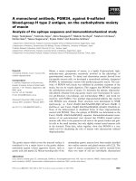
Figure 1
Histological changes of lungs (septal thickening, alveolar fibrin/edema, alveolar hemorrhage, intra-alveolar inflammatory cells) in animals placed in (a) the supine position and (b) the prone position (H&E, ×400)Histological changes of lungs (septal thickening, alveolar fibrin/edema, alveolar hemorrhage, intra-alveolar inflammatory cells) in animals placed in (a)
the supine position and (b) the prone position (H&E, ×400).
Critical Care Vol 10 No 1 Nakos et al.
Page 6 of 9
(page number not for citation purposes)
tion in comparison with the prone position, but the differences
between the two groups did not reach statistical significance
(Table 3).
An increased AI was also detected in the myocytes of the dia-
phragm (Figure 2g,h). The mean value of the AI was remarka-
bly increased in the supine position compared with the prone
position, and the difference was statistically significant (P <
0.001) (Table 3).
An increased AI was also detected in the epithelial lining of the
small intestine villi and crypts in the supine position group
compared with the prone position group. This difference was
not statistically significant, however (Figure 2i,j and Table 3).
The AI in the brain was low in both the supine position and the
prone position groups.
Discussion
The main finding in this study was the reduction of the severity
of and the extent of VILI in the prone position. This protective
result of the prone position was associated with decreased
cell apoptosis in the lung and other organs, including the liver
and the diaphragm.
We have shown that mechanical ventilation in relatively high
volumes causes injury to the lung parenchyma of animals,
which can be detected and semiquantitated using light micro-
scopy. These histologically defined changes were significantly
more extensive in the supine position than in the prone posi-
tion. Furthermore, intra-alveolar hemorrhage appeared pre-
dominantly in the dorsal areas in the supine position, while the
other histologic changes (alveolar/fibrin edema, septal thick-
ening, intra-alveolar inflammatory cells) were homogeneously
distributed throughout the lungs. All the histological changes
were homogenously distributed in the prone position.
The histologic changes in the lung were accompanied by an
increased AI at the alveolar septum. It is interesting that the AI
was significantly higher in dorsal areas compared with ventral
areas in both the prone and supine positions. We also present
evidence supporting the hypothesis that an injurious ventila-
tory strategy administered to the lungs can lead to damage of
'end organs', probably associated with apoptosis. Interest-
ingly, the prone position appears to reduce the severity and
the extent of the lung injury and is associated with a decreased
AI in the lungs and 'end organs'. The deterioration in blood
gases as well as in the respiratory system compliance was in
accordance with the lung injury and was lower in the prone
position. The increase of PCO
2
in the supine position could be
attributed to the increase of dead space due to lung injury and
basal atelectasis. Hypercapnia has been considered as a pro-
tective factor rather than a harmful one in lung injury [21]. It
was therefore not a factor favoring the deference observed
between the supine and prone positions.
When first recognized, ALI/ARDS was considered a diffuse
disease of the lungs and the injury was considered homogene-
ously distributed. Computed tomographic scanning has dem-
onstrated that alveolar filling, consolidation and atelectasis
occur predominantly in dependent lung zones, whereas other
areas may be relatively spared [22-28]. Rouby and colleagues
reported that the lung injury in ARDS is actually heterogene-
ous, with collapsed areas, areas of regional hyperinflation and
normal areas [29]. Bronchoalveolar lavage studies indicate,
however, that even radiographically spared, nondependent
areas may have substantial inflammation [30]. Our histological
findings indicate that the VILI in the supine position as well in
the prone position affects the whole lung quite homogene-
ously, except for the hemorrhage in the supine position, which
was higher in dependent areas of the lung. This phenomenon
could be due to greater tissue stresses and shearing force
induced by the inspiratory pressure in the dependent areas of
the lung, which are most subject to closure. The hemorrhage
was significantly less and was homogeneously distributed in
the prone position. This fact is probably due to expansion of
the dorsal regions resulting in a reduction of the shear stress
[15,26,31,32].
Over the past decade VILI has emerged as a clinical issue
[2,32,33]. The clinical importance of VILI has been docu-
mented in the ARDS Network study, where a reduction by
22% in the mortality of patients was noted when the mechan-
ical load exerted on the lungs was reduced with lowering of
the tidal volume [5]. Ventilation with high tidal volume results
in the release of cytokines and other proinflammatory mole-
cules [34]. In addition to inducing lung injury or worsening
existing lung injury, this cascade of mediators may also con-
tribute to extrapulmonary end-organ failure. Activation of the
Fas/Fas ligand pathway in this process could be implicated as
the apoptotic mechanism of the alveolar epithelium. Soluble
Fas ligand, a main proapoptotic factor, is considered respon-
sible for the increased apoptosis in 'end organs' [6,29,35,36].
Our results show that injurious mechanical ventilation
increases the apoptosis in the lungs as well as in 'end organs'.
These findings are consistent with those of Imai and col-
leagues [6], who demonstrated that the injurious mechanical
ventilation can lead to epithelial cell apoptosis in organs distal
to the lung, such as the kidneys. There is some evidence that
increased apoptosis is accompanied by biochemical changes
suggesting organ failure [6,7]. This could be an explanation for
the high rates of multiple organ failure in patients with ARDS
and the decrease in mortality when lung protective strategy is
applied [5,6]. The role of apoptosis and necrosis in tissue
injury and inflammation is not well understood, however. Seri-
ous lung injury could be accompanied by necrosis, while cell
death in milder situations could be due to apoptosis [37].
The prone position, under the studied conditions, appears to
decrease the severity and the extent of lung injury and is asso-
Available online />Page 7 of 9
(page number not for citation purposes)
ciated with a decrease in apoptosis of lung and 'end organ' tis-
sues. Broccard and colleagues have also shown in animal
models that, for the same pattern of ventilatory pressures, the
prone position protects better against VILI [15]. It is known
that the prone position improves oxygenation by quite complex
mechanisms: Changes in lung recruitment are definitely one
parameter contributing to improved lung oxygenation. Lung
perfusion and alveolar ventilation are more uniformly distrib-
uted in the prone position compared with the supine position
[15,22,38,39]. Our data provide another piece in the puzzle of
ventilation-induced injury of lung and 'end organs'. We pro-
pose that although there might be no regional distribution in
lung perfusion, there are definitely differences in vascular dam-
age, leading to preferential intra-alveolar hemorrhage in the
dorsal lung areas, particularly in animals in the supine position.
Pronation ameliorates these differences. From a theoretical
standpoint, shear stresses at the junction of open tissue and
closed tissue will rise to high levels that may mechanically dis-
rupt epithelial as well as endothelial membranes [30].
Conclusion
Further studies should be conducted to clarify the role of
prone ventilation on reducing oxygen toxicity, limiting VILI and
possibly leading to increased overall survival. In our study the
prone position appears to decrease the severity and the extent
of the lung injury and is associated with decreased apoptosis
in the lung and 'end organs'.
Limitations and clinical applications
The main limitations of this study are that the measure of solu-
ble pre-apoptotic and apoptosis-inducing factors was not pos-
sible and that only a single method (TUNEL) was used to
confirm apoptosis. TUNEL is a widely used method to identify
apoptotic cells in vivo. It is true that it has disadvantages, but
when supported by the light microscopic analysis of cell mor-
phology (as in this study) TUNEL is accepted in the literature
for the detection of apoptotic cell death. The number of ani-
mals was quite small, but the variability (standard deviation) in
each group of data was low enough to detect significant dif-
ferences. Furthermore, the conclusions of this study are lim-
ited to the use of a high tidal volume in noninjured lungs for a
short period of time.
The way we ventilate patients is critical to their outcomes, and
it is of high importance to focus on using gentle ventilatory
strategies in order to minimize VILI. A low tidal volume aids in
reducing the ventilator lung injury but it can also result in
dependent atelectasis. A positive end expiratory pressure
above the inflection point might attenuate this problem, and
lead to overdistention of the nondependent region [40]. A
combination of the prone position with a low tidal volume and
an optimal positive end expiratory pressure could be a mean-
ingful strategy to minimize VILI. Furthermore, it is conceivable
that at some point in the future we will be focusing on inhibition
of apoptosis with antimediator therapy.
The apparent 'clinical implication' of this study is that using an
excessively high tidal volume for even a short period of time
can have dramatic consequences on lung morphology and
function, and might be sufficient to induce cascades finally
leading to nonpulmonary organ damage. Beside that, even the
Figure 2
Apoptotic cells in the lungs [(a) supine position and (b) prone posi-tion], the liver [(c) supine position and (d) prone position], the kidneys [(e) supine position and (f) prone position], the diaphragm [(g) supine position and (h) prone position] and the small intestine [(i) supine posi-tion and (j) prone position] detected using the TUNEL method (×400)Apoptotic cells in the lungs [(a) supine position and (b) prone posi-
tion], the liver [(c) supine position and (d) prone position], the kidneys
[(e) supine position and (f) prone position], the diaphragm [(g) supine
position and (h) prone position] and the small intestine [(i) supine posi-
tion and (j) prone position] detected using the TUNEL method (×400).
Critical Care Vol 10 No 1 Nakos et al.
Page 8 of 9
(page number not for citation purposes)
application of a modest tidal volume in injured lung with an
inhomogeneous distribution could result in local damage.
Competing interests
The authors declare that they have no competing interests.
Authors' contributions
GN, AB, PK, MEL and MB were involved in the design of the
study. GN and AB wrote the final manuscript. GN performed
the statistical analysis. AB, PK and MB participated in the his-
tological studies and measurement of the AI. EG, NK, BK, AD,
AKi, AKa and MEL participated in the animal preparation. All
authors read and approved the final manuscript.
Acknowledgements
The authors thank Konstantina Grepi for expert technical assistance
with the TUNEL method.
References
1. Pinhu L, Whitehead T, Evans T, Griffiths M: Ventilator-associated
lung injury. Lancet 2003, 361:332-340.
2. Ricard J-D, Dreyfuss D, Saumon G: Ventilator-induced lung
injury. Curr Opin Crit Care 2002, 8:12-20.
3. Dos Santos CC, Slutsy AS: Cellular responses to mechanical
stress. Mechanisms of ventilator-induced lung injury: a per-
spective. J Appl Physiol 2000, 89:1645-1655.
4. Pinhu L, Whitehead T, Evans T, Griffiths M: Ventilator-associated
lung injury. Lancet 2003, 361:332-340.
5. Network ARDS: Ventilation with lower tidal volumes as com-
pared with traditional tidal volumes for acute lung injury and
the acute respiratory distress syndrome: the acute respiratory
distress syndrome network. N Engl J Med 2000,
342:1301-1308.
6. Imai Y, Parodo J, Kajikawa O, de Perrot M, Fischer S, Edwards V,
Cutz E, Liu M, Keshavjee S, Martin TR, et al.: Injurious mechani-
cal ventilation and end-organ epithelial cell apoptosis and
organ dysfunction in an experimental model of acute respira-
tory distress syndrome. JAMA 2003, 289:2104-2112.
7. Martin T, Nakamura M, Metute-Bello G: The role of apoptosis in
acute lung injury. Crit Care Med 2003, 31:S184-S188.
8. Fine A, Janssen-Heininger Y, Soultanakis RP, Swisher SG, Uhal
BD: Apoptosis in lung pathophysiology. Am J Physiol Lung Cell
Mol Physiol 2000, 279:L423-L427.
9. Piehl MA, Brown RS: Use of the extreme position changes in
acute respiratory failure. Crit Care Med 1976, 4:13-14.
10. Gattinoni L, Tognoni G, Pesenti A, Taccone P, Mascheroni D,
Labarta V, Malacrida R, Di Giulio P, Fumagalli R, Pelosi P, the
Prone-Supine Study Group, et al.: Effect of prone positioning on
the survival of patients with acute respiratory failure. N Engl J
Med 2001, 345:568-573.
11. Messerole E, Peine P, Wittkopp S, Marini JJ, Albert RK: The prag-
matics of prone positioning. Am J Respir Crit Care Med 2002,
165:1359-1363.
12. Albert RK, Leasa D, Sanderson M, Robertson HT, Hlastala MP:
The prone position improves arterial oxygenation and reduces
shunt in oleic-acid-induced acute lung injury. Am Rev Respir
Dis 1987, 135:628-633.
13. Mutoh T, Guest RJ, Lamm WJ, Albert RK: Prone position alters
the effect of volume overload on regional pleural pressures
and improves hypoxemia in pigs in vivo. Am Rev Respir Dis
1992, 146:300-306.
14. Albert RK: Prone ventilation. Clin Chest Med 2000, 21:511-517.
15. Broccard A, Shapiro RS, Schmitz LL, Adams AB, Nahum A, Marini
JJ: Prone positioning attenuates and redistributes ventilator-
induced lung injury in dogs. Crit Care Med 2000, 28:295-303.
16. Carpenter T: Novel approaches in conventional mechanical
ventilation for paediatric acute lung injury. Paediatr Respir Rev
2004, 5:231-237.
17. Sinclair SE, Albert RK: Altering ventilation-perfusion relation-
ships in ventilated patients with acute lung injury. Intens Care
Med 1997, 23:942-950.
18. Carraway MS, Welty-Wolf KE, Miller DL, Ortel TL, Idell S, Ghio AJ,
Petersen LC, Piantadosi CA: Blockade of tissue factor. Treat-
ment of organ injury in established sepsis. Am J Respir Crit
Care Med 2003, 167:1200-1209.
19. Bai M, Agnantis NJ, Kamina S, Demou A, Gagorianakou P, Katsa-
raki A, Kanavaros P: In vivo cell kinetics in breast carcinogene-
sis. Breast Cancer Res 2001, 3:276-283.
20. Bai M, Agnantis NJ, Skyrlas A, Tsanou E, Kamina S, Galani V,
Kanavaros P: Increased expression of the bcl6 and CD10 pro-
teins is associated with increased apoptosis and proliferation
in diffuse large B-cell lymphomas. Mod Pathol 2003,
16:471-480.
21. Ni Chonghaite M, Higgins B, Laffey JG: Permissive hypercapnia:
role in protective lung ventilatory strategies. Curr Opin Crit
Care 2005, 11:56-62.
22. Guerin C, Badet M, Rosselli S, Heyer L, Sab JM, Langevin B, Philit
F, Fournier G, Robert D: Effects of prone position on alveolar
recruitment and oxygenation in acute lung injury. Intens Care
Med 1999, 25:1222-1230.
23. Wenz M, Hoffmann B, Bohlender J, Kaczmarczyk : Angiotensin II
formation and endothelin clearance in ARDS petients in
supine and prone positions. Intens Care Med 2000,
26:282-298.
24. Peces-Barba G, Rodriguez-Nieto MJ, Verbanck S, Gonzalez-Man-
gado N: Lower pulmonary diffusing capacity in the prone vs
supine posture. J Appl Physiol 2004, 96:1937-1942.
25. Altemeier WA, McKinney S, Krueger MA, Glenny RW: Effect of
posture on regional gas exchange in pigs. J Appl Physiol 2004,
97:2104-2111.
26. Eisner MD, Thompson BT, Schoenfeld D, Anzueto A, Matthay MA:
Airway pressures and early barotrauma in patients with acute
lung injury and acute respiratory distress syndrome. Am J
Resp Crit Care Med 2002, 165:978-982.
27. Goodman LR: Congestive heart failure and adult respiratory
distress syndrome: new insights using computed tomogra-
phy. Radiol Clin North Am 1996, 34:33-46.
28. Gattinoni L, Bombino M, Pelosi P, Lissoni A, Pesenti A, Fumagalli
R, Tagliabue M: Lung structure and function in different stages
of severe adult respiratory distress syndrome. JAMA 1994,
271:1772-1779.
29. Rouby JJ, Lherm T, Martin de Lassale E, Poete P, Bodin L, Finet JF,
Callard P, Viars P: Histologic aspects of pulmonary barotrau-
mas in critically ill patients with acute respiratory failure. Inten-
sive Care Med 1993, 19:383-389.
30. Pittet JF, MacKersie RC, Martin TR, Matthay MA: Biological mark-
ers of acute lung injury: prognostic and pathogenetic signifi-
cance. Am J Respir Crit Care Med 1997, 155:1187-1205.
31. Marini JJ: Advances in the understanding of acute respiratory
distress syndrome: summarizing a decade of progress. Curr
Opin Crit Care 2004, 10:265-271.
32. Mead J, Takishima T, Leith D: Stress distribution in lungs: a
model of pulmonary elasticity. J Appl Physiol 1970,
28:596-608.
33. Matthay MA, Bhattacharya S, Gaver D, Ware LB, Lim LH, Syrkina
O, Eyal F, Hubmayr R: Ventilator-induced lung injury: in vivo and
in vitro mechanisms. Am J Physiol Lung Cell Mol Physiol 2002,
283:L678-L682.
34. Vlahakis NE, Hubmayr RD: Response of alveolar cells to
mechanical stress. Curr Opin Crit Care 2003, 9:2-8.
Key messages
• The utilization of an excessively high tidal volume for
even a short period of time can have dramatic conse-
quences on lung morphology and function, and might
be sufficient to induce nonpulmonary organ damage.
• The prone position appears to decrease the severity
and the extent of the lung injury.
• The prone position is associated with decreased apop-
tosis in the lung and 'end organs'.
Available online />Page 9 of 9
(page number not for citation purposes)
35. Bhatia M, Moochhala S: Role of the inflammatory mediators in
the pathophysiology of acute respiratory distress syndrome. J
Pathol 2004, 202:145-156.
36. Pugin J, Verghese G, Widmer M-C, Matthay MA: The alveolar
space is the site of intense inflammatory and profibrotic reac-
tions in the early phase of acute respiratory distress syn-
drome. Crit Care Med 1999, 27:304-312.
37. Fischer S, Cassivi SD, Xavier AM, Cardella JA, Cutz E, Edwards V,
Liu M, Keshavjee S: Cell death in human lung transplantation:
apoptosis induction in human lungs during ischemia and after
transplantation. Ann Surg 2000, 231:424-431.
38. Albert RK, Hubmayr RD: The prone position eliminates com-
pression of the lungs by the heart. Am J Respir Crit Care Med
2000, 161:1660-1665.
39. Nyren S, Mure M, Jacobsson H, Larsson SA, Lindahl SG: Pulmo-
nary perfusion is more uniform in the prone than in the supine
position: scintigraphy in healthy humans. J Appl Physiol 1999,
86:1135-1141.
40. Gattinoni L, D'Andrea L, Pelosi P, Vitale G, Pesenti A, Fumagalli R:
Regional effects and mechanism of positive end-expiratory
pressure in early adult respiratory distress syndrome. JAMA
1993, 269:2122-2127.