Báo cáo khoa học: "Activated protein C increases sensitivity to vasoconstriction in rabbit Escherichia coli endotoxin-induced shock" potx
Bạn đang xem bản rút gọn của tài liệu. Xem và tải ngay bản đầy đủ của tài liệu tại đây (292.91 KB, 8 trang )
Open Access
Available online />Page 1 of 8
(page number not for citation purposes)
Vol 10 No 2
Research
Activated protein C increases sensitivity to vasoconstriction in
rabbit Escherichia coli endotoxin-induced shock
Eric Wiel
1,2,3
, Marion Elizabeth Costecalde
1,2
, Gilles Lebuffe
1,2
, Delphine Corseaux
4
,
Brigitte Jude
4
, Régis Bordet
1
, Benoît Tavernier
1,2
and Benoît Vallet
1,2
1
EA 1046, Laboratory of Pharmacology, University Hospital of Lille, France
2
Federation of Research in Anesthesiology and Intensive Care Medicine, University Hospital of Lille, France
3
Prehospital Emergency Department (SAMU 59), University Hospital of Lille, France
4
EA 2693-INSERM-ESPRI, Laboratory of Hematology, University Hospital of Lille, France
Corresponding author: Eric Wiel,
Received: 12 Dec 2005 Revisions requested: 16 Jan 2006 Revisions received: 8 Feb 2006 Accepted: 20 Feb 2006 Published: 15 Mar 2006
Critical Care 2006, 10:R47 (doi:10.1186/cc4858)
This article is online at: />© 2006 Wiel et al.; licensee BioMed Central Ltd.
This is an open access article distributed under the terms of the Creative Commons Attribution License ( />),
which permits unrestricted use, distribution, and reproduction in any medium, provided the original work is properly cited.
Abstract
Introduction The aim of this study was to investigate the effects
of activated protein C (aPC) on vascular function, endothelial
injury, and haemostasis in a rabbit endotoxin-induced shock
model.
Method This study included 22 male New Zealand rabbits
weighing 2.5 to 3 kg each. In vitro vascular reactivity,
endothelium CD31-PECAM1 immunohistochemistry, plasma
coagulation factors and monocyte tissue factor (TF) expression
were performed 5 days (D5) after onset of endotoxic shock
(initiated by 0.5 mg/kg intravenous bolus of Escherichia coli
lipopolysaccharide (LPS)) with or without treatment with aPC
injected as an intravenous 2 mg/kg bolus 1 hour after LPS
(LPS+aPC group and LPS group, respectively).
Results LPS decreased the sensitivity to phenylephrine (PE) in
aortic rings without endothelium (E-) when compared to E- rings
from the control group (p < 0.05). This was abolished by N
G
-
nitro-L-arginine methyl ester and not observed in E- rings from
aPC-treated rabbits. Although aPC failed to decrease monocyte
TF expression in endotoxinic animals at D5, aPC treatment
restored the endothelium-dependent sensitivity in response to
PE (2.0 ± 0.2 µM in rings with endothelium (E+) versus 1.0 ±
0.2 µM in E- rings (p < 0.05) in the LPS+aPC group versus 2.4
± 0.3 µM in E+ rings versus 2.2 ± 0.2 µM in E- rings (p value
not significant), in the LPS group). Endotoxin-induced de-
endothelialisation was reduced by aPC at D5 (28.5 ± 2.3% in
the LPS+aPC group versus 40.4 ± 2.4% in the LPS group, p <
0.05).
Conclusion These data indicate that aPC increased the
sensitivity to a vasoconstrictor agent (PE) associated with
restoration of endothelial modulation, and protected against
endothelial histological injury in endotoxin-induced shock. It
failed to inhibit TF expression at D5 after LPS injection.
Introduction
Septic shock is often associated with vascular damage, hae-
mostasis activation and development of disseminated intra-
vascular coagulation leading to multiple organ dysfunction and
death [1]. In such conditions, morphological and functional
endothelial abnormalities are considered to be involved in the
development of circulatory failure [2-4].
Morphological injuries are characterized by endothelial
detachment and denudation reaching approximately 20% to
35% of the endothelial surface [5-7]. They are associated with
coagulation activation through monocyte tissue factor (TF)
expression [1,7], and with impaired contractile induction of
endothelial modulation [7]. Furthermore, sepsis alters the nitric
oxide (NO) pathway, with a reduction of endothelial constitu-
tive NO synthase (NOS) expression and overexpression of
vascular smooth muscle cell inducible NOS (iNOS). Overall,
ACh = acetylcholine; aPC = activated protein C; CTRL = control; E- = without endothelium; E+ = with endothelium; EC
50
= concentration of agonist
causing half-maximal contraction or relaxation; EPCR = endothelial protein C receptor; iNOS = inducible nitric oxide synthase; L-NAME = N
G
-nitro-
L-arginine methyl ester; LPS = lipopolysaccharide; NF = nuclear factor; NO = nitric oxide; PBS = phosphate buffer saline; PC = protein C; PE =
phenylephrine; TF = tissue factor.
Critical Care Vol 10 No 2 Wiel et al.
Page 2 of 8
(page number not for citation purposes)
these phenomena contribute to the refractory hypotension and
altered tissue perfusion observed during septic shock. In
human volunteers, it was demonstrated that endotoxin injec-
tion is associated with prolonged coagulation activation and
endothelial injury [8]. In the rabbit endotoxin shock model, we
reported that endothelial injuries and monocyte TF expression
are sustained, persisting longer than five days after a single
injection of lipopolysaccharide (LPS) [7-12]. Persistence of
inflammatory activation via the nuclear factor (NF)-κB pathway
could explain, at least in part, the prolonged endothelial and
monocyte alterations. The anatomical and functional injuries
were observed to be corrected approximately 21 days after
LPS injection [7].
This diffuse vascular injury associated with the triggered blood
coagulation cascade results in microvascular thrombosis and
disseminated intravascular coagulation responsible for multi-
ple organ failure [13]. The anticoagulant protein C (PC) path-
way controls microvascular thrombosis, limiting the
coagulation response to injury [14]. Once bound to thrombo-
modulin, thrombin loses its procoagulant properties by its ina-
bility to act upon fibrinogen as a substrate for conversion to
fibrin, and turns into an anticoagulant by activating PC. Acti-
vated PC (aPC) inactivates the coagulation cofactors Va and
VIIIa through proteolytic degradation, thereby limiting thrombin
generation. aPC produces then anti-thrombotic, pro-fibrino-
lytic and anti-inflammatory activities through several different
mechanisms [15].
During severe sepsis, PC is consumed by the process of
coagulation triggered by endothelial and/or monocyte TF
expression, and its plasma level is lowered. This correlates
with a higher mortality rate [16]. Moreover, endothelial microv-
ascular injury is associated with functional alteration of
endothelial thrombomodulin and a loss of PC activation. This
is the rationale for the use of aPC, and not only PC, as a ther-
apeutic agent for severe sepsis.
Therefore, both anti-inflammatory and anti-thrombotic actions
are of interest when studying how aPC helps prevent endothe-
lium damage and monocyte TF expression in septic shock.
This study was conducted to investigate the long-term influ-
ence of aPC on endothelial function in a well-characterized
rabbit endotoxin-induced shock model [7-12].
Materials and methods
Study protocol
The animal experiments were approved by the French Agricul-
tural Office for the care of animal subjects, and the care and
handling of the animals were in agreement with the European
legislation for animal research.
We used 22 male New Zealand White rabbits, weighing 2.5
to 3 kg each, obtained from the Charles River Laboratory (St
Aubin-lés-Elbeuf, France). Animals were maintained through-
out on a standard rabbit chow diet with 100 g of food per day
and water ad libitum.
For the endotoxin animals, conscious animals were rapidly
injected intravenously via a marginal ear vein with 0.5 mg/kg
body weight of purified LPS endotoxin (Escherichia coli sero-
type O55:B5 from a single batch; Sigma Chemical, St Louis,
MO, USA).
Animals were randomly assigned to one of the four following
groups: rabbits in the control (CTRL) group (n = 6) received
normal saline; those in the LPS group (n = 6) received LPS
alone; those in the LPS+aPC group (n = 6) received aPC (2
mg/kg) as a single bolus injection 1 hour after the LPS injec-
tion; and 4 rabbits received aPC alone 1 hour after saline
injection in the same condition. All animals were sacrificed at
5 days (D5) after LPS or saline injection under general anaes-
thesia. The number of rabbits per group was chosen on the
basis of our previous studies demonstrating that at least four
to six animals per group were necessary to show statistical dif-
ferences in the analyzed parameters [7-12].
We used recombinant human aPC because a previous study
demonstrated that its action on endothelial protein C receptor
(EPCR) was the same regardless of the animal species used,
with the half-life differing from one species to another [17]. A
dose of 2 mg/kg aPC was administered 1 hour after LPS
because Jackson and colleagues showed that aPC given to
dogs at a dose of 1 mg/kg/h for 2 hours was efficacious [18].
We injected aPC as a single intravenous bolus since adminis-
tration by infusion would have needed the placement of a cath-
eter in the anesthetized rabbits, and this procedure would
have caused modification of the haemodynamic condition.
Arterial blood gas analysis was performed four hours after LPS
or saline injection. At D5, hematological and coagulation
parameters were measured in all groups. The body weight was
assessed at D5 for each animal. In vitro vascular reactivity and
endothelium CD31-PECAM1 immunoreactivity were obtained
at D5.
In vitro vascular reactivity
The descending abdominal aorta was removed rapidly by
laparotomy under general anesthesia (pentobarbital, 30 mg/
kg; Specia, Paris, France) and immersed in iced oxygenated
Krebs-Henseleit solution of the following composition: 118
mmol/l NaCl, 4.6 mmol/l KCl, 27.2 mmol/l NaHCO
3
, 1.2
mmol/l MgSO
4
, 1.2 mmol/l KH
2
PO
4
, 1.75 mmol/l CaCl
2
, 0.03
mmol/l Na
2
EDTA and 11.1 mmol/l D-glucose (pH 7.35 to
7.45). Intravenous heparin (500 IU/kg; Panpharma, Fougéres,
France) was given before removal of the aorta to prevent coag-
ulation. Vessels were cleaned of surrounding fat and connec-
tive tissue and cut into rings 3 to 4 mm long. Four rings were
sectioned from each aorta. Two rings of each aorta were func-
tionally denuded of endothelium by lightly rubbing the luminal
Available online />Page 3 of 8
(page number not for citation purposes)
wall with a wooden applicator. As previously described [19],
all rings were mounted progressively under 8 g of resting ten-
sion (previously determined as the optimal point of their
length-tension relationship) on stainless hooks in organ cham-
bers (Radnoti Glass Technology, Monrovia, CA, USA) filled
with 40 ml warmed (37°C) and oxygenated (95% oxygen/5%
CO
2
) Krebs-Henseleit solution. Rings were connected to
force transducers, and changes in isometric force were
recorded continuously. The output from the transducers was
amplified by signal conditioners and sent to an Intel 486-
based computer for analog-to-digital conversion. After an
equilibration period of 1 hour, the presence or absence of
functional endothelium was verified by addition of acetylcho-
line (ACh; 3.10
-5
mmol/l; Sigma Chemical) to rings precon-
tracted with phenylephrine (PE; 3.10
-7
mmol/l; Sigma
Chemical). After a new 30 minute stabilization period at the
resting tension, cumulative concentration-response curves
were determined for PE (10
-9
to 3.10
-5
mmol/l). The presence
of a vascular smooth muscle cell iNOS was pharmacologically
determined by performing the same protocol in the presence
of N
G
-nitro-L-arginine methyl ester (L-NAME; 3.10
-6
mmol/l;
Sigma Chemical) in vessels without endothelium. Endothe-
lium-derived vascular reactivity was assessed by application of
the following: the receptor-dependent endothelium-depend-
ent vasodilator agonist ACh (10
-9
to 3.10
-5
mmol/l); the recep-
tor-independent endothelium-dependent vasodilator agonist
calcium ionophore A23187 (10
-9
to 3.10
-6
mmol/l; Sigma
Chemical); and the endothelium-independent vasodilator
sodium nitroprusside (10
-9
to 3.10
-5
mmol/l; Sigma Chemical).
PE, ACh, sodium nitroprusside and L-NAME were dissolved in
deionized water.
Immunohistochemical staining of vascular endothelium
Aortic segments were fixed with paraformaldehyde 4% and
then cryoprotected by immersion in sucrose 30%. Tissues
were embedded in optimal cutting temperature, frozen in iso-
pentane and stored at -80°C. Tissue sections were cut 6 µm
thick. The endothelial cell layer was stained by using an anti-
body against the endothelium-specific intercellular adhesion
molecule CD31-PECAM1. Briefly, frozen sections were air-
dried for 1 hour, incubated with peroxidase blocking reagent,
rinsed in PBS for 10 minutes, and blocked with 10% horse
serum in PBS for 10 minutes. The sections were then incu-
bated at 37°C overnight with a mouse-prepared monoclonal
primary antibody to CD31 (Dako, Carpinteria, CA, USA)
diluted 1:20 in PBS. After three washings in PBS, an anti-
mouse biotinylated secondary antibody was applied for 1 hour.
The sections were washed with PBS and then incubated with
avidin-biotin-peroxidase preformed complex (Vectastain Elite
ABC Peroxydase kit, Vector Laboratories, Burlingame, CA,
USA) for 1 hour. The peroxidase activity was revealed by using
hydrogen peroxide and diaminobenzidine as a chromogen.
Finally, sections were counterstained with hematoxylin and
mounted with Permount (Fisher Scientific, Elancourt, France).
In each experiment, negative controls without the primary anti-
body were included to check for nonspecific staining.
For quantification of endothelial injury, three non-consecutive
cross sections per aortic segment were photomicrographed
microscopically (Axioskop 20; Zeiss, Le Pecq, France). After
photographic reconstruction of each tissue section, each pic-
ture was digitalized for computerized analysis (Color Image
1.32 Software). The surface area of endothelial cell injury
(including the three types subendothelial vacuolization,
detachment of endothelial cells and endothelial denudation)
was measured and expressed as percentage of total circum-
ference of each section.
Hematological and coagulation studies
Hematological and coagulation variables
At D5, blood was sampled under sterile conditions from the
ear artery. Samples collected on EDTA were used for blood
cell counts (Coulter MAXM; Beckman Instruments, Fullerton,
CA, USA). The total white blood cell counts were verified man-
ually. Peripheral blood smears for differential white cell counts
were stained with May Grünwald Giemsa. Each count was
performed by three investigators, who were blinded to the
treatment allocation. Factor II, V and VII+X levels were deter-
mined by an automated clotting assay (STA; Stago, Asnières,
France) by using calcified rabbit brain thromboplastin and
human factor deficient plasma (Stago). Prothrombin index was
measured by an automated clotting assay by using calcified
rabbit brain thromboplastin (Stago). Fibrinogen levels were
measured by the Clauss technique (Biomérieux, Lyon,
France).
Isolation of mononuclear cells, cell culture and TF activity
assay
The mononuclear cells were isolated by gradient centrifuga-
tion (MSL, density = 1.077 ± 0.001; Laboratories Eurobio,
Les Ulis, France), washed two times, and resuspended in
RPMI 1640 (3 × 10
6
cells/ml; GIBCO Life Technologies,
Eragny, France). Cell viability was >98% as assessed by the
trypan blue test. All reagents, test tubes and culture supplies
used were free of endotoxin, as determined by the chromoge-
nic limulus amebocyte lysate assay. The sensitivity of this
assay was 0.025 endotoxin units/ml. Aliquots of cell prepara-
tions (3 × 10
6
cells/ml) suspended in RPMI 1640 without fetal
calf serum were cultured for 16 hours at 37°C in a humidified
5% CO
2
atmosphere, with or without stimulation by endotoxin
at 1 µg/ml, which corresponded to 5,000 endotoxin units/ml
(E. coli 055:B5, Sigma Chemical); these are referred to as
stimulated and unstimulated cells, respectively. By the end of
the incubation period, mononuclear cells were resuspended
and frozen at -80°C.
TF activity was determined with a modified amidolytic assay
[20,21]. Briefly, lysed cell suspensions (50 µl) were incubated
at 37°C in a microtiter plate (2 minutes) and mixed with 0.25
Critical Care Vol 10 No 2 Wiel et al.
Page 4 of 8
(page number not for citation purposes)
mol/l CaCl
2
(50 µl) (3 minutes of incubation) and prothrombin
concentrate complex (Laboratoire de Fractionnement et des
Biotechnologies, Les Ulis, France) as a source of factor VII (50
µl, 3 UI/ml) and factor X (6 UI/ml). After addition of 50 µl of the
chromogenic substrate S2765 (Biogenic, Maurin, France), the
change in optical density at 410 nm was quantified with a
microplate reader and converted to units of TF activity from
log-log plots of serial dilutions of rabbit brain thromboplastin
(Néoplastine CI Plus; Diagnostica Stago, Asnières, France).
Arbitrarily, 1 ml of thromboplastin was assigned a value of
1,000 U/ml of TF. Results were expressed as mU/1.5 × 10
5
mononuclear cells.
Statistical analysis
Results are presented as mean ± standard error of the mean.
Hematological and coagulation data were compared using the
unpaired Student's t test. The concentrations of agonist caus-
ing half-maximal contraction or relaxation (EC
50
) were calcu-
lated by using nonlinear semilogistic regression analysis. EC
50
were compared using the Mann-Whitney test. Relaxation to
the vasodilator agents is expressed as percentage reduction
of the maximal contraction to PE. Mean intergroup differences
were tested by repeated measures analysis of variance
(ANOVA), followed by Scheffé's least-significant-difference
test. Significance was accepted at p < 0.05.
Results
In vivo parameters
Because all animals were killed at D5, five-day survivors before
sacrifice were considered permanent survivors. No death was
observed in CTRL and aPC groups. The mortality rate was
similar both in the LPS group and the LPS+aPC group
(16.7%; 1 death/6), with rabbits dying within the first 4 hours
following LPS injection. Compared with the baseline values,
there was a significant body weight loss at D5 in LPS-treated
animals of 13.9 ± 2.1% in the LPS group and 11.1 ± 2.9% in
the LPS+aPC group (not significant versus the LPS group).
Ex vivo measurements
Arterial blood-gas analyses
Metabolic acidosis confirmed endotoxic shock at H4 (pH =
7.3 ± 0.1, bicarbonate = 9.6 ± 1.5 mmol/l, PaCO
2
= 17.6 ±
1.2 in the LPS group versus pH = 7.4 ± 0.0, bicarbonate =
25.5 ± 1.1 mmol/l, PaCO
2
= 40.9 ± 1.5 in the CTRL group; p
< 0.05 for all parameters). In the aPC group, the results were
pH = 7.5 ± 0.0, bicarbonate = 25.7 ± 0.7 mmol/l, PaCO
2
=
32.5 ± 1.6; p < 0.05 for pH and PaCO
2
versus CTRL group.
No difference was observed between the LPS+aPC and LPS
groups (LPS+aPC group, pH = 7.3 ± 0.1, bicarbonate = 8.0
± 2.0 mmol/l, PaCO
2
= 14.5 ± 3.5; not significant versus the
LPS group).
Hematological and coagulation parameters
Effects of in vivo LPS administration on hematological and
coagulation parameters at D5 are presented in Tables 1 and
2, respectively. aPC alone was responsible for a trend towards
a decrease in leukocytes when compared to the CTRL group
(Table 1). For coagulation variables, no difference was
observed between groups for the value of the prothrombin
index (Table 2). LPS increased fibrinogen and the plasma con-
Table 1
Hematological variables at day 5
Group Leukocytes
(10
3
/mm
3
)
Neutrophils
(10
3
/mm
3
)
Lymphocytes
(10
3
/mm
3
)
Monocytes
(10
3
/mm
3
)
Hemoglobin (g/
l)
Hematocrit (%) Platelets (10
3
/
mm
3
)
CTRL (n = 6) 7.1 ± 1.0 3.65 ± 1.09 2.64 ± 0.42 0.37 ± 0.08 12.5 ± 0.6 38.6 ± 1.1 365 ± 54
aPC (n = 4) 4.2 ± 0.6 1.98 ± 0.30 1.70 ± 0.32 0.20 ± 0.06 13.0 ± 0.4 39.8 ± 0.8 254 ± 10
LPS (n = 5) 6.3 ± 0.3 2.58 ± 0.31 3.09 ± 0.23 0.49 ± 0.08 11.3 ± 0.2 36.7 ± 1.0 495 ± 120
LPS+aPC (n = 5) 7.6 ± 1.0 2.96 ± 0.57 3.62 ± 0.76 0.65 ± 0.09 11.3 ± 0.3 35.5 ± 1.0 478 ± 65
Groups: aPC, animals that received aPC alone CTRL, control; LPS, animals that received LPS alone; LPS+aPC, animals that received LPS and
aPC. n represents the number of rabbits.
Table 2
Coagulation variables at day 5
Group PI (%) Fibrinogen (g/l) Factor II (%) Factor V (%) Factor VII+X (%)
CTRL (n = 6) 88 ± 7 3.8 ± 0.8 101 ± 8 80 ± 6 111 ± 10
aPC (n = 4) 81 ± 3 3.2 ± 0.2 90 ± 3 70 ± 2 100 ± 8
LPS (n = 5) 105 ± 2 9.8 ± 1.4
a
162 ± 14
a
138 ± 18 210 ± 22
a
LPS+aPC (n = 5) 102 ± 5 8.6 ± 1.8 154 ± 21 123 ± 24 173 ± 27
Groups: aPC, animals that received aPC alone; CTRL, control; LPS, animals that received LPS alone; LPS+aPC, animals that received LPS and
aPC. n represents the number of rabbits.
a
p < 0.05 versus CTRL. PI, prothrombin index.
Available online />Page 5 of 8
(page number not for citation purposes)
centrations of factor II and factor VII+X when compared to the
CTRL group (Table 2). These alterations were not prevented
by aPC administration (Table 2).
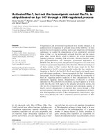
Monocyte TF expression at D5
aPC alone did not modify monocyte TF expression at D5 (Fig-
ure 1). LPS administration increased monocyte TF expression
in both unstimulated (I; in vitro) and stimulated (I+E; in vitro
with endotoxin) cells when compared to monocytes taken in
CTRL animals. In unstimulated monocytes, treatment with aPC
in septic animals failed to blunt TF expression. In stimulated
cells, the same level of TF expression was observed in the
LPS+aPC group when compared to the LPS group, suggest-
ing the ability to respond to further endotoxin stimulation.
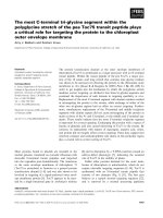
In vitro vascular reactivity
Vascular contraction
The maximal vasoconstrictor response to PE was not signifi-
cantly different between groups (data not shown).
LPS significantly modified sensitivity to PE at D5. Indeed, PE
EC
50
of rings with endothelium (E+) and rings without
endothelium (E-) were similar, suggesting endothelial dysfunc-
tion, when they were significantly different in the CTRL group
(Figure 2a). aPC treatment in LPS animals restored the differ-
ence in sensitivity between E+ and E- aortic rings (LPS+aPC
group). In the aPC group, there was persistence of endothe-
lium-dependent contraction modulation: PE EC
50
was differ-
ent in E+ and E- aortic rings, similar to the CTRL group (Figure
2a). PE EC
50
was significantly lower in E+ rings from the aPC
group compared to E+ from the CTRL group.
In E- rings, LPS decreased the sensitivity to PE (versus the
CTRL group). This difference was abolished after in vitro incu-
bation with L-NAME, and was not observed in E- rings from
aPC-treated rabbits (not significant, LPS+aPC group versus
LPS group) (Figure 2b). No difference in sensitivity to PE of E-
rings was observed between the aPC group compared to the
CTRL group (Figure 2b).
Endothelium-dependent and endothelium-independent
relaxation
Maximal endothelium-dependent receptor-dependent relaxa-
tion in response to ACh (Emax) was 78.3 ± 0.4% in the CTRL
group. This response was altered by LPS administration (Emax
= 50.0 ± 6.1%, p < 0.05 versus CTRL group). aPC treatment
failed to restore ACh-induced vascular relaxation in septic rab-
bits (Emax = 33.5 ± 4.0%; p < 0.05 versus CTRL group).
Endothelium-dependent receptor-independent relaxation in
response to calcium ionophore A23187 was not modified
between groups (data not shown). A similar observation was
recorded for endothelium-independent relaxation in response
to sodium nitroprusside (data not shown).
Figure 1
Expression of monocyte tissue factorExpression of monocyte tissue factor (TF) at day 5 with (I+E; i.e. in
vitro with endotoxin) or without (I; i.e. in vitro) stimulation in vitro (stimu-
lation obtained in culture in the presence of 1 µg/ml endotoxin). CTRL,
control group; LPS, animals that received LPS alone; LPS+aPC, ani-
mals that received LPS and aPC; aPC, animals that received aPC
alone. n represents the number of rabbits. *p < 0.05 versus CTRL
group;
§
p < 0.05 versus I.
Figure 2
Phenylephrine (PE) concentration eliciting 50% of maximal constriction response (EC50) in different groupsPhenylephrine (PE) concentration eliciting 50% of maximal constriction
response (EC50) in different groups. CTRL, control group; LPS, ani-
mals that received LPS alone; LPS+aPC, animals that received LPS
and aPC; aPC, animals that received aPC alone. n represents the
number of rabbits. (a) Aortic rings in the presence of endothelium (E+)
and in the absence of endothelium (E-). *p < 0.05 versus E+;
§
p < 0.05
versus CTRL E+;
163
p < 0.05 versus CTRL E (b) Aortic rings (E-)
incubated with or without N
G
-nitro-L-arginine methyl ester (L-NAME).
§
p
< 0.05 versus LPS E-;
163
p < 0.05 versus CTRL E
Critical Care Vol 10 No 2 Wiel et al.
Page 6 of 8
(page number not for citation purposes)
Immunohistochemical staining of vascular endothelium
For the sham groups (CTRL and aPC groups), endothelial
cells stained by immunohistochemical label (PECAM1/CD31)
appeared intact (Figure 3). LPS induced three types of
endothelial cell injury: subendothelial vacuolization, detach-
ment of endothelial cells and endothelial denudation. In the
LPS group, the percentage of injured endothelium accounted
for 40.4 ± 2.4% of total endothelial surface area in the abdom-
inal aorta at D5 (p < 0.05 versus CTRL group). aPC treatment
in LPS animals reduced these lesions, resulting in a surface
area of endothelial injury of 28.5 ± 2.3% (p < 0.05 versus LPS
group).
Discussion
In the present study, we report that aPC is able to prevent
endothelial morphological injuries and to increase the sensitiv-
ity to the vasoconstrictor agent PE in a well-documented rab-
bit endotoxin-induced shock model [7-12]. This was not
associated with any effect on monocyte TF expression,
endothelium-dependent relaxation in response to ACh, or mor-
tality.
Our mortality rate was similar in LPS and LPS+aPC animals
(16.7%). Because our model is a low mortality rate model,
explanations about the absence of the effect of aPC on mor-
tality cannot be drawn from this study. Taylor and colleagues
[22] found a decreased mortality rate in septic baboons
treated with aPC and Roback and colleagues [23] showed an
increased survival rate in rabbits with LPS-induced meningitis
that were treated with aPC. A main difference between our
study and these two previous studies is that in the latter aPC
was administered before LPS challenge whereas we decided
to give aPC 1 hour after LPS injection. Another explanation
may be related to the method of administration of aPC in our
model; we injected aPC as a single bolus but it was adminis-
tered as a continuous infusion in the two previous studies
[22,23]. It has been shown that the half-life of aPC is
decreased when administered to species different from
human [17]. This suggests that its action might not be sus-
tained over time, resulting in an absence of efficacy when LPS-
induced effects have progressed. This is consistent with the
fact that we did not observe improvement of either the in vivo
or the arterial blood gas parameters of endotoxinic animals
treated with aPC. We did not, however, assess the plasma
level of aPC in our study. This assessment is not easily
obtained in rabbit. During laparotomy for abdominal aorta
extraction, we did not observe any organ haematomas or
haemorrhage. We did not see any difference in either haema-
tocrit or haemoglobin levels between endotoxinic animals
treated or not with aPC. This suggests, as reported in the lit-
erature [24], that aPC does not cause bleeding or haemodilu-
tion.
A recent study reports that aPC binding to EPCR is a prereq-
uisite to its action on mortality [25]. When aPC binds to
EPCR, it activates a signaling pathway leading to inhibition of
NF-κB expression and pro-inflammatory cytokines via the
induction of protease activated receptor-1. Inadequate bind-
ing of aPC to EPCR could explain the absence of the anti-
inflammatory effect and be responsible for the persistence of
monocyte TF expression. This could be due to differences
between the species used. Not administering aPC as an infu-
sion and its short half-life could also explain the persistence of
the inflammatory syndrome associated with persistence of
monocyte TF expression at D5 after LPS bolus injection.
Besides these pharmacokinetic considerations, our results are
corroborated by a recent pharmacodynamic in vivo study
using an acute human endotoxemia model [26]. This model
allows studying the in vivo pharmacodynamics of drugs with
anticoagulant or anti-inflammatory properties [27-29]. The
authors concluded that aPC failed to decrease LPS-induced
monocyte TF expression and failed to have any anti-inflamma-
tory effects. They emphasized that the model used was an
inadequate severe sepsis model with concentrations of aPC
that remained above the pathological threshold. In the same
way, studies that demonstrated an effect on monocyte TF
expression were performed using in vitro experiments with
supraphysiological concentrations of aPC [30-32]. Under
these conditions, aPC might have acted as an anti-apoptotic
agent [1,33]. This anti-apoptotic effect has been demon-
strated in a human model of ischemic brain [34]. The anti-
apoptotic signaling pathway of aPC might be different from
that for NF-κB expression modulation.
A previous study demonstrated that aPC also has an anti-
apoptotic effect on human endothelial cell cultures exposed to
Figure 3
Quantification of abdominal aorta endothelial injury surface area by immunohistochemical study in endotoxic rabbitsQuantification of abdominal aorta endothelial injury surface area by
immunohistochemical study in endotoxic rabbits. LPS, animals that
received LPS alone; LPS+aPC, animals that received LPS and aPC. n
represents the number of rabbits. *p < 0.05 LPS+aPC group versus
the LPS group.
Available online />Page 7 of 8
(page number not for citation purposes)
LPS [32]. By using immunohistochemical staining, we demon-
strated that aPC restores and avoids prolonged vascular
endothelial cell injury induced by endotoxinic shock. This result
is in agreement with recent results [35], but the mechanism
remains unclear. This was associated with improvement of
endothelial cell function; in particular, aPC restores endothe-
lium-dependent sensitivity to PE. This is in accordance with
contractile induction of endothelial modulation. Indeed, as pre-
viously reported [7-12], LPS was responsible for the loss of
the endothelium-dependence of PE sensitivity of aortic rings
(the EC
50
PE was similar between E+ and E- aortic rings in the
LPS group). The sensitivity of smooth muscle cells to PE was
decreased in aortic rings after LPS injection (the EC
50
PE of
E- rings from the LPS group was higher than the EC
50
PE of
E- rings from the CTRL group). This was restored by in vitro
incubation with L-NAME, an inhibitor of NOS, suggesting the
presence of iNOS in smooth muscle cells. In the present
study, aPC treatment restores the endothelium-dependent
sensitivity to PE in LPS-treated aortic rings (as observed in the
CTRL group). This may result from reduced iNOS expression
in smooth muscle cells. It has been recently demonstrated that
aPC could inhibit excessive production of NO [35]. Moreover,
we previously reported that monocyte TF expression may
inhibit endothelial function [10,36]. Our results on restoration
of PE sensitivity by aPC in spite of persistence of monocyte TF
expression are in agreement with a recent study demonstrat-
ing that aPC has vascular protective effects independent of its
action on coagulation [37]. Our results for the contractile
response to PE, especially the increased sensitivity of aortic
rings to PE when aPC is administered to non-endotoxinic ani-
mals (the EC
50
of E+ rings of the aPC group is lower than that
of E+ rings of the CTRL group, demonstrating an increased
sensitivity to PE; Figure 2a), are consistent with a recent pub-
lication demonstrating that aPC improved vascular tone in
septic patients [38].
Despite protective effects on endothelial structure and PE
sensitivity, aPC failed to restore endothelium-dependent relax-
ation in response to ACh. These results suggest that the pro-
tective effect on endothelial function pertains to the PE
signaling pathway. In our study, endothelium-altered relaxation
specifically involves ACh, but not the endothelium-dependent
receptor-independent agent calcium ionophore A23187. This
suggests that an alteration in ACh receptor-NOS coupling
and/or a reduced production of endothelium-derived NO
causes this attenuated endothelium-mediated vasorelaxation.
Another explanation may be the absence of any effect of aPC
on similar pathways leading to expression of NF-κB and TF.
This could result, at least in part, in a modified endothelial aPC
signaling pathway due to EPCR dysfunction [15] or abnormal
binding of aPC to EPCR. A recent study confirms our result on
ACh-induced relaxation. The authors demonstrated that aPC
did not relax norepinephrine-increased vascular tone in rabbit
thoracic aorta [39].
Conclusion
We demonstrate that aPC increases the sensitivity of aortic
rings to the vasoconstrictor agent PE and restores endothelial
modulation in the PE response. This was associated with
decreased endothelial injury in endotoxin-treated animals.
These results suggest that aPC may preserve endothelial
structure via an anti-apoptotic effect. It failed to restore ACh-
induced relaxation, suggesting that aPC probably acts differ-
ently in the relaxant and contractile signaling pathways. aPC
did not modify monocyte TF expression. This suggests that
aPC may act differently on monocyte TF expression or ACh
receptor-NOS coupling. This could be caused by the lack of
binding of aPC to EPCR, explaining its lack of effect on the NF-
κB pathway, the inflammatory process, monocyte TF expres-
sion, and mortality.
Competing interests
The authors declare that they have no competing interests
(aPC was provided by Eli-Lilly).
Authors' contributions
All the authors contributed to the elaboration of the protocol,
its feasibility and the preparation of the manuscript. EW and
MEC performed the vasoreactivity study, blood gas analysis
and immunohistochemical staining. EW and GL were respon-
sible for the statistical analysis. DC and BJ performed the iso-
lation of monocytes, the determination of TF expression and
the study of hematological and coagulation variables. RB, BT
and BV participated in the elaboration of the protocol, and the
preparation and correction of the manuscript.
References
1. Hotchkiss RS, Karl IE: The pathophysiology and treatment of
sepsis. N Engl J Med 2003, 348:138-150.
2. Kang YH, Williams R: Endotoxin-induced endothelial injury and
subendothelial accumulation of fibronectin in rat aorta. Anat
Rec 1991, 229:86-102.
3. Young JS, Headrick JP, Berne RM: Endothelial-dependent and -
independent responses in the thoracic aorta during endotoxic
shock. Circ Shock 1991, 35:25-30.
Key messages
• In our model of endotoxinic shock, aPC increases the
sensitivity to vasoconstriction.
• aPC restores endothelial modulation in the PE
response.
• aPC has protective effects on endothelial structure,
probably via an anti-apoptotic effect.
• aPC did not modify coagulation activation, defined in
our model as monocyte tissue factor expression.
• aPC failed to restore ACh-induced relaxation, suggest-
ing that it probably acts differently in the relaxant and
contractile signaling pathways.
Critical Care Vol 10 No 2 Wiel et al.
Page 8 of 8
(page number not for citation purposes)
4. Parker JL, Adams HR: Selective inhibition of endothelium-
dependent vasodilator capacity by Escherichia coli endotox-
emia. Circ Res 1993, 72:539-551.
5. Reidy MA, Bowyer DE: Scanning electron microscopy: mor-
phology of aortic endothelium following injury by endotoxin
and during subsequent repair. Atherosclerosis 1977,
26:319-328.
6. Lee M, Schuessler G, Chien S: Time dependent effects of endo-
toxin on the ultrastructure of the aortic endothelium. Artery
1988, 15:71-89.
7. Leclerc J, Pu Q, Corseaux D, Haddad E, Decoene C, Bordet R, Six
I, Jude B, Vallet B: A single endotoxin injection in rabbit causes
prolonged blood vessel dysfunctions and procoagulant state.
Crit Care Med 2000, 28:3672-3678.
8. Abraham E: Effects of recombinant human activated protein C
in human models of endotoxin administration. Proc Am Thorac
Soc 2005, 2:243-7.
9. Wiel E, Pu Q, Corseaux D, Robin E, Bordet R, Lund N, Jude B, Val-
let B: Effect of L-arginine on endothelial injury and hemostasis
in rabbit endotoxin shock. J Appl Physiol 2000, 89:1811-1818.
10. Pu Q, Wiel E, Corseaux D, Bordet R, Azrin MA, Ezekowitz MD,
Lund N, Jude B, Vallet B: Beneficial effect of glycoprotein IIb/
IIIa inhibitor (AZ-1) on endothelium in Escherichia coli endo-
toxin-induced shock. Crit Care Med 2001, 29:1181-1188.
11. Wiel E, Pu Q, Leclerc J, Corseaux D, Bordet R, Lund N, Jude B,
Vallet B: Effects of the angiotensin-converting enzyme inhibi-
tor perindopril on endothelial injury and hemostasis in rabbit
endotoxic shock. Intensive Care Med 2004, 30:1652-1659.
12. Wiel E, Lebuffe G, Robin E, Gasan G, Corseaux D, Tavernier B,
Jude B, Bordet R, Vallet B: Pretreatment with peroxysome pro-
liferator-activated receptor (PPAR)-alpha agonist, fenofibrate,
protects endothelium in rabbit Escherichia coli endotoxin-
induced shock. Intensive Care Med 2005, 31:1269-1279.
13. Wiel E, Vallet B, Ten Cate H: The endothelium in intensive care.
Crit Care Clin 2005, 21:403-416.
14. Esmon CT: The protein C anticoagulant pathway. Arterioscler
Thromb 1992, 12:135-145.
15. Riewald M, Ruf W: Science review. Role of coagulation pro-
tease cascades in sepsis. Crit Care 2003, 7:123-129.
16. Yan SB, Helterbrand JD, Hartman DL, Wright TJ, Bernard GR: Low
levels of protein C are associated with poor outcomes in
severe sepsis. Chest 2001, 120:915-922.
17. Hoffmann JN, Vollmar B, Laschke MW, Inthorn D, Fertmann J,
Schildberg FW, Menger MD: Microhemodynamic and cellular
mechanism of activated protein C action during endotoxemia.
Crit Care Med 2004, 32:1011-1017.
18. Jackson CV, Bailey BD, Shetler TJ: Pharmacological profile of
recombinant human activated protein C (LY203638) in a
canine model of coronary artery thrombosis. J Pharmacol Exp
Ther 2000, 295:967-971.
19. Hamon M, Vallet B, Bauters C, Wernert N, McFadden EP,
Lablanche JM, Dupuis B, Bertrand ME: Long-term oral adminis-
tration of L-arginine reduces intimal thickening and enhances
neo-endothelium-dependent acetylcholine-induced relaxation
after arterial injury. Circulation 1994, 90:1357-1362.
20. Carson S: Continuous chromogenic tissue factor assay: com-
parison to clot-based assays and sensitivity established using
pure tissue factor. Thromb Res 1987, 47:379-387.
21. Corseaux D, Le Tourneau T, Six I, Ezekowitz MD, Mc Fadden EP,
Meurice T, Asseman P, Bauters C, Jude B: Enhanced monocyte
tissue factor response after experimental balloon angioplasty
in hypercholesterolemic rabbit: inhibition with dietary L-
arginine. Circulation 1998, 98:1776-1782.
22. Taylor FB, Chang A, Esmon CT, D'Angelo A, Vigano-D'Angelo S,
Blick KE: Protein C prevents the coagulopathic and lethal
effects of E. coli infusion in the baboon. J Clin Invest 1987,
79:918-925.
23. Roback MG, Stack AM, Thompson C, Brugnara C, Schwartz HP,
Saladino RA: Activated protein C concentrate for the treatment
of meninogococcal endotoxin shock in rabbits. Shock 1998,
9:138-142.
24. Gruber A, Griffin JH, Harker LA, Hanson SR: Inhibition of plate-
let-dependent thrombus formation by activated protein C in a
primate model. Blood 1989, 73:639-642.
25. Taylor FB, Stearns-Kurosawa DJ, Kurosawa S, Ferrell G, Chang A,
Laszik Z, Kosanke S, Peer G, Esmon CT: The endothelial cell
protein C receptor aids in host defense against Escherichia
coli sepsis. Blood 2000, 95:1680-1686.
26. Derhaschnig U, Reiter R, Knöbl P, Baumgartner M, Keen P, Jilma
B: Recombinant human activated protein C (rhAPC; drotrec-
ogin alfa [activated]) has minimal effect on markers of coagu-
lation, fibrinolysis, and inflammation in acute human
endotoxemia. Blood 2003, 102:2093-2098.
27. Hollenstein U, Homoncik M, Knöbl P, Pernerstorfer T, Graggaber
J, Eichler HG, Handler S, Jilma B: Acenocoumarol decreases tis-
sue factor-dependent coagulation during systemic inflamma-
tion in humans. Clin Pharmacol Ther 2002, 71:368-374.
28. Pernerstorfer T, Hollenstein U, Hansen J, Knechtelsdorfer M,
Stohlawetz P, Graninger W, Eichler HG, Speiser W, Jilma B:
Heparin blunts endotoxin-induced coagulation activation. Cir-
culation 1999, 100:2485-2490.
29. deJonge E, Dekkers PE, Creaseay AA, Hack CE, Paulson SK,
Karim A, Kesecioglu J, Levi M, van Deventer SJ, van der Poll T: Tis-
sue factor pathway inhibitor dose-dependently inhibits coagu-
lation activation without influencing the fibrinolytic and
cytokine response during human endotoxemia. Blood 2000,
95:1124-1129.
30. Grinnell BW, Yan SB: Novel antithrombotics based on modula-
tion of protein C pathway. Coron Artery Dis 1998, 9:89-97.
31. Shua F, Kobayashia H, Fukudome K, Tsuneyoshib N, Kimotob M,
Teraoa T: Activated protein C suppresses tissue factor expres-
sion on U937 cells in the endothelial protein C receptor-
dependent manner. FEBS Lett 2000, 477:208-212.
32. Joyce DE, Gelbert L, Ciacca A, DeHoff B, Grinnell BW: Gene
expression profile of antithrombotic protein C defines new
mechanisms modulating inflammation and apoptosis. J Biol
Chem 2001, 276:11199-11203.
33. Hotchkiss RS, Tinsley KW, Swanson PE, Karl IE: Endothelial cell
apoptosis in sepsis. Crit Care Med 2002, 30:S225-S228.
34. Cheng T, Liu D, Griffin JH, Fernandez JA, Castellino F, Rosen ED,
Fukudome K, Zlokovic BV: Activated protein C blocks p53-medi-
ated apoptosis in ischemic human brain endothelium and is
neuroprotective. Nat Med 2003, 9:338-342.
35. Isobe H, Okajima K, Uchiba M, Mizutani A, Harada N, Nagasaki A,
Okabe H: Activated protein C prevents endotoxin-induced
hypotension in rats by inhibiting excessive production of nitric
oxide. Circulation 2001, 104:1171-1175.
36. Vallet B, Wiel E: Endothelial cell dysfunction and coagulation.
Crit Care Med 2001, 29:S36-S41.
37. Riewald M, Ruf W: Protease-activated receptor-1 signaling by
activated protein C in cytokine-perturbed endothelial cells is
distinct from thrombin signaling. J Biol Chem 2005,
280:19808-19814.
38. Monnet X, Lamia B, Anguel N, Richard C, Bonmarchand G, Teboul
JL: Rapid and beneficial hemodynamic effects of activated pro-
tein C in septic shock patients. Intensive Care Med 2005,
31:1573-1576.
39. Bhattacharya A, Grinnell BW, Cohen ML: Unlike thrombin, pro-
tein C and activated protein C do not affect vascular tone. Pep-
tides 2000, 21:1231-1236.