Báo cáo y học: " Biochemical and virological analysis of the 18-residue C-terminal tail of HIV-1 integrase" pot
Bạn đang xem bản rút gọn của tài liệu. Xem và tải ngay bản đầy đủ của tài liệu tại đây (912.16 KB, 13 trang )
BioMed Central
Page 1 of 13
(page number not for citation purposes)
Retrovirology
Open Access
Research
Biochemical and virological analysis of the 18-residue C-terminal
tail of HIV-1 integrase
Mohd J Dar
1,3
, Blandine Monel
†1
, Lavanya Krishnan
†1
, Ming-Chieh Shun
1
,
Francesca Di Nunzio
1
, Dag E Helland
2
and Alan Engelman*
1
Address:
1
Department of Cancer Immunology and AIDS, Dana-Farber Cancer Institute, 44 Binney Street, Boston, MA, USA,
2
Molecular Biology
Institute, University of Bergen, N-5020 Bergen, Norway and
3
Current Address: University of Pittsburgh School of Medicine, S-427 BST, 200
Lothrop Street, Pittsburgh, PA 15213, USA
Email: Mohd J Dar - ; Blandine Monel - ;
Lavanya Krishnan - ; Ming-Chieh Shun - ; Francesca Di
Nunzio - ; Dag E Helland - ; Alan Engelman* -
* Corresponding author †Equal contributors
Abstract
Background: The 18 residue tail abutting the SH3 fold that comprises the heart of the C-terminal
domain is the only part of HIV-1 integrase yet to be visualized by structural biology. To ascertain
the role of the tail region in integrase function and HIV-1 replication, a set of deletion mutants that
successively lacked three amino acids was constructed and analyzed in a variety of biochemical and
virus infection assays. HIV-1/2 chimers, which harbored the analogous 23-mer HIV-2 tail in place
of the HIV-1 sequence, were also studied. Because integrase mutations can affect steps in the
replication cycle other than integration, defective mutant viruses were tested for integrase protein
content and reverse transcription in addition to integration. The F185K core domain mutation,
which increases integrase protein solubility, was furthermore analyzed in a subset of mutants.
Results: Purified proteins were assessed for in vitro levels of 3' processing and DNA strand
transfer activities whereas HIV-1 infectivity was measured using luciferase reporter viruses.
Deletions lacking up to 9 amino acids (1-285, 1-282, and 1-279) displayed near wild-type activities
in vitro and during infection. Further deletion yielded two viruses, HIV-1
1-276
and HIV-1
1-273
, that
displayed approximately two and 5-fold infectivity defects, respectively, due to reduced integrase
function. Deletion mutant HIV-1
1-270
and the HIV-1/2 chimera were non-infectious and displayed
approximately 3 to 4-fold reverse transcription in addition to severe integration defects. Removal
of four additional residues, which encompassed the C-terminal strand of the SH3 fold, further
compromised integrase incorporation into virions and reverse transcription.
Conclusion: HIV-1
1-270
, HIV-1
1-266
, and the HIV-1/2 chimera were typed as class II mutant viruses
due to their pleiotropic replication defects. We speculate that residues 271-273 might play a role
in mediating the known integrase-reverse transcriptase interaction, as their removal unveiled a
reverse transcription defect. The F185K mutation reduced the in vitro activities of 1-279 and 1-276
integrases by about 25%. Mutant proteins 1-279/F185K and 1-276/F185K are therefore highlighted
as potential structural biology candidates, whereas further deleted tail variants (1-273/F185K or 1-
270/F185K) are less desirable due to marginal or undetectable levels of integrase function.
Published: 19 October 2009
Retrovirology 2009, 6:94 doi:10.1186/1742-4690-6-94
Received: 15 July 2009
Accepted: 19 October 2009
This article is available from: />© 2009 Dar et al; licensee BioMed Central Ltd.
This is an Open Access article distributed under the terms of the Creative Commons Attribution License ( />),
which permits unrestricted use, distribution, and reproduction in any medium, provided the original work is properly cited.
Retrovirology 2009, 6:94 />Page 2 of 13
(page number not for citation purposes)
Background
Retrovirus replication proceeds through a series of steps
that initiate upon virus entry into a cell, followed by par-
ticle uncoating and reverse transcription. To support pro-
ductive replication, the resulting double stranded cDNA
must be integrated into a cell chromosome. The integrated
DNA provides an efficient transcriptional template for
viral gene expression and ensures for segregation of viral
genetic material to daughter cells during division. Due to
its essential nature, the integrase (IN) encoded by HIV-1 is
an intensely studied antiviral drug target [1].
Integration can be divided into three enzyme-based steps,
the first two of which are catalyzed by IN. In the initial 3'
processing reaction, IN removes the terminal pGT
OH
dinu-
cleotides from the 3' ends of the blunt-ended HIV-1
reverse transcript, yielding the precursor ends for integra-
tion [2-4]. In the second step, DNA strand transfer, IN
uses the 3'-oxygens to cut the chromosomal target DNA in
a staggered fashion and at the same time joins the viral 3'
ends to the resulting 5' phosphates [3]. The final step,
repair of single stranded gaps and joining of viral DNA 5'
ends, is accomplished by cellular enzymes [5,6]. HIV-1 IN
activities can be measured in vitro using oligonucleotide
DNA substrates that mimic the ends of the reverse tran-
script and either Mg
2+
or Mn
2+
cofactor [7-10].
IN is a multi-domain protein consisting of the N-terminal
domain (NTD, HIV-1 residues 1-49), catalytic core
domain (CCD, residues 50-212), and C-terminal domain
(CTD, residues 213-288). The NTD contains a conserved
HHCC Zn-coordination motif, and Zn-binding contrib-
utes to IN multimerization and catalytic function [11,12].
The CCD contains an invariant triad of acidic residues
(Asp-64, Asp-116, Glu-152 of HIV-1) that forms the
enzyme active site [13-16]. The CCD also contributes to
IN multimerization [17] and engages viral [18-20] and
chromosomal [21,22] DNAs during integration. The CTD,
which is the least conserved of the domains among retro-
viruses [23], also contributes to specific [24] and non-spe-
cific [25-27] DNA interactions, as well as multimerization
[28].
Insight into the mechanism of HIV-1 integration is some-
what hampered by lack of relevant 3-dimensional infor-
mation, as structures for the enzyme bound to its DNA
substrates, or the free holoenzyme, have yet to be
reported. NTD-CCD [29-31] and CCD-CTD [32-34] two-
domain x-ray crystal structures have nevertheless been
informative. Three NTD-CCD structures, containing HIV-
1, HIV-2, or maedi-visna virus domains, have revealed a
dimer-of-dimers architecture for the active IN tetramer
[29,30] and the high affinity binding mode of the com-
mon lentiviral integration cofactor LEDGFp75 [31]. An
SH3 fold comprised of five strands makes up the heart
of the CTD [35,36], and a comparison of HIV-1 [32], SIV
[33], and Rous sarcoma virus [34] CCD-CTD structures
reveals considerable flexibility in CTD positioning with
respect to the different CCDs. Nevertheless, extended viral
DNA binding surfaces were ascribed to each CCD-CTD
structure. Although residues 271-288, herein referred to as
the tail, were present in the two-domain HIV-1 construct,
they were disordered and therefore unseen in the resulting
crystal structure [32].
The roles of the C-terminal tail in IN function and HIV-1
replication are largely unexplored. The IN
1-270
deletion
mutant that lacked the tail supported 10-50% of wild-type
(WT) Mn
2+
-dependent 3' processing and DNA strand
transfer activities, whereas the activities of IN
1-279
were
largely unimpaired (50-100% of WT) [25]. HIV-1 carrying
the substitution of Ala for Lys-273 grew like the WT in Jur-
kat T cells, dispensing an obvious role for this highly con-
served tail residue in virus replication [37]. To learn more
about the role of this region in IN catalysis and HIV-1 rep-
lication, successive three amino acid deletion mutants
were constructed and analyzed in various enzymatic and
virus infection assays. The somewhat larger 23-residue
HIV-2 tail was moreover swapped for the HIV-1 sequence
to assess the activities of tail chimera enzyme and virus.,
C-terminal deletion mutants that lack all or part of the tail
could be useful structural biology candidates due to their
inability to adopt an ordered fold in previous crystal struc-
tures. Thus, one goal of this study was to evaluate the sol-
ubility-enhancing F185K CCD mutation [38] for its
potential effects on the in vitro activities of tail deletion
mutant enzymes.
Methods
Plasmid DNA constructions
Bacterial expression vector pKBIN6Hthr [39] and viral IN
shuttle vector pUCWTpol [40] were previously described.
Because the IN tail overlaps the 5' end of vif, shuttle vector
pUCWTpol3stop, which harbored three stop codons after
Vif residue Asn-19, was constructed by PCR using Pfu
Ultra DNA polymerase (Stratagene, La Jolla, CA) and
primers AE1064 (5'-ACAGGATGAGGATTAACTGATGA-
TAAGCTTTAGTAAAACACCATATG)/AE1065 (5'-
CATATGGTGTTTTACTAAAGCTTATCATCAGTTAATCCT-
CATCCTGTC). IN deletion mutations were subsequently
constructed in pUCWTpol3stop or pKBIN6Hthr by PCR.
Plasmid pUCWTpolBam-Spe, which contains unique
BamHI and SpeI sites downstream of the IN coding region
and a stop codon after Arg-17 in Vif [41], was used to swap
tail sequences as follows. AAA/CAG/ATG, which encodes
for HIV-1 residues Lys-273, Gln-274, and Met-275, was
changed to GGT/CGA/CTG to imbed a unique SalI site in
pUCWTpolSal-Bam-Spe at the HIV-1/2 tail boundary. A
linker constructed by annealing AE3697 (5'-PO
4
-
TCGACAGGAGATGGACAGCGGAAGTCACCTGGAGGG
Retrovirology 2009, 6:94 />Page 3 of 13
(page number not for citation purposes)
CGCAAGAGAGGACGGTGAGATGGCATAAG) with
AE3698 (5'-PO
4
-
GATCCTTATGCCATCTCACCGTCCTCTCTTGCGCCCTC
CAGGTGACTTCCGCTGTCCATCTCCTG) was then
ligated to SalI/BamHI-digested pUCWTpolSal-Bam-Spe.
To move the chimera tail to pKBIN6Hthr, pUCWTpolSal-
Bam-Spe was amplified using XhoI-tagged AE3699 (5'-
TGGTGCTCGAG
TGCGGACCCACGCGGGACGAGT-
GCCATCTCACCGTCCTCTCTTGC) and AflII-tagged
AE3700 (AACATCTTAAG
ACAGCAGTAC) and the result-
ing digested fragment was ligated with XhoI/AflII-cut
pKBIN6Hthr. Mutated AgeI-PflMI 1.8 kb fragments from
pUCWTpol3stop or pUCWTpolSal-Bam-Spe were
swapped for the corresponding fragment in the single
round HIV-1
NL4-3
-based vector pNLX.Luc(R-) [42]. All
plasmid regions constructed by PCR were analyzed by
DNA sequencing to verify targeted changes and lack of
unwanted secondary mutations.
Protein expression and purification
Escherichia coli strain PC2 [43] transformed with IN
expression constructs were grown for 16 h at 30°C. The
next day bacteria subcultured at 1:30 in 600 ml LB-100
g/ml ampicillin were grown at 30°C until A
600
of 0.6, at
which time expression was induced by the addition of 0.6
mM isopropyl--D-thiogalactopyranoside. Cells were har-
vested following 5 h of induction at 28°C. The bacterial
pellet resuspended in ice-cold buffer A [25 mM Tris-HCl,
pH 7.4, 1 M NaCl, 7.5 mM 3-[(3-Cholamidopro-
pyl)dimethylammonio]-2-hydroxy-1-propanesulfonate
(CHAPS)] containing 25 mM imidazole-0.5 mM phenyl-
methanesulphonylfluoride was sonicated. After centrifu-
gation for 30 min at 39,000 g, the supernatant was
incubated with 0.6 ml of buffer A-25 mM imidazole-
equilibrated Ni
2+
-nitrilotriacetic acid (Ni-NTA) agarose
beads (QIAGEN, Valencia, CA) at 4°C for 3 h. The beads
were washed twice with 20 volumes of buffer A-25 mM
imidazole followed by washing with 30 volumes of buffer
A-35 mM imidazole. IN-His
6
was eluted with buffer A-200
mM imidazole. IN containing fractions identified by Na
dodecyl sulfate (SDS)-polyacrylamide gel electrophoresis
were pooled and dialyzed overnight against buffer D [25
mM Tris-HCl, pH 7.4, 1 M NaCl, 7.5 mM CHAPS, 10%
glycerol (w/v), 10 mM dithiothreitol (DTT)]. The His-Tag
was removed using 40 U of thrombin (Sigma-Aldrich, St.
Louis, MO) per mg of protein for 3 h at room temperature,
which left the heterologous LVPR sequence at each C-ter-
minus. After removal of thrombin by incubation with
Benzamidine beads (Novagen, Madison, WI), IN was con-
centrated using Centricon-10 Concentrators (Millipore,
Billerica, MA) and dialyzed against buffer D for 4 h. Pro-
tein concentration was determined by spectrophotome-
ter, and aliquots flash frozen in liquid N
2
were stored at -
80°C. Quantitative image analysis (Alpha Innotech
FlourChem FC2, San Leandro, CA) of Coomassie-stained
gels revealed that each IN preparation was minimally 90%
pure.
Recombinant LEDGFp75 expressed in bacteria was puri-
fied as previously described [44]. LEDGFp75 concentra-
tions were determined using the Bio-Rad protein assay kit
(Hercules, CA). Exonuclease III was from New England
Biolabs (Beverley, MA).
Anti-IN monoclonal antibody 8G4 [45] was purified from
hybridoma cell supernatant using protein G sepharose
(GE Healthcare, Piscataway, NJ) following the manufac-
turer's recommendations. 500 ml of cell supernatant
loaded onto 1 ml of protein G beads were subsequently
washed with phosphate-buffered saline. Antibody eluted
with 20 mM glycine-HCl, pH 2.8 was immediately neu-
tralized by addition of 1 M Tris-HCl, pH 8.5. Pooled frac-
tions were concentrated by ultrafiltration, and resulting
antibody concentration was determined by spectropho-
tometry.
In vitro integration assays
Oligonucleotides that mimic the HIV-1 U5 end were used
as viral DNA substrates. AE143 (5'-ACTGCTAGAGATTT-
TCCACACTGACTAAAA) and AE191 (5'-TTTTAGTCAGT-
GTGGAAAATCTCTAGCAG) were annealed prior to
filling-in the 3' recess with [-
32
P]TTP (3000 Ci/mmol;
PerkinElmer, Waltham, MA) using Sequenase version 2.0
T7 DNA polymerase (GE Healthcare) to label the phos-
phodiester within the pGT
OH
dinucleotide that is cleaved
during 3' processing [3,46]. To prepare a 30 bp preproc-
essed duplex for DNA strand transfer, AE155 (5'-TTT-
TAGTCAGTGTGGAAAATCTCTAGCA) 5'-end labeled
with [-
32
P]ATP (3000 Ci/mmol; PerkinElmer) using T4
polynucleotide kinase (GE Healthcare) [46] was annealed
with AE143. Unincorporated radionuclide was removed
by passing labeled duplexes through Bio-Spin 6 columns
(Bio-Rad) equilibrated with 10 mM Tris-HCl, pH 8.0-20
mM NaCl-0.1 mM EDTA.
Reaction mixtures (16 l) contained 25 mM MOPS, pH
7.2, 10 mM DTT, 31 mM NaCl, 10 mM MgCl
2
, 5 M
ZnSO
4
, 5 nM DNA substrate, and 0.49 M IN. Reactions
stopped by addition of an equal volume of sequencing gel
sample buffer (95% formamide, 10 mM EDTA, 0.003%
xylene cyanol, 0.003% bromophenol blue) were boiled
for 2 min prior to fractionation through 20% polyacryla-
mide- (3' processing) or 15% polyacrylamide-8.3 M urea
(DNA strand transfer) sequencing gels. Reaction products
in wet gels exposed to phosphor image plates were quan-
tified using Image Quant version 1.2 (GE Healthcare).
LEDGFp75-dependent concerted integration activity was
assayed essentially as previously described [31]. A pre-
processed 32 bp U5 end was prepared by annealing
Retrovirology 2009, 6:94 />Page 4 of 13
(page number not for citation purposes)
AE3653 (5'-CCTTTTAGTCAGTGTGGAAAATCTCTAGCA)
with AE3652 (5'- ACTGCTAGAGATTTTCCACACT-
GACTAAAAGG). Reactions (36 l) were initiated by mix-
ing 0.5 M HIV-1 DNA with 0.33 g pGEM-3 target DNA
in 25.3 mM NaCl, 5.5 mM MgSO
4
, 11 mM DTT, 4.4 M
ZnCl
2
, 22 mM HEPES-NaOH, pH 7.4. IN (2 l) in dilu-
tion buffer (750 mM NaCl, 10 mM DTT, 25 mM Tris-HCl,
pH 7.4) was then added. Following 2-3 min at room tem-
perature, 2.0 l of LEDGFp75 was added, and the reac-
tions were allowed to proceed at 37°C for 1 h. The final
concentrations of IN and LEDGFp75 were both 0.8 M.
Reactions stopped by the addition of EDTA and SDS to
the final concentrations of 25 mM and 0.5%, respectively,
were deproteinized using 30 g proteinase K (Roche
Molecular Biochemicals, Indianapolis, IN) for 60 min at
37°C. DNAs recovered following precipitation with etha-
nol were separated on 1.5% agarose-TAE (40 mM Tris
base, 20 mM acetate, 1 mM EDTA) gels run in TAE at 150
V for 2 h. DNAs stained with ethidium bromide (0.5 g/
ml) were quantified using Alpha Innotech FlourChem
FC2.
Cells and viruses
293T cells were grown in Dulbecco's modified Eagle's
medium (DMEM) supplemented to contain 10% fetal
bovine serum (FBS) (Invitrogen Corporation, Carlsbad,
CA). Cells were plated at 8.6 × 10
6
/10-cm dish 24 h prior
to transfection. Virus stocks were prepared by co-transfect-
ing cells with 10 g pNLX.Luc(R-) and 1 g of envelope
expression vector pCG-VSV-G [47] using FuGene 6 as
described by the manufacturer (Roche Molecular Bio-
chemicals). Cell-free supernatants harvested at 48 h post-
transfection were passed through 0.45 m filters. Virus
titer was determined using an exogenous reverse tran-
scriptase (RT) assay as previously described [48]. For west-
ern blot analysis, viruses pelletted by ultracentrifugation
at 122,000 g for 2 h at 4°C were lysed for 15 min on ice in
40 l of buffer containing 140 mM NaCl, 8 mM
Na
2
HPO
4
, 2 mM NaH
2
PO
4
, 1% Nonidet P40, 0.5% Na
deoxycholate, 0.05% SDS. Supernatant recovered after
centrifugation at 19,800 g was stored at -80°C. Following
electrophoresis and transfer to polyvinylidene fluoride,
IN and p24 were detected using 1:100 and 1:5000 dilu-
tions of 8G4 and 13-203-000 (Advanced Biotechnologies
Inc, Columbia, MD) antibodies, respectively.
HeLa-T4 cells [49] were grown in DMEM-10% FBS con-
taining 100 IU/ml penicillin and 100 g/ml streptomycin.
For infectivity measurements, cells plated at 75,000 cells/
well of 24-well tissue culture plates 24 h prior to infection
were incubated in duplicate with 10
6
RT-cpm of virus for
17 h, after which cells washed with phosphate-buffered
saline were replenished with fresh media. At 46 h post-
infection, cells were collected, washed, and lysed using 75
l passive lysis buffer as recommended by the manufac-
turer (Promega Corp., Madison, WI). Luciferase activities
(20 l), determined in duplicate for each infection, were
normalized to total levels of cellular protein as previously
described [42]. For quantitative (Q)-PCR assays, 900,000
cells were plated per 10 cm dish the day before infection.
Cells were infected with 2.3 × 10
7
RT-cpm of TURBO
DNase-treated [42] native or heat-inactivated (65°C for
30 min) virus. 8G4 hybridoma cells were grown in DMEM
containing 10% ultra low IgG FBS (Invitrogen Corpora-
tion) with penicillin and streptomycin.
Q-PCR assays for reverse transcription and integration
Total cellular DNA was isolated at 7 or 24 h post-infection
using the QIAamp DNA mini kit (QIAGEN). Late reverse
transcription (LRT) products were detected using primers
and Taqman probe as previously described [50,51]. Two-
long terminal repeat (2-LTR) containing circles were
detected at 24 h post-infection using primers MH535/536
[50] and SYBR green (QIAGEN). Integration was meas-
ured at 24 h using a modified nested HIV-1 R-Alu format
based on reference [52]. DNA (100 ng) was amplified
using the phage lambda T-R chimera primer AE3014 [53]
and Alu-specific AE1066 (5'-TCCCAGCTACTCGGGAG-
GCTGAGG) with rTth DNA polymerase XL as recom-
mended by the manufacturer (Applied Biosystems Inc,
Foster City, CA). Samples (1 l) were then analyzed by Q-
PCR using SYBR green with primers AE989 and AE990
[51]. DNA generated from WT-infected cells was end-
point diluted in DNA prepared from uninfected cells to
generate the integration standard curve. LRT, 2-LTR, and
Alu-integration Q-PCR values obtained from samples pre-
pared using heat-inactivated virus were subtracted from
those generated using native virus.
Results and Discussion
Experimental strategy
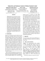
Little is known about the role of HIV-1 IN C-terminal tail
(residues 271-288, Figure 1) in integration. This region of
the protein, which overlaps the 5' end of the vif reading
frame, is fairly well conserved among different HIV-1 iso-
lates. Some clade C sequences harbor Ala in place of Asp-
278 and numerous clades as well as SIVcpz carry Gly at
position 283 (Figure 1); the remaining residues by con-
trast show little or no sequence variation [54]. To ascer-
tain the role of the tail in IN function, six nested deletions
mutants lacking 3, 6, 9, 12, 15, or 18 amino acids from the
C-terminus were constructed in the pKBIN6Hthr bacterial
expression construct [39] and luciferase-based
pNLX.Luc(R-) viral vector [42] (Figure 1). The CCD F185K
mutation, which dramatically increases the solubility of
the HIV-1 protein [38], was tested in some constructs to
assess its potential affects on IN activities in vitro. The 1-
266 deletion mutant, which lacked the C-terminal 22 res-
idues and hence the fifth strand of the CTD SH3 fold in
addition to the tail (Figure 1) [35,36], was used as a loss-
Retrovirology 2009, 6:94 />Page 5 of 13
(page number not for citation purposes)
of-function control [55]. Finally, the 23 residue HIV-2 tail
(underlined in Figure 1) was swapped for the correspond-
ing HIV-1 sequence to test the functionality of this mar-
ginally related sequence substitution. Because the viral
changes necessarily altered the overlapping vif sequence,
these constructs incorporated stop codons downstream of
the IN region within the vif frame to negate synthesis of
altered Vif proteins. Viruses were constructed in 293T
cells, which lack APOBEC3G and thus do not require
functional Vif to yield infectious particles [56].
The C-terminal tail and IN enzymatic activities
Recombinant proteins were engineered to contain C-ter-
minal hexahistidine tags to facilitate purification. Though
this might appear counterintuitive given the C-terminal
focus of the study, it was necessary to obtain relatively
pure preparations. The tail region is hypersensitive to pro-
teolysis during expression in E. coli [57], and preliminary
experiments with N-terminally tagged proteins yielded
heterogeneous populations eluted from Ni-NTA beads
whose purities were not substantially improved upon by
subsequent ion exchange or size exclusion chromatogra-
phy (data not shown). The C-terminal tag obviated this
problem, as proteolyzed variants failed to bind Ni-NTA
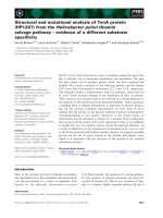
beads. Indeed, quantitative image analysis of purified WT
and mutant proteins revealed near homogeneous prepa-
rations (Figure 2A).
IN activities were measured using three different assay
designs, each of which incorporated an ~30 bp DNA
mimic of the viral U5 end (Figure 2B-D). Overall levels of
IN 3' processing and DNA strand transfer activities were
determined in two separate assays using differentially
labeled 30 bp substrates (Figure 2B and 2C). Under these
conditions, the majority of DNA strand transfer reaction
products result from the insertion of a single oligonucle-
otide end into one strand of a second target DNA mole-
cule [8]. By contrast, integration in cells proceeds via the
concerted insertion of viral U3 and U5 DNA ends into
opposing strands of chromosomal DNA. Reactions that
contain relatively low concentrations of IN protein [58],
relatively long viral DNA substrates [59], or relatively high
concentrations of oligonucleotide substrate in the pres-
ence of LEDGFp75 [31] support efficient concerted HIV-1
integration. Here, LEDGFp75 was used in a third assay
format (Figure 2D) to monitor the concerted integration
activities of IN mutant proteins. His
6
-tags were removed
from purified IN proteins by thrombin cleavage prior to
enzyme assays, yielding the remnant LVPR C-terminal
sequence. Experiments conducted with a subset of pro-
teins prior to cleavage (WT, 1-279, 1-273, 1-270,1-266,
and HIV-1/2) revealed similar levels of 3' processing activ-
ities relative to WT, indicating that the remnant sequence
did not significantly influence mutant enzyme activities
(data not shown).
IN sequence alignment and HIV-1 mutants analyzed in this studyFigure 1
IN sequence alignment and HIV-1 mutants analyzed in this study. The upper drawing indicates the three IN domains,
with amino acid residues conserved among all retroviruses noted. CTD sequences downstream of the invariant Trp are shown
below for HIV-1 (NL4-3 isolate, accession number M19921), SIVcpz (accession number AF115393), and HIV-2 (ROD isolate,
accession number M15390). Residues that appear in more than one sequence are highlighted in grey. The broad arrows
beneath the alignment indicate the strands that comprise the SH3 fold [35,36]. Numbers 266-285 above the alignment mark
the IN deletion mutant enzymes and viruses analyzed in this study. The underline indicates the region of HIV-2 IN that was
swapped for HIV-1 residues 271-288.
-1 …WKGPAKLLWKGEGAVVIQDNSDIKVVPRRKAKIIRDY.GKQMAGDDCVASRQDED
pz …WKGPARLLWKGEGAVVIKEREEVKVIPRRKAKIIRDY.GKQMAGDDSMAGGQDESQGLE
-2 …WKGPGELLWKGEGAVLVKVGTDIKIIPRRKAKIIRDYGGRQEMDSGSHLEGAREDGEMA
235
240
250
260
266
270
273
276
279
282
285
288
64
CTDNTD
288
43401612
116
235
159152
CCD
H
HCC D D K WE
HIV-1 IN
2345
HIV-1
SIVcpz
HIV-2
Retrovirology 2009, 6:94 />Page 6 of 13
(page number not for citation purposes)
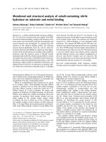
To follow the course of the 3' processing reaction, oligo-
nucleotide substrate DNA was labeled at the inter-nucle-
otide linkage of the 3'-terminal GT (Figure 2B); IN
mediated hydrolysis liberates pGT
OH
, which is readily dis-
tinguished from the 30 bp substrate following electro-
phoresis on high percentage DNA sequencing gels [3,4]
(Figure 3A, lanes 2 and 3; results quantified in panel B).
Exonuclease III-mediated hydrolysis by contrast yielded
free pT
OH
(Figure 3A, lanes 1 and 17). All IN preparations
were basically void of contaminating exonuclease activity
(Figure 3A), reflecting the relatively high degrees of pro-
tein purity (Figure 2A). IN
D64N
and IN
1-266
, which con-
tained the substitution of Asn for active site residue Asp-
64 [14] and lacked part of the CTD SH3 fold, respectively,
were predictably inactive (Figure 3A, lanes 15 and 16).
The activities of the three mutants that retained most of
the tail, IN
1-285
, IN
1-282
, and IN
1-279
, were overall similar at
65-70% of WT (Figure 3A, lanes 5-7). Mutants with fur-
ther progressive tail deletions yielded a stepwise reduction
in 3' processing activity, as IN
1-276
, IN
1-273
, and IN
1-270
supported about 51%, 26%, and 13%, respectively, of WT
function. Thus, IN
1-279
and IN
1-270
support Mg
2+
-depend-
ent 3' processing activities that do not significantly differ
from those reported using Mn
2+
[25]. The IN
HIV1/2
chimera
protein like IN
1-270
retained marginal (about 12% of WT)
activity (Figure 3A, lane 20; Figure 3B). The F185K solubil-
ity mutation marginally impacted activity, generally yield-
ing 20-25% reductions when compared to the same
protein lacking the CCD change (Figure 3B).
The preprocessed DNA strand transfer substrate was
labeled at the 5' end of the strand that becomes joined to
Integrase proteins and in vitro integration assaysFigure 2
Integrase proteins and in vitro integration assays. (A) Purified proteins (approximately 5 g each) were stained with
Coomassie blue following SDS-polyacrylamide gel electrophoresis. Migration positions of molecular mass standards in kDa are
shown on the left. (B) 3' Processing assay. The blunt-ended viral DNA substrate is shown highlighting the subterminal CA that
is conserved among all retroviruses, retrotransposons, and some bacterial transposases. During 3' processing, IN cleaves the
A/G phosphodiester bond (short vertical arrow), releasing radiolabelled pGT
OH
dinucleotide. (C) The DNA strand transfer
assay utilizes a preprocessed viral DNA end. Integration into target DNA yields products whose lengths exceed that of the
starting substrate. (D) Two different DNAs, viral donor (oligonucleotide drawn in the same orientation as in panel C, top) and
circular target, are used in the concerted integration assay. In the presence of LEDGFp75, some donor DNA is integrated into
only one strand of the target to yield a tagged, nicked circle half-site reaction product. Concerted integration across the major
groove by contrast yields a linearized product whose length exceeds that of the starting circle by twice the length of the viral
donor. For panels B-D, thin and bold lines represent viral donor and target DNAs, respectively. *, positions of
32
P label (panels
B and C).
CAG T
GT C A
*
5'
3'
CA
GTCA
pG T
OH
*
5'
3'
OH
+
IN
+
CA
GTCA
3'
OH
*
G
T
3'
C
A
*
3'
5'
C
A
IN
+
pGEM-3
half-site concerted
IN/LEDGFp75
B
CD
donor
target
25
32
47
F185K
WT
D64N
1-285
1-282
1-279
1-279/F185K
1-276/F185K
1-270/F185K
1-273/F185K
1-270
1-276
1-273
1-266
HIV1/2
A
Retrovirology 2009, 6:94 />Page 7 of 13
(page number not for citation purposes)
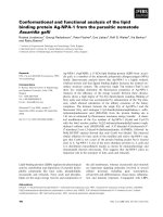
target DNA; IN activity yields a population of products
that migrate more slowly than the starting substrate on
DNA sequencing gels [8] (Figure 2C and 4A). Relative lev-
els of IN mutant DNA strand transfer activities in large
part mirrored 3' processing activities with some subtle dif-
ferences noted (compare Figure 4B to Figure 3B). IN
1-285
,
IN
1-279
, and IN
1-276
supported DNA strand transfer at basi-
cally the same level as the WT, whereas the activity of IN
1-
270
was undetectable (Figure 4A, lanes 4-6 and 13; Figure
4B). Mn
2+
can support more robust IN activity than Mg
2+
[9,60], which may have contributed to the previously
reported residual level of IN
1-270
DNA strand transfer
activity [25]. IN
HIV1/2
DNA strand transfer activity, by con-
trast to IN
1-270
, was increased from its relative level of 3'
processing activity (Figure 4B and 3B).
WT and mutant IN 3' processing activitiesFigure 3
WT and mutant IN 3' processing activities. (A) Polyacrylamide gel images reveal the migration positions of labeled 30-
mer DNA substrate (S), cleaved pGT
OH
dinucleotide, as well as pT
OH
mononucleotide. The reactions loaded in lanes 1 and 17
contained exonuclease III in place of IN, whereas lanes 2 and 18 omitted IN. The reactions in the remaining lanes contained the
indicated IN proteins. (B) Mutant 3' processing activities plotted as percentage of WT IN function. Results are mean ± SEM for
two (HIV-1/2 chimera) to four (all other mutants) independent experiments.
F185K
1-285
1-282
1-279
1-279/F185K
1-276/F185K
1-276
1-273/F185K
1-273
1-270/F185K
1-270
1-266
D64N
HIV1/2
%WT activity
A
ExoIII
F185K
WT
D64N
1-285
1-282
1-279
1-279/F185K
1-276/F185K
1-270/F185K
1-273/F185K
1-270
1-276
1-273
1-266
- IN
S
pGT
OH
pT
OH
ExoIII
HIV1/2
WT
- IN
pGT
OH
pT
OH
S
123456789101112131415 1718192016
B
IN mutant DNA strand transfer activitiesFigure 4
IN mutant DNA strand transfer activities. (A) Scanned gel images show the migration positions of preprocessed sub-
strate (S) DNA as well as the integration products (IP) of DNA strand transfer. IN was omitted from the reactions loaded in
lanes 1 and 16; the remaining lanes contained the indicated IN proteins. (B) Mean DNA strand transfer activities ± SEM for two
independent experiments plotted as percentage of WT IN activity.
Retrovirology 2009, 6:94 />Page 8 of 13
(page number not for citation purposes)
Supercoiled pGEM-3 plasmid DNA was incorporated into
the reaction mixture to help identify concerted integration
reaction products (Figure 2D and 5A). Integration of only
one donor DNA end into one plasmid DNA strand yields
a tagged circle whose mobility through agarose matches
that of starting relaxed circular plasmid (Figure 5A). Pair-
wise integration of two oligonucleotides by contrast yields
a linearized product whose size is slightly larger than lin-
ear plasmid (Figure 2D). IN DNA strand transfer activity
was barely detectable in the absence of LEDGFp75, yield-
ing slight increases in the nicked or open circular plasmid
population (Figure 5A, compare lanes 3 and 27 to lanes 2
and 26, respectively) [31]. LEDGFp75 greatly stimulated
IN activity such that the supercoiled target DNA was
largely consumed, yielding a mixture of half-site and con-
certed integration products (Figure 5A, lanes 4 and 28). IN
mutant product formation was quantified to reflect over-
all levels (half-site plus concerted, Figure 5B) of DNA
strand transfer activities or just concerted integration (Fig-
ure 5C). The overall activities of the various deletion
mutant proteins in large part mirrored their oligonucle-
otide-based DNA strand transfer activities (compare Fig-
ure 5B to 4B). Though 0.49 M IN
HIV1/2
supported about
40% of IN
WT
activity in the oligonucleotide-based assay
(Figure 4B), 0.8 M protein failed to support appreciable
product formation in the concerted assay format (Figure
5A, lane 31). Doubling the amount of input IN
HIV1/2
to
1.6 M yielded significant half-site product formation
(about 66% of IN
WT
, Figure 5A, lane 30 and Figure 5B) in
the absence of detectable concerted integration activity
(Figure 5C). Taken together, our data indicate that the C-
terminal tail does not play a specific role in concerted
DNA integration, though the introduction of a foreign
sequence for the HIV-1 tail can uncouple pairwise from
single end integration activity. Though others noted that
the F185K substitution ablated Mg
2+
-dependent integra-
tion of preprocessed oligonucleotide donor DNA into het-
erologous target DNA [61], our reaction conditions failed
to reveal an affect of the solubilizing mutation on full-
length IN activity in the presence of LEDGFp75 (Figure
5A, lane 6; panels B and C). We furthermore conclude that
the C-terminal 9 amino acids of HIV-1 IN can be removed
without dramatically effecting Mg
2+
-based single end or
concerted DNA integration activities (Figures 3, 4, 5)., We
highlight these derivatives as potential candidates for
structural biology studies despite the approximate 20-
25% reductions in IN
1-279
and IN
1-276
activities brought on
by the F185K change. We would by contrast advise against
extensive analysis of tailless IN
1-270
, due to its lack of
detectable DNA strand transfer activity under these assay
conditions (Figure 4 and 5).
Characterization of IN mutant viruses
To assess HIV-1 infectivity, HeLa-T4 cells were infected
with normalized levels of single-round viruses that carry
the luciferase reporter gene in place of nef. Two days post-
infection, cells were harvested and resulting luciferase
activities were normalized to the levels of total protein in
the different cell extracts [42,47]. Deletion of up to 9
amino acids from the IN C-terminus failed to affect HIV-
1 infectivity (Figure 6). IN mutants HIV-1
1-276
and HIV-1
1-
273
supported about 50% and 20% of the level of WT
infection, respectively, whereas HIV-1
1-270
, HIV-1
1-266
,
and the HIV-1/2 tail chimera were non-infectious (Figure
6).
IN mutations can affect multiple steps in the HIV-1 repli-
cation cycle, including particle release from virus-produc-
ing cells and/or reverse transcription during the
subsequent round of infection (reviewed in ref. [62]).
Viruses specifically blocked at integration are distin-
guished as class I, whereas class II mutants display addi-
tional stage defects. To assess potential affects on virus
particle release, RT content in HeLa cell supernatants at 2
days post-transfection was normalized to levels of cell-
associated luciferase activity. Normalized levels of mutant
virus release did not significantly differ from the WT
under this assay condition (data not shown). Defective
mutant viruses (HIV-1
1-266
, HIV-1
1-270
, HIV-1
1-273
, HIV-1
1-
276
, and HIV-1/2; Figure 6) produced from transfected
293T cells were analyzed by western blotting to assess lev-
els of virion-incorporated IN protein. Monoclonal anti-
body 8G4, which recognizes discontinuous epitopes in
the NTD and CCD [45], was utilized to avoid potential
complications from the CTD mutations. Accordingly, 8G4
effectively recognized the different forms of recombinant
IN protein (Figure 7, top panel). Based on relative levels
of p24 content (bottom panel), we conclude that HIV-1
1-
276
, HIV-1
1-273
, HIV-1
1-270
, and HIV-1
1-266
harbor signifi-
cantly less IN protein than WT HIV-1 (viral lysate panels,
compare lanes 2-5 to lane 1), with HIV-1
1-266
suffering the
most dramatic defect (lane 2). We therefore conclude that
an intact SH3 fold plays an important role in Gag-Pol
incorporation and/or IN retention in virions.
Q-PCR assays were utilized to assess defective mutant
virus reverse transcription (LRT at 7 h post-infection) and
2-LTR circle formation and integration (nested Alu-R
PCR) at 24 h. Virus stocks were treated with DNase prior
to infection to digest plasmid DNA that may persist after
transfection and hence template in the LRT reaction for-
mat. To control for potential plasmid carry-over, a parallel
set of infections was conducted using heat-inactivated
viruses. Resulting LRT values (typically 1-5%) were sub-
tracted from native viral infections. HIV-1
1-276
and HIV-1
1-
273
supported the WT levels of reverse transcription and
circle formation (Figure 8A and 8B), whereas HIV-1
1-270
,
HIV-1
1-266
, and the HIV-1/2 chimera supported about
25%, 5%, and 33% of WT LRT product formation (Figure
8A). Under these experimental conditions IN residues
Retrovirology 2009, 6:94 />Page 9 of 13
(page number not for citation purposes)
LEDGFp75-dependent concerted integration activities of WT and IN mutant proteinsFigure 5
LEDGFp75-dependent concerted integration activities of WT and IN mutant proteins. (A) The scanned ethidium-
stained agarose gels reveal the migration positions of donor, supercoiled (s.c.), and open circular (o.c.) substrate DNAs, as well
as half-site and concerted integration reaction products. Donor DNA was omitted from the reactions analyzed in lanes 1 and
25, whereas IN was omitted from lanes 2 and 26. The remaining lanes contained the indicated IN proteins and, at times,
LEDGFp75. The concentration of HIV-1/2 IN in lanes 29 and 30 was 1.6 M, whereas all other IN concentrations were 0.8 M.
The migration positions of molecular mass standards in kb are shown to the left of the gel. (B and C) Levels of overall and con-
certed DNA strand transfer activities, respectively, normalized to IN
WT
(set to 100%). Results are mean ± SEM for two inde-
pendent experiments.
0.5
1
1.5
2
3
5
donor
s.c. target
concerted
A
F185K
WT
D64N+LEDGF
1-285
1-282
1-279
1-279/F185K
1-276/F185K
1-270
1-276
1-266+LEDGF
- IN
HIV1/2 (1.6 M)
- donor
1-270+LEDGF
1-273
1-273+LEDGF
1-276/F185K+LEDGF
1-276+LEDGF
1-279/F185K+LEDGF
1-279+LEDGF
1-282+LEDGF
1-285+LEDGF
F185K+LEDGF
WT+LEDGF
half-site/
o.c. target
WT
- IN
- donor
WT+LEDGF
HIV1/2 (1.6 M)+LEDGF
HIV1/2 (0.8 M)+LEDGF
123456789101112131415 1718192016 21 22 23 24 25 26 27 28 29 30 31
%WT activity
20
40
60
80
100
120
20
40
60
80
100
120
F185K
1-285
1-282
1-279
1-279/F185K
1-276/F185K
1-276
1-273
1-270
1-266
D64N
HIV1/2 (1.6 M)
HIV1/2 (0.8 M)
F185K
1-285
1-282
1-279
1-279/F185K
1-276/F185K
1-276
1-273
1-270
1-266
D64N
HIV1/2 (1.6 M)
HIV1/2 (0.8 M)
BC
Retrovirology 2009, 6:94 />Page 10 of 13
(page number not for citation purposes)
271-273 contribute to reverse transcription. Due to the
pleiotropic nature of HIV-1 IN mutations these results
were not entirely unexpected. Residues 271-273 might
influence the interaction between IN and RT [63], which
occurs via the CTD [64,65]. An RT binding interface was
recently mapped to strands 2-4 of the SH3 fold [66] and
though residues 271-273 abut -5 (Figure 1), it is not
unreasonable to suspect the disordered tail could affect RT
binding. Alternatively, a number of NTD and CCD muta-
tions in addition to CTD changes can impair DNA synthe-
sis (see [62] for review), indicating that the C-terminal tail
changes might perturb reverse transcription via global
affects on IN and/or the preintegration complex.
HIV-1
1-276
and HIV-1
1-273
supported about 40% and 20%
of WT integration, respectively (Figure 8C), indicating
that their partial infectivities (Figure 6) were due to spe-
cific integration defects attributable to the intrinsic activi-
ties of the deletion mutant enzymes (Figure 3, 4, 5).
Consistent with their non-infectious phenotypes and ina-
bilities for recombinant IN proteins to catalyze concerted
integration activity, neither HIV-1
1-270
nor the HIV-1/2
chimera supported a detectable level of integration during
infection (Figure 8C). As both of these viruses supported
the formation of detectable 2-LTR circles (Figure 8B), we
group them as class II defective IN mutants that display
marginal (3 to 4-fold) reverse transcription in additional
to prominent integration defects. HIV-1
1-266
was a more
IN mutant viral infectivityFigure 6
IN mutant viral infectivity. Normalized levels of IN
mutant infectivities are shown relative to WT HIV-1 (set at
100%). Each experiment amassed duplicate luciferase assays
of duplicate infections. Shown is the mean ± SEM of five inde-
pendent experiments. RLU, relative light units.
WT and IN mutant virus protein contentFigure 7
WT and IN mutant virus protein content. Top panel, 2
ng of the indicated recombinant IN protein was analyzed by
western blotting. Lower panels, viral lysates. The primary
blotting antibody is indicated to the right of each panel.
-p24
8G4
8G4
WT
HIV1/2
1-266
1-270
1-273
1-276
Recombinant IN
Viral
lysates
123456
Reverse transcription and integration profiles of IN mutant virusesFigure 8
Reverse transcription and integration profiles of IN
mutant viruses. (A) Mutant viral LRT levels, graphed as
percentages of the WT (leftward bar). (B) 2-LTR circle levels
at 24 h post-infection. (C) Mutant viral integration in com-
parison to the WT. Panels A and B average results of two dif-
ferent infection experiments (mean ± SEM). Mean ± SEM of
duplicate Q-PCR assays of one infection experiment is
shown in panel C. The panel C data are representative of
those obtained from a duplicate set of infections.
Retrovirology 2009, 6:94 />Page 11 of 13
(page number not for citation purposes)
severe class II mutant virus, harboring a significant reverse
transcription as well as integration defect.
Conclusion
The results of this study revealed that nine amino acids
can be removed from the HIV-1 IN C-terminus without
significantly affecting the activity of the enzyme or infec-
tivity of the virus. Additional removal of up to six amino
acids impacted infectivity by up to 80%, yielding viruses
that were specifically defective for integration due to the
compromised activities of the associated IN
1-276
and IN
1-
273
enzymes. Heuer and Brown [67] reported that residues
271-288 crosslink to viral and target DNA sequences
within junctional disintegration substrates. We would
therefore surmise that tail residues 271-279 interact with
substrate DNA during integration. HIV-1
1-270
was non-
infectious and harbored an approximate fourfold reverse
transcription defect. This suggests IN residues 271, 272,
and 273 might impact its physical association with RT.
HIV-1
1-266
, which lacked the fifth strand of the fold,
failed to incorporate significant levels of IN protein and
was in large part defective for reverse transcription. Thus,
an intact SH3 fold apparently contributes to Gag-Pol
packaging and subsequent viral DNA synthesis. Our
results moreover highlight partial tailed variants 1-279/
F185K and 1-276/F185K as viable candidates for struc-
tural biology studies, as they retained >20% of IN enzy-
matic activities yet lacked at least half of the disordered
region.
List of abbreviations used
CCD: catalytic core domain; CHAPS: 3-[(3-Cholamido-
propyl)dimethylammonio]-2-hydroxy-1-propanesul-
fonate; CTD: C-terminal domain; DMEM: Dulbecco's
modified Eagle's medium; DTT: dithiothreitol; FBS: fetal
bovine serum; IN: integrase; LRT: late reverse transcrip-
tion; LTR: long-terminal repeat; Ni-NTA: Ni
2+
-nitrilot-
riacetic acid; NTD: N-terminal domain; Q: quantitative;
RT: reverse transcriptase; SDS: Na dodecyl sulfate; WT:
wild type.
Competing interests
The authors declare that they have no competing interests.
Authors' contributions
MJD constructed molecular clones, purified recombinant
IN proteins, and conducted in vitro integration assays. BM
performed the brunt of virological measurements includ-
ing infectivity, LRT, and 2-LTR circle Q-PCRs. LK purified
8G4 antibody, performed western blotting, and per-
formed some IN purifications and enzyme assays. MCS
performed Alu-PCR and quantified virus release from
transfected HeLa cells. FDN devised the western blotting
procedure, and trained and supervised BM. DEH supplied
essential reagents. AE conceived of the study, supervised
and interpreted experimental results, and wrote the man-
uscript. All authors read and approved the final manu-
script.
Acknowledgements
The authors thank Nan Yan for valuable technical advice. This work was
supported by US NIH grants AI039394 and AI070042 (to AE) and the Har-
vard University Center for AIDS Research (CFAR), an NIH funded program
(P30AI060354) that is supported by the following NIH Institutes and Cent-
ers: NIAID, NCI, NIMH, NIDA, NICHD, NHLBI, NCCAM. The contents
of this manuscript do not necessarily reflect the views of the Department
of Health and Human Services, nor does the mention of trade names, com-
mercial products, or organizations imply endorsement by the US Govern-
ment.
References
1. Vandegraaff N, Engelman A: Molecular mechanism of HIV inte-
gration and therapeutic intervention. Expert Rev Mol Med 2007,
9:1-19.
2. Pauza C: Two bases are deleted from the termini of HIV-1 lin-
ear DNA during integrative recombination. Virology 1990,
179:886-889.
3. Engelman A, Mizuuchi K, Craigie R: HIV-1 DNA integration:
mechanism of viral DNA cleavage and DNA strand transfer.
Cell 1991, 67:1211-1221.
4. Vink C, Yeheskiely E, Marel GA van der, Van Boom JH, Plasterk RHA:
Site-specific hydrolysis and alcoholysis of human immunode-
ficiency virus DNA termini mediated by the viral integrase
protein. Nucleic Acids Res 1991, 19:6691-6698.
5. Brin E, Yi J, Skalka AM, Leis J: Modeling the late steps in HIV-1
retroviral integrase-catalyzed DNA integration. J Biol Chem
2000, 275:39287-39295.
6. Yoder KE, Bushman FD: Repair of gaps in retroviral DNA inte-
gration intermediates. J Virol 2000, 74:11191-11200.
7. Sherman PA, Fyfe JA: Human immunodeficiency virus integra-
tion protein expressed in Escherichia coli possesses selective
DNA cleaving activity. Proc Natl Acad Sci USA 1990, 87:5119-5123.
8. Bushman FD, Craigie R: Activities of human immunodeficiency
virus (HIV) integration protein in vitro: Specific cleavage and
integration of HIV DNA. Proc Natl Acad Sci USA 1991,
88:1339-1343.
9. Engelman A, Craigie R: Efficient magnesium-dependent human
immunodeficiency virus type 1 integrase activity. J Virol 1995,
69:5908-5911.
10. Delelis O, Carayon K, Saib A, Deprez E, Mouscadet J-F: Integrase
and integration: biochemical activities of HIV-1 integrase.
Retrovirology 2008, 5:114.
11. Zheng R, Jenkins TM, Craigie R: Zinc folds the N-terminal
domain of HIV-1 integrase, promotes multimerization, and
enhances catalytic activity. Proc Natl Acad Sci USA 1996,
93:13659-13664.
12. Lee SP, Xiao J, Knutson JR, Lewis MS, Han MK: Zn2+ promotes the
self-association of human immunodeficiency virus type-1
integrase in vitro. Biochemistry 1997, 36:173-180.
13. Drelich M, Wilhelm R, Mous J: Identification of amino acid resi-
dues critical for endonuclease and integration activities of
HIV-1 IN protein in vitro. Virology 1992, 188:459-468.
14. Engelman A, Craigie R: Identification of conserved amino acid
residues critical for human immunodeficiency virus type 1
integrase function in vitro. J Virol 1992, 66:6361-6369.
15. Kulkosky J, Jones KS, Katz RA, Mack JP, Skalka AM: Residues critical
for retroviral integrative recombination in a region that is
highly conserved among retroviral/retrotransposon inte-
grases and bacterial insertion sequence transposases. Mol Cell
Biol 1992, 12:2331-2338.
16. van Gent DC, Groeneger AAMO, Plasterk RHA: Mutational anal-
ysis of the integrase protein of human immunodeficiency
virus type 2. Proc Natl Acad Sci USA 1992, 89:9598-9602.
17. Hickman AB, Palmer I, Engelman A, Craigie R, Wingfield P: Biophys-
ical and enzymatic properties of the catalytic domain of HIV-
1 integrase. J Biol Chem 1994, 269:29279-29287.
Retrovirology 2009, 6:94 />Page 12 of 13
(page number not for citation purposes)
18. Gerton JL, Brown PO: The core domain of HIV-1 integrase rec-
ognizes key features of its DNA substrates. J Biol Chem 1997,
272:25809-25815.
19. Jenkins TM, Esposito D, Engelman A, Craigie R: Critical contacts
between HIV-1 integrase and viral DNA identified by struc-
ture-based analysis and photo-crosslinking. EMBO J 1997,
16:6849-6859.
20. Esposito D, Craigie R: Sequence specificity of viral end DNA
binding by HIV-1 integrase reveals critical regions for pro-
tein-DNA interaction. EMBO J 1998, 17:5832-5843.
21. Appa RS, Shin C-G, Lee P, Chow SA: Role of the nonspecific
DNA-binding region and alpha helices within the core
domain of retroviral integrase in selecting target DNA sites
for integration. J Biol Chem 2001, 276:45848-45855.
22. Harper AL, Skinner LM, Sudol M, Katzman M: Use of patient-
derived human immunodeficiency virus type 1 integrases to
identify a protein residue that affects target site selection. J
Virol 2001, 75:7756-7762.
23. Esposito D, Craigie R: HIV integrase structure and function. Adv
Virus Res 1999, 52:319-333.
24. Gao K, Butler SL, Bushman F: Human immunodeficiency virus
type 1 integrase: arrangement of protein domains in active
cDNA complexes. EMBO J 2001, 20:3565-3576.
25. Vink C, Oude Groeneger AM, Plasterk RHA: Identification of the
catalytic and DNA-binding region of the human immunode-
ficiency virus type I integrase protein. Nucleic Acids Res 1993,
21:1419-1425.
26. Woerner AM, Marcus-Sekura CJ: Characterization of a DNA
binding domain in the C-terminus of HIV-1 integrase by
deletion mutagenesis. Nucleic Acids Res 1993, 21:3507-3511.
27. Engelman A, Hickman AB, Craigie R: The core and carboxyl-ter-
minal domains of the integrase protein of human immuno-
deficiency virus type 1 each contribute to nonspecific DNA
binding. J Virol 1994, 68:5911-5917.
28. Jenkins TM, Engelman A, Ghirlando R, Craigie R: A soluble active
mutant of HIV-1 integrase: involvement of both the core and
the C-terminal domains in multimerization. J Biol Chem 1996,
271:7712-7718.
29. Wang J-Y, Ling H, Yang W, Craigie R: Structure of a two-domain
fragment of HIV-1 integrase: implications for domain organ-
ization in the intact protein. EMBO J 2001, 20:7333-7343.
30. Hare S, Di Nunzio F, Labeja A, Wang J, Engelman A, Cherepanov P:
Structural basis for functional tetramerization of lentiviral
integrase. PLoS Pathog 2009, 5:1000515.
31. Hare S, Shun MC, Gupta SS, Valkov E, Engelman A, Cherepanov P: A
novel co-crystal structure affords the design of gain-of-func-
tion lentiviral integrase mutants in the presence of modified
PSIP1/LEDGF/p75. PLoS Pathog 2009, 5:e1000259.
32. Chen JC-H, Krucinski J, Miercke LJW, Finer-Moore JS, Tang AH, Leav-
itt AD, Stroud RM: Crystal structure of the HIV-1 integrase
catalytic core and C-terminal domains: A model for viral
DNA binding. Proc Natl Acad Sci USA 2000, 97:8233-8238.
33. Chen Z, Yan Y, Munshi S, Li Y, Zugay-Murphy J, Xu B, Witmer M,
Felock P, Wolfe A, Sardana V: X-ray structure of simian immun-
odeficiency virus integrase containing the core and C-termi-
nal domain (residues 50-293) - an initial glance of the viral
DNA binding platform. J Mol Biol 2000, 296:521-533.
34. Yang Z-N, Mueser TC, Bushman FD, Hyde CC: Crystal structure
of an active two-domain derivative of rous sarcoma virus
integrase. J Mol Biol 2000, 296:535-548.
35. Eijkelenboom AP, Lutzke RA, Boelens R, Plasterk RHA, Kaptein R,
Hård K: The DNA-binding domain of HIV-1 integrase has an
SH3-like fold. Nat Struct Biol 1995, 2:807-810.
36. Lodi PJ, Ernst JA, Kuszewski J, Hickman AB, Engelman A, Craigie R,
Clore GM, Gronenborn AM: Solution structure of the DNA
binding domain of HIV-1 integrase. Biochemistry 1995,
34:9826-9833.
37. Lu R, Ghory HZ, Engelman A: Genetic analyses of conserved res-
idues in the carboxyl terminal domain of human immunode-
ficiency virus type 1 integrase. J Virol 2005, 79:10356-10368.
38. Jenkins T, Hickman A, Dyda F, Ghirlando R, Davies D, Craigie R:
Cat-
alytic domain of human immunodeficiency virus type 1 inte-
grase: Identification of a soluble mutant by systematic
replacement of hydrophobic residues. Proc Natl Acad Sci USA
1995, 92:6057-6061.
39. McKee CJ, Kessl JJ, Shkriabai N, Dar MJ, Engelman A, Kvaratskhelia M:
Dynamic modulation of HIV-1 integrase structure and func-
tion by cellular lens epithelium-derived growth factor
(LEDGF) protein. J Biol Chem 2008, 283:31802-31812.
40. Limón A, Devroe E, Lu R, Ghory HZ, Silver PA, Engelman A: Nuclear
localization of human immunodeficiency virus type 1 pre-
integration complexes (PICs): V165A and R166A are pleio-
tropic integrase mutants primarily defective for integration,
not PIC nuclear import. J Virol 2002, 76:10598-10607.
41. Belshan M, Schweitzer CJ, Donnellan MR, Lu R, Engelman A: In vivo
biotinylation and capture of HIV-1 matrix and integrase pro-
teins. J Virol Methods 2009, 159:178-184.
42. Lu R, Limón A, Devroe E, Silver PA, Cherepanov P, Engelman A:
Class II integrase mutants with changes in putative nuclear
localization signals are primarily blocked at a post-nuclear
entry step of human immunodeficiency virus type 1 replica-
tion. J Virol 2004, 78:12735-12746.
43. Cherepanov P: LEDGF/p75 interacts with divergent lentiviral
integrases and modulates their enzymatic activity in vitro.
Nucleic Acids Res 2007, 35:113-124.
44. Vandegraaff N, Devroe E, Turlure F, Silver PA, Engelman A: Bio-
chemical and genetic analyses of integrase-interacting pro-
teins lens epithelium-derived growth factor (LEDGF)/p75
and hepatoma-derived growth factor related protein 2
(HRP2) in preintegration complex function and HIV-1 repli-
cation. Virology 2006, 346:415-426.
45. Nilsen BM, Haugan IR, Berg K, Olsen L, Brown PO, Helland DE: Mon-
oclonal antibodies against human immunodeficiency virus
type 1 integrase: epitope mapping and differential effects on
integrase activities in vitro. J Virol 1996, 70:1580-1587.
46. Craigie R, Hickman AB, Engelman A: Integrase. In HIV: Biochemistry,
Molecular Biology, and Drug Discovery Volume 2. Edited by: Karn J. New
York, NY: IRL Press; 1995:53-71.
47. Shun M-C, Daigle JE, Vandegraaff N, Engelman A: Wild-type levels
of human immunodeficiency virus type 1 infectivity in the
absence of cellular emerin protein. J Virol 2007, 81:166-172.
48. Engelman A, Englund G, Orenstein JM, Martin MA, Craigie R: Multi-
ple effects of mutations in human immunodeficiency virus
type 1 integrase on viral replication. J Virol 1995, 69:2729-2736.
49. Maddon PJ, Dalgleish AG, McDougal JS, Clapham PR, Weiss RA, Axel
R: The T4 gene encodes the AIDS virus receptor and is
expressed in the immune system and the brain. Cell 1986,
47:333-348.
50. Butler SL, Hansen MST, Bushman FD: A quantitative assay for
HIV DNA integration in vivo. Nat Med 2001, 7:631-634.
51. Shun M-C, Raghavendra NK, Vandegraaff N, Daigle JE, Hughes S, Kel-
lam P, Cherepanov P, Engelman A: LEDGF/p75 functions down-
stream from preintegration complex formation to effect
gene-specific HIV-1 integration. Genes Dev 2007, 21:1767-1778.
52. Brussel A, Sonigo P: Analysis of early human immunodeficiency
virus type 1 DNA synthesis by use of a new sensitive assay for
quantifying integrated provirus. J Virol 2003, 77:10119-10124.
53. Engelman A, Oztop I, Vandegraaff N, Raghavendra NK: Quantita-
tive analysis of HIV-1 preintegration complexes. Methods
2009, 47:283-290.
54. Rhee SY, Liu TF, Kiuchi M, Zioni R, Gifford RJ, Holmes SP, Shafer RW:
Natural variation of HIV-1 group M integrase: implications
for a new class of antiretroviral inhibitors. Retrovirology 2008,
5:74.
55. Engelman A, Bushman FD, Craigie R: Identification of discrete
functional domains of HIV-1 integrase and their organization
within an active multimeric complex. EMBO J 1993,
12:3269-3275.
56. Sheehy AM, Gaddis NC, Malim MH: The antiretroviral enzyme
APOBEC3G is degraded by the proteasome in response to
HIV-1 Vif. Nat Med 2003, 9:1404-1407.
57. Hickman AB, Dyda F, Craigie R: Heterogeneity in recombinant
HIV-1 integrase corrected by site-directed mutagenesis: the
identification and elimination of a protease cleavage site.
Protein Eng 1997, 10:601-606.
58. Sinha S, Grandgenett DP: Recombinant human immunodefi-
ciency virus type 1 integrase exhibits a capacity for full-site
integration in vitro that is comparable to that of purified pre-
integration complexes from virus-infected cells. J Virol 2005,
79:8208-8216.
Publish with BioMed Central and every
scientist can read your work free of charge
"BioMed Central will be the most significant development for
disseminating the results of biomedical research in our lifetime."
Sir Paul Nurse, Cancer Research UK
Your research papers will be:
available free of charge to the entire biomedical community
peer reviewed and published immediately upon acceptance
cited in PubMed and archived on PubMed Central
yours — you keep the copyright
Submit your manuscript here:
/>BioMedcentral
Retrovirology 2009, 6:94 />Page 13 of 13
(page number not for citation purposes)
59. Li M, Craigie R: Processing of viral DNA ends channels the
HIV-1 integration reaction to concerted integration. J Biol
Chem 2005, 280:29334-29339.
60. Marchand C, Johnson AA, Karki RG, Pais GCG, Zhang X, Cowansage
K, Patel TA, Nicklaus MC, Burke TR Jr, Pommier Y: Metal-depend-
ent inhibition of HIV-1 integrase by {beta}-diketo acids and
resistance of the soluble double-mutant (F185K/C280S). Mol
Pharmacol 2003, 64:600-609.
61. Podtelezhnikov AA, Gao K, Bushman FD, McCammon JA: Modeling
HIV-1 integrase complexes based on their hydrodynamic
properties. Biopolymers 2003, 68:110-120.
62. Engelman A: In vivo analysis of retroviral integrase structure
and function. Adv Virus Res 1999, 52:411-426.
63. Wu X, Liu H, Xiao H, Conway JA, Hehl E, Kalpana GV, Prasad V, Kap-
pes JC: Human immunodeficiency virus type 1 integrase pro-
tein promotes reverse transcription through specific
interactions with the nucleoprotein reverse transcription
complex. J Virol 1999, 73:2126-2135.
64. Hehl EA, Joshi P, Kalpana GV, Prasad VR: Interaction between
human immunodeficiency virus type 1 reverse transcriptase
and integrase proteins. J Virol 2004, 78:5056-5067.
65. Zhu K, Dobard C, Chow SA: Requirement for integrase during
reverse transcription of human immunodeficiency virus type
1 and the effect of cysteine mutations of integrase on its
interactions with reverse transcriptase. J Virol 2004,
78:5045-5055.
66. Wilkinson TA, Januszyk K, Phillips ML, Tekeste SS, Zhang M, Miller JT,
Le Grice SFJ, Clubb RT, Chow SA: Identifying and characterizing
a functional HIV-1 reverse transcriptase-binding site on inte-
grase. J Biol Chem 2009, 284:7931-7939.
67. Heuer TS, Brown PO: Mapping features of HIV-1 integrase
near selected sites on viral and target DNA molecules in an
active enzyme-DNA complex by photo-cross-linking. Bio-
chemistry 1997, 36:10655-10665.