Báo cáo y học: " Encapsidation of APOBEC3G into HIV-1 virions involves lipid raft association and does not correlate with APOBEC3G oligomerization" potx
Bạn đang xem bản rút gọn của tài liệu. Xem và tải ngay bản đầy đủ của tài liệu tại đây (877.12 KB, 12 trang )
BioMed Central
Page 1 of 12
(page number not for citation purposes)
Retrovirology
Open Access
Research
Encapsidation of APOBEC3G into HIV-1 virions involves lipid raft
association and does not correlate with APOBEC3G
oligomerization
Mohammad A Khan, Ritu Goila-Gaur, Sandra Kao, Eri Miyagi,
Robert C Walker Jr and Klaus Strebel*
Address: Laboratory of Molecular Microbiology, Viral Biochemistry Section, National Institute of Allergy and Infectious Diseases, National
Institutes of Health, Building 4, Room 310, 4 Center Drive, MSC 0460, Bethesda, MD 20892-0460, USA
Email: Mohammad A Khan - ; Ritu Goila-Gaur - ; Sandra Kao - ;
Eri Miyagi - ; Robert C Walker - ; Klaus Strebel* -
* Corresponding author
Abstract
Background: The cellular cytidine deaminase APOBEC3G (A3G), when incorporated into the
human immunodeficiency virus type 1 (HIV-1), renders viral particles non-infectious. We previously
observed that mutation of a single cysteine residue of A3G (C100S) inhibited A3G packaging. In
addition, several recent studies showed that mutation of tryptophan 127 (W127) and tyrosine 124
(Y124) inhibited A3G encapsidation suggesting that the N-terminal CDA constitutes a viral
packaging signal in A3G. It was also reported that W127 and Y124 affect A3G oligomerization.
Results: Here we studied the mechanistic basis of the packaging defect of A3G W127A and Y124A
mutants. Interestingly, cell fractionation studies revealed a strong correlation between
encapsidation, lipid raft association, and genomic RNA binding of A3G. Surprisingly, the presence
of a C-terminal epitope tag affected lipid raft association and encapsidation of the A3G W127A
mutant but had no effect on wt A3G encapsidation, lipid raft association, and interaction with viral
genomic RNA. Mutation of Y124 abolished A3G encapsidation irrespective of the presence or
absence of an epitope tag. Contrasting a recent report, our co-immunoprecipitation studies failed
to reveal a correlation between A3G oligomerization and A3G encapsidation. In fact, our W127A
and Y124A mutants both retained the ability to oligomerize.
Conclusion: Our results confirm that W127 and Y124 residues in A3G are important for
encapsidation into HIV-1 virions and our data establish a novel correlation between genomic RNA
binding, lipid raft association, and viral packaging of A3G. In contrast, we were unable to confirm a
role of W127 and Y124 in A3G oligomerization and we thus failed to confirm a correlation
between A3G oligomerization and virus encapsidation.
Background
APOBEC3G (A3G) is a cellular cytidine deaminase with
potent antiretroviral activity that severely limits replica-
tion of vif-defective HIV-1 in human cells [1]. A3G is
expressed in most if not all natural human HIV-1 target
cells; yet HIV-1 efficiently infects humans and has caused
Published: 3 November 2009
Retrovirology 2009, 6:99 doi:10.1186/1742-4690-6-99
Received: 15 June 2009
Accepted: 3 November 2009
This article is available from: />© 2009 Khan et al; licensee BioMed Central Ltd.
This is an Open Access article distributed under the terms of the Creative Commons Attribution License ( />),
which permits unrestricted use, distribution, and reproduction in any medium, provided the original work is properly cited.
Retrovirology 2009, 6:99 />Page 2 of 12
(page number not for citation purposes)
a worldwide pandemic. This ability of HIV-1 to infect and
replicate in A3G-positive human cells is made possible by
the viral accessory protein Vif, which was found to prevent
the packaging of A3G into progeny virions. Inhibition of
A3G packaging is accomplished either by proteasome-
mediated degradation of A3G or by other degradation-
independent mechanisms (reviewed in [2]). Inhibition of
A3G encapsidation may also require Vif dimerization
since peptide antagonists to Vif dimerization blocked A3G
packaging without affecting its intracellular stability [3].
The antiviral effect of A3G generally requires encapsida-
tion of the deaminase into viral particles. Interestingly,
the antiviral effects of A3G are not limited to HIV-1 but
extend to other retroviruses including murine leukemia
virus, mouse mammary tumor virus, simian immunodefi-
ciency virus, equine infectious anemia virus, and hepatitis
B virus (for review see [2]). Packaging of A3G into such
diverse viruses suggests that virus encapsidation is either
relatively nonspecific or involves signals shared by these
viruses. Interestingly, although A3G selectively targets sin-
gle stranded DNA for deamination it also binds RNA.
RNA binding of A3G has been shown to contribute to
virus encapsidation [4-11]. A3G also interacts with the NC
component of the viral Gag precursor protein [7,12-21].
This interaction likely also contributes to the packaging of
A3G into viral particles. In vitro studies using purified
recombinant NC and A3G found that the two proteins do
not competitively bind RNA but instead form an RNA-
protein ternary complex [5].
Several reports have investigated domains in A3G
required for packaging into HIV-1 virions. We and others
have recently reported that mutations in the A3G catalytic
domain 1 (CD1) can impair A3G packaging [21,22].
Characterization of in-frame deletion mutants implicated
a linker region located C-terminal to the CD1 domain
(residues 121-161) as critical for A3G packaging into HIV-
1 virus-like particles [12,20]. These findings were sup-
ported by other studies that identified residues 122 to 127
in the linker domain as important for A3G encapsidation
[9,23-26]) It is interesting to note that the adjacent D128
plays an important role in the species specific sensitivity
of A3G to Vif [27-30]. Thus, the N-terminal linker region
appears to be an important contact point for Vif as well as
a requirement for A3G encapsidation. However, there is
no conclusive evidence that these regions in A3G consti-
tute direct Vif and/or Gag binding sites as of yet. It is
equally possible that these regions impose conforma-
tional constraints on the protein that indirectly affect A3G
encapsidation or modulate binding of Vif to other regions
of the protein. In support of the latter possibility, Steng-
lein et al. have recently found that the W127A mutation
has profound effects on A3G's intracellular localization
only in conjunction with simultaneous mutation of Y19
[31]. Based on structural predictions, W127 is located at
the protein surface [26,31]. and might therefore be avail-
able for a variety of functions including protein-protein
and protein-nucleic acid interactions. Indeed, the packag-
ing defect of the A3G W127A mutant was explained by an
inability of this mutant to interact with 7SL RNA [9,24].
More recently, the packaging defect of W127A and Y124A
mutants was correlated with a defect in A3G oligomeriza-
tion and the authors proposed that RNA-dependent oli-
gomerization of APOBEC3G was required for restricting
HIV-1 [32].
Here we further characterized the role of W127 and Y124
for the packaging of A3G into HIV-1 virions and for A3G
oligomerization. Consistent with previous reports we
found that packaging of A3G-HA was severely affected by
the W127A mutation. Similarly, packaging of Myc
epitope-tagged A3G-Myc W127A was severely restricted
suggesting that the packaging defect imposed by the
W127A mutation is not epitope tag specific. Of note, the
effect of the W127A mutation on virus encapsidation was
much less severe in the context of untagged A3G. This is
surprising and implies that the effects of mutations
around position W127 are sensitive to and exacerbated by
changes at the C-terminus of the protein. In contrast,
mutation of Y124A imposed a severe packaging defect
irrespective of the presence or absence of an epitope tag.
A3G-HA was previously found to associate with cellular
raft structures [14]. Interestingly, our results identified a
novel correlation between A3G raft association and virus
encapsidation. We analyzed a total of nine A3G variants
and found that all packaging competent A3G variants
associated with lipid rafts while all packaging incompe-
tent A3G variants failed to do so. We further found that all
packaging competent A3G variants interacted with
genomic viral RNA as well as 7SL RNA while all packaging
incompetent variants interacted with 7SL RNA but failed
to interact with viral genomic RNA. Finally, all of our A3G
variants analyzed in this study retained the ability to oli-
gomerize irrespective of whether the A3G variant was
packaging competent or not. Thus, our data clearly estab-
lish a positive correlation between packaging competence
of A3G and the ability to associate with lipid rafts and to
interact with viral genomic RNA. In contrast, our data
failed to verify a correlation between A3G oligomerization
and packaging competence. Finally, our results suggest
that the presence of C-terminal epitope tags in A3G can
impose conformational constraints on A3G that appear to
be functionally inconsequential in the context of wild
type protein but can exacerbate defects induced by
changes to other regions of the protein such as mutation
of W127.
Retrovirology 2009, 6:99 />Page 3 of 12
(page number not for citation purposes)
Methods
Plasmids
The vif-defective molecular clone pNL4-3Vif(-)[33] was
used for the production of virus. Wild type human A3G
carrying a C-terminal Myc epitope tag was described pre-
viously [34]. For the expression of untagged human A3G,
a stop codon was introduced into pcDNA-A3G-Myc by
PCR-directed mutagenesis as reported elsewhere [35].
Mutation of tryptophan residue W127 and tyrosine resi-
due Y124 to alanine in Myc-tagged and untagged human
A3G was accomplished by PCR-based mutagenesis of
pcDNA-APO3G-Myc and pcDNA-APO3G vector, respec-
tively. The presence of the desired mutation was verified
by sequence analysis. Both tagged and untagged A3G were
detected by the A3G-specific ApoC17 rabbit polyclonal
antibody and were distinguishable by their different
mobilities in the gel. Plasmids pA3G, pA3G-HA, pA3G
W127A and pA3G-HA W127A expressing untagged and
C-terminally HA-tagged A3G wt and W127A mutants in
the backbone of pCMV4-HA were a gift of Michael Malim
[23].
Tissue culture and transfection
HeLa cells were propagated in Dulbecco's modified
Eagle's medium containing 10% fetal bovine serum
(FBS). For transfection, HeLa cells were grown in 25 cm
2
flasks to about 80% confluence. Cells were transfected
using LipofectAMINE PLUS (Invitrogen Crop., Carlsbad
CA) following the manufacturer's recommendations. A
total of 5 to 6 μg of plasmid DNA per 25 cm
2
flasks (~5 ×
10
6
cells) was used. Total amounts of transfected DNA
were kept constant in all samples of any given experiment
by adding empty vector DNA (pUC18 or pcDNA3.1(-
)MycHis) as appropriate. Unless stated otherwise, cells
were harvested 24 h post-transfection.
Antisera
A3G was identified using a polyclonal rabbit serum
against a synthetic peptide comprising the 17 C-terminal
residues of A3G (anti-ApoC17; available through the NIH
AIDS Research and Reagent Program, Cat # 10082).
Serum from an HIV-positive patient (APS) was used to
detect HIV-1-specific capsid (CA) proteins. Tubulin was
identified using a monoclonal antibody to α-tubulin
(Sigma-Aldrich, Inc., St. Louis MO; Cat # T9026). For
immunoprecipitation of tagged and untagged A3G, poly-
clonal ApoC17 antibody was used. Raft associated marker
protein caveolin was identified by polyclonal anti-caveo-
lin antibody (BD Bioscience Pharmingen, San Diego CA;
Cat # 610060). Transferrin receptor (TfR) was included as
a non raft marker protein and was identified using a TfR-
specific monoclonal antibody (BD Bioscience Pharmin-
gen, San Diego CA; Cat # 612125).
Preparation of virus stocks
Virus stocks were prepared by transfection of HeLa cells
with appropriate plasmid DNAs of pNL4-3Vif(-) in the
presence of tagged and untagged variants of wild type and
mutant (W127A, Y124A) A3G as indicated in the text.
Virus-containing supernatants were harvested 24 h after
transfection. Cellular debris was removed by centrifuga-
tion (3 min, 3,000 × g) and clarified supernatants were fil-
tered (0.45 μm) to remove residual cellular contaminants.
For determination of viral infectivity, unconcentrated fil-
tered supernatants were used for the infection of LuSIV
indicator cells. For immunoblot analysis of viral protein,
virus-containing supernatants (7 ml) were concentrated
by ultracentrifugation through 4 ml of 20% sucrose in
phosphate-buffered saline (PBS) as described previously
[34].
Infectivity assay
To determine viral infectivity, virus stocks were normal-
ized for equal levels of reverse transcriptase activity and
used to infect LuSIV cells (5 × 10
5
) in a 24-well plate in a
total volume of 1.2 to 1.4 ml. LuSIV cells are derived from
CEMx174 cells and contain a luciferase indicator gene
under the control of the SIVmac239 long terminal repeat
[36]. These cells were obtained from Janice Clements
through the NIH AIDS Research and Reference Reagent
Program (catalog # 5460) and were maintained in com-
plete RPMI 1640 medium supplemented with 10% FBS
and hygromycin B (300 μg/ml). Cells were infected for 24
h at 37°C. Cells were then harvested and lysed in 150 μl
of Promega 1× reporter lysis buffer (Promega Crop., Mad-
ison WI). To determine the luciferase activity in the
lysates, 50 μl of each lysate was combined with luciferase
substrate (Promega. Corp., Madison WI) by automatic
injection and light emission was measured for 10 seconds
at room temperature in a luminometer (Opticomp II;
MGM instruments, Hamden CT).
Immunoblotting
For immunoblot analysis of intracellular proteins, whole-
cell lysates were prepared as follows. Cells were washed
once with PBS, suspended in PBS (400 μl/10
7
cells), and
mixed with an equal volume of sample buffer (4%
sodium dodecyl sulfate [SDS], 125 mM Tris-HCL, pH 6.8,
10% 2-mercaptoethanol, 10% glycerol and 0.002%
bromophenol blue). Proteins were solubilized by boiling
for 10 to 15 min at 95°C, with occasional vortexing of the
samples to shear cellular DNA. Residual insoluble mate-
rial was removed by centrifugation (2 min, 15,000 rpm, in
an Eppendorf Minifuge). For immunoblot analysis of
virus-associated proteins, concentrated viral pellets were
suspended in a 1:1 mixture of PBS and sample buffer and
boiled. Cell lysates and viral extracts were subjected to
SDS-polyacrylamide gel electrophoresis; proteins were
transferred to polyvinylidene diflouride membranes and
Retrovirology 2009, 6:99 />Page 4 of 12
(page number not for citation purposes)
reacted with appropriate antibodies as described in the
text. Membranes were then incubated with horseradish
peroxidase (HRP)-conjugated secondary antibodies (GE
Healthcare Biosciences, Piscataway NJ) and visualized by
enhanced chemiluminescence (GE Healthcare Bio-
sciences).
Immunoprecipitation
For immunoprecipitation of tagged and untagged A3G,
A3G W127A, and A3G Y124A, lysates of transfected cells
were prepared as follows. Cells were washed once with
PBS and lysed in 300 μl of lysis buffer (50 mM Tris, pH
7.5, 150 mM NaCl, 0.5% Triton X-100). Cell extracts were
clarified at 13,000 × g for 3 min, and the supernatant was
incubated on a rotating wheel for 1 h at 4°C with protein
A-Sepharose coupled with anti-ApoC17 antibody.
Immune complexes were washed three times with 50 mM
Tris, 300 mM NaCl, and 0.1% Triton X-100, pH 7.4.
Bound proteins were eluted from beads by heating in
sample buffer for 5 min at 96°C and analyzed by immu-
noblotting.
Co-immunoprecipitation analysis
HeLa cells were transfected with 2.5 μg each of vectors
expressing untagged or C-terminally Myc tagged A3G pro-
teins in various combinations. Cells were lysed in 600 ml
lysis buffer (0.5% Triton X-100, 287 mM NaCl, 2.68 mM
KCl, 1.47 mM KH
2
PO
4
, Na
2
HPO
4
, pH 7.2) as described
[32]. Lysates were immunoprecipitated with a Myc-spe-
cific monoclonal antibody (clone 9E10; Sigma-Aldrich,
Inc., St. Louis MO; Cat # M 4439) as described above.
Immunoprecipitates were subjected to immunoblot anal-
ysis using an A3G-specific rabbit polyclonal antibody
(Apo-C17).
Membrane floatation analysis (raft association)
Raft association of A3G was assessed by membrane float-
ation analyses essentially as described by Ono et al [37].
HeLa cells were transfected with 5 μg of wild type and
mutant A3G expression constructs DNA (pcDNA-APO3G,
pcDNA-APO3G-Myc, pcDNA-APO3G-W127A, pcDNA-
APO3G-W127A-Myc, and pcDNA-APO3G-C100S-Myc,
respectively). Cells were harvested 20 h later by scraping
and washed three times with ice-cold PBS. Cells were pel-
leted (2,000 × g for 2 min) and resuspended in 300 μl of
10 mM Tris-HCl pH 7.5 supplemented with 4 mM EDTA
and Complete™ protease inhibitor cocktail (Roche Diag-
nostics Corp., Indianapolis IN). After 10 min incubation
on ice cells were sonicated for 10 sec and centrifuged for 3
min at 2,000 × g at 4°C in a microcentrifuge to remove
insoluble material and nuclei. The postnuclear superna-
tants (120 μl) were mixed with 120 μl of TNE lysis buffer
(100 mM Tris-HCl, 600 mM NaCl and 16 mM EDTA)
containing 0.5% Triton X-100 and incubated on ice for 20
min. A total of 200 μl of each lysate was mixed with 1 ml
of 85.5% sucrose (w/v) in TNE lysis buffer, placed at the
bottom of ultracentrifuge tubes, and overlaid with 2.5 ml
of 65% (w/v) sucrose and 1.5 ml of 10% sucrose (w/v) in
TNE lysis buffer. The samples were centrifuged at 4°C in a
SW55 rotor for 16 hours at 35,000 rpm to obtain Triton
X-100 resistant and sensitive fractions. Ten equal fractions
(500 μl each) were collected from the top, mixed with 4×
sample buffer (180 μl) and boiled. Samples were analyzed
by immunoblotting.
RNA extraction
Total cellular RNA was extracted from untransfected and
transfected HeLa cells using the RNeasy RNA extraction kit
(QIAGEN, Valencia CA) following the manufacturer's
instructions. To isolate RNA form immune complexes,
beads were washed three times with RNA-protein binding
buffer (20 mM HEPES, 25 mM KCl, 7 mM 2-Mercaptoeh-
anol, 5% Glycerol and 0.1% NP-40). RNA was then
extracted using the RNeasy RNA extraction kit. For isola-
tion of genomic RNA precipitated with the A3G complex,
vif-defective HIV-1 proviral vector DNA (1 μg) was co-
transfected into HeLa cells with A3G vectors (4 μg) as
indicated in the text. RNA was then extracted from the
immunocomplexes as above.
QRT-PCR
qRT-PCR was performed using the one tube SYBR green
method as per manufacturer instruction (AB Biosystems,
Warrington UK). Briefly, each 16 μl reaction mixture con-
tained 0.08 μl of Reverse Transcriptase, 0.04 μl of RNase
inhibitor, 300 μM of forward and 50 μM of reverse spe-
cific primers, 8 μl of 2× SYBR green PCR Master Mix, 2.6
μl of RNase-free water, and 5 μl of template RNA. RNaseA
treated RNA from A3G wt samples were used as a negative
control. The reactions were performed on a AB Biosystems
7300 Real Time PCR System (AB Biosystems) using the
following conditions: 48°C for 30 min followed by 95°C
for 10 min and then 40 cycles of 95°C for 15 s and 60°C
for 1 min with a dissociation protocol. The target
sequences amplified by the SYBR green method used the
following primer pairs: 7SL RNA, forward (5'-CCCG-
GGAGGTCACCATATT-3'), reverse (5'-CTGTAGTC-
CCAGCTACTCG-3'); HIV-1 genomic RNA, forward
(5'TCAGCATTATCAGAAGGAGCCACC-3'), reverse (5'-
TCATCCATCCTATTTGTTCCTGAAG-3').
Results
Mutation of W127 induces a packaging defect in A3G that
is exacerbated by C-terminal epitope tags
Previous work indicated that tagged and untagged vari-
ants of wild type A3G (A3G wt) are efficiently incorpo-
rated into vif-defective HIV-1 virions and exhibit strong
antiviral activity. In contrast, HA-tagged A3G W127A and
W127L mutants were reported to be packaging incompe-
tent [9,23,26,32]. suggesting that W127 is part of a pack-
Retrovirology 2009, 6:99 />Page 5 of 12
(page number not for citation purposes)
aging motif. Similarly, Y124A mutations were found to be
poorly packaged [23,26,32]. We first constructed an
untagged W127A mutant to study the mechanism of A3G
encapsidation into HIV-1 virions. Viral encapsidation of
this mutant was compared to untagged A3G wt by coex-
pression in HeLa cells together with vif-deficient NL4-3.
Cells and virus containing supernatants from transfected
cultures were prepared for immunoblot analysis as
described in Methods and probed with antibodies to A3G
and an HIV-1-positive patient serum (Fig. 1A). A3G and
capsid (CA)-specific bands were quantified by optical
scanning and the encapsidation efficiency of A3G was cal-
culated taking into consideration fluctuations in intracel-
lular A3G expression and viral capsid protein (Fig. 1B).
Results are expressed relative to untagged A3G wt (Fig. 1B,
lane 1), which was defined as 100%. We found that pack-
aging of untagged A3G W127A was reduced 3-5-fold rela-
tive to untagged A3G wt (Fig. 1, compare lanes 1 & 3 to
lanes 5 & 7.). However, the effect of the W127A mutation
on packaging appeared to be relatively modest when com-
pared to previously published data, which showed a
much stronger effect [9,23,26,32].
To address potential effects of epitope tags on packaging
of A3G, we performed a side-by-side comparison of
untagged and epitope tagged A3G wt, A3G W127A, as well
as A3G Y124A. For the Y124A mutant the presence or
absence of a C-terminal Myc tag was investigated; A3G
W127A constructs encoding either a C-terminal Myc tag
or an HA tag were analyzed. Amounts of transfected A3G
vectors were adjusted as described in the legend to figure
1 to minimize differences in expression or stability of the
proteins (Fig. 1A, cell). All DNAs were cotransfected into
HeLa cells together with an equal amount of vif-defective
proviral DNA (pNL4-3Vif(-)) and total amounts of trans-
fected DNA were kept constant. Surprisingly, epitope-
tagged A3G W127A variants were much less efficiently
packaged into virions than their untagged counterparts
(Fig. 1B, compare lanes 5 & 7 to lanes 6 & 8). In contrast,
A3G Y124A was packaging incompetent irrespective of the
present or absence of a C-terminal Myc tag (Fig. 1B, lanes
9 & 10). Thus, the presence of a C-terminal epitope tag,
irrespective of its nature (i.e Myc versus HA), reduced the
packaging efficiency of W127A mutants by 40- to 60-fold
when compared to untagged A3G wt.
Consistent with their poor packaging efficiency, tagged
and untagged Y124A mutants as well as epitope tagged
A3G W127A mutants exhibited only modest antiviral
activity in the context of Vif-defective viruses (Fig. 2, lanes
8, 10, 11 & 12), while untagged A3G W127A strongly
inhibited viral infectivity (Fig. 2, lanes 7 & 9). As expected,
viruses produced in the presence of A3G wt were non-
infectious, irrespective of the presence or absence of a C-
terminal epitope tag (Fig. 2, lanes 3-6). These results sug-
gest that Y124 is critical for A3G packaging while the
importance of W127 in A3G for virus encapsidation is
strongly influenced by the presence or absence of a C-ter-
minal epitope tag.
Packaging of A3G correlates with lipid raft association
A3G was reported to associate with lipid rafts, presumably
on intracellular membranes [14]. Since lipid rafts are
important for HIV-1 assembly and release [37] we investi-
gated a possible correlation between lipid raft-association
and packaging competence of A3G. For this purpose,
HeLa cells were transfected with A3G expression vectors
encoding untagged and epitope-tagged A3G wt, Y124A,
and W127A mutants (Fig. 3). In addition, we included the
A3G-Myc C100S mutant as an independent control since
it was previously found to be poorly packaged into HIV-1
virions [22]. Cells were harvested 20 h after transfection
and processed for floatation analysis as described in the
Methods section. As judged from the migration of the
lipid raft marker protein caveolin, detergent-insensitive
raft-associated proteins were enriched in fractions 2-4 of
our floatation gradient (Fig. 3, panel 10). Soluble, deter-
gent-sensitive proteins remained at the bottom of the gra-
dient (fractions 9-10) as exemplified by the migration of
transferrin receptor protein (Fig. 3, panel 11 TfR). We
found that A3G wt associated with lipid rafts irrespective
of the presence or absence of a C-terminal epitope tag
(Fig. 3, panels 1 - 3); however, not all of the protein was
detergent resistant, consistent with a previous report [14].
Similarly, untagged A3G W127A partitioned with lipid
rafts (Fig. 3, panel 4). Interestingly, all five packaging-
defective A3G variants, i.e. A3G-Myc W127A, A3G-HA
W127A, A3G Y124A, A3G-Myc Y124A, and A3G-Myc
C100S, failed to associate with lipid rafts (Fig. 3, panels 5
- 8). Thus, 4 out of 4 packaging competent A3G variants
associated with lipid rafts while 5 out of 5 packaging-
incompetent variants failed to associate with lipid rafts.
These results establish a strong correlation between lipid
raft association and packaging competence of A3G.
A3G packaging competence correlates with the ability to
interact with viral genomic RNA
Previous reports suggested that A3G packaging into HIV-1
virions requires interaction with RNA [7-10,24,32,38,39].
However, there was some discussion about the nature of
the RNA mediating A3G encapsidation. One study
reported that the packaging defect of A3G W127A was
caused by a lack of interaction with 7SL RNA [9]. Other
reports including our own argued against a role of 7SL
RNA in the packaging of A3G and identified viral genomic
RNA as a critical cofactor for A3G encapsidation [8,24]. To
assess the importance of W127 and Y124 for interaction
with 7SL RNA and/or genomic RNA, we performed a pull-
down experiment to identify 7SL RNA and genomic RNA
in immunocomplexes of A3G. The impact of a C-terminal
Retrovirology 2009, 6:99 />Page 6 of 12
(page number not for citation purposes)
Figure 1 (see legend on next page)
Retrovirology 2009, 6:99 />Page 7 of 12
(page number not for citation purposes)
Expression and packaging of A3G variants into vif-deficient HIV-1 virionsFigure 1 (see previous page)
Expression and packaging of A3G variants into vif-deficient HIV-1 virions. (A) HeLa cells were transfected with
pcDNA-APO3G (1 μg), pcDNA-APO3G-MycHis (1 μg), pcDNA-APO3G-W127A (1 μg), pcDNA-APO3G-W127A-MycHis (2
μg), pCMV4-APO3G (1 μg), pCMV4-APO3G-HA (2 μg), pCMV4-APO3G-W127A (2 μg), pCMV4-APO3G-W127A-HA (2 μg),
pcDNA-APO3G-Y124A (2 μg), and pcDNA-APO3G-Y124A-MycHis (1 μg), together with the vif-defective proviral construct
pNL43Vif(-) (3 μg). Total amounts of transfected DNA were adjusted to 5 μg using empty vector DNA as appropriate. Cells
and virus-containing supernatants were harvested 24 h post transfection and processed for immunoblotting as described in
Methods. Blots were probed with antibodies to A3G or an HIV-positive patient serum (APS) to identify viral capsid (CA) pro-
tein. Samples in lanes 1 & 3 and 5 & 7 are replicates derived from independent transfections. (B) A3G and capsid-specific bands
in panel A were quantified by densitometric scanning of the gel and the encapsidation efficiency of A3G was calculated for each
variant taking into consideration fluctuations in intracellular A3G expression and viral capsid protein. Results are expressed rel-
ative to untagged A3G wt (Fig. 1B, lane 1), which was defined as 100%. Actual values are shown above each column.
epitope tag was assessed using Myc-tagged A3G variants.
A3G variants were individually transfected into HeLa cells
together with pNL4-3vif(-) DNA as a source of genomic
RNA. Transfection conditions were adjusted to ascertain
equal expression of all four A3G variants. Cell lysates were
prepared 24 h after transfection and used for immunopre-
cipitation with an A3G-specific antibody. Input material
(total cell lysates) and immunoprecipitates were subse-
quently subjected to immunoblot analysis using an A3G-
specific antibody (Fig. 4A, top panel). A sample lacking
A3G was included as control (Fig. 4A, lane 1). All A3G var-
iants were expressed at similar levels (Fig. 4A, lanes 2-7)
and were precipitated by the A3G-specific antibody with
similar efficiency (Fig. 4A, lower panel). Equal samples of
the cell lysates and of the immunoprecipitates were used
for extraction of total RNA. 7SL RNA and genomic RNA
levels in total lysates (Fig. 4B) or in immunoprecipitates
(Fig. 4C) were determined by qRT-PCR as described in
Methods. As expected, amplification of input samples
resulted in very similar signals for 7SL RNA and genomic
RNA in all samples (Fig. 4B). Immunoprecipitation of the
A3G-deficient sample with the A3G-specific antibody nei-
ther pulled down 7SL RNA nor genomic RNA, attesting to
the specificity of the immunoselection (Fig. 4C, Ctrl).
A3G wt interacted with both 7SL and viral genomic RNA
and this interaction was independent of the presence or
absence of an epitope tag (Fig. 4C, A3G +/-). Importantly,
treatment of A3G wt samples with RNaseA abolished
amplification of 7SL and genomic RNA attesting to the
absence of contaminating DNA in the RNA preparations
(Fig. 4C, RNase). All four A3G variants including A3G-
Myc W127A immunoprecipitated similar levels of 7SL
RNA (Fig. 4C, W127A & Y124A, grey bars). In contrast,
only untagged A3G W127A precipitated wild type levels
of viral RNA (Fig. 4C, W127A (-)Myc, black bar) while
packaging incompetent A3G variants were severely com-
promised in their ability to bind viral genomic RNA.
These results suggest that viral genomic RNA selectively
associates with packaging competent A3G, which is con-
sistent with our previous observations on the importance
of viral genomic RNA in the packaging of A3G [7,8].
Lack of correlation between A3G oligomerization and
packaging competence
We previously reported that mutation of C97 in the N-ter-
minal enzymatically inactive deaminase domain of A3G
affected oligomerization of the protein but did not abol-
ish packaging or antiviral activity [22]. In contrast, a more
recent study concluded that RNA-dependent oligomeriza-
Incorporation of APO3G inversely correlates with viral infec-tivityFigure 2
Incorporation of APO3G inversely correlates with
viral infectivity. Cell free virus particles from figure 1 were
normalized for reverse transcriptase activity and used to
infect LuSIV indicator cells. Virus-induced activation of luci-
ferase was determined 24 h later in a standard luciferase
assay as described in Methods. Mock transfected cells were
included as a negative control (mock). Vif-defective virus pro-
duced in the absence of A3G served as positive control
(Ctrl) and was defined as 100%. Infectivities of the A3G-con-
taining virus preparations were calculated relative to the
A3G-negative virus. Error bars reflect standard error calcu-
lated from duplicate infections.
A3G wt
W127A
0
20
40
60
80
100
mock
Ctrl
no tag 1
Myc
no tag 2
HA
no tag 1
Myc
no tag 2
HA
viral infectivity
(% of Ctrl)
123456789101112
no tag
Myc
Y124A
Retrovirology 2009, 6:99 />Page 8 of 12
(page number not for citation purposes)
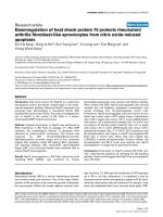
Floatation analysis of A3GFigure 3
Floatation analysis of A3G. HeLa cells were transfected with vectors encoding untagged and C-terminally Myc- or HA-
tagged A3G wt (panels 1 - 3), A3G W127A (panels 4 - 6), or A3G Y124 (panels 7-8). The packaging incompetent A3G C100S-
Myc variant was included for comparison (panel 9). Cellular caveolin was used as a raft marker (panel 10) and transferrin
receptor (TfR) was included as a non-raft associated control (panel 11). Samples were processed for floatation analysis as
described in Methods and 10 equal fractions were collected from the top of the gradient. The position of raft and non-raft pro-
teins is indicated at the bottom. Proteins are identified on the right.
Top
Bottom
A3G-Myc C100S
caveolin
TfR
A3G wt
A3G W127A
A3G-Myc W127A
A3G-Myc wt
rafts non-rafts
(detergent-resistant) (detergent-sensitive)
1
2
3
4
10
11
A3G-HA wt
A3G-HA W127A
5
6
A3G Y124A
A3G-Myc Y124A
7
8
9
12345678910
Retrovirology 2009, 6:99 />Page 9 of 12
(page number not for citation purposes)
Figure 4 (see legend on next page)
A3G W127A
A3G
W127A
Ctrl
Ctrl
RNase
+-
+-+-+-
+-
IP:
A3Ga-
(+/- Myc tag)
(+/- Myc tag)
WB: a-A3G
IgG
A3G
+-
Y124A
Y124A
total
lysate
12345 67
0.0
0.1
0.2
0.3
0.4
0.5
0.6
0.7
0.8
0.9
1.0
0.0
0.1
0.2
0.3
0.4
0.5
0.6
0.7
0.8
0.9
1.0
real-time PCR
real-time PCR
7SL RNA
HIV RNA
A
B
C
A3G
Retrovirology 2009, 6:99 />Page 10 of 12
(page number not for citation purposes)
tion of A3G is required for packaging and for restriction of
HIV-1 [32]. This conclusion is based on the observation
that the packaging defective A3G Y124 and W127 mutants
failed to interact with A3G wt in co-immunoprecipitation
studies.
Since our own packaging studies revealed an impact of C-
terminal epitope tags for packaging of A3G W127A, we
decided to analyze the correlation between A3G dimeriza-
tion and packaging competence. To that end we cotrans-
fected A3G wt, A3G W127A, or A3G Y124A mutants in
various combinations using untagged or Myc-tagged wild
type or mutant A3G constructs. Proteins were immuno-
precipitated by a Myc-specific monoclonal antibody fol-
lowed by immunoblotting with a polyclonal A3G-specific
antibody (Fig. 5, middle panel). Total lysates were also
probed with an A3G-specific antibody as an input control
(Fig. 5, top panel). A tubulin blot was included as loading
control (Fig. 5, lower panel). As expected, untagged A3G
proteins were not precipitated in the absence of Myc-
tagged A3G (Fig. 5, lanes 7-9). Consistent with our previ-
ous report [22]. A3G wt interacted with A3G-Myc wt to
form homo-oligomers (Fig. 5, lane 2). On the other hand,
A3G wt did not seem to interact well with Myc-tagged
A3G W127A (Fig. 5, lane 3). However, the reverse combi-
nation, i.e. A3G-Myc wt plus untagged A3G W127A,
revealed significant interaction (Fig. 5, lane 4). Impor-
tantly, the severely packaging impaired Y124A mutants
exhibited strong interaction with A3G wt irrespective of
which partner in the pull-down assay was tagged (Fig. 5,
lanes 5-6). Thus, our data suggest that mutation of W127
and Y124 does not prohibit A3G oligomerization. There-
fore, we failed to observe a correlation between A3G oli-
gomerization and packaging competence.
Discussion
The mechanism of A3G encapsidation into HIV-1 virions
has attracted significant attention since it offers a potential
target for therapeutic interference with virus replication.
There is increasing evidence that encapsidation of A3G
into virions requires interactions with the viral nucleocap-
sid domain in the Gag precursor and involves RNA
although the nature of the RNA involved in A3G packag-
ing, i.e. cellular versus viral, remains under investigation
[8,9,12-15,18-20,38,41-43]. There is only limited infor-
mation concerning sequences in A3G that are necessary
Packaging incompetent A3G variants are defective for binding viral RNAFigure 4 (see previous page)
Packaging incompetent A3G variants are defective for binding viral RNA. (A-C) HeLa cells were transfected with 4
μg of empty vector DNA (lane 1), 2 μg of either Myc-tagged or untagged A3G wt (lanes 2 & 3, respectively), 4 μg of A3G-Myc
W127A (lane 4), 2 μg of A3G W127A (lane 5), 2 μg of Y124A (lane 6), or 3 μg of A3G-Myc Y124A (lane 7). All samples were
co-transfected with 1 μg of vif-defective pNL4-3Vif(-) as a source of genomic RNA. Total amounts of DNA were adjusted to 5
μg using empty vector DNA as appropriate. Cells were harvested 24 h after transfection and divided into four fractions. Frac-
tion 1 was used for immunoblot analysis of whole cell extracts (panel A, top); fraction 2 was used for total RNA extraction and
qRT-PCR (panel B). Fractions 3 & 4 were first immunoprecipitated as a pool with an A3G-specific rabbit antibody as described
in Methods. Part of the immunoprecipitate (fraction 3) was then used for immunoblotting (panel A, bottom); the other part
(fraction 4) was used for RNA extraction and qRT-PCR. (A) Fractions 1 & 3 were analyzed by immunoblotting for the pres-
ence of A3G using an A3G specific rabbit polyclonal antibody. Proteins are identified on the right. IgG = rabbit immunoglobulin
heavy chain. Total cellular RNA (B) or RNA present in the immune complexes (C) was extracted and subjected to qRT-PCR
analysis of 7SL and genomic RNA as described in Methods. 7SL and genomic RNA levels detected in the presence of untagged
A3G wt were used as reference and defined as 1.0. RNA levels from all other samples were calculated relative to the reference
sample. An RNA sample treated with DNase-free RNaseA (1 mg/ml; 30 min, 37°C) was used as a control for the absence of
contaminating DNA.
Lack of correlation of A3G oligomerization and packaging competenceFigure 5
Lack of correlation of A3G oligomerization and pack-
aging competence. HeLa cells were transfected with 2.5
μg each of plasmids expressing A3G wt (wt), W127A, or
Y124A mutants in a combination of two as indicated above
the figure. Cell lysates were analyzed either directly by
immunoblotting with antibodies to A3G (top panel) or tubu-
lin (bottom panel) or subsequent to immunoprecipitation
with a Myc-specific monoclonal antibody (middle panel).
Untagged A3G variants (no tag) have a faster mobility in the
gel than the Myc-tagged variants. The position of the
untagged proteins co-immunoprecipitated by the Myc-tagged
variants is indicated on the right (co-IP).
mock
wt + wt-Myc
wt + W127A-Myc
wt-Myc + W127A
wt + Y124A-Myc
wt-Myc + Y124A
wt
W127A
Y124A
IP: a-Myc
WB: -APO-C17a
total
lysate
WB: -tuba
123456789
no tag
co-IP
Retrovirology 2009, 6:99 />Page 11 of 12
(page number not for citation purposes)
and sufficient for viral encapsidation. Several studies
implicated the N-terminal catalytic domain (CD1) in A3G
in RNA interaction and virus encapsidation [5,21,22].
Other studies found that sequences in the N-terminal
linker domain downstream of the CD1 domain encom-
passing residues 122 to 127 were critical for A3G packag-
ing [9,12,20,23,25,26]. However, it remains unclear
whether this latter domain represents a direct contact
point for protein-protein or protein-RNA interactions or
simply represents a conformationally sensitive area in the
protein.
Our current study focused on two amino acid residue,
W127 and Y124, in A3G to demonstrate that the require-
ments for A3G packaging are complex and can be influ-
enced by the presence or absence of terminal epitope tags.
Our finding that untagged A3G W127A was packaged 10
times more efficient than Myc- or HA-tagged A3G W127A
was surprising and unexpected since the interfering
epitope tags were not located adjacent to residue W127
but were more than 250 amino acids away at the C-termi-
nus of the protein. There is currently no structure of full-
length A3G available. However, computer modelling sug-
gests that W127 is located at the surface of the protein
[31]. It is therefore possible that in 3-dimensional space
the C-terminus of A3G is in close proximity to the N-ter-
minal linker region surrounding residue W127. Therefore,
changes in this region could, in the context of an epitope-
tag, induce changes in the protein resulting in mislocali-
zation in the cell - as evidenced by differential raft associ-
ation - and culminating in the exclusion of A3G from
virions. Importantly, the epitope tag effect was not tag
specific since identical results were obtained with C-termi-
nal Myc and HA tags. Based on these data we consider it
possible that W127 in A3G is critical for proper protein
folding/conformation and/or cellular localization of the
protein rather than representing a motif required for viral
encapsidation.
Conclusion
Our data reveal an interesting correlation between A3G's
propensity to associate with lipid raft structures, viral RNA
interaction, and packaging competence. The reason for
the inability of the epitope tagged A3G W127A mutants as
well as tagged and untagged Y124A mutants to associate
with lipid rafts and with viral RNA remains to be investi-
gated. Since A3G is not a membrane protein it is likely
that its affinity to raft structures is due to an interaction
with other raft associated host factors. Our floatation
studies were done in the absence of viral proteins, ruling
out the possible contribution of Gag protein in the raft
targeting of A3G. Thus, the lack of raft association could
be indicative of altered intracellular localization or traf-
ficking of A3G or could be due to a conformational
change resulting in the loss of protein-protein interaction.
Competing interests
The authors declare that they have no competing interests.
Authors' contributions
MAK conceived the study, performed the molecular and
biochemical studies, and drafted the manuscript. RG, EM,
and RCW assisted with biochemical studies and helped
with data analysis. SK assisted with A3G mutagenesis and
biochemical analyses. KS coordinated and supervised the
project and wrote the final manuscript.
Acknowledgements
We thank Amy Andrew for critical comments on the manuscript and
Melissa Gilden and Sandrine Opi for plasmid construction and help with
reagents. We are grateful to Michael Malim for providing pA3G, pA3G
W127A, pA3G-HA, and pA3G-HA W127A vectors. This work was sup-
ported by a Grant from the NIH Intramural AIDS Targeted Antiviral Pro-
gram to K.S. and by the Intramural Research Program of the NIH, NIAID.
References
1. Sheehy AM, Gaddis NC, Choi JD, Malim MH: Isolation of a human
gene that inhibits HIV-1 infection and is suppressed by the
viral Vif protein. Nature 2002, 418:646-650.
2. Goila-Gaur R, Strebel K: HIV-1 Vif, APOBEC, and intrinsic
immunity. Retrovirology 2008, 5:51.
3. Miller JH, Presnyak V, Smith HC: The dimerization domain of
HIV-1 viral infectivity factor Vif is required to block virion
incorporation of APOBEC3G. Retrovirology 2007, 4:81.
4. Jarmuz A, Chester A, Bayliss J, Gisbourne J, Dunham I, Scott J, Navar-
atnam N: An anthropoid-specific locus of orphan C to U RNA-
editing enzymes on chromosome 22. Genomics 2002,
79:285-296.
5. Iwatani Y, Takeuchi H, Strebel K, Levin JG: Biochemical Activities
of Highly Purified, Catalytically Active Human APOBEC3G:
Correlation with Antiviral Effect. J Virol 2006, 80:5992-6002.
6. Yu Q, Konig R, Pillai S, Chiles K, Kearney M, Palmer S, Richman D,
Coffin JM, Landau NR: Single-strand specificity of APOBEC3G
accounts for minus-strand deamination of the HIV genome.
Nat Struct Mol Biol 2004, 11:435-442.
7. Khan MA, Kao S, Miyagi E, Takeuchi H, Goila-Gaur R, Opi S, Gipson
CL, Parslow TG, Ly H, Strebel K: Viral RNA is required for the
association of APOBEC3G with human immunodeficiency
virus type 1 nucleoprotein complexes. J Virol 2005,
79:5870-5874.
8. Khan MA, Goila-Gaur R, Opi S, Miyagi E, Takeuchi H, Kao S, Strebel
K: Analysis of the contribution of cellular and viral RNA to
the packaging of APOBEC3G into HIV-1 virions. Retrovirology
2007, 4:48.
9. Wang T, Tian C, Zhang W, Luo K, Sarkis PT, Yu L, Liu B, Yu Y, Yu
XF: 7SL RNA mediates virion packaging of the antiviral cyti-
dine deaminase APOBEC3G. J Virol 2007, 81:13112-13124.
10. Tian C, Wang T, Zhang W, Yu XF: Virion packaging determi-
nants and reverse transcription of SRP RNA in HIV-1 parti-
cles. Nucleic Acids Res 2007, 35:7288-7302.
11. Wang T, Tian C, Zhang W, Sarkis PT, Yu XF: Interaction with 7SL
RNA but not with HIV-1 genomic RNA or P bodies is
required for APOBEC3F virion packaging. J Mol Biol 2008,
375:
1098-1112.
12. Cen S, Guo F, Niu M, Saadatmand J, Deflassieux J, Kleiman L: The
interaction between HIV-1 Gag and APOBEC3G. J Biol Chem
2004, 279:33177-33184.
13. Schafer A, Bogerd HP, Cullen BR: Specific packaging of
APOBEC3G into HIV-1 virions is mediated by the nucleo-
capsid domain of the gag polyprotein precursor. Virology 2004,
328:163-168.
14. Alce TM, Popik W: APOBEC3G is incorporated into virus-like
particles by a direct interaction with HIV-1 Gag nucleocapsid
protein. J Biol Chem 2004, 279:34083-34086.
Publish with BioMed Central and every
scientist can read your work free of charge
"BioMed Central will be the most significant development for
disseminating the results of biomedical research in our lifetime."
Sir Paul Nurse, Cancer Research UK
Your research papers will be:
available free of charge to the entire biomedical community
peer reviewed and published immediately upon acceptance
cited in PubMed and archived on PubMed Central
yours — you keep the copyright
Submit your manuscript here:
/>BioMedcentral
Retrovirology 2009, 6:99 />Page 12 of 12
(page number not for citation purposes)
15. Zennou V, Perez-Caballero D, Gottlinger H, Bieniasz PD:
APOBEC3G incorporation into human immunodeficiency
virus type 1 particles. J Virol 2004, 78:12058-12061.
16. Mariani R, Chen D, Schrofelbauer B, Navarro F, Konig R, Bollman B,
Munk C, Nymark-McMahon H, Landau NR: Species-specific exclu-
sion of APOBEC3G from HIV-1 virions by Vif. Cell 2003,
114:21-31.
17. Mangeat B, Turelli P, Caron G, Friedli M, Perrin L, Trono D: Broad
antiretroviral defence by human APOBEC3G through lethal
editing of nascent reverse transcripts. Nature 2003,
424:99-103.
18. Burnett A, Spearman P: APOBEC3G Multimers Are Recruited
to the Plasma Membrane for Packaging into Human Immu-
nodeficiency Virus Type 1 Virus-Like Particles in an RNA-
Dependent Process Requiring the NC Basic Linker. J Virol
2007, 81:5000-5013.
19. Douaisi M, Dussart S, Courcoul M, Bessou G, Vigne R, Decroly E:
HIV-1 and MLV Gag proteins are sufficient to recruit
APOBEC3G into virus-like particles. Biochem Biophys Res Com-
mun 2004, 321:566-573.
20. Luo K, Liu B, Xiao Z, Yu Y, Yu X, Gorelick R, Yu XF: Amino-termi-
nal region of the human immunodeficiency virus type 1
nucleocapsid is required for human APOBEC3G packaging.
J Virol 2004, 78:11841-11852.
21. Navarro F, Bollman B, Chen H, Konig R, Yu Q, Chiles K, Landau NR:
Complementary function of the two catalytic domains of
APOBEC3G. Virology 2005, 333:374-386.
22. Opi S, Takeuchi H, Kao S, Khan MA, Miyagi E, Goila-Gaur R, Iwatani
Y, Levin JG, Strebel K: Monomeric APOBEC3G is catalytically
active and has antiviral activity. J Virol 2006, 80:4673-4682.
23. Huthoff H, Malim MH: Identification of Amino Acid Residues in
APOBEC3G Required for Regulation by Human Immunode-
ficiency Virus Type 1 Vif and Virion Encapsidation. J Virol
2007, 81:3807-3815.
24. Bach D, Peddi S, Mangeat B, Lakkaraju A, Strub K, Trono D: Charac-
terization of APOBEC3G binding to 7SL RNA. Retrovirology
2008, 5:54.
25. Gooch BD, Cullen BR: Functional domain organization of
human APOBEC3G. Virology 2008, 379:118-124.
26. Zhang KL, Mangeat B, Ortiz M, Zoete V, Trono D, Telenti A, Michie-
lin O: Model Structure of Human APOBEC3G. PLoS ONE 2007,
2:e378.
27. Bogerd HP, Doehle BP, Wiegand HL, Cullen BR: A single amino
acid difference in the host APOBEC3G protein controls the
primate species specificity of HIV type 1 virion infectivity fac-
tor. Proc Natl Acad Sci USA 2004, 101:3770-3774.
28. Schrofelbauer B, Chen D, Landau NR: A single amino acid of
APOBEC3G controls its species-specific interaction with vir-
ion infectivity factor (Vif). Proc Natl Acad Sci USA 2004,
101:3927-3932.
29. Mangeat B, Turelli P, Liao S, Trono D: A single amino acid deter-
minant governs the species-specific sensitivity of
APOBEC3G to Vif action. J Biol Chem 2004, 279:14481-14483.
30. Xu H, Svarovskaia ES, Barr R, Zhang Y, Khan MA, Strebel K, Pathak
VK: A single amino acid substitution in human APOBEC3G
antiretroviral enzyme confers resistance to HIV-1 virion
infectivity factor-induced depletion. Proc Natl Acad Sci USA 2004,
101:5652-5657.
31. Stenglein MD, Matsuo H, Harris RS: Two regions within the
amino-terminal half of APOBEC3G cooperate to determine
cytoplasmic localization. J Virol 2008, 82:9591-9599.
32. Huthoff H, Autore F, Gallois-Montbrun S, Fraternali F, Malim MH:
RNA-dependent oligomerization of APOBEC3G is required
for restriction of HIV-1. PLoS Pathog 2009, 5:e1000330.
33. Karczewski MK, Strebel K: Cytoskeleton association and virion
incorporation of the human immunodeficiency virus type 1
Vif protein. J Virol 1996, 70:494-507.
34. Kao S, Khan MA, Miyagi E, Plishka R, Buckler-White A, Strebel K: The
human immunodeficiency virus type 1 Vif protein reduces
intracellular expression and inhibits packaging of
APOBEC3G (CEM15), a cellular inhibitor of virus infectivity.
J Virol 2003, 77:11398-11407.
35. Opi S, Kao S, Goila-Gaur R, Khan MA, Miyagi E, Takeuchi H, Strebel
K: Human immunodeficiency virus type 1 Vif inhibits packag-
ing and antiviral activity of a degradation-resistant
APOBEC3G variant. J Virol
2007, 81:8236-8246.
36. Roos JW, Maughan MF, Liao Z, Hildreth JE, Clements JE: LuSIV cells:
a reporter cell line for the detection and quantitation of a
single cycle of HIV and SIV replication. Virology 2000,
273:307-315.
37. Ono A, Freed EO: Plasma membrane rafts play a critical role
in HIV-1 assembly and release. Proc Natl Acad Sci USA 2001,
98:13925-13930.
38. Bogerd HP, Cullen BR: Single-stranded RNA facilitates nucleo-
capsid: APOBEC3G complex formation. RNA 2008,
14:1228-1236.
39. Wang T, Zhang W, Tian C, Liu B, Yu Y, Ding L, Spearman P, Yu XF:
Distinct viral determinants for the packaging of human cyti-
dine deaminases APOBEC3G and APOBEC3C. Virology 2008,
377:71-79.
40. Zhang L, Li X, Ma J, Yu L, Jiang J, Cen S: The incorporation of
APOBEC3 proteins into murine leukemia viruses. Virology
2008, 378:69-78.
41. Goila-Gaur R, Khan MA, Miyagi E, Kao S, Strebel K: Targeting
APOBEC3A to the viral nucleoprotein complex confers anti-
viral activity. Retrovirology 2007, 4:61.
42. Svarovskaia ES, Xu H, Mbisa JL, Barr R, Gorelick RJ, Ono A, Freed EO,
Hu WS, Pathak VK: Human apolipoprotein B mRNA-editing
enzyme-catalytic polypeptide-like 3G (APOBEC3G) is incor-
porated into HIV-1 virions through interactions with viral
and nonviral RNAs. J Biol Chem 2004, 279:35822-35828.
43. Soros VB, Yonemoto W, Greene WC: Newly synthesized
APOBEC3G is incorporated into HIV virions, inhibited by
HIV RNA, and subsequently activated by RNase H. PLoS
Pathog 2007, 3:e15.