Báo cáo y học: "Identification of an endogenous retroviral envelope gene with fusogenic activity and placenta-specific expression in the rabbit: a new "syncytin" in a third order of mammals" ppt
Bạn đang xem bản rút gọn của tài liệu. Xem và tải ngay bản đầy đủ của tài liệu tại đây (2.41 MB, 11 trang )
BioMed Central
Page 1 of 11
(page number not for citation purposes)
Retrovirology
Open Access
Research
Identification of an endogenous retroviral envelope gene with
fusogenic activity and placenta-specific expression in the rabbit: a
new "syncytin" in a third order of mammals
Odile Heidmann
†
, Cécile Vernochet
†
, Anne Dupressoir and
Thierry Heidmann*
Address: Unité des Rétrovirus Endogènes et Eléments Rétroïdes des Eucaryotes Supérieurs, CNRS UMR 8122, Institut Gustave Roussy, 39 rue
Camille Desmoulins, F-94805 Villejuif, and Université Paris-Sud, Orsay, F-91405, France
Email: Odile Heidmann - ; Cécile Vernochet - ; Anne Dupressoir - ;
Thierry Heidmann* -
* Corresponding author †Equal contributors
Abstract
Background: Syncytins are envelope genes of retroviral origin that have been co-opted by the host to
mediate a specialized function in placentation. Two of these genes have already been identified in primates,
as well as two distinct, non orthologous genes in rodents.
Results: Here we identified within the rabbit Oryctolagus cuniculus-which belongs to the lagomorpha
order- an envelope (env) gene of retroviral origin with the characteristic features of a bona fide syncytin,
that we named syncytin-Ory1. An in silico search for full-length env genes with an uninterrupted open reading
frame within the rabbit genome first identified two candidate genes that were tested for their specific
expression in the placenta by quantitative RT-PCR of RNA isolated from a large set of tissues. This
resulted in the identification of an env gene with placenta-specific expression and belonging to a family of
endogenous retroelements present at a limited copy number in the rabbit genome. Functional
characterization of the identified placenta-expressed env gene after cloning in a CMV-driven expression
vector and transient transfection experiments, demonstrated both fusogenic activity in an ex vivo cell-cell
fusion assay and infectivity of pseudotypes. The receptor for the rabbit syncytin-Ory1 was found to be the
same as that for human syncytin-1, i.e. the previously identified ASCT2 transporter. This was
demonstrated by a co-culture fusion assay between hamster A23 cells transduced with an expression
vector for ASCT2 and A23 cells transduced with syncytin-Ory1. Finally, in situ hybridization of rabbit
placenta sections with a syncytin-Ory1 probe revealed specific expression at the level of the junctional zone
between the placental lobe and the maternal decidua, where the invading syncytial fetal tissue contacts the
maternal decidua to form the labyrinth, consistent with a role in the formation of the syncytiotrophoblast.
The syncytin-Ory1 gene is found in Leporidae but not in Ochotonidae, and should therefore have entered
the lagomorpha order 12-30 million years ago.
Conclusion: The identification of a novel syncytin gene within a third order of mammals displaying
syncytiotrophoblast formation during placentation strongly supports the notion that on several occasions
retroviral infections have resulted in the independent capture of genes that have been positively selected
for a convergent physiological role.
Published: 27 November 2009
Retrovirology 2009, 6:107 doi:10.1186/1742-4690-6-107
Received: 22 October 2009
Accepted: 27 November 2009
This article is available from: />© 2009 Heidmann et al; licensee BioMed Central Ltd.
This is an Open Access article distributed under the terms of the Creative Commons Attribution License ( />),
which permits unrestricted use, distribution, and reproduction in any medium, provided the original work is properly cited.
Retrovirology 2009, 6:107 />Page 2 of 11
(page number not for citation purposes)
Background
Previous studies have identified two pairs of envelope
(env) genes of retroviral origin that have been independ-
ently captured by their host for a role in placentation. In
simians, syncytin-1 [1-3] and syncytin-2 [4,5] entered the
primate genome 25 and >40 million years (My) ago,
respectively. They retained their coding capacity in all the
subsequent branches; syncytin-1 and syncytin-2 display pla-
centa-specific expression, are fusogenic in ex vivo cell-cell
fusion assays, and one of them displays immunosuppres-
sive activity [6]. A pair of env genes from endogenous ret-
roviruses (ERVs) were then identified in the mouse,
named syncytin-A and -B, which share closely related func-
tional properties although they have a completely distinct
origin, showing a divergent sequence and a different
genomic location compared to primate syncytins [7]. As
found for the latter, syncytin-A and -B have the status of
bona fide genes. They have been conserved since their
entry into the Muridae genome, approximately 20 million
years (My) ago; they display placenta-specific expression,
mediate cell-cell fusion in ex vivo assays [7], and one of
them is immunosuppressive [6]. Recently we have further
unambiguously demonstrated via the generation of syncy-
tin-A knockout mice that these genes are indeed essential
for placentation, with a lack of cell-cell fusion observed in
vivo at the level of the placenta of the knockout embryos,
resulting in impaired maternal-fetal exchanges and death
of the embryos at mid-gestation [8]. Therefore, it appears
that on some occasions in the course of mammalian evo-
lution, env genes from endogenous retroviruses have been
"co-opted" by their host to participate in the formation of
the syncytiotrophoblast layer, at the maternal-fetal inter-
face, by mediating the fusion of mononucleated cytotro-
phoblasts.
A further question that we wanted to answer was whether
mammals belonging to orders other than rodents and pri-
mates but possessing a placenta with a related architec-
ture, i.e. with a syncytiotrophoblast layer in direct contact
with maternal blood at the maternal-fetal interface, have
also "captured" retroviral env genes to generate, in a con-
vergent manner, this specific placental structure. Among
mammals whose placenta displays such a structural
organization at the maternal-fetal interface, i.e. with a
haemochorial placenta, the lagomorpha order was
selected over the two other orders which also possess a
haemochorial placenta -i.e. the Insectivora (hedgehog)
and Chiroptera (bats) [9]; this was done because one of its
representatives, the rabbit (Oryctolagus cuniculus), has an
already sequenced genome, and can be reared and inves-
tigated easily at different stages of gestation, and has a pla-
cental physiology that has been appropriately described
[9-11].
Here, by combining in silico search for env genes within
the rabbit genome, RT-PCR assays for their in vivo tran-
scriptional activity in a large panel of tissues including the
placenta, cloning of the candidate genes, ex vivo assays for
their fusogenicity and, ultimately, in situ hybridization of
placenta sections, we identify a new fusogenic and pla-
centa-specific endogenous env gene, displaying all the
characteristic features of a bona fide syncytin gene, that we
named syncytin-Ory1. Although we demonstrate that the
syncytin-Ory1 protein shares the same ASCT2 receptor in
common with human syncytin-1, it is divergent from all
four syncytins previously identified in rodents and pri-
mates and must therefore have been captured independ-
ently from a distinct ancestral retrovirus. The occurrence
in a third order of Mammals of a new syncytin gene that is
specifically expressed at the maternal-fetal interface
within the placental junctional zone provides strong sup-
port to the notion that ERVs have played a convergent role
in the recurrent emergence of syncytiotrophoblast-con-
taining haemochorial placentae in the course of evolu-
tion.
Results and Discussion
In silico search for retroviral env genes within the rabbit
(Oryctolagus cuniculus) genome
To identify putative env-derived syncytin genes, we made
use of the available rabbit genome sequence (low cover-
age 2× assembly of the Oryctolagus cuniculus genome,
Ensembl May 2005 assembly, updated version 49) and of
the method that we previously devised to screen the
whole human and mouse genomes for such genes [7,12].
Basically, it makes use of the degenerate CKS17u consen-
sus motif, associated with the immunosuppressive
domain of retroviral envelope proteins, and is designed to
match the majority of env genes of exogenous and endog-
enous origin [12]. Rabbit sequences from the Ensembl
database were screened with this motif using the BIOMO-
TIF program, and only sequences with open reading
frames (ORFs) longer than 1.5 kb were considered. Five
ORF-containing sequences were obtained, four of which
disclosed >98% nt identity. We named these four
sequences Env-Ory1. The fifth sequence that was obtained
was unrelated to Env-Ory1. We named this sequence Env-
Ory2 (Figure 1). Analysis of the scaffold database identi-
fied the former ORF as belonging to a low-copy family of
ERVs, most of which had an env gene that was interrupted
by stop codons, deletions and/or truncations. Due to the
low coverage of the available assembly, it could not be
determined whether the identified ORF corresponds to
distinct loci or to a single locus - in that case with distinct
alleles.
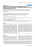
Transcription profile and identification of a placenta-
specific envelope
Quantitative RT-PCR analysis of transcript levels for the
two identified candidate syncytins was performed using
primers specific for each family of elements. As illustrated
in Figure 2, the Env-Ory1-encoding gene has the charac-
Retrovirology 2009, 6:107 />Page 3 of 11
(page number not for citation purposes)
teristic profile of a bona fide syncytin gene, with high levels
of expression in the placenta and very limited expression
in other tissues. Expression in the placenta decreases with
gestational age, with a four-fold reduction from day 12 to
26 (i.e. 4 days before delivery). The other candidate gene
encoding Env-Ory2 is expressed only limitedly (at least
100-fold lower), with no specific expression in the pla-
centa, and was not considered further.
Some of the sequence scaffolds reveal the Env-Ory1-
encoding gene to be part of a proviral structure with
degenerate but identifiable LTR, gag and pol gene
sequences. A phylogenetic tree based on the envelope TM
subunit (Figure 1) shows that this envelope protein is
related to that of type-D retroviruses such as MPMV, BaEV
and RD114, as similarly observed on the basis of a pol-
based tree (not shown). A putative donor and acceptor
splice site for the generation of a subgenomic env tran-
script can be identified according to http://
www.cbs.dtu.dk/services/NetGene2, as classically
observed for retroviruses. Their position and functionality
were further ascertained by RT-PCR analysis of Env-Ory1-
encoding transcripts in the placenta, using appropriate
primers (see Methods and Figure 3).
Analysis of the amino-acid sequence of Env-Ory1 (con-
sensus in Figure 3 of the four sequences in the database,
also corresponding to that PCR-amplified, see below) dis-
plays the characteristic features of retroviral envelope pro-
teins, with a putative signal peptide at the N-terminus,
and a furin cleavage site (RQKR, consensus R/K-X-R/K-R)
at amino acid 380 to generate the SU and TM subunits.
The hydrophobicity plot of the TM subunit reveals a puta-
tive fusion peptide at the TM N-terminus, and a hydro-
phobic transmembrane domain.
Assay for the fusogenic activity of Env-Ory1
Fusogenic activity of Env-Ory1 was assayed as previously
described [4,5,7], by ex vivo assays in cells in culture for
both detection of syncytia formation (cell-cell membrane
fusion) and generation of infectious pseudotypes (virus-
cell membrane fusion). The Env-Ory1-encoding sequence
Retroviral envelope protein-based phylogenic tree with posi-tions of the identified rabbit Env-Ory1- and Env-Ory2, and of the human and mouse syncytinsFigure 1
Retroviral envelope protein-based phylogenic tree
with positions of the identified rabbit Env-Ory1- and
Env-Ory2, and of the human and mouse syncytins.
The tree was determined by the neighbor-joining method
using envelope TM subunit sequences (see ref [12]) from
murine and human ERVs, and infectious retroviruses. The
horizontal branch length and the scale indicate the percent-
age of amino acid substitutions from the node. Percent boot-
strap values obtained from 1,000 replicates are indicated.
endoMMTV
99
100
61
77
100
98
29
100
95
75
99
42
100
35
55
42
38
94
76
59
100
Real-time quantitative RT-PCR analysis of Env-Ory1 and Env-Ory2 transcripts in rabbit tissuesFigure 2
Real-time quantitative RT-PCR analysis of Env-Ory1
and Env-Ory2 transcripts in rabbit tissues. Transcript
levels were normalized relative to the amount of 18S rRNA
(arbitrary units). At least 3 samples per organ type were ana-
lyzed for Env-Ory1 (from different adult animals for non-fetal
tissues; from a given litter for the embryos and placentae);
one sample per organ type was analyzed for Env-Ory2.
Retrovirology 2009, 6:107 />Page 4 of 11
(page number not for citation purposes)
was first PCR-amplified from genomic DNA of Oryctolagus
cuniculus and inserted into a CMV promoter-containing
expression vector (see Methods). The plasmids were
sequenced and those containing a full-length env gene
ORF (that were actually >99.7% identical, and therefore
most probably corresponding to a single sequence ele-
ment) were assayed. As illustrated in Figure 4A, transient
transfection of human SH-SY5Y neuroblastoma cells with
the Env-Ory1-expressing vector triggers cell-cell fusion, as
expected for a bona fide syncytin expressed in a cell line
Characterization of Env-Ory1Figure 3
Characterization of Env-Ory1. (A) Schematic representation of the Env-Ory1-associated ERV with the LTRs and the splice
sites for the sub-genomic env transcript indicated. (B) Schematic structure, hydrophobicity profile and primary sequence of the
Env-Ory1 glycoprotein (deposited in GenBank [GenBank:GU196371
]). The SU and TM subunits of the envelope protein are
delineated, with a canonical furin cleavage site (RQKR; consensus: R/K-N-R/K-R) between the two subunits and the CWLC
domain involved in SU-TM interaction indicated in red; the hydrophobic signal peptide and fusion peptide and the transmem-
brane domain are shaded in light gray, and the putative immunosuppressive domain (ISU) in dark gray.
Retrovirology 2009, 6:107 />Page 5 of 11
(page number not for citation purposes)
Fusogenic activity of syncytin-Ory1Figure 4
Fusogenic activity of syncytin-Ory1. (A) Assay for cell-cell fusion mediated by syncytin-Ory1. The indicated cell lines were
transfected with an expression vector for syncytin-Ory1 or an empty vector (none) together with a LacZ expression vector.
Cells were cultured for 1-2 days after transfection, fixed and X-gal-stained. Syncytia (arrows) were detected in syncytin-Ory1-
transfected SH-SY5Y cells, with only mononucleated cells visible in the other cases. (B) Assay for cell infection mediated by
syncytin-Ory1-pseudotyped virus particles. Pseudotypes were produced by cotransfection of human 293T cells with expres-
sion vectors for the SIV core, the syncytin-Ory1 protein (or an empty vector) and a LacZ-containing retroviral transcript.
Supernatants were used to infect the indicated target cells, which were X-gal stained 3 days after infection. Abbreviation: Syn-
Ory1, syncytin-Ory1.
Retrovirology 2009, 6:107 />Page 6 of 11
(page number not for citation purposes)
carrying its cognate receptor. Cell-cell fusion is not
observed with the hamster A23 cells, which were therefore
used as a control in the following assays for receptor iden-
tification.
Env-Ory1 can also form infectious pseudotypes, as
expected from its retroviral origin. As illustrated in Figure
4B, pseudotypes generated with an SIV core are able to
infect SH-SY5Y cells that are positive in the cell-cell fusion
assay above, but not A23 cells. The profile of cells positive
for infection was found to be similar to that observed for
human syncytin-1 (data not shown), thus suggesting that
Env-Ory1 could possibly use the same receptor (i.e. the
neutral amino acid trasporter ASCT2, [2]). This point was
investigated in the experiments illustrated in Figure 5 in
which distinct pools of A23 cells transfected with either an
ASCT2 or an Env-Ory1 expression vector (supplemented
with a β-galactosidase expression vector) were mixed and
assayed for cell-cell fusion. As shown in the figure, cell-cell
fusion (as revealed by the presence of large LacZ+ syncy-
tia) could be observed with the Env-Ory1 and ASCT2 pair
(as well as with the ASCT2/syncytin-1 and MFSD2/syncy-
tin-2 [13] pairs, used as positive controls) but not with
any of the other combinations. This strongly suggests that
Env-Ory1 uses the ASCT2 receptor, as does human syncy-
tin-1. It is rather unexpected for two independently
acquired - and distinct - retroviral envelopes to use the
same cellular receptor. ASCT2 seems, however, to be a
rather "successful" receptor being also the one used by a
series of type-D infectious retroviruses, such as the pri-
mate MPMV, feline RD114, and avian SNV viruses [14,15]
whose Env actually clusters with Env-Ory1 in phylogenic
trees (Figure 1 for the TM domain and data not shown for
SU). Finally, database screening indeed reveals the pres-
ence of an ASCT2 gene in the rabbit genome (mRNA
accession number NM_001082378; 85% amino-acid
identity with human ASCT2), and qRT-PCR demonstrates
its expression in the rabbit placenta (data not shown). In
conclusion, Env-Ory1 can be considered as a bona fide
syncytin owing to its fusogenic activity and specific
expression in the placenta, and be named Syncytin-Ory1.
In situ hybridization of placenta sections
To further assess the physiological relevance of syncytin-
Ory1 expression in the placenta, in situ hybridization
experiments were performed on paraffin sections of pla-
centa at day 12 of gestation, i.e. the stage showing maxi-
mum expression of syncytin-Ory1 by qRT-PCR (Figure 2).
Figure 6A shows the representative architecture of rabbit
placenta at day 12. Three main zones can be distin-
Fusion assay between ASCT2-transduced and syncytin-Ory1-transduced co-cultured cells demonstrates that ASCT2 is the syncy-tin-Ory1 receptorFigure 5
Fusion assay between ASCT2-transduced and syncytin-Ory1-transduced co-cultured cells demonstrates that
ASCT2 is the syncytin-Ory1 receptor. Left panel: Cell-cell fusion was assayed upon independent transfections of a set of
A23 cells with an empty vector (none) or an expression vector for either the syncytin-Ory1, syncytin-1 or syncytin-2 protein
together with an nls-LacZ gene-expression vector, and another set of A23 cells with an expression vector for the syncytin-1
receptor ASCT2, the syncytin-2 receptor MFSD2 [13] or an empty vector (none). One day after transfection, cells were resus-
pended and pairs of transfected cells from each set were cocultured for 1-2 days, fixed and X-Gal stained. Right panel: Syncytia
can be easily detected (arrows) for the syncytin-Ory1/ASCT2, syncytin-1/ASCT2 and syncytin-2/MFSD2 pairs, with only mono-
nucleated cells visible in the other cases. Abbreviations: syn-Ory1, syncytin-Ory1; syn1, syncytin-1; syn2, syncytin-2.
Retrovirology 2009, 6:107 />Page 7 of 11
(page number not for citation purposes)
guished. The maternal decidua results from the modifica-
tion of the uterus after implantation, and the placental
lobe consists in a labyrinthine structure where fetal-mater-
nal exchanges take place. In the labyrinth, fetal and mater-
nal blood circulations are separated by 2 layers of
trophoblasts (haemodichorial placenta). Between the
decidua and placental lobe, a junctional zone can be
observed where fetal vessels surrounded by invading fetal
tissue contact the maternal decidua to form the labyrinth
(Figure 6A, B). The invading fetal tissue has been
described as a broad syncytial front toward the decidua,
backed by and presumably formed from cellular cytotro-
phoblasts [9]. Thus, as soon as the fetal processes reach
the maternal blood spaces, a haemodichorial structure is
formed, with both a cellular and a syncytial trophophob-
last layer separating maternal and fetal blood spaces
(maternal lacuna, ml, and fetal vessels, fv). All of these
characterize the definitive labyrinthine placenta [9,11].
A specific digoxigenin-marked antisense probe was syn-
thesized for syncytin-Ory1 transcript detection, as well as
the corresponding sense probe as a negative control. As
shown in Figure 6C, specific labeling was observed only
with the antisense probe (upper panels), but not with the
control probe (lower panels). Syncytin-Ory1 expression is
restricted to the junctional zone, where its expression (of
variable intensity depending on the area, even within the
same placental section) is limited to trophoblast cells sur-
rounding the invading fetal vessels (brackets for heavily
marked zones and arrows for punctuated domains in Fig-
ure 6C). Although we cannot formally discriminate
between the syncytial trophoblast and the cellular cytotro-
phoblasts, the labeling profile is compatible with syncytin-
Ory1 being expressed in the cytotrophoblast just before
fusion takes place and/or in the newly formed syncytio-
trophoblast (no labeling was detected in the placental
lobe outside the junctional zone). These observations are
consistent with a role for syncytin-Ory1 in the formation of
the syncytiotrophoblast.
Syncytin-Ory1 sequences are present in Leporidae but
not in Ochotonidae
Phylogenetic relationships in the order of Lagomorpha
are illustrated in Figure 7 (adapted from [16]). The order
includes 2 families, Ochotonidae (pikas) and Leporidae
(hares and rabbits). We searched for syncytin-Ory1 genes
in lagomorph species belonging to the Ochotonidae fam-
ily (Ochotona princeps) and the Leporidae family (genus
Lepus: Lepus americanus, Lepus europaeus and Lepus starcki;
genus Sylvilagus: Sylvilagus brasiliensis, Sylvilagus florida-
nus). The domestic rabbit, Oryctolagus cuniculus, analyzed
in this study, belongs to the genus Oryctolagus within the
Leporidae.
Genomic DNA from these different species was PCR-
amplified using a pair of primers for the syncytin-Ory1
ORF. A 1900 bp specific amplicon could be obtained from
the 3 Lepus species, but neither from the Sylvilagus nor
from the Ochotona genus members tested. However, a pair
of primers internal to the ORF allowed the amplification
of a 1430 bp PCR product from the 2 Sylvilagus species
analyzed. Again, no specific amplification could be
obtained from the Ochotona princeps genomic DNA. The
PCR products obtained from Lepus and Sylvilagus were
cloned and sequenced and showed >88% nucleotide
identity with the Oryctolagus cuniculus syncytin-Ory1 gene
sequence, and therefore most probably correspond to
amplification of the same genes.
The absence of a syncytin-Ory1 gene in the Ochotona (as
well as in the more ancestrally diverged rodent (mouse)
and primate (human) orders) was confirmed by screening
(using the BLAST program) the corresponding Ensembl
databases.
Conclusively, the identification of syncytin-Ory1 genes in
Lepus and Sylvilagus in addition to Oryctolagus indicates
that gene capture most probably occurred prior to the
divergence of the Lepus and Oryctolagus/Sylvilagus genera
~12 My ago [16]. The absence of a syncytin-Ory1 gene in
Ochotona princeps suggests that this integration took place
after the Leporidae and Ochotonidae divergence ~30 My
ago [16].
Conclusion
Here we have shown that within a placental mammal that
has developed a haemochorial placenta with a syncytio-
trophoblast layer at the maternal-fetal interface, such as
the rabbit, a gene of retroviral origin can be identified
which displays all the characteristic features of a bona fide
syncytin gene. The identified syncytin-Ory1 has a cell-cell
fusion activity and is expressed specifically in the placenta
at a location consistent with a direct role in syncytiotro-
phoblast formation. syncytin-Ory1 can be found in a series
of leporidae. Notably, the identified env gene is divergent
from any other previously described genes. Thus, for a
newly investigated mammalian order, we provide evi-
dence that a retroviral env gene has been captured and has
gained the status of a "syncytin" according to a process sim-
ilar to that previously observed in the two other major
orders where haemochorial placentae have emerged: the
primates (syncytin-1 and -2) and the rodents (syncytin-A
and -B).
It is therefore likely that such gene captures have arisen
recurrently during the evolution of placental mammals
and that syncytin genes will be found in other placental
mammals displaying a syncytiotrophoblast organization.
Ongoing investigations carried out on representative ani-
mals of the Carnivora order strongly support this hypoth-
esis. An interesting question which remains to be
answered is to determine which specific properties of the
Retrovirology 2009, 6:107 />Page 8 of 11
(page number not for citation purposes)
Structure and in situ hybridization for syncytin-Ory1 expression of day 12 rabbit placenta: (A) Schematic representation of a rab-bit placenta (right) and haematoxylin and eosin staining of a day 12 placenta section (left) with the 3 main layers of the placenta indicatedFigure 6
Structure and in situ hybridization for syncytin-Ory1 expression of day 12 rabbit placenta: (A) Schematic repre-
sentation of a rabbit placenta (right) and haematoxylin and eosin staining of a day 12 placenta section (left)
with the 3 main layers of the placenta indicated. (B) Higher magnification of the areas framed in A. Abbreviations: frbc:
fetal red blood cell, fv: fetal blood vessel, ml: maternal blood lacuna, mrbc: maternal red blood cell. (C) In situ hybridization on
sections of a day 12 rabbit placenta (serial sections of the HES in B) with digoxigenin-labeled syncytin-Ory1 sense (lower panel,
negative control) and antisense (upper panel) riboprobes, revealed with an alkaline phosphatase-conjugated anti-digoxigenin
antibody. Brackets and arrows highlight the positive labeling of trophoblast cells surrounding the invading fetal vessels in the
junctional zone.
Retrovirology 2009, 6:107 />Page 9 of 11
(page number not for citation purposes)
captured syncytins are responsible for the relative diversity
observed in the physiology of mammalian placentation.
Among species with hemochorial placentation in particu-
lar, most of the species (including human, rabbit and
most rodents) have a single layer of syncytiotrophoblast,
whereas a few others (Muridae) have a two-layered syncy-
tiotrophoblast. Moreover, it will be of interest to deter-
mine whether placental mammals [such as Suidae (pig) or
Equidae (horse)] which do not possess a syncytiotro-
phoblast layer at their maternal-fetal interface, are
deprived of syncytins, or whether other functions of retro-
viral envelope proteins such as their immunosuppressive
activity have driven the capture of a non-fusogenic syncy-
tin-like gene. In this case, retroviral envelope proteins
would be co-opted solely for an immunological role in
relation with maternal-fetal tolerance. Experiments in
progress with knock-in mice where syncytin genes have
been mutated for immunosuppressive activity without
impairment of fusogenicity may also help answer these
questions.
Methods
Database screening and sequence analyses
Retroviral env gene sequences were searched by using the
Biomotif program />menes/bioMotif_html_doc/ref_Run.html and the degen-
erate universal CKS17u consensus motif [12] as a query.
We made use of the available rabbit genome sequence
(low coverage 2× assembly of the Oryctolagus cuniculus
genome, Ensembl RABBIT May 2005 assembly, updated
version 49). ORF-containing scaffolds (scaffold number
and ORF-coordinates indicated) were:
131951(25124:26887), 15025(429:2192 reverse strand),
90528(17499:19256), 163359(7744:9486 reverse
strand) for Env-Ory1, and 1093(1017:3035 reverse
strand) for Env-Ory2. Multiple alignments were carried
out by using the CLUSTALW program http://bio
info.hku.hk/services/analyseq/cgi-bin/clustalw_in.pl.
Phylogenic trees were constructed from alignments by
using the neighbor-joining program within CLUSTALW
and were viewed with the NJPLOT program.
The Ochotona princeps (Pika) genome low coverage 1.93×
assembly (Ensembl OchPri2.0 June 2007, updated ver-
sion 53), as well as of the human and mouse assemblies
(from the Genome Reference Consortium) were also
screened for the presence of the identified rabbit Env-
Ory1 ORF sequence, using the BLAST programs at the
National Center for Biotechnology Information http://
www.ncbi.nlm.nih.gov/BLAST.
Real-time RT-PCR
Env-Ory1 and Env-Ory2 mRNA expression was deter-
mined by real-time quantitative RT-PCR. Pregnant New
Zealand white rabbit females obtained from INRA (Jouy-
en-Josas, France) at various stages of gestation were sacri-
ficed, and dissected organs were stored in liquid nitrogen.
Total RNA was extracted from the frozen organs using the
RNeasy RNA isolation kit (Qiagen). Reverse transcription
was performed with 1 μg of DNase-treated RNA as in [17].
Real-time qPCR was with 5 μl of diluted (1:10) cDNA in a
final volume of 25 μl by using SYBR Green PCR Master
Mix (or Taqman Universal PCR Master Mix for 18S rRNA
detection) (Applied Biosystems). PCR was carried out
using an ABI PRISM 7000 sequence detection system.
Primer sequences were as follows: 5'-GCTGTTTTTAT-
GCTAACAAGTCC and 5'-GATAAAGGTCATCAGC CT
ATTGA for Env-Ory1 and 5'-CCTCTAAATGTCATCTTCAC-
CAG and 5'-CTATTGGGACAGCAGTTCTAGTC for Env-
Ory2,. The transcript levels were normalized relative to
the amount of 18S rRNA (as determined with the primers
and Taqman probe from Applied Biosystems). Samples
were assayed in duplicate.
Syncytin-Ory1 expression vector
The syncytin-Ory1 expression vector was constructed as fol-
lows: syncytin-Ory1 was PCR-amplified from genomic
DNA of New Zealand white rabbit using the Accuprime
DNA polymerase (Invitrogen) for 30 cycles. XhoI-contain-
ing primer sequences were: 5'-ATCACCTCGAGTGCT-
GGAATTGTTGTCATTGTTG and 5'-ATCACCTCGAGCG
TCATTGGCTTACTGCTCATTT. After restriction with XhoI,
the PCR product was cloned into the phCMV-G vector
(GenBank accession AJ318514
, gift F L. Cosset) opened
with XhoI. Constructs were verified by sequencing.
Cell fusion assay
Cell lines described in [13,18] were grown in DMEM
medium supplemented with 10% fetal calf serum (Invit-
rogen).
Putative entry date of syncytin-Ory1 during lagomorph evolu-tionFigure 7
Putative entry date of syncytin-Ory1 during lago-
morph evolution. Schematized phylogenetic tree with the
evolutionary timeline of four lagomorph genus (adapted from
[16]) and the rodent and primate outgroups depicted, with
average divergence times indicated for the nodes. The pres-
ence of syncytin-Ory1 sequences in each genus, detected
either by PCR experiments (a) or database screening (b), is
indicated on the right.
Ochotona
Lepus
Oryctolagus
Sylvilagus
60MY
29MY
12MY
10MY
Mus
a,b
+
a
+
a
+
a
b
b
Leporidae
Rodentia
Primates
Ochotonidae
Homo
Lagomorpha
Retrovirology 2009, 6:107 />Page 10 of 11
(page number not for citation purposes)
For the self-fusion assay, cells seeded at 10
4
- 5 × 10
4
cells
per well in 24-well plates were transfected by using the
Lipofectamine kit (Invitrogen) with 0.2 μg of either the
syncytin-Ory1 expressing or an empty vector, supple-
mented with 0.2 μg of a LacZ-expression vector (pCMV-β,
Clontech). Syncytia were visualized by X-Gal staining 24
to 48 h after coculture.
For cell-cell fusion by the coculture assay, A23 cells were
seeded at 5 × 10
5
cells per 60-mm dish. A set of dishes
were transfected by using the Lipofectamine LTX kit (Inv-
itrogen) with 5 μg of either an ASCT2 or an MFSD2
expression vector [13] or an empty vector, and another set
were transfected with 2.5 μg of either a syncytin-Ory1 or a
syncytin-1 or a syncytin-2 expression vector or an empty
vector, each cotransfected with 2.5 μg of the nls-LacZ
expression vector (R9SA, [19]). One day after transfection,
3.5 × 10
5
cells from each group of transfected cells were
cocultured in 6-well plates. Syncytia were visualized by X-
Gal staining 24 to 48 h after coculture.
Pseudotyping assay
SIV virions pseudotyped with syncytin-Ory1 were pro-
duced by cotransfecting 8 × 10
5
293T cells with: 2.25 μg
pSIV3+ (encoding SIV retroviral proteins except Env) [20];
2.25 μg R9SA (a LacZ-marked defective SIV retroviral vec-
tor) [19]; and 0.5 μg of syncytin-Ory1 expression vector,
using the Lipofectamine LTX transfection kit (Invitrogen).
Supernatants from the transfected cells were harvested 48
h after transfection, filtered through 0.45 μm pore-size
PVDF membranes, supplemented with Polybrene (4 μg/
ml), transferred to target cells seeded in 24-well plates (5
× 10
4
- 8 × 10
4
cells per well) the day before infection, fol-
lowed by spinoculation at 1200 × g for 2 h 30 min at room
temperature. X-Gal staining was performed 3 days later.
In situ hybridization
Freshly collected rabbit placentae (at day 12 of gestation)
were fixed in 4% paraformaldehyde at 4°C, embedded in
paraffin, and serial sections (4 μm) were either stained
with haematoxylin and eosin or used for in situ hybridiza-
tion. A PCR-amplified 1135 bp syncytin-Ory1 fragment
(primers: 5'-AGACTGCGGAGATAAAACTGC and 5'-
GTGGACCGCGATTCCTAGTC) was cloned into pGEM-T
Easy (Promega) for in vitro synthesis of the antisense and
sense riboprobes, generated with SP6 RNA polymerase
and digoxygenin 11-UTP (Roche Applied Science). Sec-
tions were processed, hybridized at 42°C overnight with
the riboprobes and incubated further at room tempera-
ture for 2 h with alkaline phosphatase-conjugated anti-
digoxygenin antibody Fab fragments (Roche Applied Sci-
ence). Staining was revealed with NBT and BCIP phos-
phatase alkaline substrates as indicated by the
manufacturer (Roche Applied Science).
Search for syncytin-Ory1 in other lagomorphs
Genomic DNAs from Lepus americanus, Lepus europaeus,
Ochotona princeps, Sylvilagus brasiliensis and Sylvilagus flori-
danus were a gift from A. Surridge (Department of Zool-
ogy, University of Cambridge, UK). Genomic DNA from
Lepus starcki was extracted from tissue given by F. Catzeflis
(Laboratoire de Paléontologie, Université de Montpellier
2, France). Genomic DNAs were digested by Not I, which
does not cut within the syncytin-Ory1 gene. PCRs were per-
formed on 100 ng of DNA, using Accuprime Taq DNA
Polymerase (Invitrogen) for 40 cycles (30 sec at 94°C, 30
sec at 50°C, 2 min at 68°C). The primers used were: 5'-
TTCCTGAGGGCTCACTGATTAAC and 5'-GAAGGGGA-
GAGTCAGTTGTTGGAG (external to the ORF) or 5'-
AGACTGCGGAGATAAAACTGC and 5'-gataaaggtcat-
cagcctattga (internal to the ORF). PCR products were then
cloned in pGEM-T Easy vector (Promega) for subsequent
sequencing. Primer sequences for splice site determina-
tion were: 5'-CTTGGGGTTCGAGCCTGT and 5'-TTGAG-
CACGGCCACGGCCAC, on each side of the putative
splice donor (SD) and acceptor (SA) sequences, respec-
tively (see Figure 3A).
Competing interests
The authors declare that they have no competing interests.
Authors' contributions
OH, CV, AD and TH designed research and drafted the
manuscript. OH, CV and AD performed research. All
authors read and approved the final manuscript.
Acknowledgements
The authors wish to acknowledge A. Surridge and F. Catzeflis for the gift of
tissues, P. Opolon and O. Bawa for histological analyses, P. Dessen at the
Institut Gustave Roussy Bioinformatic Center for help in the computing
work and the Institut Gustave Roussy - Service Commun d'Expérimenta-
tion Animale and INRA (Jouy en Josas, France) for animal care. We thank
Christian Lavialle for comments and critical reading of the manuscript. This
work was supported by the CNRS and a fellowship to C.V. from the Fon-
dation pour la Recherche Médicale.
References
1. Mi S, Lee X, Li X, Veldman GM, Finnerty H, Racie L, LaVallie E, Tang
XY, Edouard P, Howes S, Keith JC Jr, McCoy JM: Syncytin is a cap-
tive retroviral envelope protein involved in human placental
morphogenesis. Nature 2000, 403:785-789.
2. Blond JL, Lavillette D, Cheynet V, Bouton O, Oriol G, Chapel-Fern-
andes S, Mandrand B, Mallet F, Cosset FL: An envelope glycopro-
tein of the human endogenous retrovirus HERV-W is
expressed in the human placenta and fuses cells expressing
the type D mammalian retrovirus receptor. J Virol 2000,
74:3321-3329.
3. Mallet F, Bouton O, Prudhomme S, Cheynet V, Oriol G, Bonnaud B,
Lucotte G, Duret L, Mandrand B: The endogenous retroviral
locus ERVWE1 is a bona fide gene involved in hominoid pla-
cental physiology. Proc Natl Acad Sci USA 2004, 101:1731-1736.
4. Blaise S, de Parseval N, Bénit L, Heidmann T: Genomewide screen-
ing for fusogenic human endogenous retrovirus envelopes
identifies syncytin 2, a gene conserved on primate evolution.
Proc Natl Acad Sci USA 2003, 100:13013-13018.
Publish with BioMed Central and every
scientist can read your work free of charge
"BioMed Central will be the most significant development for
disseminating the results of biomedical researc h in our lifetime."
Sir Paul Nurse, Cancer Research UK
Your research papers will be:
available free of charge to the entire biomedical community
peer reviewed and published immediately upon acceptance
cited in PubMed and archived on PubMed Central
yours — you keep the copyright
Submit your manuscript here:
/>BioMedcentral
Retrovirology 2009, 6:107 />Page 11 of 11
(page number not for citation purposes)
5. Blaise S, Ruggieri A, Dewannieux M, Cosset F-L, Heidmann T: Iden-
tification of an envelope protein from the FRD family of
Human Endogenous Retroviruses (HERV-FRD) conferring
infectivity on retroviral particles and functional conservation
among simians. J Virol 2004, 78:1050-1054.
6. Mangeney M, Renard M, Schlecht-Louf G, Bouallaga I, Heidmann O,
Letzelter C, Richaud A, Ducos B, Heidmann T: Placental syncytins:
Genetic disjunction between the fusogenic and immunosup-
pressive activity of retroviral envelope proteins. Proc Natl Acad
Sci USA 2007, 104:20534-20539.
7. Dupressoir A, Marceau G, Vernochet C, Bénit L, Kanellopoulos C,
Sapin V, Heidmann T: Syncytin-A and syncytin-B, two fusogenic
placenta-specific murine envelope genes of retroviral origin
conserved in Muridae. Proc Natl Acad Sci USA 2005, 102:725-730.
8. Dupressoir A, Vernochet C, Bawa O, Harper F, Pierron G, Opolon
P, Heidmann T: Syncytin-A knockout mice demonstrate the
critical role in placentation of a fusogenic, endogenous retro-
virus-derived, envelope gene. Proc Natl Acad Sci USA 2009,
106:12127-12132.
9. Wooding P, Burton GJ: Haemochorial Placentation: Mouse,
Rabbit, Man, Apes, Monkeys. In Comparative Placentation Struc-
tures, Functions and Evolution Springer; 2008:196-202.
10. Larsen JF: Electron microscopy of the implantation site in the
rabbit. Am J Anat 1961, 109:319-334.
11. Larsen JF: Electron Microscopy of the Chorioallantoic Pla-
centa of the Rabbit. I. The Placental labyrinth and the Multi-
nucleated Giant Cells of the Intermediate Zone. J
Ultrastructure Research 1962, 7:535-549.
12. Benit L, Dessen P, Heidmann T: Identification, phylogeny, and
evolution of retroviral elements based on their envelope
genes. J Virol 2001, 75:11709-11719.
13. Esnault C, Priet S, Ribet D, Vernochet C, Bruls T, Lavialle C, Weis-
senbach J, Heidmann T: A placenta-specific receptor for the
fusogenic, endogenous retrovirus-derived, human syncytin-
2. Proc Natl Acad Sci USA 2008, 105:17532-17537.
14. Rasko JE, Battini JL, Gottschalk RJ, Mazo I, Miller AD: The RD114/
simian type D retrovirus receptor is a neutral amino acid
transporter. Proc Natl Acad Sci USA 1999, 96:2129-2134.
15. Tailor CS, Nouri A, Zhao Y, Takeuchi Y, Kabat D: A sodium-
dependent neutral-amino-acid transporter mediates infec-
tions of feline and baboon endogenous retroviruses and sim-
ian type D retroviruses. J Virol 1999, 73:4470-4474.
16. Matthee CA, van Vuuren BJ, Bell D, Robinson TJ: A molecular
supermatrix of the rabbits and hares (Leporidae) allows for
the identification of five intercontinental exchanges during
the Miocene. Syst Biol 2004, 53:433-447.
17. de Parseval N, Lazar V, Bénit L, Casella J, Heidmann T: Survey of
human genes of retroviral origin: identification and tran-
scriptome of the genes with coding capacity for complete
envelope proteins. J Virol 2003, 77:10414-10422.
18. Blaise S, de Parseval N, Heidmann T: Functional characterization
of two newly identified Human Endogenous Retrovirus cod-
ing envelope genes. Retrovirology 2005, 2:19.
19. Negre D, Cosset FL: Vectors derived from simian immunode-
ficiency virus (SIV). Biochimie 2002, 84:1161-1171.
20. Negre D, Mangeot PE, Duisit G, Blanchard S, Vidalain PO, Leissner P,
Winter AJ, Rabourdin-Combe C, Mehtali M, Moullier P, Darlix JL,
Cosset FL: Characterization of novel safe lentiviral vectors
derived from simian immunodeficiency virus (SIVmac251)
that efficiently transduce mature human dendritic cells. Gene
Ther 2000, 7:1613-1623.