Báo cáo y học: "Hematopoietic stem cells and retroviral infection" potx
Bạn đang xem bản rút gọn của tài liệu. Xem và tải ngay bản đầy đủ của tài liệu tại đây (2.87 MB, 17 trang )
REVIEW Open Access
Hematopoietic stem cells and retroviral infection
Prabal Banerjee
1,2†
, Lindsey Crawford
1†
, Elizabeth Samuelson
1
, Gerold Feuer
1,2*
Abstract
Retroviral induced malignancies serve as ideal models to help us better understand the molecular mechanisms
associated with the initia tion and progression of leukemogenesis. Numerous retroviruses including AEV, FLV, M-
MuLV and HTLV-1 have the ability to infect hematopoietic stem and progenitor cells, resulting in the deregulation
of normal hematopoiesis and the development of leukemia/lymphoma. Research over the last few decades has
elucidated similarities between retroviral-induced leukemogenesis, initiated by deregulation of innate hematopoie-
tic stem cell traits, and the cancer stem cell hypothesis. Ongoing research in some of these models may provide a
better understanding of the processes of normal hematopoiesis and cancer stem cells. Research on retroviral
induced leukemias and lymphomas may identify the molecular events which trigger the initial cellular transforma-
tion and subsequent maintenance of hematologic malignancies, including the generation of cancer stem cells. This
review focuses on the role of retroviral infection in hematopoietic stem cells and the initiation, maintenance and
progression of hematological malignancies.
Introduction
Hematopoiesis is a highly regulated and hierarchical
process wherein hematopoietic stem cells (HSCs) differ-
entiate into mature hematopoietic cells [1]. It is a pro-
cess controlled by complex interactions between
numerous genetic processes in blood cells and their
environment. The fundamental processes of self-renewal
and quiescence, proliferation and differentiation, and
apoptosis are governed by these interactions within both
hematopoietic stem cells and mature blood cell lineages.
Under normal physiologic conditions, hematopoietic
homeostasi s is maintained by a delicate balance between
processes such as self-renewal, proliferation and differ-
entiation versus apoptosis or cell-cycle arrest in hemato-
poietic progenitor/hematopoietic stem cells (HP/HSCs).
Under stress conditions, such as bl eeding or infection,
fewer HP/HSCs undergo apoptosis while increased
levels of cytokines and growth factors enhance prolifera-
tion and differentiation. In a normally functioning
hematopoietic system, the kinetics of hematopoiesis
return to baseline levels when the stress conditions end.
Deregulation of the signaling pathways that control the
various hematopoietic processes leads to abnormal
hematopoiesis and is associated with the development of
cancer, including leukemia (reviewed in [2]).
Although not fully charac terized, deregulation of nor-
mal hematopoietic signaling pathways in HP/HSCs fol-
lowing viral infection has previously b een documented
[3-5]. Previous studies demonstrated productive infec-
tion of HP/HSCs by re troviruses and suggested that ret-
roviral mediated leukemogenesis shares similarities with
the development of other types of cancer, including the
putative existence of cancer stem cells (CSCs) [6,7].
Here we discuss the evidence demonstrating that retro-
viruses can infect HP/HSCs, and we speculate on the
ability of Human T-cell lymphotropic virus type 1
(HTLV-1) to generate an “infectious” leukemic/cancer
stem cell (ILSC/ICSC).
What Defines a HSC?
HSCs are pluripotent stem cells that can generate all
hemato-lymphoid cells. A cell must meet four basic
functional requirements to be defined as a HSC: 1) the
capability for self-renewal, 2) the capability to undergo
apoptosis, 3) the maintenance of multilineage hemato-
poiesis, and 4) the mobiliza tion out of the bone marrow
into the circulating blood. The ability of HSCs to per-
manently reconstitute an irradiated recipient host is the
most stringent test to evaluate if a population is a true
HSC. Long-term transplantation experiments suggest a
clonal diversity model of HSCs where the HSC
* Correspondence:
† Contributed equally
1
Department of Microbiology and Immunology, SUNY Upstate Medical
University, Syracuse, NY, 13210, USA
Banerjee et al. Retrovirology 2010, 7:8
/>© 2010 Banerjee et al; li censee BioMed Central Ltd. This is an Open Access ar ticle distributed under the terms of the Creative
Commons Attribution License ( which permits unrestricted use, distribution, and
reproduction in any me dium, pr ovided the original work is properly cited.
compartment consists of a fixed number of different
types of HSCs, each with an epigenetically prepro-
grammed fate. The HP/HSC population is typically
def ined by surface expression of CD34 and represents a
heterogeneous cell population encompassing stem cells,
early pluripotent progenitor cells, multipotent progeni-
tor cells, and uncommitted differentiating cells [8].
HSCs have the potential to proliferate indefinitely and
can differentiate into mature hematopoietic lineage spe-
cific cells.
In adults, HSCs are maintained within the bone mar-
row and differentiate to produce the requisite n umber
of highly specialized cells of the hematopoietic system.
HSCs differentiate into two distinctive types of hemato-
poietic progenitors: 1) a common lymphoid progenitor
(CLP) population that generates B-cells, T-cells and NK
cells, and 2) a common myeloid progenitor (CMP)
population that generates granulocytes, neutrophils,
eosinophils, macrophages and erythrocytes (Figure 1).
Lineage commitment of these progenitors involves a
complex process that can be induced in response to a
variety of factors, including the modulation of hemato-
poietic-associated cytokines and transcription factors.
These factors serve dual purposes both by maintaining
pluripotency and by actively inducing lineage commit-
ment and differentiation of HSCs [9-18]
Leukemia Stem Cells/Cancer Stem Cells (LSC/CSC)
The cancer stem cell hypothesis postulates that cancer
can be initiated, sustained and maintained by a small
number of malignant cells that have HSC-like properties
including self-renewal and pluripotency [19-21]. The
hier archi cal organization of leukemia was first proposed
by Fialkow et al. in the 1970s, and it was later demon-
strated that acute myeloid leukemia (AML) contains a
diversity of cells of various lineages but of monoclonal
origin [22]. It is now well established that HSCs are not
only responsible for the generation of the normal hema-
topoietic system but can also initiate and sustain the
development of leukemia, including AML [2,7,23]. This
hematopoietic progenitor, termed a leukemic/cancer
stem cell (LSC/CSC), is the result of an accumulation of
mutations in normal HSCs that affect proliferation,
apoptosis, self-renewal and differentiation [24]. One of
the most well established models for this theory came
from the seminal work of John Dick and colleagues that
established cancer stem cells at the top of a hierarchical
pyramid for the establishment of AML [25]. Many sig-
naling pathways, such as the Wnt signaling pathway,
that have been classically associated with solid cancers
are now also associated with HSC development and dis-
ease [26,27]. CSCs have been unequivocally identified in
AMLandarealsosuspectedtoplayaroleinother
leukemias, including chronic myel ogenous leukemia
(CML) and acute lymphoblastic leukemia (ALL) [28-30].
In order to be defined as a LSC/CSC, cells must have
the ability to generate the variety of differentiated leuke-
mic cells present in the original tumor and must
demonstrate self-renewal. The classical experiment to
define a cancer stem cell is its ability to reproduce the
disease phenotype of the original malignancy in immu-
nocompromised mice. LSC/CSC have the ability to reca-
pitulate the original disease phenotype following
transplantation into NOD/SCID mice as illustrated by
the transplantation of CD34
+
CD38
-
LSC/CSC obtained
from AML patients [25,31,32]. Interestingly, the CD34
+
CD38
-
cell surface phenotype of LSC/CSC is shared by
immature hematopoietic precursors including HSCs,
raising the possibility that LSC/CSC arise from HSCs.
Indeed, the transplantation of mature CD34
+
CD38
+
cells
fails to recapitulate AML in N OD/SCID mice indicating
that the HSC rather than the more mature CD34
+
CD38
+
progenitor cell, is the LSC/CSC. The identification and
characterization of LSC/CSC is critical for designing
specific therapies since LSC/CSCs are relatively resistant
to traditional radiation and chemotherapy [33-35]. This
theory provides an attractive model for leukemogenesis
because the self-renewal of HSCs allows for multiple
genetic mutations to occur within their long life span.
For HSCs to become LSC/CSC, fewer genetic mutations
may be required than in mature hematopoietic cells,
which must also acquire self-renewal capacity [36].
The Cancer Stem Cell Hypothesis
There are currently three hypotheses that address the
question of which target cell in cancer undergoes leuke-
mic tr ansformation (Figure 2) [34]. The first hypothesis
proposes that multiple cell types within the stem and
progenitor cell hierarchy are susceptible t o transforma-
tion. Mutati onal events alter normal differenti ation pat-
terns and promote clonal expansion of leukemic cells
from a specific differentiation state. The second hypoth-
esis proposes that the mutations responsible for trans-
formation and progression to leukemia occur in
primitive multipotent stem cells and result in the devel-
opment of a LSC/CSC. Thus, disease heterogeneity
results from the ability of the LSC/ CSC to differentiate
and acquire specific phenotypic lineage markers [37].
The final hypot hesis proposes that progression to acute
leukemia may require a s eries of genetic ev ents begin-
ning with clonal expansion of a transformed LSC/CSC.
This “two-hit” model of leukemogenesis suggests that
there is a pre-leukemic stem cell that has undergone an
initial transformation event, but has not yet acquired
the additional mutations necessary to progress to leuke-
mia [38].
Banerjee et al. Retrovirology 2010, 7:8
/>Page 2 of 17
Deregulation of genes involved in normal HSC self-
renewal and differentiation in human cancer suggests an
overlap in the regula tory pathways used by normal and
malignant stem cells. Emerging evidence suggests that
both normal and cancer stem cells share common devel-
opmental pathways. Since the signaling pathways that
normally regulate HSC self-renewal and differentiation
are also associated with tumorigenesis, it has been pro-
posed that HSCs can be the target for transformation in
certain types of cancer [20]. HSCs already have the
inherent ability for self-renewal and persist for long per-
iods of time in comparison to the high turnover rat e of
mature, differentiated cells. HSCs possess two distinctive
properties that can be deregulated to initiate and sustain
neoplastic malignancies, namely self-renewal and prolif-
eration. Retroviral infe ction in HSCs may therefore
result in the accumulation of mutations and in the mod-
ulation of key hematopoiesis-associated gene expression
patterns. The alteration of normal hematopoietic
signaling pathways, including those related to self-
renewal a nd differentiation, may lead t o the ge neration
of a LSC/CSC population. During normal hematopoiesis,
the HSC undergoes self-rene wal or enters a committed,
lineage specific differentiation and maturation pathway.
Once HSCs commit to a lineage specific pathway and
become terminally differentiated, they lose the capac ity
to undergo self-renewal [39,40]. LSC/CSC however can
undergo lon g-term proliferation without entering term-
inal differentiation resulting in the manifestation of
hematological malignancies.
Retroviral Infection and Hematopoiesis
Recent evidenc e suggests that viral infection may have a
profound influence on normal hematopoiesis [41]. Viral
infection of HP/HSCs may adversely affect the levels of
cytokines and transcription factors vital for proliferation
and differentiation. Alternatively, viral infection may
induce cytolysis, apoptosis and/or the destruction of
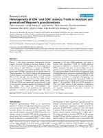
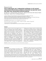
Figure 1 Hematopoiesis and retroviral infection:CD34
+
hematopoietic stem cells (HSCs) can undergo self-renewal as well as undergoing
maturation to give rise to common lymphoid progenitor (CLP) and common myeloid progenitor (CMP) cells, which serve as precursors to all
lymphoid and myeloid cells respectively. HSCs as well as other lineage specific progenitors are permissive for infection by a variety of murine
and human retroviruses including HIV-1 and HTLV-1.
Banerjee et al. Retrovirology 2010, 7:8
/>Page 3 of 17
progenitor cells, resulting in perturbation of hematopoi-
esis. Additionally, infected HPCs may differentiate
resulting in dissemination of pathogens into diverse ana-
tomical sites and to an effective spread of infection.
HP/HSCs can also serve as targets for cellular trans-
formation by specific viruses partly because of their
innate ability for self-renewal. CD34
+
HP/HSCs are sus-
ceptible to infection with a number of viruses including
HIV-1, HTLV-1, Hepatitis C virus, JC virus, Parvovirus,
Human Cytomegalovirus (HCMV), and the Human Her-
pesviruses (HHV): HHV-5, HHV-6, HHV-7, HHV-8
[3-5,42-52]. The concept that viruses can invade, infect
and establish a latent infection in the bone marrow was
firstdemonstratedinstudieswithHCMV.HCMV
infects a va riety of cell types, including hematopoietic
and stromal cells of the bone marrow, endothelial cells,
epithelial cells, fibroblasts,neuronalcells,andsmooth
muscle cells [3,53-57]. The bone marrow is a site of
HCMV latency [5,58], but the primary cellular reservoir
harboring latent virus within the bone marrow is con-
troversial. Latent viral genomes are detected in CD14
+
monocytes and CD33
+
myeloid precursor cells [59,60].
However HCMV can also infect CD34
+
hematopoietic
progenitor populations, and viral DNA sequences can be
detected in CD34
+
cells from healthy seropositive indivi-
duals [45,46,58,61], suggesting that a primitive cell
pop ulation serves as a renewable primary cellular reser-
voir for latent HCMV. The finding that HCMV DNA
sequences are present in CD34
+
cells of seropositive
individuals is consistent with the hypothesis that HCMV
resides in a HPC which subsequently gives rise to multi-
ple blood ce ll lineages. Recently, it has also been pro-
posedthatothervirusessuchasHTLV-1andKaposi’ s
Sarcoma Herpesvirus (KSHV) can also infect CD34
+
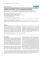
Figure 2 Generation of Leukemi c Stem Cells. Thre e hypotheses have been proposed that lead to t he development of leukemic stem cells
(LSC/CSC): (A) LSC/CSC might arise from either a hematopoietic stem cell (HSC), hematopoietic progenitor cell (HPC), committed lymphoid
progenitor (CLP) or committed myeloid progenitor (CMP), (B) from a multipotent HSC or HPC into LSC/CSC through a single transformation
event or, (C) from HSC or HPCs through a series of transformation events initiated by the generation of a pre-LSC/CSC.
Banerjee et al. Retrovirology 2010, 7:8
/>Page 4 of 17
HP/HSCs and establish latent infection within the BM
resident cells [52,62].
Apart from the establishment of latent infection
within the bone marrow (BM), suppression o f hemato-
poiesis has been documented to occur following infec-
tion of HPCs with HCMV, HHV-5, HHV-6, HIV-1, and
measles virus either as a result of direct infection of
HPCs or by indirect mechanisms such as disruption of
the cytokine milieu within the stem cell niche following
infection of bone marrow stromal cells. Our laboratory
has reported that HTLV-1 and KSHV infection of CD34
+
HP/HSCs suppresses hematopoiesis in vitro and that
viral infection can be disseminated into mature lym-
phoid cell lineages in vivo when monitored in huma-
nized SCID mice ( HU-SCID) [52,63,64]. HTLV-1 and
KSHV are both associated with hematological malignan-
cies and it is plausible that CSCs can be generated fol-
lowing infection of HP/HSCs with these viruses.
Multiple retroviruses establish latent infections in HP/
HSCs resulting in perturbation of hematopoiesis and
indu ction of viral pathogenesis [65-69]. Retroviral infec-
tions of HSCs can have adverse effects includ ing induc-
tion of cell-cycle arrest and increased susceptibility to
apoptosis, both would manifest in the suppression of
hematopoiesis. Additionally, mutations and transcrip-
tional deregulation of specific hematopoiesis-associated
genes can skew normal hematopoiesis toward s pecific
lineages and ha ve been demonstrated to occur following
infection o f HP/HSCs with HIV-1, HTLV-1 and Friend
Leukemia virus (FLV) [64,70,71].
Hematopoiesis occurs in the bone marrow microenvir-
onment, a complex system comprised of many cell types
including stromal cells that produce cytokines, growth
factors and adhesion molecules vital for the mainte-
nance, differentiation and maturation of HP/HSCs
[9,11]. Apart from infection of HSCs, retroviruses such
as HIV-1 and Moloney Murine leukemia virus (M-
MuLV) have been shown to infect bone marrow stromal
cells, compromising their ability to support hematopoi-
esis and resulting in multilineage hematopoietic failure
[72,73].
Retroviruses and Leukemogenesis: The “two-hit”
Hypothesis
Studies of retroviral induced leukemia have proven very
useful in understanding the multi-step processes asso-
ciated with leukemogenesis. Moreover, these models
have broadened our understanding of hematopoiesis and
hematopoietic stem cell biology. Retroviral infection
models such as FLV and M-MuLV, which induce leuke-
mic states in mice, have emerged as powerful tools to
study the molecular mechanisms associated with leuke-
mogenesis and the generation of LSC/CSCs [74-78].
The emerging concept from these murine models is that
acute leukemia arises from cooperation between two
distinctive mutagenic events; one interfering with differ-
entiation and another conferring a proliferative advan-
tage to HP/HSCs (Figure 2C) [79,80]. Studies from
Avian Erythroblastosis virus (AEV), FLV and M-MuLV-
induced leukemia/lymphoma models demonstrate that
leukemia/lymphoma development depends on: (1) a
mutation that impairs differentiation and blocks matura-
tion, (2) a mutation that promotes autonomous cell
growth, and (3) that neither mutational event is able to
induce acute leukemia by i tself [68,81]. Thus, these
models provide direct evidence for t he “two-hit model”
of leukemogenesis as has been proposed for some LSC/
CSC induced hematological malignancies, including
AML [79]. This concept is perhaps be st illustrated by
AEV infection in birds, FLV and MuLV infection in
mice and in HTLV-1 infection in humans (Figure 3).
During AEV infection, the oncogenic tyrosin e kinase
v-Erb-b, together with the aberrant nuclear transcription
factor v-Erb-A are transduced. The mutated thyroid hor-
mone receptor a, v-Erb-A, becomes unresponsive to the
ligand and actively recruits tyrosine kinases. These
kinases, such as stem-cell factor activated c-kit, cause
arrest of erythroid differentiation at the B FU-E/CFU-E
stage. Additionally, v-Erb-b encodes a mutated epider-
mal growth factor receptor that induces extensive ery-
throblast self-renewal [69,82]. These two virally-induced
events promote the abnormal proliferation of erythroid
progenitors and lead to the development of leukemia.
Another relevant leukemogenesis model induced by
retroviral infection of HPCs is acute erythroleukemia
caused by t he infection of mice with FLV [83-85]. FLV
has two distinct viral components, a re plication-compe-
tent Friend Murine Leukemia virus (F-MuLV) and a
replication defective pathogenic component known as
the Friend Spleen Focus Forming virus (F-SFFV)
[85-87]. The pathogenic component of FLV (F-SFFV)
can infect a variety of hematopoietic cells, though early
erythroid progenitors are the primary target for infection
[86,88]. F-SFFV can alter the normal growth and differ-
entiation profile of erythroid progenitor cells leading to
leukemog enesis. The induction of multistage erythroleu-
kemiabyFLVisalsoatwostageprocess:apre-leuke-
mic stage known as “ erythroid hyperplasia” and a
leukemic phase referred to as “erythroid cell transforma-
tion” (Figure 3B). The pre-leukemic stage is character-
ized by the infection and random i ntegration of F-SFFV
virus into erythroid precursor cells, forming an infected
stem cell population, followed by the expression of the
viral envelope glycoprotein gp55 on the cell surface.
gp55 subsequently binds to the cellular receptor of ery-
thropoietin (Epo-R) and interacts with the sf-Stk tyro-
sine kinase signaling pathway leading to a constitutive
activation signal for the p roliferation of undifferentiated
Banerjee et al. Retrovirology 2010, 7:8
/>Page 5 of 17
erythroid progenitor cells independent of erythropoietin
[83, 89,90]. Within the proliferating erythr oid progenitor
cell population are infected cells with randomly inte-
grated virus in the sp-1 locus, which leads to the activa-
tion and overexpression of PU.1.Originallyisolatedby
Moreau-Gache lin and co-workers as a gene targe ted for
recurrent insertions of SFFV, PU.1 has subsequently
been shown to be involved in terminal myeloid differen-
tiation, B and T-cell development, as well as maint e-
nance of normal erythropoiesis and HSC development
[91,92]. The over-expression of PU.1 in erythroid pre-
cursor cells as a result of SFFV integration leads to a
block in erythroid differentiation and, i n conjunction
with the inactivation of p53, clonal expansion of these
leukemic cells in susceptible mice [71,91]. Thus FLV-
mediated erythroleukemia is associated with two distinc-
tive p hases, “thepre-leukemicphase” mediated by gp55
binding to Epo-R and the “leukemic phase” mediated by
SFFV integration and the subsequent over-expression of
PU.1. T his demonstrates that both AEV and FLV infec-
tion follow the two-hit model of the cancer stem cell
hypothesis.
M-MuLV is a non-acute retrovirus that typically
induces a T-cell lymphoma after a latency period of 3-6
months [67]. The tumor cells typically have the pheno-
type of immature T-cells (CD4
-
/CD8
-
or CD4
+
/CD8
+
)
although some tumors show a more mature surface
phenotype (CD4
+
/CD8
-
or CD4
-
/CD8
+
) [72,93]. This led
to the hypo thesis that the virus might originally infect
an immature T-c ell or a HPC to form a ICSC/ILSC
which then continues to differentiate post-infection,
initially in the bone marrow and then in the thymus
[67,94]. Because T-lymphocytes develop in the thymus
from bone marrow-derived immature precursors (pro-
thymocytes), it has been proposed by several investiga-
tors that a bone marrow-thymus axis plays an important
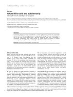
Figure 3 The “ Two-Hit” Model of Retrovirus-Induced Leukemogenesis. (A) HTLV-1 infection of CD34
+
hematopoietic progenitor and st em
cells (HP/HSCs) leads to the development of Adult T-cell leukemia/lymphoma (ATLL). (B) FLV infection of erythroid progenitors leads to
erythroleukemia. (C) M-MuLV infection of pro-T cells leads to T-cell lymphoma. The dotted line indicates the separation between the early and
late phase of infection.
Banerjee et al. Retrovirology 2010, 7:8
/>Page 6 of 17
role in the development of T-cell lymphoma by M-
MuLV [93,95-97]. Although the identity of the initial
target cell for M-MuLV infection is still unknown, a
two-st age leukemogenesis model for the development of
M-MuLV-induced leukemia has been proposed [67]. In
this model the animal is infected with MuLV on two
separate occasions ; the first infection occurs in the bone
marrow at the pre-leukemic (early) phase which leads to
hyperplasia and migration of infected lymphoid progeni-
tors into the thymus where a subsequent infecti on leads
to insertional activation of proto-oncogenes and out-
growth of the tumor resulting in the leukemic (late)
phase of infection (Figure 3C). Early infection of the
bone marrow is thought to be essential for establish-
ment of the pre-leukemic state and for development of
spleen hyperpl asia. The late phase splenic hyperplasia is
the result of a compensatory hematopoiesis due to
diminished normal hematopoiesis in the bone marrow
resulting from the establishment of the preleukemic
phase and plays an integral role in the establishment of
malignancy [98-100].
Bovine leukemia virus (BLV) is a deltaretrovirus
which causes leukemia/lymphoma in cattle [101]
(reviewed by [102,103]) and has been used as a model
of HTLV-1 infection and disease. While B-cells are the
primary target of BLV infection in contrast to the T-
cell tropism displayed by HTLV-1, BLV-infected B
lymphocytes are similarly arrested in G
0
/G
1
and pro-
tected from apoptosi s similar to properties demon-
strated following HTLV-1 infection HP/HSCs [64,104].
It has been suggested that CD5
+
B-cell progenitors are
more susceptible to BLV infection [105] and that t here
is a relationship between the B-cell phenotype and
BLV tropism [106]. More recently, the existence of a
pre-malignant clone has been proposed. This infected
progenitor is detectab le early after viral infectio n and
could contribute t o both genetic instability and clonal
expansion, both characteristics of cancer cells [107]. It
can therefore be speculated that the infection of pro-
genitor populati ons by BLV may result in the estab-
lishment of an ILSC/ICSC and subsequent
development o f leukemia.
Much of the current knowledge about leukemic
mechanisms originates with studies on AML. AML is
characterized by the uncontrolled self-renewal of
hematopoietic progenitors that fail to differentiate nor-
mally. Induction of AML is associated with a variety of
mutations that can be broadly classified into two dis-
tinctive categories; mutations in genes encoding tran-
scription factors involved with hematopoietic
regulation and mutations in genes encoding proteins
linked to survival and proliferation signaling pathways
[74-78,108,109]. Studiesinmicehaveshownthat
neither type of mutation alone is sufficient for the
induction of AML and that cooperative mutagenic
events are required for disease initiation [69,79]. The
leukemogenesis models of AEV, FLV and MuLV vali-
date this concept and underline the importance of
these models for the study of down-stream molecular
events associated with these mutagenic events. The
emergence of LSC/CSC as a result of these oncogenic
events would explain the complexity assoc iated with
hematological malignancy developme nt such as AML
and CML in humans.
Human T-cell Leukemia Virus Type-1 (HTLV-1) and
Adult T-cell Leukemia/Lymphoma (ATLL)
Human T-cell leukemia/lymphoma virus type-1
(HTLV-1) is the causative agent of Adult T-cell Leuke-
mia/Lymphoma (ATLL), an aggressive CD4
+
leukemia/
lymphoma [110]. ATLL is a rare T-cell malignancy
characterized by hypercalcemia, hepatomegaly, spleno-
megaly, lymphadenopathy, t he presence of a monoclo-
nal expansion of malignant CD4
+
CD25
+
T-cells that
evolve from a polyclonal population of HTLV-1
infected CD4
+
T-cells, and infiltration of lymphocytes
into the skin and liver. HTLV-1 causes ATLL in a
small percentage of infected individuals after a pro-
longed latency period of up to 20-40 years [111].
Although HTLV-1 can replicate by reverse transcrip-
tion during the initial phase of infection, the integrated
provirus is effectively replicated during proliferation of
infected cells [112]. Typically, HTLV-1 infected cells
can persist for d ecades in patients, a nd the infected
cell population transits from a polyclonal phase into a
monoclonal expansion during development and pro-
gression to ATLL.
There are four ATLL subtypes; acute, lymphomatous,
chronic, and smoldering. The first two subtypes are
associated with a rapidly progressing clinical course
with a mean survival time of 5-6 months. Smoldering
and chronic ATLL have a more indolent course and
may represent transitional states towards acute ATLL.
Clinical features of ATLL include leukemic cells with
multi-lobulated nuclei called ‘flower cells’ which infil-
trate into various tissues including the skin and the
liver, abnormally high blood calcium levels, and con-
current opportunistic infections in patients [113,114].
Although considerable progress has been made in
understanding ATLL biology, the exact sequence of
events occurring during the initial stages of malignancy,
including the types of cells infected with HTLV-1,
remain unclear. The primary target cells for HTLV-1
infection may not only influence HTLV-1 pathogenesis,
but the sequestration of these cells in anatomical sites
such as the bone marrow may also allow the virus to
effectively evade the primary immune response against
infection.
Banerjee et al. Retrovirology 2010, 7:8
/>Page 7 of 17
The Role of HSCs in HTLV-1 Infection and
Pathogenesis
It has been previously reported by our laboratory and
other investigators that HTLV-1 can infect human HP/
HSCs [65,115]. It has been hypothesized that HTLV-1
can specifically induce a late nt infection in CD34
+
HP/
HSCs an d can initiate preleukemic events in these pro-
genitor cells [62]. These cells could potentially provide a
durable reservoir for latent virus in infected individuals.
It has been speculated that HTLV-1 infection of CD34
+
HPCs may result in the generation of an ILSC/ICSC
and may also induce perturbation of normal hematopoi-
esis, ultimately resulting in the outgrowth of malignant
clones and the development of ATLL.
The development of ATLL correlates with neonatal or
perinatal transmission of HTLV-1. HTLV-1 carries no
cellular proto-oncogenes, and the oncogenic potential of
the virus is linked to Tax1, a 40 kDa protein that func-
tions as a trans-activator of viral gene expression and as a
key component of HTLV-1-mediated transformation
[116,117]. Tax1 is a relatively promiscuous transactivator
of both viral and cellular gene transcription and has been
closely linked to the initiation of leukemogenesis. Apart
from regulating viral gene expression through the 5’ long
terminal repeat (LTR), Tax1 can modulate the expression
of a large variety of cellular genes and proteins including
those encoding cytokines, apoptosis inhibitors, cell cycle
regulators, transcriptio n factors, and intracellular signal-
ing molec ules [116 ,118-120]. Tax1 usually indu ces cellu-
lar gene e xpression by the a ctivation of transcrip tion
factorssuchasNF-B and cyclic AMP response ele-
ment-binding protein/activating transcription factor
(CREB/ATF) [121]. Tax1 has also bee n shown to trans-
repress transcription of c ertain cellular genes, including
bax [122], human b-polymerase [119], cyclin A [123], lck
[124], MyoD [125], INK4 [126], and p53 [127].
Transgenic mouse models of Tax1 expression have
resulted in the generation of murine malignancies,
including a mature T-cell malignancy, underlying the
critical role of Tax1 in the manifestation of T-cell leuke-
mia [128,129]. Transgenic mice constructed to target
expression of Tax1 to both immature and mature thy-
mocytes using a Lck (Leukocyte-specific protein tyrosine
kinase) promoter reproducibly develop immature and
mature T-cell leukemia/lym phomas with immunological
and pathological similarities to human ATLL [128,129].
In a recent study by Yamazaki et al., splenic lymphoma-
tous cells were harvested and purified from Tax-trans-
genic mice using a combination of immunological and
physiological markers for CSCs a nd were injected into
NOD/SCID mice using a limiting-dilution assay [6].
Injection with as few as 1 × 10
2
CSCs was sufficient to
recapitulate the original lymphoma and reestablish CSCs
in recipient NOD/SCID mice implicating a role for
LSC/CSC in the establishment of ATLL.
LSC/CSCs have the ability to self-renew, are seques-
tered in the bone marrow microenvironment and are
relatively resistant to conventional chemotherapeutic
treatment regimens. The recent focus and characteriza-
tion of the role of LSC/CSC in the induction of AML has
generated a paradigm for LSC/CSC-generated cancers
and has resulted in a re-evaluation of therapeutic strate-
gies for successful targeting and elimination of leukemic
cells in patients [31]. Although the Tax-transgenic mouse
model is not a complete representation of ATLL manifes-
tation in humans, this finding is intriguing particularly
since other investigators have suggested that HTLV-1
infection in the human bone marrow and in human HP/
HSCs specifically, may facilitate the early events initiating
ATLL development [62]. Since a limited number of
ATLL cases display phenotypes indicative of immature
hematopoietic cells, HTLV-1 infection and transforma-
tion of HP/HSCs in humans may result in the generation
of virally-infected ATLL LSC/CSC [130]. Lymphoma
cells and LSC/CSC from Tax-transgenic mice were also
demonstrated to sequester in the osteoblastic and vascu-
lar niches of the bone marrow in transplanted NOD/
SCID mice. It is interesting to speculate that if ATLL
arises from a LSC/CSC, then the sequestration of HTLV-
1-infected HP/HSCs in the bone marrow microenviron-
mentmaybeacontributingfactorintheresistanceof
this leukemia to treatment with conventional che-
motherapies. It remains to be determined if the recent
results from the Tax-transgenic model are truly illustra-
tive of t he human disease. However, the Tax-transgenic
murine model does provide several interest ing clues into
the mechanisms of HTLV-1 pathogenesis, and this may
eventually group ATLL along with other hematological
malignancies that have a LSC/CSC origin.
Recapitulating ATLL in ‘humanized’ SCID (HU-SCID)
mice has been challenging, and previous attempts to
directly infect mature human T -cells in the human thy-
mus-liver conjoint organ in HU-SCID mice with HTLV -
1 failed to induce a malignancy [65]. Recent data from
our laboratory demonstrates that ex vivo infection of
CD34
+
HP/HSCs with HTLV-1 reproducibly and consis-
tently results in development of a CD4
+
T-cell lym-
phoma in H U-SCID mice [131]. Clearly, H TLV-1
infection of HP/HSCs plays a pivotal role in the initia-
tion and accelerated progression of malignancy during
the course of HTLV-1 pathogenesis.
HTLV-1 Infected CD34
+
HP/HSCs: Notch, PU.1 and
micro-RNA Deregulation
Manifestation of ATLL in patients generally occurs dec-
ades after infection, suggesting that HTLV-1 latently
Banerjee et al. Retrovirology 2010, 7:8
/>Page 8 of 17
infects bone marrow stem cells that are sequestered
from immunological surveillance. It is conceivable that
the initiation of l eukemogenesisinHSCsinvolvesthe
generation of a CSC/LSC that will eventually manifest
into the monoclonal ATLL malignancy. Several path-
ways that regulate HSC self-renewal are also associated
with human cancers, including hematopoietic malignan-
cies such as T-cell leukemia [132] and T-ALL [133,134].
It has previously been shown that disruption of normal
HSC self-renewal signaling pathways can induce hema-
topoietic neoplasms [132,135]. Two main reasons sug-
gest that HSCs can serve as target cells for virally-
induc ed leukemia/lymphoma. First, stem cells have con-
stitutively activated self-renewal pathways, requiring
maintenance of activation in contrast to the de novo
activation required in a more di fferentiated cell. Second,
self-renewal provides a persistent target for repeated
viral infection and/or continual replication of integrated
proviral DNA. HTLV-1 infection of CD34
+
HP/HSCs
deregulates normal HSC self-renewal pathways through
a variety of potential mechanisms suggesting that
HTLV-1 infection may generate ILSC/ICSC.
The Notch signaling pathway regulates self-renewal
and differentiation of HSCs and h as been implicated as
a key regulator of human T and B-cell derived lympho-
mas [135,136]. Studies using adult bone m arrow trans-
plantation into NOD/SCID mice demonstrate that
inactivation of Notch1 arrests T-cell development at the
earliest precursor stage [134] and promotes B-cell devel-
opment in the thymus [137]. The modulation of Notch
levels in LSC/CSC derived from Tax-transgenic mice
suggests that Notch may contribute in the development
of ATLL similar to its role in other T-cell malignancies
such as T-ALL [133,134].
The sp1 gene encodes for the transcription factor
PU.1, whic h is a member of the ets family of transcrip-
tion factors, is expressed at various levels in all hemato-
poietic cells. PU.1 expression has been shown to play an
important role in the regulation of hematopoiesis
[138,139]. Specifically, expressi on of PU.1 is tightly con-
trolled in HSCs and regulates the fate of cells di fferen-
tiating into lymphocyte, macrophage or granulocyte
lineages [140,141]. Deregulation of PU.1 expression has
been linked to the developm ent of hematopoietic malig-
nancies including the transformation of myeloid c ells
[92]. During hematopoiesis, PU.1 is required for h ema-
topoietic development along both the lymphoid and
myeloid lineages, but is down-regulated during erythro-
poiesis. In AML patients, mutations in Flt3 decrease
PU.1 expression and block differentiation [141] while
mutations in PU.1 impair development within bo th
myeloid and lymphoid lineages [142]. Knockout mouse
studies have shown that perturbation of PU.1 expression
results not only in the loss of B-cells and macrophage
developm ent, but also delays T lymphopo iesis [143,144].
Additionally, PU.1 supports the sel f-renewal of HSCs by
regulating the multilineage commitment of multipotent
progenitors, thereby maintaining a pool o f pluripotent
HSCs within the bone marrow [145,146].
Notably the reduc tion in PU.1 expression in bone
marrow derived CD34
+
HP/HSCs has been shown to
induce an intermediate stage o f poorly differentiated
pre-leukemic cells which, with the accumulation of addi-
tional genetic mutations, results in an aggressive form of
AML [147]. The HTLV-I accessory protein p30 has also
been shown to interact with the ets domain of PU.1
resulting in impairment of the DNA binding activity of
PU.1 and subsequent inhibition of PU.1-dependent tran-
scription [148]. HTLV-1 p30-mediated alteration of
PU.1 expression may be a contributing factor in the
deregulation of hematopoiesis due to HTLV-1 infection
of HSCs and may contribute to the establishment of
ILSC/ICSC.
Bmi-1 (B-lymphoma Mo-MuLV i nsertion region),
which belongs to the polycomb group of epigenetic
chromatin modifiers, was originally identified as an
oncogene [149]. Bmi-1 is required for the maintenance
of HSC sel f-renewal in mice and is also involved in reg-
ulation of g enes controlling cell prolife ration, survival
and differentiation of HSCs [149-152]. Deficiency of
Bmi-1 results in a progressive loss of HS Cs and in
defects in the stem cell compartment of the nervous sys-
tem [153]. Bmi-1 expression is e levated in HP/HSCs in
contrast to differentiated hematopoietic cells, and both
self-renewal as well as the in vivo repopulation potential
of HSCs is dependent on Bmi-1 [152,154-156]. It has
been reported that Bmi-1 is required for the activation
and survival of pre-T-cells and during transition from
DN to DP T-cells [157]. Bmi-1 is required for the prolif-
eration of LSC/CSCs, and the deregulation of Bmi-1 is
linked to human cancers [155,158]. Notably LSC/CSCs
from Tax1-transgenic mice show a robust down-regu la-
tion of Bmi-1, providing a mechanistic link between
HTLV-1 infection and deregulation of hematopoiesis.
Micro-RNAs (miRNAs) are a class of non-coding
RNAs, 20-25 nucleotides long, that play an important
role in both normal and malignant hematopoiesis,
including self-renewal, differentiat ion and line age speci-
ficity of HPCs [159-163] (reviewed in [164]). Loss of
miRNAs has also been reported in a variety of cancers
indicating that alteration of miRNA levels might play a
critical role in tumorigen esis [165-167]. miR-150 is pre-
ferentially expressed in the megakaryocytic lineage and
has been recently shown to drive the differentiation of
megakaryocyte-erythrocyte precursors toward megakar-
yocyte development at the expense of erythroid differen-
tiation [168]. Over-expression of miR-221 and miR-222
interferes with the kit receptor and blocks engraftment
Banerjee et al. Retrovirology 2010, 7:8
/>Page 9 of 17
of HSCs in humanized mice [169]. Over-expression of
miRNA-181a has been linked to the development of
AML and CLL [170,171]. These studies highlight the
role of miRNA in regulating normal hematopoiesis and
suggest that miRNA expression may modulate the mani-
festation of hematopoietic malignancies.
Retroviruses such as HIV-1 and HTLV-1 have been
rece ntly shown to target miRNAs for modulation of key
cellular pathways including cell-cycle regulation and
immune responses [172-174]. Specifically, miRNAs that
are involved in the regulation of cell proliferation, apop-
tosis and immune responses are up-regulated in ATL
cells [175,176]. Bellon et al. recently demonstrate d that
miRNAs involved in normal hematopoiesis and immune
responses are also profoundly deregulated in ATLL cells
indicating a possible l ink between modulation of cellular
miRNA expression and dere gulation of hematopoiesis
by HTLV-1 [177]. Specifically, signific ant changes in the
expression of miR-223 and miR-150 in ATLL patient
samples were identified. miR-223 controls the terminal
differentiation pathway of HSCs and is upregulated fol-
lowing differentiation into myeloid and lymphoid pro-
genitors [162]. The differential expression of miR-150
regulates lineage deci sion between T and B-cells. Ecto-
pic expression of miR-150 in lymphoid progenitors
enhances T lymphopoiesis with respect to B lymphopoi-
esis [178]. The deregulation of cellular miRNAs might
contribute to the transformation process resulting in the
development of ATLL.
HTLV-1 infection in HP/HSC could result in aberrant
miRNA expression ultimately predisposing HSC devel-
opment toward T l ymphopoiesis. Since expression o f
these miRNAs (223 and 150) are restricted to HP/HSCs,
CLPs and CMPs, patient derived primary ATLL cells
may originated from an infected HPC population in
contrast to in vitro-established HTLV-1 infected CD4
+
T cell lines. This supports the hypothesis that ATLL
cells are derived from HTLV-1 infected CD34
+
HP/
HSCs rather than virally transformed mature T-cells
[64,128,129]. Upon differentiation of an HTLV-1
infected CD34
+
HPC, the alteration of miRNA levels
may favor T-cell differentia tion, as recently demon-
strated by the exclusive development of CD4
+
mature
T-cell lymphomas in HU-S CID mice reconstituted with
CD34
+
HPCs infected ex vivo with HTLV-1[131].
Tax1 and Cell Cycle Regulation in HP/HSCs
HTLV-1 Tax1 has been shown to induce G
0
/G
1
cell
cycle arrest leading to quiescence in both cultured
mammalian cell lines and primary human CD34
+
HPCs
[116,179,180]. Likewise, the expression of Tax1 in Sac-
charomyces cerevisiae leads to growth arrest and loss of
cell viability [181,182]. Intriguingly, in addition to
increasing the levels of cyclins and CDKs, Tax1 also
increases the levels of CDK inhibitors p 16
Ink4
,p21
cip1/
waf1
(p21) and p27
kip
(p27)ininfectedcells
[63,64,179,183,184]. Over-expression of p21 inhibits two
critical checkpoints in the mammalian cell cycle, namely
G
1
/S and S/G
2
, through p53-independent and depen-
dent pathways [185]. Moreover, p21 and p27 are the key
contributors in th e cell-cycle regulation of CD34
+
HPCs
[186-188]. Tax1 has also been shown to suppress
human mitotic checkpoint protein MAD1 resulting in
deregulation of the G2/M phase of the cell cycle result-
ing in aneuploidy [189]
Cell cycle progression is highly regulated in CD34
+
HPCs with a majority of CD34
+
HPCs residing in quies-
cence and demonstrating a unique expression pattern of
CDKs, cyclins, and CDK inhibitors. The CDK inhibitors
p21 and p27, in particular, have b een shown to be key
contributors in restricting cell cycle entry from G
0
and
maintaining quiescence in CD34
+
HPCs [186-188]. We
have previously shown that during HTLV-1 infection,
induction of G
0
/G
1
cell cycle arrest and suppression of
multilineage hematopoiesis in HPCs is attributed to the
concomitant activation of p21 and p27 in these cells by
Tax1 [63,64,180]. Although Tax1 usually induces cellu-
lar gene expression by activation of transcription factors
such as NF- B, CREB/ATF and Akt [190], it has
recently been suggested that Tax1 deregulation of p21
and p 27 may also be mediated independently of NF-B
activation [191] and p53 [184]. Moreover, the reported
absence of NF-B activity in CD34
+
CD38
-
HSCs [192]
suggests that HP/HSCs provide a unique microenviron-
ment for HTLV-1 infection which stands in stark con-
trast to the cellular environment provided by mature
CD4
+
T lymphocytes. It may be inferred that Tax1-
mediated cell cycle deregulation is cell-type specific,
inducing cell cycle arrest in HPCs while concurrently
maintaining the ability to activate cell proliferation in
mature CD4
+
T-lymphocytes.
Survivin, originally identified as a member o f the inhi-
bitor of apoptosis protein family, has recently been
implicated in regulating hematopoiesis, cell cycle control
and transformation [193-196]. Survivin is expressed in
normal adult bone m arrow cells and in CD34
+
HPCs
where it regulates proliferation and/or survival, and sur-
vivin expression is upregulated by hematopoietic growth
factors [197]. Notably, survivin has been shown to be a
key mediator of early cell cycle entry in CD34
+
HPCs
and regulates progenitor cell proliferation through p21-
dependent and independent pathways [198], in addition
to regulating apoptosis of HSCs [199]. This implicates
survivin as an integral cellular factor, regulating multiple
aspects of hematopoiesis. HTLV-1 mediated suppression
of hematopoiesis in CD34
+
HPCs is regulated, in part,
bydown-regulationofsurvivinexpressioninthesecells
by Tax1 [64]. Notably, CD34
+
CD38
-
HSCs demonstrate
Banerjee et al. Retrovirology 2010, 7:8
/>Page 10 of 17
elevated sensitivity to cell-cycle arrest following HTLV-1
infection in comparison to more mature CD34
+
CD38
+
HPCs, suggesting that HTLV-1 may target stem cells to
facilitate a latent infection in vivo by inducing cell cycle
arrest to induce cellular quiescence (Figure 4).
HTLV-1 Interaction with CD34
+
HP/HSCs:
Emerging Views
Emerging evidence has led to a new view of HTLV-1
mediated leukemogenesis that correlates neonat al trans-
mission o f HTLV-1 with viral infection targeting of HP/
HSCs and immature human thymocytes [63,65,115].
This hypothesis challenges the current view that mature
differentiated CD4
+
T-cells are the exclusive target for
HTLV-1 infection and for the initiation of ATLL.
Analysis of bone marrow samples from pediatric HTLV-
1 infections would confirm the hypothesis that HTLV-1
infection enters and is sequestered in the CD34
+
HP/
HSCs in the bone marrow. It is noteworthy that pre-
vious reports have demonstrated HTLV-1 transmission
following a bone marrow transplantation procedure
from a HTLV- 1 infected donor [200]. HTLV-1 infection
of HP/HSCs can result in skewing of hematopoiesis
toward distinct cellular lineages and outgrowth of malig-
nant clones leading to ATLL. HP/HSCs may be critic al
target cell for HTLV-1 infection and for establishment
of latency in vivo providing a reservoir of inf ecte d cells
which progresses, after the accumulation of additional
molecular events, to the develo pment of ATLL [62,65].
This hypothesis is supported by recent identification of
Figure 4 The Role of HTLV-1 Inf ection of HSCs: Potential Mechanisms for Generation of an Infectious Leukemic Stem Cell (ILSC/ICSC).
HTLV-1 infection and subsequent Tax1 expression can lead to either cell cycle arrest or generation of pre-leukemic stem cells (pre-LSC/CSC)
from infected CD34
+
hematopoietic progenitor and stem cells (HP/HSCs).
Banerjee et al. Retrovirology 2010, 7:8
/>Page 11 of 17
a rare CSC population in Tax-transgenic mice [6,128].
Notably, our laboratory has detected a high incidence of
HTLV-1 proviral sequences in CD34
+
HP/HSCs from
HTLV-1-infected patient peripheral blood lymphocyte
samples, suggesting that HP/HSCs are a natural cellular
reservoir for HTLV-1 infection [131]. The down-regula-
tion of key hematopoietic genes, including Notch1 and
Bmi-1, in CSCs from Tax-transgenic mice indicates that
the CSC potentially emerges from primitive HPCs or
immature thymocytes and highlights the role of Tax1
expression in the induction of lymphoproliferative dis-
ease (Figure 4). The role of HTLV-1 Tax in HP/HSCs
includes cell cycle deregulation and perturbation of
hematopoiesis, as we have previously reported [63,180].
Clearly many parameters defining how HTLV-1 and its
associated viral genes (including Tax1, p30 and HBZ
[201]), may contribute to the development of a ILSC/
ICSC in ATLL have yet to be established. The role of
the HTLV-1 antisens e enco ded protein HBZ is of parti-
cular interest as it is consistently expressed in all ATLL
patient cells examined in contrast to Tax1 which is
usually silenced in ATLL cells [202,203]. Emerging in
vivo murine models, particularly the HU-SCID mouse
models, will help characterize the pathobiology of
HTLV-1 infection and establish the existence of ILSC/
ICSC. Moreover, these models will allow for the identifi-
cation of events resulting in leukemia-initiation and pro-
gression and for the pre-clinical therapeutic evaluation
for this fatal malignancy which currently lacks effective
treatment regimens.
Perspectives
Retroviral infection of HP/HSCs in the bone marrow
clearly provides a reservoir for infected cells and results
in dramatically altered patterns of hematopoiesis. Deter-
mining and identifying whether retroviruses, such as
HTLV-1, exploit this cellular trait to establish an ILSC
would present a new paradigm in the pathobiology of
HTLV-1 infection and would allow novel targeted treat-
ments to be designed in order to intervene and treat ret-
roviral mediated neoplasms.
Abbreviations
LSC/CSC: leukemic stem cell/cancer stem cell; HP/HSC: hematopoietic
progenitor/stem cell; HTLV-1: human T cell lymphotropic virus type 1; ILSC/
ICSC: infectious leukemic/cancer stem cell; CLP: common lymphoid
progenitor; CMP: common myeloid progenitor; AML: acute myeloid
leukemia; ALL: acute lymphoblastic leukemia; CML: chronic myelogenous
leukemia; HU-SCID mouse: humanized severe combined immunodeficient
mouse; HIV-1: human immunodeficiency virus type-1; FLV: Friend leukemia
virus; M-MuLV: Moloney murine leukemia virus; AEV: Avian erythroblastosis
virus; BLV: Bovine leukemia virus; ATLL: Adult T cell leukemia/lymph oma;
CREB/ATF: cyclic AMP response element-binding protein/activating
transcription factor; Bmi-1: B-lymphoma Mo-MuLV insertion region; DN:
double negative; DP: double positive; miRNAs: micro-RNAs.
Acknowledgements
This work was supported by grants from the US National Institutes of Health
(CA124595) and by the Empire State Stem Cell Fund through New York
State Department of Health Contract (NYSTEM #C023059 and #N08G-127) to
G.F. Opinions expressed here are solely those of the author and do not
necessarily reflect those of the Empire State Stem Cell Board, the New York
State Department of Health, or the State of New York.
Author details
1
Department of Microbiology and Immunology, SUNY Upstate Medical
University, Syracuse, NY, 13210, USA.
2
Center for Humanized SCID Mice and
Stem Cell Processing Laboratory, SUNY Upstate Medical University, Syracuse,
NY, 13210, USA.
Authors’ contributions
PB and LC were responsible for drafting and revising the manuscript as well
as organizing the content. ES created Figures 1, 2, 3 and 4 and their legends
and proofread the final version of the manuscript for content and
consistency. GF assisted in all aspects of writing the manuscript from
revisions to final approval of the version to be published. All authors read
and approved the final manuscript.
Competing interests
The authors declare that they have no competing interests.
Received: 1 October 2009
Accepted: 4 February 2010 Published: 4 February 2010
References
1. Kondo M, Wagers AJ, Manz MG, Prohaska SS, Scherer DC, Beilhack GF,
Shizuru JA, Weissman IL: Biology of Hematopoietic Stem Cells and
Progenitors: Implications for Clinical Application. Annual Review of
Immunology 2003, 21:759-806.
2. Reya T, Morrison SJ, Clarke MF, Weissman IL: Stem cells, cancer, and
cancer stem cells. Nature 2001, 414:105-111.
3. Maciejewski JP, Bruening EE, Donahue RE, Mocarski ES, Young NS, St
Jeor SC: Infection of hematopoietic progenitor cells by human
cytomegalovirus. Blood 1992, 80:170-178.
4. Manchester M, Smith KA, Eto DS, Perkin HB, Torbett BE: Targeting and
hematopoietic suppression of human CD34+ cells by measles virus. J
Virol 2002, 76:6636-6642.
5. Goodrum F, Jordan CT, Terhune SS, High K, Shenk T: Differential outcomes
of human cytomegalovirus infection in primitive hematopoietic cell
subpopulations. Blood 2004, 104:687-695.
6. Yamazaki J, Mizukami T, Takizawa K, Kuramitsu M, Momose H, Masumi A,
Ami Y, Hasegawa H, Hall WW, Tsujimoto H, Hamaguchi I, Yamaguchi K:
Identification of cancer stem cells in a Tax-transgenic (Tax-Tg) mouse
model of adult T- cell leukemia/lymphoma (ATL). Blood 2009,
114(13):2709-20.
7. Jordan CT, Guzman ML, Noble M: Cancer Stem Cells. The New England
Journal of Medicine 2006, 355:1253-1261.
8. Huang S, Terstappen LW: Lymphoid and myeloid differentiation of single
human CD34+, HLA-DR+, CD38- hematopoietic stem cells. Blood 1994,
83:1515-1526.
9. Allen TD, Dexter TM: The essential cells of the hemopoietic
microenvironment. Exp Hematol 1984, 12:517-521.
10. Laiosa CV, Stadtfeld M, Graf T: Determinants of lymphoid-myeloid lineage
diversification. Annu Rev Immunol 2006, 24:705-738.
11. Ogawa M: Differentiation and proliferation of hematopoietic stem cells.
Blood 1993, 81:2844-2853.
12. Galy A, Travis M, Cen D, Chen B: Human T, B:natural killer, and dendritic
cells arise from a common bone marrow progenitor cell subset.
Immunity 1995, 3:459-473.
13. Ikawa T, Kawamoto H, Fujimoto S, Katsura Y: Commitment of common T/
Natural killer (NK) progenitors to unipotent T and NK progenitors in the
murine fetal thymus revealed by a single progenitor assay. J Exp Med
1999, 190:1617-1626.
14. Akashi K, Traver D, Miyamoto T, Weissman IL: A clonogenic common
myeloid progenitor that gives rise to all myeloid lineages. Nature 2000,
404:193-197.
Banerjee et al. Retrovirology 2010, 7:8
/>Page 12 of 17
15. Weissman IL, Shizuru JA: The origins of the identification and isolation of
hematopoietic stem cells, and their capability to induce donor-specific
transplantation tolerance and treat autoimmune diseases. Blood 2008,
112:3543-3553.
16. Metcalf D: The molecular control of cell division, differentiation
commitment and maturation in haemopoietic cells. Nature 1989,
339:27-30.
17. Chao MP, Seita J, Weissman IL: Establishment of a Normal Hematopoietic
and Leukemia Stem Cell Hierarchy. Cold Spring Harb Symp Quant Biol
2008.
18. Wright DE, Wagers AJ, Gulati AP, Johnson FL, Weissman IL: Physiological
migration of hematopoietic stem and progenitor cells. Science 2001,
294:1933-1936.
19. Sell S, Pierce GB: Maturation arrest of stem cell differentiation is a
common pathway for the cellular origin of teratocarcinomas and
epithelial cancers. Lab Invest 1994, 70:6-22.
20. Sawyers CL, Denny CT, Witte ON: Leukemia and the disruption of normal
hematopoiesis. Cell 1991, 64:337-350.
21. Ellisen LW, Carlesso N, Cheng T, Scadden DT, Haber DA: The Wilms tumor
suppressor WT1 directs stage-specific quiescence and differentiation of
human hematopoietic progenitor cells. Embo J 2001, 20:1897-1909.
22. Fialkow PJ, Singer JW, Adamson JW, Vaidya K, Dow LW, Ochs J, Moohr JW:
Acute nonlymphocytic leukemia: heterogeneity of stem cell origin. Blood
1981, 57:1068-1073.
23. Wang JC, Dick JE: Cancer stem cells: lessons from leukemia. Trends Cell
Biol 2005, 15:494-501.
24. Renneville A, Roumier C, Biggio V, Nibourel O, Boissel N, Fenaux P,
Preudhomme C: Cooperating gene mutations in acute myeloid leukemia:
a review of the literature. Leukemia 2008, 22:915-931.
25. Lapidot T, Sirard C, Vormoor J, Murdoch B, Hoang T, Caceres-Cortes J,
Minden M, Paterson B, Caligiuri MA, Dick JE: A cell initiating human acute
myeloid leukaemia after transplantation into SCID mice. Nature 1994,
367:645-648.
26. Zhu AJ, Watt FM: beta-catenin signalling modulates proliferative
potential of human epidermal keratinocytes independently of
intercellular adhesion. Development 1999, 126:2285-2298.
27. Chan EF, Gat U, McNiff JM, Fuchs E: A common human skin tumour is
caused by activating mutations in beta-catenin. Nat Genet 1999,
21:410-413.
28. Jamieson CH, Ailles LE, Dylla SJ, Muijtjens M, Jones C, Zehnder JL, Gotlib J,
Li K, Manz MG, Keating A, Sawyers CL, Weissman IL: Granulocyte-
macrophage progenitors as candidate leukemic stem cells in blast-crisis
CML. N Engl J Med 2004, 351:657-667.
29. Sirard C, Lapidot T, Vormoor J, Cashman JD, Doedens M, Murdoch B,
Jamal N, Messner H, Addey L, Minden M, Laraya P, Keating A, Eaves A,
Lansdorp PM, Eaves CJ, Dick JE: Normal and leukemic SCID-repopulating
cells (SRC) coexist in the bone marrow and peripheral blood from CML
patients in chronic phase, whereas leukemic SRC are detected in blast
crisis. Blood 1996, 87:1539-1548.
30. Bjerkvig R, Tysnes BB, Aboody KS, Najbauer J, Terzis AJ: Opinion: the origin
of the cancer stem cell: current controversies and new insights. Nat Rev
Cancer 2005, 5:899-904.
31. Bonnet D, Dick JE: Human acute myeloid leukemia is organized as a
hierarchy that originates from a primitive hematopoietic cell. Nat Med
1997, 3:730-737.
32. Custer RP, Basma GC, Bosma MJ: Severe Combined Immunodeficiency
(SCID) in the Mouse: Pathology, Reconstitution, Neoplasms. American
Journal of Pathology 1985, 120:464-477.
33. Deshpande AJ, Buske C: Lymphoid Progenitors as Candidate Cancer Stem
Cells in AML. Cell Cycle 2007, 6:543-545.
34. Hope KJ, Jin L, Dick JE: Human Acute Myeloid Leukemia Stem Cells.
Archives of Medical Research 2003, 34:507-514.
35. Chan W-I, Huntly BJP: Leukemia Stem Cells in Acute Myeloid Leukemia.
Seminars in Oncology 2008, 35:326-335.
36. Tan BT, Park CY, Ailles LE, Weissman IL: The cancer stem cell hypothesis: a
work in progress. Lab Invest 2006, 86:1203-1207.
37. McCulloch EA: Stem cells in normal and leukemic hemopoiesis (Henry
Stratton Lecture, 1982). Blood 1983, 62:1-13.
38. Kosmider O, Moreau-Gachelin F: From mice to human: the “two-hit
model” of leukemogenesis. Cell Cycle 2006, 5:569-570.
39. Keller G: Hematopoietic stem cells. Curr Opin Immunol 1992, 4:133-139.
40. Till JE, McCulloch EA: Hemopoietic stem cell differentiation. Biochim
Biophys Acta 1980, 605:431-459.
41. Kolb-Maurer A, Goebel W: Susceptibility of hematopoietic stem cells to
pathogens: role in virus/bacteria tropism and pathogenesis. FEMS
Microbiol Lett 2003, 226:203-207.
42. Segovia JC, Guenechea G, Gallego JM, Almendral JM, Bueren JA: Parvovirus
infection suppresses long-term repopulating hematopoietic stem cells. J
Virol 2003, 77
:8495-8503.
43. Sansonno D, Lotesoriere C, Cornacchiulo V, Fanelli M, Gatti P, Iodice G,
Racanelli V, Dammacco F: Hepatitis C virus infection involves CD34(+)
hematopoietic progenitor cells in hepatitis C virus chronic carriers. Blood
1998, 92:3328-3337.
44. Monaco MC, Atwood WJ, Gravell M, Tornatore CS, Major EO: JC virus
infection of hematopoietic progenitor cells, primary B lymphocytes, and
tonsillar stromal cells: implications for viral latency. J Virol 1996,
70:7004-7012.
45. Movassagh M, Gozlan J, Senechal B, Baillou C, Petit JC, Lemoine FM: Direct
infection of CD34+ progenitor cells by human cytomegalovirus:
evidence for inhibition of hematopoiesis and viral replication. Blood
1996, 88:1277-1283.
46. Sindre H, Tjoonnfjord GE, Rollag H, Ranneberg-Nilsen T, Veiby OP, Beck S,
Degre M, Hestdal K: Human cytomegalovirus suppression of and latency
in early hematopoietic progenitor cells. Blood 1996, 88:4526-4533.
47. Zhuravskaya T, Maciejewski JP, Netski DM, Bruening E, Mackintosh FR, St
Jeor S: Spread of human cytomegalovirus (HCMV) after infection of
human hematopoietic progenitor cells: model of HCMV latency. Blood
1997, 90:2482-2491.
48. Khaiboullina SF, Maciejewski JP, Crapnell K, Spallone PA, Dean Stock A,
Pari GS, Zanjani ED, Jeor SS: Human cytomegalovirus persists in myeloid
progenitors and is passed to the myeloid progeny in a latent form. Br J
Haematol 2004, 126:410-417.
49. Isomura H, Yamada M, Yoshida M, Tanaka H, Kitamura T, Oda M, Nii S,
Seino Y: Suppressive effects of human herpesvirus 6 on in vitro colony
formation of hematopoietic progenitor cells. J Med Virol 1997, 52:406-412.
50. Isomura H, Yoshida M, Namba H, Fujiwara N, Ohuchi R, Uno F, Oda M,
Seino Y, Yamada M: Suppressive effects of human herpesvirus-6 on
thrombopoietin-inducible megakaryocytic colony formation in vitro. J
Gen Virol 2000, 81:663-673.
51. Mirandola P, Secchiero P, Pierpaoli S, Visani G, Zamai L, Vitale M, Capitani S,
Zauli G: Blood 2000, 96:126-131.
52. Wu W, Vieira J, Fiore N, Banerjee P, Sieburg M, Rochford R, Harrington W Jr,
Feuer G: KSHV/HHV-8 infection of human hematopoietic progenitor
(CD34+) cells: persistence of infection during hematopoiesis in vitro and
in vivo. Blood 2006, 108:141-151.
53. Sinzger C, Grefte A, Plachter B, Gouw AS, The TH, Jahn G: Fibroblasts,
epithelial cells, endothelial cells and smooth muscle cells are major
targets of human cytomegalovirus infection in lung and gastrointestinal
tissues. J Gen Virol 1995, 76(Pt 4):741-750.
54. Soderberg C, Larsson S, Bergstedt-Lindqvist S, Moller E: Definition of a
subset of human peripheral blood mononuclear cells that are
permissive to human cytomegalovirus infection. J Virol 1993,
67:3166-3175.
55. Soderberg C, Larsson S, Bergstedt-Lindqvist S, Moller E: Identification of
blood mononuclear cells permissive of cytomegalovirus infection in
vitro. Transplant Proc 1993,
25:1416-1418.
56. Schrier RD, Nelson JA, Oldstone MB: Detection of human cytomegalovirus
in peripheral blood lymphocytes in a natural infection. Science 1985,
230:1048-1051.
57. Maciejewski JP, St Jeor SC: Human cytomegalovirus infection of human
hematopoietic progenitor cells. Leuk Lymphoma 1999, 33:1-13.
58. Mendelson M, Monard S, Sissons P, Sinclair J: Detection of endogenous
human cytomegalovirus in CD34+ bone marrow progenitors. J Gen Virol
1996, 77(Pt 12):3099-3102.
59. Taylor-Wiedeman J, Sissons JG, Borysiewicz LK, Sinclair JH: Monocytes are a
major site of persistence of human cytomegalovirus in peripheral blood
mononuclear cells. J Gen Virol 1991, 72(Pt 9):2059-2064.
60. Hahn G, Jores R, Mocarski ES: Cytomegalovirus remains latent in a
common precursor of dendritic and myeloid cells. Proc Natl Acad Sci USA
1998, 95:3937-3942.
Banerjee et al. Retrovirology 2010, 7:8
/>Page 13 of 17
61. von Laer D, Meyer-Koenig U, Serr A, Finke J, Kanz L, Fauser AA, Neumann-
Haefelin D, Brugger W, Hufert FT: Detection of cytomegalovirus DNA in
CD34+ cells from blood and bone marrow. Blood 1995, 86:4086-4090.
62. Grant C, Barmak K, Alefantis T, Yao J, Jacobson S, Wigdahl B: Human T cell
leukemia virus type I and neurologic disease: events in bone marrow,
peripheral blood, and central nervous system during normal immune
surveillance and neuroinflammation. J Cell Physiol 2002, 190:133-159.
63. Tripp A, Banerjee P, Sieburg M, Planelles V, Li F, Feuer G: Induction of cell
cycle arrest by human T-cell lymphotropic virus type 1 Tax in
hematopoietic progenitor (CD34+) cells: modulation of p21cip1/waf1
and p27kip1 expression. J Virol 2005, 79:14069-14078.
64. Banerjee P, Sieburg M, Samuelson E, Feuer G: Human T-cell lymphotropic
virus type 1 infection of CD34+ hematopoietic progenitor cells induces
cell cycle arrest by modulation of p21(cip1/waf1) and survivin. Stem Cells
2008, 26:3047-3058.
65. Feuer G, Fraser JK, Zack JA, Lee F, Feuer R, Chen IS: Human T-cell leukemia
virus infection of human hematopoietic progenitor cells: maintenance of
virus infection during differentiation in vitro and in vivo. J Virol 1996,
70:4038-4044.
66. Folks TM, Kessler SW, Orenstein JM, Justement JS, Jaffe ES, Fauci AS:
Infection and replication of HIV-1 in purified progenitor cells of normal
human bone marrow. Science 1988, 242:919-922.
67. Fan H: Leukemogenesis by Moloney murine leukemia virus: a multistep
process. Trends Microbiol 1997, 5:74-82.
68. Moreau-Gachelin F, Wendling F, Molina T, Denis N, Titeux M, Grimber G,
Briand P, Vainchenker W, Tavitian A: Spi-1/PU.1 transgenic mice develop
multistep erythroleukemias. Mol Cell Biol 1996, 16:2453-2463.
69. Beug H, Bauer A, Dolznig H, von Lindern M, Lobmayer L, Mellitzer G,
Steinlein P, Wessely O, Mullner E: Avian erythropoiesis and
erythroleukemia: towards understanding the role of the biomolecules
involved. Biochim Biophys Acta 1996, 1288:M35-47.
70. Zauli G, Vitale M, Re MC, Furlini G, Zamai L, Falcieri E, Gibellini D, Visani G,
Davis BR, Capitani S, et al: In vitro exposure to human immunodeficiency
virus type 1 induces apoptotic cell death of the factor-dependent TF-1
hematopoietic cell line. Blood 1994, 83:167-175.
71. Moreau-Gachelin F, Robert-Lezenes J, Wendling F, Tavitian A, Tambourin P:
Integration of spleen focus-forming virus proviruses in Friend tumor
cells. J Virol 1985, 53:292-295.
72. Brightman BK, Davis BR, Fan H: Preleukemic hematopoietic hyperplasia
induced by Moloney murine leukemia virus is an indirect consequence
of viral infection. J Virol 1990, 64:4582-4584.
73. Moses AV, Williams S, Heneveld ML, Strussenberg J, Rarick M, Loveless M,
Bagby G, Nelson JA: Human immunodeficiency virus infection of bone
marrow endothelium reduces induction of stromal hematopoietic
growth factors. Blood 1996, 87
:919-925.
74. Speck NA, Gilliland DG: Core-binding factors in haematopoiesis and
leukaemia. Nat Rev Cancer 2002, 2:502-513.
75. Gilliland DG, Griffin JD: The roles of FLT3 in hematopoiesis and leukemia.
Blood 2002, 100:1532-1542.
76. Tenen DG: Disruption of differentiation in human cancer: AML shows the
way. Nat Rev Cancer 2003, 3:89-101.
77. Reilly JT: Pathogenesis of acute myeloid leukaemia and inv(16)(p13;q22):
a paradigm for understanding leukaemogenesis?. Br J Haematol 2005,
128:18-34.
78. Rosenbauer F, Koschmieder S, Steidl U, Tenen DG: Effect of transcription-
factor concentrations on leukemic stem cells. Blood 2005, 106:1519-1524.
79. Gilliland DG: Hematologic malignancies. Curr Opin Hematol 2001,
8:189-191.
80. Moreau-Gachelin F: Lessons from models of murine erythroleukemia to
acute myeloid leukemia (AML): proof-of-principle of co-operativity in
AML. Haematologica 2006, 91:1644-1652.
81. Rulli K, Lenz J, Levy LS: Disruption of hematopoiesis and thymopoiesis in
the early premalignant stages of infection with SL3-3 murine leukemia
virus. J Virol 2002, 76:2363-2374.
82. Schroeder C, Gibson L, Nordstrom C, Beug H: The estrogen receptor
cooperates with the TGF alpha receptor (c-erbB) in regulation of chicken
erythroid progenitor self-renewal. Embo J 1993, 12:951-960.
83. Ruscetti SK: Erythroleukaemia induction by the Friend spleen focus-
forming virus. Baillieres Clin Haematol 1995, 8:225-247.
84. Moreau-Gachelin F: Multi-stage Friend murine erythroleukemia: molecular
insights into oncogenic cooperation. Retrovirology 2008, 5:99.
85. Friend C: Cell-free transmission in adult Swiss mice of a disease having
the character of a leukemia. J Exp Med 1957, 105:307-318.
86. Tambourin P, Wendling F: Malignant transformation and erythroid
differentiation by polycythaemia-inducing Friend virus. Nat New Biol
1971, 234:230-233.
87. Axelrad AA, Steeves RA: Assay for Friend Leukemia Virus: Rapid
Quantitative Method Based on Enumeration of Macroscopic Spleen Foci
in Mice. Virology 1964, 24:513-518.
88. Fredrickson T, Tambourin P, Wendling F, Jasmin C, Smajda F: Target cell of
the polycythemia-inducing Friend virus: studies with myleran.
J Natl
Cancer Inst 1975, 55:443-446.
89. Zon LI, Moreau JF, Koo JW, Mathey-Prevot B, D’Andrea AD: The
erythropoietin receptor transmembrane region is necessary for
activation by the Friend spleen focus-forming virus gp55 glycoprotein.
Mol Cell Biol 1992, 12:2949-2957.
90. Li JP, Hu HO, Niu QT, Fang C: Cell surface activation of the erythropoietin
receptor by Friend spleen focus-forming virus gp55. J Virol 1995,
69:1714-1719.
91. Moreau-Gachelin F, Tavitian A, Tambourin P: Spi-1 is a putative oncogene
in virally induced murine erythroleukaemias. Nature 1988, 331:277-280.
92. Kastner P, Chan S: PU.1: a crucial and versatile player in hematopoiesis
and leukemia. Int J Biochem Cell Biol 2008, 40:22-27.
93. Brightman BK, Chandy KG, Spencer RH, Gupta S, Pattengale PK, Fan H:
Characterization of lymphoid tumors induced by a recombinant murine
retrovirus carrying the avian v-myc oncogene. Identification of novel (B-
lymphoid) tumors in the thymus. J Immunol 1988, 141:2844-2854.
94. Lazo PA, Klein-Szanto AJ, Tsichlis PN: T-cell lymphoma lines derived from
rat thymomas induced by Moloney murine leukemia virus: phenotypic
diversity and its implications. J Virol 1990, 64:3948-3959.
95. Buckheit RW Jr, Kurtzberg J, Bolognesi DP, Weinhold KJ: The coenrichment
of stem cells, prothymocytes, and stromal elements with ecotropic
retrovirus-producing cells from the bone marrow of leukemia-prone AKR
mice. Virology 1988, 162:354-361.
96. Haran-Ghera N: Potential leukemic cells among bone marrow cells of
young AKR/J mice. Proc Natl Acad Sci USA 1980, 77:2923-2926.
97. Haran-Ghera N, Rubio N, Leef F, Goldstein G: Characteristics of
preleukemia cells induced in mice. Cell Immunol 1978, 37:308-314.
98. Belli B, Fan H: The leukemogenic potential of an enhancer variant of
Moloney murine leukemia virus varies with the route of inoculation. J
Virol 1994, 68:6883-6889.
99. Fan H, Brightman BK, Belli B, Okimoto M, Tao M: Early (preleukemic)
events in Moloney murine leukemia virus leukemogenesis. Leukemia
1997, 11(Suppl 3):149-151.
100. Li QX, Fan H: Combined infection by Moloney murine leukemia virus and
a mink cell focus-forming virus recombinant induces cytopathic effects
in fibroblasts or in long-term bone marrow cultures from preleukemic
mice. J Virol 1990, 64:3701-3711.
101. Burny A, Bruck C, Cleuter Y, Couez D, Deschamps J, Ghysdael J, Gregoire D,
Kettmann R, Mammerickx M, Marbaix G, et al: Bovine Leukemia Virus, a
Versatile Agent with Various Pathogenic Effects in Various Animal
Species. Cancer Research
1985, 45:4578s-4582.
102. Gillet N, Florins A, Boxus M, Burteau C, Nigro A, Vandermeers F, Balon H,
Bouzar A-B, Defoiche J, Burny A, Reichert M, Kettmann R, Willems L:
Mechanisms of leukemogenesis induced by bovine leukemia virus:
prospects for novel anti-retroviral therapies in human. Retrovirology 2007,
4:18.
103. Gallo RC, Wong-Staal F: Retroviruses as Etiologic Agents of Some Animal
and Human Leukemias and Lymphomas and as Tools for Elucidating
the Molecular Mechanism of Leukemogenesis. Blood 1982, 60:545-555.
104. Stone DM, Norton LK, Davis WC: Spontaneously proliferating lymphocytes
from bovine leukaemia virus-infected, lymphocytic cattle are not the
virus-expressing lymphocytes, as these cells are delayed in G0/G1 of the
cell cycle and are spared from apoptosis. Journal of General Virology 2000,
81:971-981.
105. Mirsky ML, Olmstead CA, Da Y, Lewin HA: The Prevalence of Proviral
Bovine Leukemia Virus in Peripheral Blood Mononuclear Cells at Two
Subclinical Stages of Infection. Journal of Virology 1996, 70:2178-2183.
106. Meirom R, Moss S, Brenner J: Bovine leukemia virus-gp51 antigen
expression is associated with CD5 and IgM markers on infected
lymphocytes. Veterinary Immunology and Immunopathology 1997,
59:113-119.
Banerjee et al. Retrovirology 2010, 7:8
/>Page 14 of 17
107. Moules V, Pomier C, Sibon D, Gabet A-S, Reichert M: Fate of Premalignant
Clones during the Asymptomatic Phase Preceeding Lymphoid
Malignancy. Cancer Research 2005, 65:1234-1243.
108. Reilly JT: FLT3 and its role in the pathogenesis of acute myeloid
leukaemia. Leuk Lymphoma 2003, 44:1-7.
109. Michaud J, Scott HS, Escher R: AML1 interconnected pathways of
leukemogenesis. Cancer Investigation 2003, 21:105-136.
110. Poiesz BJ, Ruscetti FW, Gazdar AF, Bunn PA, Minna JD, Gallo RC: Detection
and isolation of type C retrovirus particles from fresh and cultured
lymphocytes of a patient with cutaneous T-cell lymphoma. Proc Natl
Acad Sci USA 1980, 77:7415-7419.
111. Matsuoka M, Jeang KT: Human T-cell leukaemia virus type 1 (HTLV-1)
infectivity and cellular transformation. Nat Rev Cancer 2007, 7:270-280.
112. Mortreux F, Gabet AS, Wattel E: Molecular and cellular aspects of HTLV-1
associated leukemogenesis in vivo. Leukemia 2003, 17:26-38.
113. Matsuoka M: Human T-cell leukemia virus type I and adult T-cell
leukemia. Oncogene 2003, 22:5131-5140.
114. Takatsuki K: Discovery of adult T-cell leukemia. Retrovirology 2005, 2:16.
115. Wencker M, Sausse C, Derse D, Gazzolo L, Duc Dodon M: Human T-cell
leukemia virus type 1 Tax protein down-regulates pre-T-cell receptor
alpha gene transcription in human immature thymocytes. J Virol 2007,
81:301-308.
116. Boxus M, Twizere JC, Legros S, Dewulf JF, Kettmann R, Willems L: The
HTLV-1 Tax interactome. Retrovirology 2008, 5:76.
117. Jeang KT, Giam CZ, Majone F, Aboud M: Life, death, and tax: role of HTLV-
I oncoprotein in genetic instability and cellular transformation. J Biol
Chem 2004, 279:31991-31994.
118. Sun SC, Ballard DW: Persistent activation of NF-kappaB by the tax
transforming protein of HTLV-1: hijacking cellular IkappaB kinases.
Oncogene 1999, 18:6948-6958.
119. Jeang KT, Widen SG, Semmes OJt, Wilson SH: HTLV-I trans-activator
protein, tax, is a trans-repressor of the human beta-polymerase gene.
Science 1990, 247:1082-1084.
120. Kasai T, Jeang KT: Two discrete events, human T-cell leukemia virus type
I Tax oncoprotein expression and a separate stress stimulus, are
required for induction of apoptosis in T-cells. Retrovirology 2004, 1:7.
121. Yoshida M: Multiple viral strategies of HTLV-1 for dysregulation of cell
growth control. Annu Rev Immunol
2001, 19:475-496.
122. Brauweiler A, Garrus JE, Reed JC, Nyborg JK: Repression of bax gene
expression by the HTLV-1 Tax protein: implications for suppression of
apoptosis in virally infected cells. Virology 1997, 231:135-140.
123. Kibler KV, Jeang KT: CREB/ATF-dependent repression of cyclin a by
human T-cell leukemia virus type 1 Tax protein. J Virol 2001,
75:2161-2173.
124. Lemasson I, Robert-Hebmann V, Hamaia S, Duc Dodon M, Gazzolo L,
Devaux C: Transrepression of lck gene expression by human T-cell
leukemia virus type 1-encoded p40tax. J Virol 1997, 71:1975-1983.
125. Riou P, Bex F, Gazzolo L: The human T cell leukemia/lymphotropic virus
type 1 Tax protein represses MyoD-dependent transcription by
inhibiting MyoD-binding to the KIX domain of p300. A potential
mechanism for Tax-mediated repression of the transcriptional activity of
basic helix-loop-helix factors. J Biol Chem 2000, 275:10551-10560.
126. Suzuki T, Narita T, Uchida-Toita M, Yoshida M: Down-regulation of the
INK4 family of cyclin-dependent kinase inhibitors by tax protein of
HTLV-1 through two distinct mechanisms. Virology 1999, 259:384-391.
127. Uittenbogaard MN, Giebler HA, Reisman D, Nyborg JK: Transcriptional
repression of p53 by human T-cell leukemia virus type I Tax protein. J
Biol Chem 1995, 270:28503-28506.
128. Hasegawa H, Sawa H, Lewis MJ, Orba Y, Sheehy N, Yamamoto Y,
Ichinohe T, Tsunetsugu-Yokota Y, Katano H, Takahashi H, Matsuda J, Sata T,
Kurata T, Nagashima K, Hall WW: Thymus-derived leukemia-lymphoma in
mice transgenic for the Tax gene of human T-lymphotropic virus type I.
Nat Med 2006, 12:466-472.
129. Ohsugi T, Kumasaka T, Okada S, Urano T: The Tax protein of HTLV-1
promotes oncogenesis in not only immature T cells but also mature T
cells. Nat Med 2007, 13:527-528.
130. Yamada Y, Kamihira S, Amagasaki T, Kinoshita K, Kusano M, Chiyoda S,
Yawo E, Ikeda S, Suzuyama J, Ichimaru M: Adult T cell leukemia with
atypical surface phenotypes: clinical correlation. J Clin Oncol 1985,
3:782-788.
131. Banerjee P, Tripp A, Lairmore M, Crawford L, Sieburg M, Ramos JC,
Harrington WJ, Beilke MA, Feuer G: Adult T Cell Leukemia/Lymphoma
Development in HTLV-1-Infected Humanized SCID Mice. Blood 2010.
132. Ellisen LW, Bird J, West DC, Soreng AL, Reynolds TC, Smith SD, Sklar J: TAN-
1, the human homolog of the Drosophila notch gene, is broken by
chromosomal translocations in T lymphoblastic neoplasms. Cell 1991,
66:649-661.
133. Weng AP, Ferrando AA, Lee W, Morris JPt, Silverman LB, Sanchez-Irizarry C,
Blacklow SC, Look AT, Aster JC: Activating mutations of NOTCH1 in
human T cell acute lymphoblastic leukemia. Science 2004, 306:269-271.
134. Radtke F, Wilson A, Mancini SJC, MacDonald HR: Notch regulation of
lymphocyte development and function. Nat Immunol 2004, 5:247-253.
135. Jundt F, Schwarzer R, Dorken B:
Notch signaling in leukemias and
lymphomas. Curr Mol Med 2008, 8:51-59.
136. Stier S, Cheng T, Dombkowski D, Carlesso N, Scadden DT: Notch1
activation increases hematopoietic stem cell self-renewal in vivo and
favors lymphoid over myeloid lineage outcome. Blood 2002,
99:2369-2378.
137. Wilson A, MacDonald HR, Radtke F: Notch 1-deficient Common Lymphoid
Precursors Adopt a B Cell Fate in the Thymus. J Exp Med 2001,
194:1003-1012.
138. Fisher RC, Scott EW: Role of PU.1 in hematopoiesis. Stem Cells 1998,
16:25-37.
139. Klemsz MJ, McKercher SR, Celada A, Van Beveren C, Maki RA: The
macrophage and B cell-specific transcription factor PU.1 is related to the
ets oncogene. Cell 1990, 61:113-124.
140. Orkin SH: Diversification of Haematopoietic Stem Cells to Specific
Lineages. Nature Reviews Genetics 2000, 1:57-64.
141. Stirewalt DL: Fine-tuning PU.1. Nature Genetics 2004, 36:550-551.
142. Ikuta K, Uchida N, Friedman J, Weissman IL: Lymphocyte Development
from Stem Cells. Annual Review of Immunology 1992, 10:759-783.
143. McKercher SR, Torbett BE, Anderson KL, Henkel GW, Vestal DJ, Baribault H,
Klemsz M, Feeney AJ, Wu GE, Paige CJ, Maki RA: Targeted disruption of
the PU.1 gene results in multiple hematopoietic abnormalities. Embo J
1996, 15:5647-5658.
144. Celada A, Borras FE, Soler C, Lloberas J, Klemsz M, van Beveren C,
McKercher S, Maki RA: The transcription factor PU.1 is involved in
macrophage proliferation. J Exp Med 1996, 184:61-69.
145. Iwasaki H, Somoza C, Shigematsu H, Duprez EA, Iwasaki-Arai J, Mizuno S,
Arinobu Y, Geary K, Zhang P, Dayaram T, Fenyus ML, Elf S, Chan S,
Kastner P, Huettner CS, Murray R, Tenen DG, Akashi K: Distinctive and
indispensable roles of PU.1 in maintenance of hematopoietic stem cells
and their differentiation. Blood 2005, 106:1590-1600.
146. Dakic A, Metcalf D, Di Rago L, Mifsud S, Wu L, Nutt SL: PU.1 regulates the
commitment of adult hematopoietic progenitors and restricts
granulopoiesis. J Exp Med 2005, 201:1487-1502.
147. Rosenbauer F, Wagner K, Kutok JL, Iwasaki H, Le Beau MM, Okuno Y,
Akashi K, Fiering S, Tenen DG: Acute myeloid leukemia induced by
graded reduction of a lineage-specific transcription factor, PU.1. Nat
Genet 2004, 36:624-630.
148. Datta A, Sinha-Datta U, Dhillon NK, Buch S, Nicot C: The HTLV-I p30
interferes with TLR4 signaling and modulates the release of pro- and
anti-inflammatory cytokines from human macrophages. J Biol Chem 2006,
281:23414-23424.
149. Valk-Lingbeek ME, Bruggeman SW, van Lohuizen M: Stem cells and cancer;
the polycomb connection. Cell 2004, 118:409-418.
150. Buszczak M, Spradling AC: Searching chromatin for stem cell identity. Cell
2006, 125:233-236.
151. Haupt Y, Alexander WS, Barri G, Klinken SP, Adams JM: Novel zinc finger
gene implicated as myc collaborator by retrovirally accelerated
lymphomagenesis in E mu-myc transgenic mice. Cell 1991, 65:753-763.
152. Park IK, Qian D, Kiel M, Becker MW, Pihalja M, Weissman IL, Morrison SJ,
Clarke MF: Bmi-1 is required for maintenance of adult self-renewing
haematopoietic stem cells. Nature 2003, 423:302-305.
153. Lugt van der NM, Domen J, Linders K, van Roon M, Robanus-Maandag E, te
Riele H, Valk van der M, Deschamps J, Sofroniew M, van Lohuizen M, et al:
Posterior transformation, neurological abnormalities, and severe
hematopoietic defects in mice with a targeted deletion of the bmi-1
proto-oncogene. Genes Dev 1994, 8:757-769.
154. Lessard J, Baban S, Sauvageau G: Stage-specific expression of polycomb
group genes in human bone marrow cells. Blood 1998, 91:1216-1224.
Banerjee et al. Retrovirology 2010, 7:8
/>Page 15 of 17
155. Lessard J, Sauvageau G: Bmi-1 determines the proliferative capacity of
normal and leukaemic stem cells. Nature 2003, 423:255-260.
156. Iwama A, Oguro H, Negishi M, Kato Y, Morita Y, Tsukui H, Ema H, Kamijo T,
Katoh-Fukui Y, Koseki H, van Lohuizen M, Nakauchi H: Enhanced self-
renewal of hematopoietic stem cells mediated by the polycomb gene
product Bmi-1. Immunity 2004, 21:843-851.
157. Miyazaki M, Miyazaki K, Itoi M, Katoh Y, Guo Y, Kanno R, Katoh-Fukui Y,
Honda H, Amagai T, van Lohuizen M, Kawamoto H, Kanno M: Thymocyte
proliferation induced by pre-T cell receptor signaling is maintained
through polycomb gene product Bmi-1-mediated Cdkn2a repression.
Immunity 2008, 28:231-245.
158. Raaphorst FM: Deregulated expression of Polycomb-group oncogenes in
human malignant lymphomas and epithelial tumors. Hum Mol Genet
2005, 14(Spec No 1):R93-R100.
159. Kluiver J, Kroesen BJ, Poppema S, Berg van den A: The role of microRNAs
in normal hematopoiesis and hematopoietic malignancies. Leukemia
2006, 20:1931-1936.
160. Garcia P, Frampton J: Hematopoietic lineage commitment: miRNAs add
specificity to a widely expressed transcription factor. Dev Cell 2008,
14:815-816.
161. Garzon R, Volinia S, Liu CG, Fernandez-Cymering C, Palumbo T, Pichiorri F,
Fabbri M, Coombes K, Alder H, Nakamura T, Flomenberg N, Marcucci G,
Calin GA, Kornblau SM, Kantarjian H, Bloomfield CD, Andreeff M, Croce CM:
MicroRNA signatures associated with cytogenetics and prognosis in
acute myeloid leukemia. Blood 2008, 111:3183-3189.
162. Chen CZ, Li L, Lodish HF, Bartel DP: MicroRNAs Modulate Hematopoietic
Lineage Differentiation. Science 2004, 303:83-86.
163. Chen CZ, Lodish HF: MicroRNAs as regulators of mammalian
hematopoiesis. Semin Immunol 2005, 17:155-165.
164. Gangaraju VK, Lin H: MicroRNAs: key regulators of stem cells. Nat Rev Mol
Cell Biol 2009, 10:116-125.
165. Calin GA, Croce CM: MicroRNA signatures in human cancers. Nat Rev
Cancer 2006, 6:857-866.
166. Calin GA, Croce CM: MicroRNA-cancer connection: the beginning of a
new tale. Cancer Res 2006, 66:7390-7394.
167. Garzon R, Garofalo M, Martelli MP, Briesewitz R, Wang L, Fernandez-
Cymering C, Volinia S, Liu CG, Schnittger S, Haferlach T, Liso A, Diverio D,
Mancini M, Meloni G, Foa R, Martelli MF, Mecucci C, Croce CM, Falini B:
Distinctive microRNA signature of acute myeloid leukemia bearing
cytoplasmic mutated nucleophosmin. Proc Natl Acad Sci USA 2008,
105:3945-3950.
168. Lu J, Guo S, Ebert BL, Zhang H, Peng X, Bosco J, Pretz J, Schlanger R,
Wang JY, Mak RH, Dombkowski DM, Preffer FI, Scadden DT, Golub TR:
MicroRNA-mediated control of cell fate in megakaryocyte-erythrocyte
progenitors. Dev Cell 2008, 14
:843-853.
169. Felli N, Fontana L, Pelosi E, Botta R, Bonci De, Facchiano F, Liuzzi F, Lulli V,
Morsilli O, Santoro S, Valtieri M, Calin GA, Liu CG, Sorrentino A, Croce CM,
Peschle C: MicroRNAs 221 and 222 inhibit normal erythropoiesis and
erythroleukemic cell growth via kit receptor down-modulation.
Proceedings of the National Academy of Sciences of the United States of
America 2005, 102:18081-18086.
170. Pallasch CP, Patz M, Park YJ, Hagist S, Eggle D, Claus R, Debey-Pascher S,
Schulz A, Frenzel LP, Claasen J, Kutsch N, Krause G, Mayr C, Rosenwald A,
Plass C, Schultze JL, Hallek M, Wendtner CM: miRNA deregulation by
epigenetic silencing disrupts suppression of the oncogene PLAG1 in
chronic lymphocytic leukemia. Blood 2009, 114(15):3255-64.
171. Pons A, Nomdedeu B, Navarro A, Gaya A, Gel B, Diaz T, Valera S, Rozman M,
Belkaid M, Montserrat E, Monzo M: Hematopoiesis-related microRNA
expression in myelodysplastic syndromes. Leuk Lymphoma 2009, 1-6.
172. Strebel K, Luban J, Jeang KT: Human cellular restriction factors that target
HIV-1 replication. BMC Med 2009, 7:48.
173. Bouzar AB, Willems L: How HTLV-1 may subvert miRNAs for persistence
and transformation. Retrovirology 2008, 5:101.
174. Ouellet DL, Plante I, Barat C, Tremblay MJ, Provost P: Emergence of a
complex relationship between HIV-1 and the microRNA pathway.
Methods Mol Biol 2009, 487:415-433.
175. Pichler K, Schneider G, Grassmann R: MicroRNA miR-146a and further
oncogenesis-related cellular microRNAs are dysregulated in HTLV-1-
transformed T lymphocytes. Retrovirology 2008, 5:100.
176. Yeung ML, Yasunaga J, Bennasser Y, Dusetti N, Harris D, Ahmad N,
Matsuoka M, Jeang KT: Roles for microRNAs, miR-93 and miR-130b, and
tumor protein 53-induced nuclear protein 1 tumor suppressor in cell
growth dysregulation by human T-cell lymphotrophic virus 1. Cancer Res
2008, 68:8976-8985.
177. Bellon M, Lepelletier Y, Hermine O, Nicot C: Deregulation of microRNA
involved in hematopoiesis and the immune response in HTLV-I adult T-
cell leukemia. Blood 2009, 113:4914-4917.
178. Zhou B, Wang S, Mayr C, Bartel DP, Lodish HF: miR-150, a microRNA
expressed in mature B and T cells, blocks early B cell development
when expressed prematurely. Proc Natl Acad Sci USA 2007, 104:7080-7085.
179. Liu M, Yang L, Zhang L, Liu B, Merling R, Xia Z, Giam CZ: HTLV-1 Infection
Leads to Arrest in the G1 phase of the Cell Cycle. J Virol 2008,
82(17):8442-55.
180. Tripp A, Liu Y, Sieburg M, Montalbano J, Wrzesinski S, Feuer G: Human T-
cell leukemia virus type 1 tax oncoprotein suppression of multilineage
hematopoiesis of CD34+ cells in vitro. J Virol 2003, 77:12152-12164.
181. Liu B, Liang MH, Kuo YL, Liao W, Boros I, Kleinberger T, Blancato J, Giam CZ:
Human T-lymphotropic virus type 1 oncoprotein tax promotes
unscheduled degradation of Pds1p/securin and Clb2p/cyclin B1 and
causes chromosomal instability. Mol Cell Biol 2003, 23:5269-5281.
182. Liu B, Hong S, Tang Z, Yu H, Giam CZ: HTLV-I Tax directly binds the
Cdc20-associated anaphase-promoting complex and activates it ahead
of schedule. Proc Natl Acad Sci USA 2005,
102:63-68.
183. Kuo YL, Giam CZ: Activation of the anaphase promoting complex by
HTLV-1 tax leads to senescence. Embo J 2006, 25:1741-1752.
184. Zhang L, Zhi H, Liu M, Kuo YL, Giam CZ: Induction of p21(CIP1/WAF1)
expression by human T-lymphotropic virus type 1 Tax requires
transcriptional activation and mRNA stabilization. Retrovirology 2009, 6:35.
185. Macleod KF, Sherry N, Hannon G, Beach D, Tokino T, Kinzler K, Vogelstein B,
Jacks T: p53-dependent and independent expression of p21 during cell
growth, differentiation, and DNA damage. Genes Dev 1995, 9:935-944.
186. Furukawa Y: Cell cycle control genes and hematopoietic cell
differentiation. Leuk Lymphoma 2002, 43:225-231.
187. Cheng T, Rodrigues N, Dombkowski D, Stier S, Scadden DT: Stem cell
repopulation efficiency but not pool size is governed by p27(kip1). Nat
Med 2000, 6:1235-1240.
188. Cheng T, Rodrigues N, Shen H, Yang Y, Dombkowski D, Sykes M,
Scadden DT: Hematopoietic stem cell quiescence maintained by
p21cip1/waf1. Science 2000, 287:1804-1808.
189. Jin DY, Spencer F, Jeang KT: Human T cell leukemia virus type 1
oncoprotein Tax targets the human mitotic checkpoint protein MAD1.
Cell 1998, 93:81-91.
190. Peloponese JM, Kinjo T, Jeang KT: Human T-cell leukemia virus type 1 tax
and cellular transformation. Int J Hematol 2007, 86:101-106.
191. Merling R, Chen C, Hong S, Zhang L, Liu M, Kuo YL, Giam CZ: HTLV-1 Tax
mutants that do not induce G1 arrest are disabled in activating the
anaphase promoting complex. Retrovirology 2007, 4:35.
192. Guzman ML, Neering SJ, Upchurch D, Grimes B, Howard DS, Rizzieri DA,
Luger SM, Jordan CT: Nuclear factor-kappaB is constitutively activated in
primitive human acute myelogenous leukemia cells. Blood 2001,
98:2301-2307.
193. Ambrosini G, Adida C, Altieri DC: A novel anti-apoptosis gene, survivin,
expressed in cancer and lymphoma. Nat Med 1997, 3:917-921.
194. Li F, Ackermann EJ, Bennett CF, Rothermel AL, Plescia J, Tognin S, Villa A,
Marchisio PC, Altieri DC: Pleiotropic cell-division defects and apoptosis
induced by interference with survivin function. Nat Cell Biol 1999,
1:461-466.
195. Li F, Ambrosini G, Chu EY, Plescia J, Tognin S, Marchisio PC, Altieri DC:
Control of apoptosis and mitotic spindle checkpoint by survivin. Nature
1998, 396:580-584.
196. Fukuda S, Pelus LM: Elevation of Survivin levels by hematopoietic growth
factors occurs in quiescent CD34+ hematopoietic stem and progenitor
cells before cell cycle entry. Cell Cycle 2002,
1:322-326.
197. Fukuda S, Foster RG, Porter SB, Pelus LM: The antiapoptosis protein
survivin is associated with cell cycle entry of normal cord blood CD34(+)
cells and modulates cell cycle and proliferation of mouse hematopoietic
progenitor cells. Blood 2002, 100:2463-2471.
198. Fukuda S, Mantel CR, Pelus LM: Survivin regulates hematopoietic
progenitor cell proliferation through p21WAF1/Cip1-dependent and
-independent pathways. Blood 2004, 103:120-127.
199. Leung CG, Xu Y, Mularski B, Liu H, Gurbuxani S, Crispino JD: Requirements
for survivin in terminal differentiation of erythroid cells and
Banerjee et al. Retrovirology 2010, 7:8
/>Page 16 of 17
maintenance of hematopoietic stem and progenitor cells. J Exp Med
2007, 204:1603-1611.
200. Kikuchi H, Ohtsuka E, Ono K, Nakayama T, Saburi Y, Tezono K, Ogata M,
Iwahashi M, Nasu M: Allogeneic bone marrow transplantation-related
transmission of human T lymphotropic virus type I (HTLV-I). Bone Marrow
Transplant 2000, 26:1235-1237.
201. Matsuoka M, Green PL: The HBZ gene, a key player in HTLV-1
pathogenesis. Retrovirology 2009, 6:71.
202. Usui T, Yanagihara K, Tsukasaki K, Murata K, Hasegawa H, Yamada Y,
Kamihira S: Characteristic expression of HTLV-1 basic zipper factor (HBZ)
transcripts in HTLV-1 provirus-positive cells. Retrovirology 2008, 5:34.
203. Tamiya S, Matsuoka M, Etoh K, Watanabe T, Kamihira S, Yamaguchi K,
Takatsuki K: Two types of defective human T-lymphotropic virus type I
provirus in adult T-cell leukemia. Blood 1996, 88:3065-3073.
doi:10.1186/1742-4690-7-8
Cite this article as: Banerjee et al.: Hematopoietic stem cells and
retroviral infection. Retrovirology 2010 7:8.
Submit your next manuscript to BioMed Central
and take full advantage of:
• Convenient online submission
• Thorough peer review
• No space constraints or color figure charges
• Immediate publication on acceptance
• Inclusion in PubMed, CAS, Scopus and Google Scholar
• Research which is freely available for redistribution
Submit your manuscript at
www.biomedcentral.com/submit
Banerjee et al. Retrovirology 2010, 7:8
/>Page 17 of 17