Báo cáo y học: " Expression of Nef from unintegrated HIV-1 DNA downregulates cell surface CXCR4 and CCR5 on T-lymphocytes" docx
Bạn đang xem bản rút gọn của tài liệu. Xem và tải ngay bản đầy đủ của tài liệu tại đây (1.07 MB, 10 trang )
Sloan et al. Retrovirology 2010, 7:44
/>Open Access
RESEARCH
BioMed Central
© 2010 Sloan et al; licensee BioMed Central Ltd. This is an Open Access article distributed under the terms of the Creative Commons
Attribution License ( which permits unrestricted use, distribution, and reproduction in
any medium, provided the original work is properly cited.
Research
Expression of Nef from unintegrated HIV-1 DNA
downregulates cell surface CXCR4 and CCR5 on
T-lymphocytes
Richard D Sloan
1
, Daniel A Donahue
1,2
, Björn D Kuhl
1,3
, Tamara Bar-Magen
1
and Mark A Wainberg*
1,2,3
Abstract
Background: Transcription of HIV-1 cDNA prior to, or in the absence of, integration leads to synthesis of all classes of
viral RNA transcripts. Yet only a limited range of viral proteins, including Nef, are translated in this context. Nef
expression from unintegrated HIV-1 DNA has been shown to reduce cell surface CD4 levels in T-cells. We wished to
determine whether Nef expressed from unintegrated DNA was also able to downregulate the chemokine coreceptors
CXCR4 and CCR5.
Viral integration was blocked through use of an inactive integrase or by using the integrase inhibitor raltegravir. Infected
cells bearing unintegrated DNA were assayed by flow cytometry in the GFP reporter cell line, Rev-CEM, for cell surface
levels of CD4, CXCR4 and CCR5.
Results: In cells bearing only unintegrated HIV-1 DNA, we found that surface levels of CXCR4 were significantly
reduced, while levels of CCR5 were also diminished, but not to the extent of CXCR4. We also confirmed the
downregulation of CD4. Similar patterns of results were obtained with both integrase-deficient virus or with wild-type
infections of cells treated with raltegravir. The Alu-HIV qPCR assay that we used for detection of proviral DNA did not
detect any integrated viral DNA.
Conclusions: Our results demonstrate that Nef can be expressed from unintegrated DNA at functionally relevant levels
and suggest a role for Nef in downregulation of CXCR4 and CCR5. These findings may help to explain how
downregulation of CXCR4, CCR5 and CD4 might restrict superinfection and/or prevent signal transduction involving
HIV-1 infected cells.
Background
Integration of the reverse transcribed HIV-1 genome into
host cell chromatin is one of the defining features of ret-
roviral replication and is mediated by the virally encoded
integrase enzyme. During natural infections, uninte-
grated forms of HIV-1 cDNA can be detected in abun-
dance in vivo [1-5] and in great excess relative to
integrated DNA, despite normal integrase function [1,5].
Such unintegrated DNA can be found in three forms: lin-
ear cDNA that is the precursor to integrated proviral
DNA, and 1- and 2-LTR circles that are the products of
non-homologous end joining, autointegration, or recom-
bination of linear cDNAs [6-8].
Although HIV-1 unintegrated DNA cannot itself sup-
port viral replication [9,10], it is transcriptionally active
resulting in all classes of viral transcripts [8,11,12]. Trans-
lation of the early viral gene products such as Nef [13,14],
Tat [10,15-17] and Rev [11] from viral mRNA of uninte-
grated DNA origin has been well documented; however, a
key limitation in translation of late transcripts is low lev-
els of Rev produced by unintegrated templates [11].
A detailed study of transcription using Rev-CEM cells,
a CEM-SS derived cell line that had been transduced with
a Rev and Tat dependent GFP expression vector [18],
thereby allowing GFP analysis of infected cells [19],
showed them to be permissive for transcription from
unintegrated templates to approximately 70% of wild-
type (wt) levels [20]. Earlier studies, using the Tat induced
HeLa-CD4-LTR-β-galactosidase cell line, suggested that
* Correspondence:
1
McGill University AIDS Centre, Lady Davis Institute, Jewish General Hospital,
Montréal, QC, Canada
Full list of author information is available at the end of the article
Sloan et al. Retrovirology 2010, 7:44
/>Page 2 of 10
unintegrated transcription occurred to about 10% of wild
type levels [16]. Other work identified a viral RNA tran-
script arising from across the LTR-LTR junction of 2-LTR
circles [21], although its biological function, if any,
remains undefined. Initial transcription from uninte-
grated DNA appears to be mediated by virally imported
Vpr, as the presence of Vpr increased transcription from
unintegrated DNA templates by 10-20 fold, and this pro-
cess was found to be independent of Tat [8,22].
Although unintegrated DNA can be transcribed, it pos-
sesses no origin of replication and so is not maintained
upon cell division. Therefore, the stability of unintegrated
DNA in dividing cells is governed by the rate of cell divi-
sion [23,24]. Insertion of an SV40 origin of replication
into integrase-defective HIV-1 molecular clones or lenti-
viral vector genomes allowed the maintenance and tran-
scription of unintegrated DNA in dividing cell
populations [25,26]. It has also been shown that uninte-
grated DNA is stable in growth-arrested T-cells for 5-7
days [23,27,28]. Non-dividing macrophages were shown
to contain unintegrated DNA for up to 21 days post infec-
tion, and transcription of a viral-borne luciferase reporter
gene was detectable throughout [29]. Further work dem-
onstrated that multiple unintegrated DNA forms were
present in macrophages for up to 30 days post-infection,
with viral RNA transcripts and Nef being detectable dur-
ing this period in a manner that correlated with altered
levels of cytokine expression [12].
Nef synthesized from unintegrated DNA has also been
linked to the downregulation of cell surface CD4 in pri-
mary CD4
+
T-lymphocytes [14]. This was confirmed in
the SupT1 cell line, in which cell surface CD4 downregu-
lation by Nef of unintegrated DNA origin was shown to
be dependent on Vpr-mediated Nef expression [8]. In
other studies, pre-integration translation of Nef and Tat
was shown to increase the activation state of resting T-
lymphocytes, thereby rendering them more amenable to
productive infection [13].
The expression of early gene products from uninte-
grated DNA seems to be a natural feature of the HIV-1
replication cycle [30,31]. In addition, the use of integrase
strand transfer inhibitors (INSTIs), such as raltegravir,
also leads to elevated levels of unintegrated HIV-1 DNA
[32,33]. Unintegrated DNA derived from integration-
competent virus blocked by INSTIs shows the same pat-
tern of transcription as preintegrated virus or integrase-
deficient virus [11].
When integration does occur, Nef-mediated downreg-
ulation of each of cell surface CD4 and the CXCR4
[34,35] and CCR5 [36] coreceptors has the benefit of
restricting superinfection. This may protect the virus
within the cell from cellular toxicities associated with
superinfection, due to over-accumulation of unintegrated
HIV genomes [37,38]. Additionally, downregulation of
CD4, CXCR4 and CCR5 may reduce signaling via these
receptors, which might otherwise trigger apoptosis, mod-
ulate viral transcription, and alter cellular chemotaxis in
infected cells [39,40].
Downregulation of cell surface CD4 by Nef in primary
CD4+ T-cells by unintegrated DNA is well established
[8,14]. We now show that Nef derived from unintegrated
DNA can also downregulate cell surface CXCR4 and
CCR5.
Results
Nef is expressed from unintegrated DNA
We first sought to confirm that we could identify the
expression of Nef in infections in which integration had
not occurred [13]. Using an Alu-HIV qPCR for integrated
provirus, levels of integration were expressed relative to
those measured from infections using virus with a wild-
type integrase at 72 h post infection. Neither infections
with integrase deficient virus, bearing the D116N muta-
tion, or wild-type integrase in the presence of 1 μM ralte-
gravir, displayed measurable integration, i.e. the signal
discernable from unintegrated cDNA was greater than
that for the Alu-HIV amplification (Figure 1A).
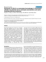
Expression of Nef was analyzed by Western blot. In the
absence of integration, i.e. infection with either integrase-
deficient D116N virus or with wt virus in the presence of
raltegravir, Nef expression still occurred at readily detect-
able levels (Figure 1B), thus confirming the translation of
Nef from unintegrated DNA templates. Additionally, we
confirmed that the introduction of two stop codons in the
first three codons of the Nef gene was sufficient to pre-
vent Nef synthesis
Integrated virus downregulates cell surface CXCR4, CCR5
and CD4 expression on Rev-CEM cells
The Rev-CEM cell line was derived by transducing the
Rev and Tat dependent GFP vector pNL-RRE(SA) [18]
into CEM-SS cells, resulting in a CXCR4-and CCR5-
bearing cell line that expresses GFP in response to the
simultaneous presence of Tat and Rev [19]. Downregula-
tion of CD4, CXCR4 and CCR5 by Nef is well established
in the context of replication competent viruses [34-36]. In
order to confirm that the Rev-CEM cell line was suitable
for the study of Nef-mediated downregulation of cell sur-
face receptors from cells bearing unintegrated viral DNA
only, we first needed to confirm that Nef-mediated recep-
tor downregulation was measurable following viral inte-
gration.
Infected cells (i.e. GFP positive) were assayed by flow
cytometry for cell surface expression of CD4, CXCR4 and
CCR5 (Figure 2). Potent downregulation of CD4 by Nef
was shown to occur, with cell surface levels being only
≈5% of those seen with Δ-nef viruses (p < 0.001) The
CXCR4 coreceptor was also downregulated by Nef, to
Sloan et al. Retrovirology 2010, 7:44
/>Page 3 of 10
Figure 1 Nef expression in the absence of integration. A. Viral integration was measured by an Alu-HIV qPCR assay for provirus. Cells were infected
with wild-type (wt) virus or D116N integrase-containing virus bearing either wt nef or Δ-nef mutations. Repeat infections were also performed for wt
integrase virus in the presence of 1 μM raltegravir. At 72 h post-infection, DNA was extracted and qPCR analysis was performed. Results were expressed
relative to those obtained with wt virus (levels of expression set at 100%). B. Expression of Nef was confirmed by Western blot analysis of lysates from
infections with wt virus (IN +) or D116N integrase-containing virus (IN -), bearing either wt nef (nef +) or Δ-nef (nef -) mutations. Repeat infections were
also performed for wt integrase virus in the presence of 1 μM raltegravir, a concentration shown to be completely inhibitory to integration.
A
B
NL 4-3
++ - -++-
IN
+-+-+
nef
++-
raltegravir
Sloan et al. Retrovirology 2010, 7:44
/>Page 4 of 10
below 50% of levels attained with the Δ-nef virus (p <
0.001), whereas CCR5 downregulation was less, i.e. ≈83%
of Δ-nef levels (p = 0.04).
Integration deficient D116N virus downregulates cell
surface CXCR4, CCR5 and CD4 expression
Having established the suitability of the Rev-CEM cell
line to measure Nef-mediated downregulation, we next
wished to study integrase-deficient virus, taking advan-
tage of the capacity of unintegrated DNA to express Tat
and Rev and thereby induce GFP expression [20]. Intro-
duction of the D116N mutation into the integrase
domain renders integrase inactive, and so cells infected
with such virus will bear unintegrated viral DNA only
[17]. Detection by Rev-CEM cells was sensitive for the
detection of unintegrated infections by flow cytometry.
With integrating virus, the infection rate inferred from
GFP expression was typically 10%, and for integrase defi-
cient virus typically 7% of the total population studied.
Infected cells were measured by flow cytometry for cell
surface expression of CD4, CXCR4 and CCR5. A pattern
of downregulation, similar to that of integrating virus was
observed. These findings confirm that Nef derived from
unintegrated HIV-1 DNA can downregulate cell surface
CD4 to levels ≈ 11% of those attained with Δ-nef virus (p
< 0.001) (Figure 3). As the data were normalized to inter-
nal controls, direct comparisons between integrating vs.
non-integrating viruses were not made.
We have also shown that Nef expressed from uninte-
grated DNA also diminished levels of expression of
CXCR4 to ≈ 42% of those attained with Δ-nef virus, (p <
0.001). In contrast, downregulation of CCR5 in the same
system only occurred to a level of ≈ 80% of that seen with
the Δ-nef virus (p < 0.02).
Integration competent virus downregulates cell surface
CXCR4, CCR5 and CD4 expression in the presence of
inhibitory concentrations of raltegravir
Having established that integrase-deficient virus could
express Nef and downregulate levels of expression of
entry receptors (Figure 3), we next wished to establish
whether such down-modulation would also occur in the
presence of an INSTI such as raltegravir. Previous work
had established that 1 μM of raltegravir was sufficient to
prevent measurable integration in the Rev-CEM cell line
by qPCR for proviral DNA (Figure 1A). We therefore per-
formed a series of infections with wt nef and Δ-nef virus
to determine patterns of receptor downregulation in the
presence of raltegravir.
Similar results to those for integrase-deficient virus
were obtained (Figure 4), with cell surface levels of CD4
being reduced to 17% of levels attained with wild-type Δ-
nef virus (p < 0.001). CXCR4 and CCR5 levels were
reduced to 60% and 79% of those attained with Δ-Nef
virus (p < 0.001 and p = 0.03, respectively). Direct com-
parisons between integrase-deficient and integrase com-
petent viruses in the pressure of raltegravir were not
made, as the experiment was internally controlled.
Finally, the results of Figure 4 show that there was no
direct effect of raltegravir on expression of any of CD4,
CXCR4 or CCR5 in this system.
Discussion
We herein provide the first evidence of chemokine core-
ceptor downregulation mediated by Nef derived from
unintegrated DNA. In addition, we confirm the findings
of other groups that Nef expressed from unintegrated
DNA can downregulate cell surface CD4 [8,14]. It may
not be possible to make direct comparisons between our
and other studies, due to different methods of flow
cytometry employed.
In our studies, levels of downregulation of Nef-medi-
ated CXCR4 derived from unintegrated DNA correlated
well with results obtained in productive infection and are
also in agreement with the finding that such downregula-
tion occurs to a lesser extent than is seen for CD4 [34,35].
Although we observed a slightly lesser degree of down-
regulation of CCR5 by Nef from unintegrated DNA than
has been reported for productive infection of activated
primary human peripheral blood lymphocytes, our
results are broadly consistent with the ≈ 25% downregu-
Figure 2 Nef mediated downregulation of CXCR4, CCR5 and CD4
by integrating virus. Infected (GFP positive) cells were analyzed rela-
tive to uninfected cells for cell surface CD4, CXCR4 and CCR5 after in-
fection with wt integrase-containing virus, either wt nef or a Δ-nef
mutation. The results of geometric means of fluorescence for each re-
ceptor are expressed relative to Δ-nef virus infection receptor levels.
Results are from 3-5 independent experiments, each with two repli-
cate infections. Error bars indicate standard deviations. For each recep-
tor, statistical comparisons between wt nef and Δ-nef were performed
by two-tailed unpaired t-tests, p < 0.001 (***) p < 0.05 (*).
Sloan et al. Retrovirology 2010, 7:44
/>Page 5 of 10
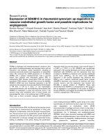
Figure 3 Nef-mediated downregulation of cell surface CXCR4, CCR5 and CD4 by integrase-deficient (D116N) virus. A. Flow cytometry dot
plots demonstrating analysis of GFP positive cells in gate R2, depicting cells infected with integrase-deficient D116N virus. Cells infected with integrase
deficient virus bearing the Δ-nef mutation demonstrate higher expression of CXCR4 than cells infected with wt nef virus. The histogram shows a direct
comparison of CXCR4 levels for wt nef virus (shaded grey) and Δ-nef virus (black line, white background). B. Cells infected with integrase-deficient virus
bearing the Δ-nef mutation demonstrate higher levels of expression of CCR5 than those infected by wt nef virus. The histogram shows a direct com-
parison of CCR5 levels after infection by wt nef virus (shaded grey) vs. Δ-nef virus (black line, white background). C. Cells infected with the integrase-
deficient D116N virus were analyzed relative to uninfected cells for the presence of CD4, CXCR4 and CCR5 after infection with wt integrase virus con-
taining either a wt nef or the Δ-nef mutation. The geometric means of fluorescence for each receptor are expressed relative to Δ-nef virus infection
receptor levels. Results are from 3-5 independent experiments, each with two replicate infections. Error bars indicate standard deviations. For each
receptor, statistical comparisons between wt nef and Δ-nef virus were performed by two-tailed unpaired t-tests, p < 0.001 (***), p < 0.05 (*).
A
GFP
CXCR4-PE
10
0
10
1
10
2
10
3
10
4
10
0
10
1
10
2
10
3
10
4
R2
CXCR4-PE
Count
10
0
10
1
10
2
10
3
10
4
0
1
2
3
4
GFP
CXCR4-PE
10
0
10
1
10
2
10
3
10
4
10
0
10
1
10
2
10
3
10
4
R2
GFP
CCR5-PE-Cy5
10
0
10
1
10
2
10
3
10
4
10
0
10
1
10
2
10
3
10
4
R2
CCR5-PE-Cy5
Count
10
0
10
1
10
2
10
3
10
4
0
4
9
13
17
GFP
CCR5-PE-Cy5
10
0
10
1
10
2
10
3
10
4
10
0
10
1
10
2
10
3
10
4
R2
B
wt nef
wt nef
¨ nef
¨ nef
wt nef vs. ¨ nef
wt nef vs. ¨ nef
GFP
CXCR4-PE
10
0
10
1
10
2
10
3
10
4
10
0
10
1
10
2
10
3
10
4
R2
uninfected
GFP
CCR5-PE-Cy5
10
0
10
1
10
2
10
3
10
4
10
0
10
1
10
2
10
3
10
4
R2
uninfected
C
Sloan et al. Retrovirology 2010, 7:44
/>Page 6 of 10
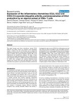
Figure 4 Nef-mediated downregulation of cell surface CXCR4, CCR5 and CD4 by IV-1 infection in the presence of raltegravir. A. FACs plots
demonstrating analysis of cells infected with wt integrase virus in the presence of 1 μM raltegravir. GFP-positive (infected) cells are depicted in gate
R2. Cells infected with Δ-nef virus demonstrate higher expression of CXCR4 than occurs for wt nef virus. The histogram shows a direct comparison of
CXCR4 levels for wt nef virus (shaded grey) vs. Δ-nef virus (black line, white background). B. Cells infected with Δ-nef virus in the presence of 1 μM ralte-
gravir demonstrate higher-level expression of CCR5 than cells infected by wt nef virus. The histogram shows a direct comparison of CCR5 levels for
wild-type nef virus (shaded grey) vs. Δ-nef virus (black line, white background). C. Cells infected with wt virus containing either a wt nef or the Δ-nef
mutation in the presence of raltegravir were analyzed relative to uninfected cells for the presence of cell surface CD4, CXCR4 and CCR5. Geometric
means of fluorescence for each receptor are expressed relative to infection by the Δ-nef virus. Results are from 3-5 independent experiments, each
performed in duplicate. Error bars indicate standard deviations. For each receptor, statistical comparisons between wt nef and Δ-nef viruses were made
by two-tailed unpaired t-tests, p < 0.001 (***), p < 0.05 (*). D. 1 μM raltegravir does not directly influence cell surface levels of CD4, CXCR4 and CCR5.
Uninfected cells were treated in the presence or absence of 1 μM raltegravir and then stained for CD4, CXCR4 or CCR4. Plots display expression levels
relative to untreated cells. Results are from 3-5 independent experiments, each performed in duplicate. For each receptor, statistical comparisons be-
tween untreated and treated cells were made by two-tailed unpaired t-tests. No statistically significant differences were found.
GFP
CXCR4-PE
10
0
10
1
10
2
10
3
10
4
10
0
10
1
10
2
10
3
10
4
R2
CXCR4-PE
Count
10
0
10
1
10
2
10
3
10
4
0
1
1
2
2
GFP
CXCR4-PE
10
0
10
1
10
2
10
3
10
4
10
0
10
1
10
2
10
3
10
4
R2
GFP
CCR5-PE-Cy5
10
0
10
1
10
2
10
3
10
4
10
0
10
1
10
2
10
3
10
4
R2
CCR5-PE-Cy5
Count
10
0
10
1
10
2
10
3
10
4
0
3
5
8
10
GFP
CCR5-PE-Cy5
10
0
10
1
10
2
10
3
10
4
10
0
10
1
10
2
10
3
10
4
R2
A
B
C
wt nef ¨ nef
¨ nefwt nef wt nef vs. ¨ nef
wt nef vs. ¨ nef
D
GFP
CXCR4-PE
10
0
10
1
10
2
10
3
10
4
10
0
10
1
10
2
10
3
10
4
R2
GFP
CCR5-PE-Cy5
10
0
10
1
10
2
10
3
10
4
10
0
10
1
10
2
10
3
10
4
R2
uninfected
uninfected
Sloan et al. Retrovirology 2010, 7:44
/>Page 7 of 10
lation of CCR5 seen with integrated infections of TZM/bl
cells [36]. Studies with 293-Affinofile cells, a cell line that
is quantitatively inducible for both CD4 and CCR5 cell
surface expression, revealed that even modest downregu-
lation of CCR5 from the cell surface was sufficient to
impact infectibility, particularly in the context of reduced
CD4 levels [41-43]. In our system as well, the levels of
downregulation of CCR5 and CD4 that we report can
probably limit viral entry.
With our methodology, one would not expect that Env
would contribute to receptor downregulation, as we used
env deleted pseudovirus bearing a VSV-G envelope.
Additionally, although Vpu can act to downregulate CD4,
there is currently no evidence that Vpu can modulate lev-
els of CXCR4 and CCR5. Further, Vpu is not synthesized
from unintegrated cDNA, and is therefore unlikely to
affect cell surface CD4 levels.
An important consideration is that patterns of tran-
scription and translation from unintegrated virus, arising
from D116N integrase mutations or INSTI-treated cells,
are identical to those observed in infections of quiescent
T-cells prior to integration [13]. Further, there are insuffi-
cient levels of 2-LTR circles in integrase-deficient infec-
tions of Rev-CEM cells to account for the numbers of
transcriptionally active cells, the inference being that
unintegrated linear cDNA molecules, rather than 2-LTR
circles, are the likely template for transcription [20].
Thus, blockage of integration can be informative in
regard to transcription from linear cDNAs. Slowly repli-
cating cells such as resting T-cells [13,44] and non-repli-
cating cells such as macrophages [12] display a lag in
transcription prior to integration. Cells in this state com-
prise the pre-integration latent reservoir [45]; and tran-
scription during this period may be beneficial in regard to
restricting superinfection, that may in turn be deleterious
for cell viability and hence the likelihood of productive
infection [37,38]. Rev can regulate integration [46] and, in
addition, Rev generated from unintegrated DNA can act
to restrict superinfection [47]. Downregulation of entry
receptors may provide similar benefit, as is also seen with
integrating infections [34,36]. Of course, recombination
between unintegrated DNA and superinfecting virus
might still occur as has been observed in vitro [48,49].
Downregulation of coreceptors by unintegrated DNA
may also reduce cell-signaling due to stimulation by natu-
ral ligands or viral envelope. This may help to avert
adverse effects such as chemotaxis, apoptosis, and
changes in viral transcription [50-54]. Further, there may
be an immunological benefit for Nef-mediated downreg-
ulation of MHC-I by unintegrated DNA, which may
result in evasion from cytotoxic T-cell mediated lysis
[55].
In summary, we have provided further evidence that
Nef translation from unintegrated DNA can occur at
functionally relevant levels, and leads to reduced cell sur-
face expression of CXCR4 and CCR5 as well as CD4.
Additional work to determine the benefits of coreceptor
downregulation for virus-infected cells is now in prog-
ress.
Methods
Plasmids and cloning
The HIV-1 molecular clone pNL4-3 was altered through
site-directed mutagenesis (Stratagene) to introduce ter-
mination codons in the first and third amino acids of the
env gene (construct termed pNL4-3-ΔE). Further modifi-
cations by mutagenesis included the substitution D116N
in the integrase coding sequence of the pol gene (con-
struct termed pNL4-3-ΔE-D116N) and the introduction
of termination codons into the first and third codons of
the nef gene (constructs termed pNL4-3-ΔE-ΔN and
pNL4-3-D116N-ΔE-ΔN).
Virus production
Pseudovirus was produced by cotransfection via lipo-
fectamine (Invitrogen) of 7 × 10
6
293T cells with 4 μg
pVPack-VSV-G (Stratagene), a vesicular stomatitis virus
G protein (VSV-G) envelope-encoding construct, in com-
bination with 12 μg of a pNL4-3 derivative (either pNL4-
3-ΔE, pNL4-3-D116N ΔE, pNL4-3-ΔE-ΔN or pNL4-3-
D116N-ΔE-ΔN).
All transfection supernatants were harvested at 72 h
post transfection, clarified by centrifugation for 5 min at
470 g, and passed through a 0.45 μm filter. Virus was
treated with 50 U/ml benzonase at 37°C for 20 minutes to
digest contaminating plasmid DNA [56] and then stored
at -80°C until use.
Cell culture and viral infections
CXCR4-and CCR5 bearing Rev-CEM cells [19] were
obtained through the NIH AIDS Research and Reference
Reagent Program (courtesy of Professor Yuntao Wu) and
were maintained in RPMI 1640 medium (Invitrogen), and
293T cells were maintained in DMEM (Invitrogen), each
supplemented with 10% fetal bovine serum, 1% L-glu-
tamine and 1% penicillin/streptomycin.
1.25 × 10
5
Rev-CEM cells were infected with 500 ng p24
of virus in 24 well plates by spinoculation at 1200 g at
25°C for 2 h followed by 2 h at 37°C, after which medium
was replaced, resulting in a multiplicity of infection
(MOI) of 0.1 for wt virus as determined by GFP expres-
sion. Cells were infected with wt nef or Δ-nef virus that
was either integrase competent (wt) or that contained a
defective D116N mutated integrase. Additional infections
were performed with wt integrase-containing pseudovi-
ruses. In some cases, media were pre-treated with 1 μM
final concentration raltegravir (a gift from Merck Canada,
Inc) for 1 h prior to infection; after spinoculation, ralte-
Sloan et al. Retrovirology 2010, 7:44
/>Page 8 of 10
gravir-containing media were again used at a concentra-
tion of 1 μM.
Integrated DNA qPCR
For the integrated DNA qPCR assays, cellular DNA was
extracted with a DNeasy blood and tissue kit (Qiagen).
PCR was performed with Platinum qPCR SuperMix-
UDG (Invitrogen) on a Corbett Rotor-Gene 6000 ther-
mocycler.
A previously described Alu-gag PCR analysis was used
[57] with the following modifications [58]. The first
round reaction was performed on undiluted samples (100
ng template) and 1:10 dilutions of each sample (10 ng
template diluted with uninfected DNA; 100 ng DNA
total) in the presence of 2 mM MgCl
2
and 200 μM dNTPs.
9 μl of the resulting first round product were used as tem-
plate for the second round nested reaction in the pres-
ence of 5 mM MgCl
2
(final concentration including
MgCl
2
carryover from first round) and 200 μM dNTPs,
using the "wild-type" probe only. Second round cycling
conditions were 50°C for 2 min, 95°C for 1 min, and 45
cycles of 95°C for 15 sec and 60°C for 30 sec. Dual-labeled
probes were obtained from Biosearch Technologies
(Novato, CA, USA). To generate a standard curve for rel-
ative quantification of integrated DNA, Alu-gag PCR was
first performed on a dilution series of DNA from infected
Rev-CEM cells (diluted with DNA from uninfected cells).
Western Blot
2 × 10
5
Rev-CEM cells were infected with pNL4-3-ΔE,
pNL4-3-D116N ΔE, pNL4-3-ΔE-ΔN or pNL4-3-D116N-
ΔE-ΔN, in the presence or absence of raltegravir. The
cells were collected after 72 hours and pelleted by low
speed centrifugation at 470 g. The pellet was resuspended
in RIPA buffer (0.15 M NaCl, 20 mM Tris pH 7.4, 2 mM
EDTA, 1% Triton X-100 and 1% deoxycholate). Cell
lysates were normalized by Bradford assay to 1 mg/ml
total protein and resolved in a 12% SDS-PAGE gel. The
blot was incubated for 60 minutes with 1:4000 polyclonal
anti-HIV-1 Nef antibody obtained from the NIH AIDS
Research and Reference Reagent Program (catalog num-
ber 2949) and anti-rabbit IgG AP conjugate (secondary
antibody) (1:10,000). A chemoluminescent reagent West-
Pico (Pierce) was used to develop the blots.
Cell surface CXCR4, CCR5 and CD4 staining
Rev-CEM cells that had been infected with pseudovirus
were stained at 72 h post-infection in PBS containing 3%
fetal bovine serum and 0.05% sodium azide for 30 min-
utes at 4°C with the following mouse monoclonal anti-
bodies (MAbs): allophycocyanin (APC)-conjugated anti-
human CD4 (clone RPA-T4; BD PharMingen); phyco-
erythrin (PE)-conjugated anti-human CXCR4 MAb
(clone 12G5; BD PharMingen); PE-Cy5-conjugated anti-
human CCR5 MAb (clone 2D7 BD PharMingen). Cells
were then fixed in a final concentration of 1% paraformal-
dehyde, and then resuspended in PBS containing 3% fetal
bovine serum and 0.05% sodium azide. 10,000 events
were assayed on a FACSCalibur instrument (BD
PharMingen); analysis was performed with BD CellQuest
Pro 4.0.2 (BD PharMingen) and FCS Express 3 software
(DeNovo). Levels of receptors were quantified relative to
those found after infection by Δ-nef virus. These studies
were controlled for by subtracting background isotype
fluorescence values from antibody-receptor fluorescence
measurements.
Statistical analysis
All statistical analyses were performed with GraphPad
Prism 4.0 software. To test for statistically significant dif-
ferences between groups, unpaired two-tailed t-tests
were performed with confidence intervals set at 95%.
Abbreviations
INSTI: Integrase strand transfer inhibitor.
Competing interests
The authors declare that they have no competing interests.
Authors' contributions
RDS designed the study, performed the experiments and drafted the manu-
script. DAD helped design the study and performed qPCR optimization. BDK
helped with plasmid construction and flow cytometry analysis. TB-M per-
formed Western blots and assisted in the study design. MAW provided overall
supervision for the project, secured funding, and helped write the manuscript.
All authors read and approved the final manuscript.
Acknowledgements
This work was supported by grants from the Canadian Institutes of Health
Research (CIHR), and Merck Canada Inc. RDS is the recipient of a postdoctoral
fellowship jointly funded by the CIHR Canadian HIV Trials Network (CTN) and
the Canadian Foundation for AIDS Research (CANFAR). DAD is the recipient of a
predoctoral fellowship from the Fonds de la Recherche en Santé du Québec
(FRSQ).
We thank Daria Hazuda of Merck Inc. for helpful comments and Drs Yuntao Wu
and Jon Marsh for the kind provision of the Rev-CEM cell line. We also thank
Cesar Collazos and Susan Colby-Germinario of the McGill AIDS Centre and
Christian Young of the Lady Davis Institute flow cytometry core facilities for
providing valuable technical assistance.
Author Details
1
McGill University AIDS Centre, Lady Davis Institute, Jewish General Hospital,
Montréal, QC, Canada,
2
Department of Microbiology and Immunology, McGill
University, Montréal, QC, Canada and
3
Department of Experimental Medicine,
McGill University, Montréal, QC, Canada
References
1. Chun T, Carruth L, Finzi D, Shen X, DiGiuseppe J, Taylor H, Hermankova M,
Chadwick K, Margolick J, Quinn TC, Kuo YH, Brookmeyer R, Zeiger MA,
Barditch-Crovo P, Siliciano RF: Quantification of latent tissue reservoirs
and total body viral load in HIV-1 infection. Nature 1997, 387:183-188.
2. Pang S, Koyanagi Y, Miles S, Wiley C, Vinters H, Chen I: High levels of
unintegrated HIV-1 DNA in brain tissue of AIDS dementia patients.
Nature 1990, 343:85-89.
3. Sharkey M, Teo I, Greenough T, Sharova N, Luzuriaga K, Sullivan J, Bucy R,
Kostrikis L, Haase A, Veryard C, Davaro RE, Cheeseman SH, Daly JS, Bova C,
Received: 17 March 2010 Accepted: 13 May 2010
Published: 13 May 2010
This article is available from: 2010 Sloan et al; licensee BioMed Central Ltd. This is an Open Access article distributed under the terms of the Creative Commons Attribution License ( which permits unrestricted use, distribution, and reproduction in any medium, provided the original work is properly cited.Retrovirology 2010, 7:44
Sloan et al. Retrovirology 2010, 7:44
/>Page 9 of 10
Ellison RT, Mady B, Lai KK, Moyle G, Nelson M, Gazzard B, Shaunak S,
Stevenson M: Persistence of episomal HIV-1 infection intermediates in
patients on highly active anti-retroviral therapy. Nat Med 2000, 6:76-81.
4. Teo I, Veryard C, Barnes H, An S, Jones M, Lantos P, Luthert P, Shaunak S:
Circular forms of unintegrated human immunodeficiency virus type 1
DNA and high levels of viral protein expression: association with
dementia and multinucleated giant cells in the brains of patients with
AIDS. J Virol 1997, 71:2928-2933.
5. Bukrinsky M, Stanwick T, Dempsey M, Stevenson M: Quiescent T
lymphocytes as an inducible virus reservoir in HIV-1 infection. Science
1991, 254:423-427.
6. Farnet C, Haseltine W: Circularization of human immunodeficiency virus
type 1 DNA in vitro. J Virol 1991, 65:6942-6952.
7. Khiytani D, Dimmock N: Characterization of a human
immunodeficiency virus type 1 pre-integration complex in which the
majority of the cDNA is resistant to DNase I digestion. J Gen Virol 2002,
83:2523-2532.
8. Poon B, Chang M, Chen I: Vpr is required for efficient Nef expression
from unintegrated human immunodeficiency virus type 1 DNA. J Virol
2007, 81:10515-10523.
9. Englund G, Theodore T, Freed E, Engelman A, Martin M: Integration is
required for productive infection of monocyte-derived macrophages
by human immunodeficiency virus type 1. J Virol 1995, 69:3216-3219.
10. Wiskerchen M, Muesing M: Human immunodeficiency virus type 1
integrase: effects of mutations on viral ability to integrate, direct viral
gene expression from unintegrated viral DNA templates, and sustain
viral propagation in primary cells. J Virol 1995, 69:376-386.
11. Wu Y, Marsh J: Early transcription from nonintegrated DNA in human
immunodeficiency virus infection. J Virol 2003, 77:10376-10382.
12. Kelly J, Beddall M, Yu D, Iyer S, Marsh J, Wu Y: Human macrophages
support persistent transcription from unintegrated HIV-1 DNA.
Virology 2008, 372:300-312.
13. Wu Y, Marsh J: Selective transcription and modulation of resting T cell
activity by preintegrated HIV DNA. Science 2001, 293:1503-1506.
14. Gillim-Ross L, Cara A, Klotman M: Nef expressed from human
immunodeficiency virus type 1 extrachromosomal DNA
downregulates CD4 on primary CD4+ T lymphocytes: implications for
integrase inhibitors. J Gen Virol 2005, 86:765-771.
15. Ansari-Lari M, Donehower L, Gibbs R: Analysis of human
immunodeficiency virus type 1 integrase mutants. Virology 1995,
211:332-335.
16. Stevenson M, Haggerty S, Lamonica C, Meier C, Welch S, Wasiak A:
Integration is not necessary for expression of human
immunodeficiency virus type 1 protein products. J Virol 1990,
64:2421-2425.
17. Engelman A, Englund G, Orenstein J, Martin M, Craigie R: Multiple effects
of mutations in human immunodeficiency virus type 1 integrase on
viral replication. J Virol 1995, 69:2729-2736.
18. Wu Y, Beddall M, Marsh J: Rev-dependent lentiviral expression vector.
Retrovirology 2007, 4:12.
19. Wu Y, Beddall M, Marsh J: Rev-dependent indicator T cell line. Curr HIV
Res 2007, 5:394-402.
20. Iyer S, Yu D, Biancotto A, Margolis L, Wu Y: Measurement of human
immunodeficiency virus type 1 preintegration transcription by using
Rev-dependent Rev-CEM cells reveals a sizable transcribing DNA
population comparable to that from proviral templates. J Virol 2009,
83:8662-8673.
21. Brussel A, Sonigo P: Evidence for gene expression by unintegrated
human immunodeficiency virus type 1 DNA species. J Virol 2004,
78:11263-11271.
22. Poon B, Chen I: Human immunodeficiency virus type 1 (HIV-1) Vpr
enhances expression from unintegrated HIV-1 DNA. J Virol 2003,
77:3962-3972.
23. Butler S, Johnson E, Bushman F: Human immunodeficiency virus cDNA
metabolism: notable stability of two-long terminal repeat circles. J
Virol 2002, 76:3739-3747.
24. Pierson T, Kieffer T, Ruff C, Buck C, Gange S, Siliciano R: Intrinsic stability of
episomal circles formed during human immunodeficiency virus type 1
replication. J Virol 2002, 76:4138-4144.
25. Vargas JJ, Gusella G, Najfeld V, Klotman M, Cara A: Novel integrase-
defective lentiviral episomal vectors for gene transfer. Hum Gene Ther
2004, 15:361-372.
26. Lu R, Nakajima N, Hofmann W, Benkirane M, Jeang K, Sodroski J, Engelman
A, Teh-Jeang K: Simian virus 40-based replication of catalytically
inactive human immunodeficiency virus type 1 integrase mutants in
nonpermissive T cells and monocyte-derived macrophages. J Virol
2004, 78:658-668.
27. Mannioui A, Schiffer C, Felix N, Nelson E, Brussel A, Sonigo P, Gluckman J,
Canque B: Cell cycle regulation of human immunodeficiency virus type
1 integration in T cells: antagonistic effects of nuclear envelope
breakdown and chromatin condensation. Virology 2004, 329:77-88.
28. Bushman F: Measuring covert HIV replication during HAART: the
abundance of 2-LTR circles is not a reliable marker. AIDS 2003,
17:749-750.
29. Gillim-Ross L, Cara A, Klotman M: HIV-1 extrachromosomal 2-LTR circular
DNA is long-lived in human macrophages. Viral Immunol 2005,
18:190-196.
30. Wu Y: The second chance story of HIV-1 DNA: Unintegrated? Not a
problem! Retrovirology 2008, 5:61.
31. Cara A, Klotman M: Retroviral E-DNA: persistence and gene expression
in nondividing immune cells. J Leukoc Biol 2006, 80:1013-1017.
32. Hazuda D, Felock P, Witmer M, Wolfe A, Stillmock K, Grobler J, Espeseth A,
Gabryelski L, Schleif W, Blau C, Miller M: Inhibitors of strand transfer that
prevent integration and inhibit HIV-1 replication in cells. Science 2000,
287:646-650.
33. Butler S, Hansen M, Bushman F: A quantitative assay for HIV DNA
integration in vivo. Nat Med 2001, 7:631-634.
34. Venzke S, Michel N, Allespach I, Fackler O, Keppler O: Expression of Nef
downregulates CXCR4, the major coreceptor of human
immunodeficiency virus, from the surfaces of target cells and thereby
enhances resistance to superinfection. J Virol 2006, 80:11141-11152.
35. Wildum S, Schindler M, Münch J, Kirchhoff F: Contribution of Vpu, Env,
and Nef to CD4 down-modulation and resistance of human
immunodeficiency virus type 1-infected T cells to superinfection. J
Virol 2006, 80:8047-8059.
36. Michel N, Allespach I, Venzke S, Fackler O, Keppler O: The Nef protein of
human immunodeficiency virus establishes superinfection immunity
by a dual strategy to downregulate cell-surface CCR5 and CD4. Curr
Biol 2005, 15:714-723.
37. Pauza C, Galindo J, Richman D: Reinfection results in accumulation of
unintegrated viral DNA in cytopathic and persistent human
immunodeficiency virus type 1 infection of CEM cells. J Exp Med 1990,
172:1035-1042.
38. Robinson H, Zinkus D: Accumulation of human immunodeficiency virus
type 1 DNA in T cells: results of multiple infection events. J Virol 1990,
64:4836-4841.
39. Wu Y, Yoder A: Chemokine coreceptor signaling in HIV-1 infection and
pathogenesis. PLoS Pathog 2009, 5:e1000520.
40. Lama J: The physiological relevance of CD4 receptor down-modulation
during HIV infection. Curr HIV Res 2003, 1:167-184.
41. Johnston S, Lobritz M, Nguyen S, Lassen K, Delair S, Posta F, Bryson Y, Arts
E, Chou T, Lee B: A quantitative affinity-profiling system that reveals
distinct CD4/CCR5 usage patterns among human immunodeficiency
virus type 1 and simian immunodeficiency virus strains. J Virol 2009,
83:11016-11026.
42. Lassen K, Lobritz M, Bailey J, Johnston S, Nguyen S, Lee B, Chou T, Siliciano
R, Markowitz M, Arts E: Elite suppressor-derived HIV-1 envelope
glycoproteins exhibit reduced entry efficiency and kinetics. PLoS
Pathog 2009, 5:e1000377.
43. Pugach P, Ray N, Klasse P, Ketas T, Michael E, Doms R, Lee B, Moore J:
Inefficient entry of vicriviroc-resistant HIV-1 via the inhibitor-CCR5
complex at low cell surface CCR5 densities. Virology 2009, 387:296-302.
44. Pierson T, Zhou Y, Kieffer T, Ruff C, Buck C, Siliciano R: Molecular
characterization of preintegration latency in human
immunodeficiency virus type 1 infection. J Virol 2002, 76:8518-8531.
45. Petitjean G, Al Tabaa Y, Tuaillon E, Mettling C, Baillat V, Reynes J, Segondy
M, Vendrell J: Unintegrated HIV-1 provides an inducible and functional
reservoir in untreated and highly active antiretroviral therapy-treated
patients. Retrovirology 2007, 4:60.
46. Levin A, Rosenbluh J, Hayouka Z, Friedler A, Loyter A: Integration of HIV-1
DNA is regulated by interplay between viral rev and cellular LEDGF/
p75 proteins. Mol Med 16:34-44.
Sloan et al. Retrovirology 2010, 7:44
/>Page 10 of 10
47. Levin A, Hayouka Z, Friedler A, Brack-Werner R, Volsky D, Loyter A: A novel
role for the viral Rev protein in promoting resistance to Super-infection
by Human Immunodeficiency Virus type 1. J Gen Virol 2010 in press.
48. Gelderblom H, Vatakis D, Burke S, Lawrie S, Bristol G, Levy D: Viral
complementation allows HIV-1 replication without integration.
Retrovirology 2008, 5:60.
49. Quan Y, Liang C, Brenner B, Wainberg M: Multidrug-resistant variants of
HIV type 1 (HIV-1) can exist in cells as defective quasispecies and be
rescued by superinfection with other defective HIV-1 variants. J Infect
Dis 2009, 200:1479-1483.
50. Khan M, Brandimarti R, Patel J, Huynh N, Wang J, Huang Z, Fatatis A,
Meucci O: Apoptotic and antiapoptotic effects of CXCR4: is it a matter
of intrinsic efficacy? Implications for HIV neuropathogenesis. AIDS Res
Hum Retroviruses 2004, 20:1063-1071.
51. Cocchi F, DeVico A, Garzino-Demo A, Arya S, Gallo R, Lusso P:
Identification of RANTES, MIP-1 alpha, and MIP-1 beta as the major HIV-
suppressive factors produced by CD8+ T cells. Science 1995,
270:1811-1815.
52. Davis C, Dikic I, Unutmaz D, Hill C, Arthos J, Siani M, Thompson D,
Schlessinger J, Littman D: Signal transduction due to HIV-1 envelope
interactions with chemokine receptors CXCR4 or CCR5. J Exp Med 1997,
186:1793-1798.
53. Zaitseva M, Peden K, Golding H: HIV coreceptors: role of structure,
posttranslational modifications, and internalization in viral-cell fusion
and as targets for entry inhibitors. Biochim Biophys Acta 2003,
1614:51-61.
54. Tremblay M, Meloche S, Gratton S, Wainberg M, Sékaly R: Association of
p56lck with the cytoplasmic domain of CD4 modulates HIV-1
expression. EMBO J 1994, 13:774-783.
55. Collins K, Chen B, Kalams S, Walker B, Baltimore D: HIV-1 Nef protein
protects infected primary cells against killing by cytotoxic T
lymphocytes. Nature 1998, 391:397-401.
56. Sastry L, Xu Y, Cooper R, Pollok K, Cornetta K: Evaluation of plasmid DNA
removal from lentiviral vectors by benzonase treatment. Hum Gene
Ther 2004, 15:221-226.
57. Yu J, Wu T, Liszewski M, Dai J, Swiggard W, Baytop C, Frank I, Levine B, Yang
W, Theodosopoulos T, O'Doherty U: A more precise HIV integration
assay designed to detect small differences finds lower levels of
integrated DNA in HAART treated patients. Virology 2008, 379:78-86.
58. Donahue D, Sloan R, Kuhl B, Bar-Magen T, Schader S, Wainberg M: Stage-
Dependent Inhibition of HIV-1 Replication by Antiretroviral Drugs in
Cell Culture. Antimicrob Agents Chemother 2009, 54:1047-1054.
doi: 10.1186/1742-4690-7-44
Cite this article as: Sloan et al., Expression of Nef from unintegrated HIV-1
DNA downregulates cell surface CXCR4 and CCR5 on T-lymphocytes Retrovi-
rology 2010, 7:44